D
EPARTMENT OF BIOCHEMISTRYThesis
Presented by
Selma HOUCHI
Submitted for the fulfillment of the requirements for the degree of
Doctorate of Sciences
In Biology
OPTION: Biochemistry
Theme
Characterization of ß-lactamase genes in clinical isolates and
determination of the inhibitory effects of Algerian seaweeds
extracts on recombinant ß-lactamases GES-22 and OXA-1
Discussed on 22/ 07/ 2019
Board of Examiners
Chairman Abdelhalim KHENCHOUCHE Associated professor (A). UFA Setif-1
Supervisor Rachid MAHDADI Pr. University Ferhat Abbas Setif-1
Examiner Karim HOUALI Pr.Univ. Mouloud Mammeri Tizi Ouzou
Examiner El-Hafidh NABTI Pr. Univ. Abderrahmane Mira Bejaia
Examiner Kamel AISSET Pr. Univ. Hadj Lakhdar Batna-1
Examiner Widad SOBHI Associated professor (A). UFA-Setif-1
Invited member Cemal SANDALLI Pr. Recep Tayyip Erdogan University, Turkey
University of Ferhat Abbas Setif 1 Faculty of Life and Nature
Sciences
ةيبعشلا ةيطارقميدلا ةيرئازجلا ةيروهمجلا
ملعلا ثحبلا و يلاعلا ميلعتلا ةرازو
ي
فيطس ،سابع تاحرف ةعماج
1ةيلك
مولع
ةايحلا و ةعيبطلا
N°……….…………..…….……/SNV/2019
In the name of Allah, the
Beneficent, the
Dedication
I am dedicating this thesis, First and foremost,
to my loving parents
my father, Ahmed,
May Allah grant him
Jannah Firdaws.
and My mother MAIZA Hafsa
for their love, endless support and encouragement
To the people who helped me a lot and for their support
moral and their sacrifices along my PhD;
My dear brothers
Abdelkarim, Faycel and Mounir
Acknowledgements
First and foremost, I would like to thank ALLAH Almighty for giving me the strength, ability and opportunity to undertake this modest research study.
There are many people who have helped me during this study. There is not enough space, nor words, to acknowledge everyone to the extent they deserve. I hope people understand the brevity of this part. First of all, I would like to thank my doctorate advisor and research guide at Ferhat Abbas SETIF-1 University, (ALGERIA) Professor Rachid MAHDADI. Under his guidance, I was given the freedom to pursue my research in my own way and I greatly appreciated that freedom. It was a great pleasure to work with him. Thanks for supporting and encouraging me through these years.
Additionally, I would like to thank the jury members for agreeing to judge this work and to be the examiners of this thesis:
Dr. Abdelhalim KHENCHOUCHE, for agreeing to judge this work and to do me the honor of
chairing this thesis jury. Pr. Karim HOUALI, Pr. El-Hafidh NABTI and Pr. Kamel AISSET,
Dr. Widad SOBHI to have accepted to judge this work. I am deeply grateful to all members of
the jury for agreeing to read the manuscript and to participate in the defense of this thesis.
The realization of this work was only possible due to the several people's collaboration, to which desire to express my gratefulness:
To Pr. Cemal sandalli at Recep tayyib Erdogan University, (TURKEY) and to all his team at the Microbiology and Molecular Biology Research Laboratory. With deep sense of gratitude, I owe sincere thanks to him for agreeing to participate in the defense of this thesis as invited member.
Pr. Guanhua DUGH and Pr. ZHANG Li at Institute of Materia Medica, Chinese Academy of
Medical Sciences and Peking Union Medical College (CHINA), and all the staff of the Laboratory of Bioactive Substances and Functions of Natural Medicines.
Pr. Hassiba TALI-MAAMAR at the Medical bacteriology laboratory in Pasteur institute, Dely
Brahim (ALGERIA) and all members of this Laboratory
Dr. Wahid REFES. Associated Professor at the National Higher School of Marine Sciences
and Coastal Management (ENSSMAL). The Marine and Littoral Ecosystems Laboratory (ECOSYSMarL), (ALGERIA) and to his student my dear colleague BAHRI Nabila.
Dr. Moussa MENNAD. Elected representative of the research staff of the National Center for
Research and Development of Fisheries and Aquaculture (CNRDPA), Bou Ismail, Tipaza (ALGERIA).
I also take a real pleasure to thank Dr. Abdelhakim AOUF at Laboratory of Cellular and Molecular Biology, Faculty of Biological Sciences, University of Science and Technology Houari Boumediene, Algiers, (ALGERIA).
I would also like to thank all the members of the Laboratory of Applied Biochemistry (Ferhat
Abbas SETIF-1 University, ALGERIA). Particularly: Pr.Lekhmici ARRAR and Pr.Noureddine
CHAREF.
Besides this, I would like to thank Dr. Azeddine TEMAMNA may ALLAH grant him
vast paradise; Pr. Abdelouahab BOUZIDI; Pr.Rachid BELHATTAB;Pr. Hammama
صخلم
فده هذه ت ةساردلا ديدحت ىلا عون لا تانيج امسملا تاميزنلاا ءانبل ةرفشملا ة زامتكلااتيب اهعويش اذك و يف تلازع تكب ي ير ة س ا مارغلا ةبل ةيرئازج تايفشتسم لخاد و جراخ نم تعمج . تاصلختسم ةردق نم ققحتلا ىلا اضيأ تفده ةيلوناثيم عبسل ة ةيمان بلاحط ىلع ا يرئازجلا لحاسل [Ulva intestinalis, Codium tomentosum, Bryopsis pulmosa,Sargassum vulgare, Dictyota dichtoma, Halopteris scoparia, Cystoseira compressa]
طيبثت ىلع نم نيعون زامتكلااتيب امه و 22 -GES و 1 -OXA . تنيب تلازعلا نأ جئاتنلا حت ىلع يو نيج لقلأا ا دحاو ا ءانبل ارفشم زامتكلااتيب ) M (23,24,51,58) -OXA bla bla , NDM bla , 2 -PER bla , GES bla , M2 -M1 -CTX bla SHV, bla , TEM bla ( نم يأ يوحت لا و تانيجلا ) IMP bla , VIM bla , KPC bla ( مسق تانورجتنلال ةينيجلا تابيلعلاب ةظوفحملا ءازجلأا يوحت لباقملاب اهنكل I و II وا ةلصفنم ةعمتجم . نم هتميق ريغتت يذلا لونيفلا تاديدع نم اهاوتحم ثيح نم ةعبسلا بلاحطلا فلتخت 0.93 ± 0.65 و 3.22 ± 0.91 mgGAEs/g DW . هذه قوفت ةينبلا بلاحطلل كلت نيترمب ءارضخلا بلاحطلا دنع ةميقلا . نيح يف وتحم ناك ، نم ةريخلأا هذه ى نيماتيف E نم ىلعأ بلاحطلا ءارضخلا . تنيب ةساردلا ليلحتلا ةي تابكرم دوجو و ةعبسلا بلاحطلل ةيلوناثملا تاصلختسملا نيب ةكرتشم يه ضمح افلا كينيلونيل (C18: 3 ω3) يلونيللا ضمح ، ي ( ك C18: 2 ω 6 يلولأا ضمح ، ) ي ( ك C18: 1 ω9 كينوديكارلأا ضمحو ، ) (C20: 4) ω6 .) ىلع ةولاع اذه يوحت ، intestinalis U. و D. dichtoma ع ( نيلاكيب ىل 5 O 10 H 15 C ( نيمكركو ) 6 O 20 H 21 C .) يوحي B.pulmosa ( نيزديادو نيلاكياب ىلع 4 O 10 H 15 C يوحي ؛) C. tomentosum ( نيتيسريك ىلع 7 O 10 H 15 C يوحي و ) C.compressa ( ىلع S ( نيجنيران ) 5 O 12 H 15 C ميق تحوارت .) 50 IC ةددحملا ةقلاعلا ىحنم ىلع Logit/Log نيب 10.31 ± 3..0 µg/mL ( D. dichtoma ىلإ ) .1.1. ± 3.10 µg/mL ( C. compressa و ) نم 10.11 ± 3.90 µg/mL ( B.pulmosa ) ىلإ 01.09 ± 1.90 µg/mL ( C. compressa عم ) GES-22 و OXA-1 يلاوتلا ىلع ، رهظأ . بلحطلل يلوناثملا صلختسملا D. dichtoma ىلع اًطبثم ا ًريثأت GES-22 كأ ب تاطبثم نم ر β-lactam لاكلا ةيكيس تانلاوفلاك ةامسملا ، ماتكابوزات و ماتكابلوس ( IC50= 68.38±0.17µg/mL, 52.68±0.64µg/mL, 29.94±0.01 ) يلاوتلا ىلع . بثي مل ط ماتكابلوس و ميزنلاا تانلاوفلاك 1 -OXA ، ماتكابوزاتلل افلاخ =243.03± 4.53 μg/mL) 50 (IC . ـل يلوناثيملا صلختسملا رهظأ B. pulmosa ىلع اًطبثم ا ًريثأت g/mL) μ =13.22 ± 0.9 50 (IC . 1 -OXA نم جئاتن تارابتخلاا سفن ت نص ف لك ةيلوناثملا تاصلختسملا ريغ تاطبثمك ـل ةيسفانت OXA-1 و GES-22 ءانثتساب ، صلختسملا ني ـل B.pulmosa و C. compressa لا اذل ن ناطبثي GES-22 و ةيسفانت ةقيرطب لا ، ةيسفانت تلا ىلع بيتر . تنيب تاسارد ةاكاحملا ةوق نأ ىلإ لا ثت ور نيوكت ىلإ عجرت امبر طيب طبا نيجورديه ةي و ءاملل ةهراك نيب تابكرملا و ةيلوناثملا تاصلختسملا يف اهنع فشكلا مت يتلا تابكرم ىرخا ةيعيبط ةهج نم لأاو ضامح ةينيملأا لل عقوم طشنلا نم لك يف Serine و Lactamases -β -metallo ةهج نم ىرخا تابكرملا عيمج ترهظأ . مت يتلا ةيلوناثملا تاصلختسملا يف اهنع فشكلا بلاحطلل score libdock و interaction energy CDOCKER كأ ب تاطبثم نم ر β-lactam ةيكيسلاكلا عم 11 ß-lactamases حارتقا نكمي . hesperidin و dihydromyrecitin و curcumin تاطبثمك ساسأ ىلع ةديج score libdock و interaction energy CDOCKER . :حيتافملا تاملكلا ،ةاكاحم ،زاماتكلااتيب
Ulva intestinalis, Codium intestinalis, Bryopsis pulmosa,
Abstract
The main aim of this study was to characterize the type and the prevalence of β-lactamases coding genes in community and nosocomial Algerian Gram negative bacterial isolates, as well as to investigate the inhibitory effects of methanolic extracts of seven seaweeds Of Algerian coast [Ulva intestinalis, Codium
tomentosum, Bryopsis pulmosa, Sargassum vulgare, Dictyota dichtoma, Halopteris scoparia, and Cystoseira compressa] against GES-22 and OXA-1 β-lactamases variants. The results showed that isolates harboring at
least, one β-lactamase coding gene (blaTEM, blaSHV, blaCTX-M1-M2, blaGES, blaPER-2, blaNDM, bla blaOXA-M
(23,24,51,58)); neither blaKPC nor blaVIM, blaIMP gene was detected. The conserved regions of class-I and II
integron gene cassettes were determined alone and together in these isolates. The TPC varied among the seven seaweeds species, ranging between 0.93 ± 0.65 and 3.22 ± 0.91 mg GAEs/g DW. TPC in the MEs of green seaweed species were found to be nearly two times greater than that in brown seaweeds. However, vitamin E contents of brown seaweed MEs were higher than those of green seaweeds. Furthermore, qualitative analysis showed the existance of common compounds in the MEs including α-linolenic acid (C18:3 ω3), linoleic acid (C18:2 ω6), oleic acid (C18:1 ω9), and arachidonic acid (C20:4 ω6). Moreover, U.
intestinalis and D. dichtoma contain baicalein (C15H10O5) and curcumin (C21H20O6). B. pulmosa contains baicalein and daidzein (C15H10O4); C. tomentosum contains quercetin (C15H10O7) and C. compressa contains (S) naringenin (C15H12O5). Values of IC50 of MEs determined by linear computerized regression analysis after logit/log transformation, are ranged from 13.01 ± 0.46 μg/mL (D. dichtoma) to 41.24 ± 0.23 μg/mL (C.
compressa) and from 13.22 ± 0.96 μg/mL (B. pulmosa) to 62.39 ± 1.96 μg/mL (C. compressa) with GES-22
and OXA-1, respectively. ME of D.dichtoma exhibited significant inhibitory effect on GES-22 more than classical β-lactam inhibitors, clavulanate, sulbactam and tazobactam (IC50 = 68.38 ± 0.17 μg/mL, 52.68 ± 0.64 μg/mL, and 29.94 ± 0.01 μg/mL, respectively). OXA-1 was not inhibited by sulbactam and clavulanate and was moderately inhibited by tazobactam (IC50 =243.03± 4.53 μg/mL). However, ME of B. pulmosa exhibited significant inhibitory effect on OXA-1 (IC50 =13.22 ± 0.96 μg/mL). Data from the same tests categorizing all MEs as mixed inhibitors of OXA-1 and GES-22, except B. pulmosa and C. compressa wich inhibit GES-22 with competitive and non-competitive manner, respectively. Docking studies indicated that the potency of inhibition is probably due to the formation of hydrogen bonds and hydrophobic interactions between ligands (compounds identified in MEs of seaweeds and other natural compounds) and active amino-acids site of both Serine and metallo-β-Lactamases. All compounds identified in the MEs of seaweeds have shown libdock score and CDOCKER interaction energy higher than the classical inhibitors clavulanate and sulbactam towards 15 ß-lactamases. Hesperidin, dihydromyrecitin and curcumin can be suggested as good ligands on the basis of libdock score and CDOCKER interaction energy.
Key words: β-lactamases, GES-22, OXA-1, docking, Ulva intestinalis, Codium tomentosum, Bryopsis
Résumé
Le but principal de cette étude était de déterminer le type et la prévalence des gènes codants pour les β-lactamases dans des isolats bactériens Gram négatif algériens communautaires et nosocomiaux, ainsi que de rechercher les effets inhibiteurs d'extraits méthanoliques (EMs)de sept algues de la cote algérienne [Ulva
intestinalis, Codium tomentosum, Bryopsis pulmosa, Sargassum vulgare, Dictyota dichtoma, Halopteris scoparia et Cystoseira compressa] sur deux variants de β-lactamases, GES-22 et OXA-1. Les résultats ont
montré que les isolats contenant au moins un gène codant pour la β-lactamase (blaTEM, blaSHV, blaCTX-M1-M2,
blaGES, blaPER-2, blaNDM, bla blaOXA-M (23,24,51,58)); ne portent ni le gène blaKPC ni le gène blaIMP ni le gène
blaVIM . Les régions conservées des cassettes de gènes des intégrons de classe I et II ont été déterminées seules et ensemble dans ces isolats. Le contenu en polyphénols totaux des sept espèces d'algues variait entre 0,93 ± 0,65 et 3,22 ± 0,91 mg GAEs/g poids sec. Ceux des EMs des algues vertes étaient presque deux fois plus élevés que ceux des algues brunes. Cependant, les teneurs en vitamine E de EMs des algues brunes étaient plus élevées que celles de MEs des algues vertes. En outre, une analyse qualitative a montré l’existence de composés communs aux MEs des sept algues, incluant l’acide α-linolénique (C18: 3 3), l’acide linoléique (C18: 2 ω6), l’acide oléique (C18: 1 ω9) et l’acide arachidonique (C20: 4). ω6). De plus, U. intestinalis et D.
dichtoma contiennent de la baicaleine (C15H10O5) et de la curcumine (C21H20O6). C. tomentosum contient de la quercetine (C15H10O7) et C. compressa contient de la (S) naringenine (C15H12O5). Les valeurs de IC50 des ME déterminées par analyse de régression linéaire informatisée après transformation logit / log, variaient de 13,01 ± 0,46 μg/mL (D. dichtoma) à 41,24 ± 0,23 μg/mL (C. compressa) et de 13,22 ± 0,96 μg/mL. (B. pulmosa) à 62,39 ± 1,96 µg/mL (C. compressa) avec GES-22 et OXA-1, respectivement. L’EM de Dichtoma a montré un effet inhibiteur plus significatif sur GES-22 que les inhibiteurs β-lactames classiques, le clavulanate, le sulbactam et le tazobactam (IC50 = 68,38 ± 0,17 μg/mL, 52,68 ± 0,64 μg/mL et 29,94 ± 0,01 μg/mL, respectivement). Le sulbactam et l’acide clavulanique n'ont pas inhibé l'OXA-1, mais le tazobactam l'a inhibé modérément (IC50 = 243,03 ± 4,53 μg/mL). Cependant, l'EM de B. pulmosa a montré un effet inhibiteur significatif sur OXA-1 (IC50 = 13,22 ± 0,96 μg/mL). Des données des memes tests classaient tous les EMs comme inhibiteurs non compétitifs d'OXA-1 et de GES-22, à l'exception de l’EM de B. pulmosa et de C. compressa, qui inhibent GES-22 de manière compétitive et non compétitive, respectivement. Des études d'amarrage ont montré que la puissance de l'inhibition est probablement due à des liaisons hydrogène et aux interactions hydrophobes entre les ligands (composés identifiés dans les EMs d'algues et d'autres composés naturels) et les acides aminés du site actif des sérine et des métallo-β-lactamases. Tous les composés identifiés dans les EMs d'algues ont montré un score de libdock et une énergie d'interaction CDOCKER supérieurs à ceux du clavulanate et du sulbactam pour les 15 β-lactamases. L’hespéridine, la dihydromyrécitine et la curcumine peuvent être suggérées comme de bons ligands sur la base de leur score de libdock et de l'énergie d'interaction CDOCKER.
Mots clés: β-lactamases, GES-22, OXA-1, docking, Ulva intestinalis, Codium tomentosum, Bryopsis
LIST OF ABBREVIATIONS
ABTS : 2,2′-azino-bis (3-ethylbenzothiazoline-6-sulfonic acid);
AMPc : Chromosomal located cephalosporinase ATCC : Amiricain Type Collection culture C3G : Third-generation cephalosporins CA-Ab : Community acquired A. baumanni
CA-SFM : Comité français de l’antibiogramme de la société française de microbiologie CDOCKER : CHARMM-based DOCKER
CHARMM : Chemistry at HARvard Macromolecular Mechanics) CLSI : Clinical and Laboratory Standards Institute
CTX : Cefotaximase
DDST : Double Disc Synergy test DPPH : 2,2'-diphenyl-1-picrylhydrazyl DS : Discovery Studio
EDTA : Ethylene diamine tetraacetic acid ESβLs : Extended Spectrum β-lactamases GES : Guyana extended spectrum β-lactamases. HAIs : Hospital acquired infections
HPLC-ESI-MS : High Performance Liquid Chromatography Time of Flight- Mass Spectrometry HPLC-TOF-MS : High Performance Liquid Chromatography electrospray ionisation tandem-
Mass Spectrometry
IC50 : half maximal inhibitory concentration
ICUs: intensive care units IMP : Imipenemase and
IPTG: Isopropyl-β-D-thiogalactopyranoside KM : Michaelis constant
MβLs : metallo-β-lactamases MEs : Methanolic extracts MHT : Modified Hodge Test MDR : Multidrug-resistant
MDR-GNB : Multi-drug resistant Gram negative bacteria MROs : Multidrug-resistant organisms
NDM : New Delhi MβL
NFGNB : Non-fermenting gram-negative bacilli OprD : Outer membrane porin D
OXA : oxacillinase PBA : Phenylboronic acid
PCDDT : Phenotypic Confirmatory Disc Diffusion Test PDB : Protein Data Bank
PER : Pseudomonas Extended Resistance RMSD : Root Mean Square Deviation S : Substrate
SβLs : Serine β-lactamases SHV : Sulfhydryl variable SD : Standard deviation
SDS-PAGE : Sigle anglophone de sodium dodecyl sulfate polyacrylamide gel electrophoresis TEM : Temoneira
VEB : Vietnam Extended-spectrum β-Lactamase. VIM : Verona integron-encoded
Vmax : maximum velocity
LIST OF FIGURES
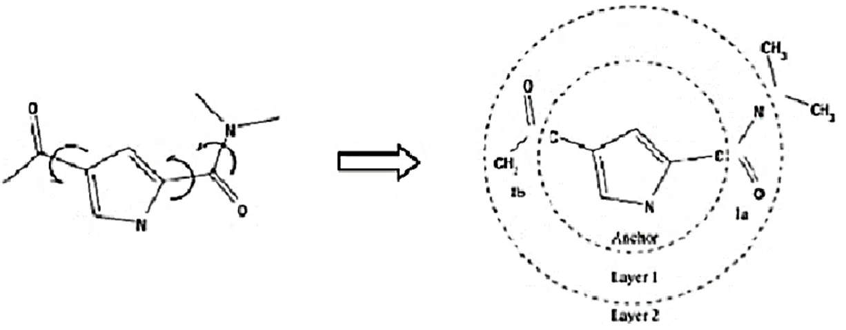
Figure 1. Demonstration of an incremental reconstruction whose molecule is fragmented
and then docked and rebuilt layer by layer in the active site ………. 22
Figure 2. Photograph of Ulva intestinalis ……… 26
Figure 3. Photograph of Codium tomentosum ……….. 27
Figure 4. Photograph of Bryopsis plumosa ……….. 28
Figure 5. Photograph of Sargassum vulgare ………. 29
Figure 6. Photograph of Dictyota dichotoma ……… 30
Figure 7. Photograph of Halopteris scoparia ………... 31
Figure 8. Photograph of Cystoseira compressa ……… 33
Figure 9. Geographical map centered on the sampling site (Kouali, Algeria) …………... 34
Figure 10. Binding sites (cavities) detected in the receptor ……….. 48
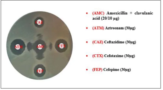
Figure 11. Positive test of synergy obtained with an E.coli strain ……… 58
Figure 12. Positive combined disc diffusion test obtained with a K.pneumoniae strain ... 58
Figure 13. Positive modified Hodge test ……… 59
Figure 14. Positive IMP-EDTA Combined Disc test obtained with P.aeruginosa strain 60 Figure 15. Polymerase chain reaction (PCR); Lane M Molecular size marker (100 bp); lane 1, blaTEM; lane 2, blaSHV; lane 3, blaPER; lane 4, blaCTX-M1; lane 5, blaGES; lane 6, blaNDM; lane 7, blaOXA-48; lane 8, blaOXA-51 ……….. 65
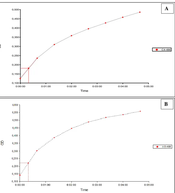
Figure 16. SDS-PAGE gel analysis and purification of (A) GES-22 and (B) OXA-1 expression in Escherichia coli. GES-22 and OXA-1 have a molecular weight of around 31,19 and 30,88 kDa, respectively. The band darkens denoting protein transcription and growth due to IPTG induction. Lane 1: supernatant, lane 2: flow, lane 3: 25 µM, lane 4: 100 µM, lane 5: 300 µM , lane 6: 300 µM ……….. 68 Figure 17. Evolution of the absorbance of 60 μM nitrocefin in the presence of 07 μl of a solution of OXA-1 (A) and GES-22 (B) proteins recombinants ……….. 72
Figure 18. Lineweaver-burk curve and values of KM (μM) and Vm (μmol/min/mg) obtained with GES-22 and OXA-1 and nitrocefin as substrate ……… 73
Figure 19. Lineweaver burk plots obtained when activity of GES-22 is started with increasing concentration of nitrocefin in presence ( ) and absence ( ) of each ME of seaweeds ………... 76
Figure 20. Lineweaver burk plots obtained when activity of OXA-1 is started with increasing concentration of nitrocefin in presence ( ) and absence ( ) of each ME of seaweeds ………... 77
Figure 21. Interaction of Dihydromyrecitin and CTX-M-9. A: 2D interaction diagram. B:
Fisher projection ………... 86
Figure 22. Interaction of Hesperidin and GES-1. A: 2D interaction diagram. B: Fisher
projection ………. 86
Figure 23. Interaction of Hesperidin and SHV-1. A: 2D interaction diagram. B: Fisher
projection ………. 87
Figure 24. Interaction of Paeoniflorin and TEM-72. A: 2D interaction diagram. B: Fisher
projection ………. 87
Figure 25. Interaction of Baicalin and TEM-1. A: 2D interaction diagram. B: Fisher
projection ………. 88
Figure 26. Interaction of Myrecitin and KPC-2. A: 2D interaction diagram. B: Fisher
projection ………. 88
Figure 27. Interaction of Esculin and OXA-10. A: 2D interaction diagram. B: Fisher
projection ………. 89
Figure 28. Interaction of Hesperidin and OXA-23. A: 2D interaction diagram. B: Fisher
projection ………. 89
Figure 29. Interaction of Rutin and OXA-24. A: 2D interaction diagram. B: Fisher
projection ………. 90
Figure 30. Interaction of Dihydromyrecitin and OXA-48. A: 2D interaction diagram. B:
Fisher projection ……….. 90
Figure 31. Interaction of Hesperidin and OXA-51. A: 2D interaction diagram. B: Fisher
projection ………. 91
Figure 32. Interaction of Dihydromyrecitin and OXA-58. A: 2D interaction diagram. B:
Fisher projection ………... 91
Figure 33. Interaction of Paeoniflorin and VIM-2. A: 2D interaction diagram. B: Fisher
projection ………. 92
Figure 34. Interaction of Rutin and IMP-1. A: 2D interaction diagram. B: Fisher
projection ………... 92
Figure 35. Interaction of Rhein and NDM-1. A: 2D interaction diagram. B: Fisher
LIST OF TABLES
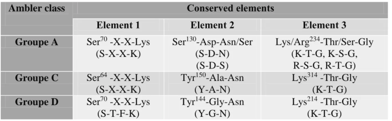
Table 1. Comparison of the three conserved regions in group A, C and D active-site serine
β-lactamases according to the Ambler classification ……….. 19
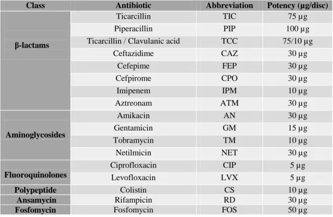
Table 2. Abbreviation and Potency of antibiotic discs used with Enterobacteriaceae …… 36
Table 3. Abbreviation and Potency of antibiotic discs used with P. aeruginosa …………. 37
Table 4. Abbreviation and Potency of antibiotic discs used with A.baumannii …………... 37
Table 5. Resistance genes with sequences of primers ……….. 41
Table 6. Specific PCR programs for the different ESβL genes ……… 42
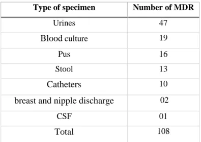
Table 7. Distribution of MDR strains according to the biological sample ………... 50
Table 8. Distribution of MDR strains according to the age, both from out and in-patients.. 51
Table 9. Distribution of MDR strains according to the sex, both from out and in-patients.. 51
Table 10. Distribution of MDR strains according to services ………... 52
Table 11. Antibiotic susceptibility of Enterobacteriaceae (n=68) ……… 55
Table 12. Antibiotic susceptibility of A.baumannii (n=20) ……….. 56
Table 13. Antibiotic susceptibility of P. aeruginosa (n=20) ………. 57
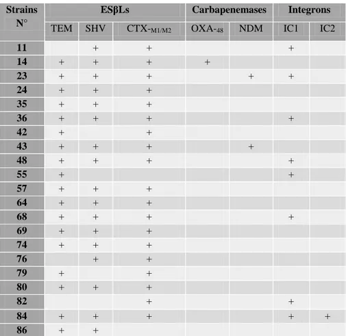
Table 14. β-lactamase coding genes in E. coli strains (n=36) ……… 61
Table 15. β-lactamase coding genes in K. pneumonaie strains (n=21) ………. 62
Table 16. β-lactamase coding genes were investigated in the other Enterobacterial species (n=11) ……… 62
Table 17. β-lactamase coding genes in P. aeruginosa strains (n=20) ……… 63
Table 18. β-lactamase coding genes in A. baumannii strains (n=20) ………... 64
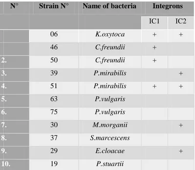
Table 19. Integrons class I and II in the other Enterobacterial species (n=11) ………. 66
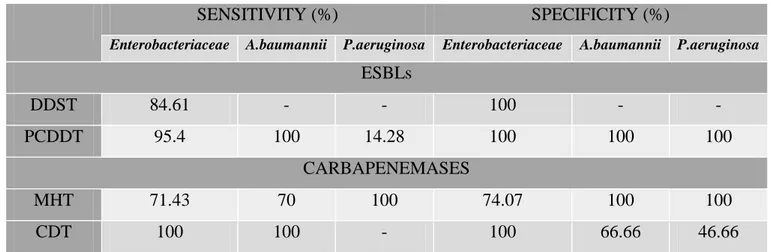
Table 20. Sensitivities and specificities of phenotypic methods for detection of ESβLs and carbapenemases production with molecular identification (PCR) as the reference method for Enterobacteriaceae, A.baumannii and P.aeruginosa isolates (n=108) ………. 67
Table 21. Total phenolic content in methanolic extracts of studied seaweeds ……….. 70
Table 22. Compounds identified in methanolic extracts from ME of seaweeds by HPLC-ESI-MS and HPLC-HPLC-ESI-MS-TOF ………. 71
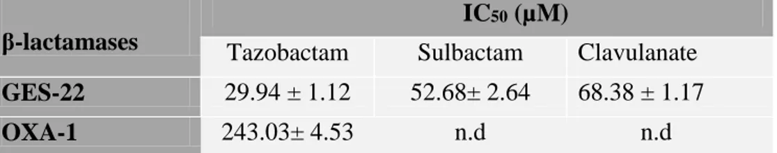
Table 23. Values of IC50 of clavulanic acid, tazobactam or sulbactam obtained with GES-22 and OXA-1 ……… 73
Table 24. IC50 of methanolic extract of seaweeds ………. 74
Table 25. Apparent kinetic parameters (KM and Vmax) of GES-22 and OXA-1 and type of their inhibition by ME of seaweeds ………. 75
Table 26. RMSD of different β-lactamases complexes studied ………. 79
Table 27. Libdock score ENERGY of the best 5 compounds and standards binding to β-lactamases ………... 80
Table 28. –CDOCKER energy/–CDOCKER inreraction energy of the best 5 compounds 83 Table 29. Amino acid residues of β-lactamases targets showing more frequent interactions in the company of the high score ligands predicted using Discovery studio ……….... 85
INTRODUCTION ………..……… 01
LITERATURE REVIEW
I. Nosocomial infections ………... 03I.1. Definition ………... 03
I.2. Causative Agents ………... 03
II. Gram-negative bacilli ……….……… 04
II.1. Enterobacteriaceae ………... 04
II.1.1. History and Taxonomy ………. 05
II.1.2. Biological characteristics and Habitat ……….. 05
II.1.3. Enterobacteriaceae infections ………... 05
II.1.4. Species belonging to the family Enterobacteriaceae ………... 05
II.1.3.1. E.coli ………... 06
II.1.3.2. K.pneumonaie ………. 06
II.2. Nonfermenting gram-negative bacilli (NFGNB) ……….. 07
II.2.1. Genus Acinetobacter ……… 07
II.2.1.1. History and Taxonomy ………... 07
II.2.1.2. Biological characteristics and Habitat ………. 08
II.2.1.3. Acinetobacter baumannii infections ……….. 08
II.2.2. Genus Pseudomonas ……… 09
II.2.2.1. History and Taxonomy ………... 09
II.2.2.2. Biological characteristics and Habitat ………. 09
II.2.2.3. Pseudomonas aeruginosa infections ……….. 10
III. Resistance of Gram-negative bacilli to β-lactams ………. 10
III.1. Types ………... 11
III.1.1. Inherent (natural) resistance ……….. 11
III.1.2. Acquired resistance ……… 11
III.2. β-lactamases………... 12
III.2.1. Classification ……….. 12
III.2.1.1. Extended-spectrum ß-lactamases (ESβLs) ………. 13
III.2.1.1.1. History and Definition ……….. 13
III.2.1.1.2. Types ……… 13
a. Ambler class-A ESβLs ………. 13
b. Ambler class-D ESβLs ………. 16
III.2.1.2. Carbapenemases ………... 17
III.2.1.2.1. History and Definition ………. 17
III.2.1.2.2. Types ……… 17
a. Ambler class-A carbapenemases ……….. 17
b. Ambler class-B carbapenemases ……….. 18
c. Ambler class-D carbapenemases ……….. 18
III.2.2. Active site and mechanism of action ……… 19
III.2.2.1. Serine β-lactamases ………... 19
III.2.2.2. Metallo-β-lactamases ……… 20
IV. Molecular Docking ………... 21
TABLE OF CONTENTS
IV.1. Docking methods and scoring functions ……….. 21
IV.1.1. Docking methods ……… 21
IV.1.1.1 Systematic search ……… 22
IV.1.1.2. Stochasticsearch ………. 23
IV.1.1.3. Simulationsearch ……… 23
IV.1.2. Scoring functions ………... 23
IV.1.2.1. Force-field-based ……… 24
IV.1.2.2. Empirical ……… 24
IV.1.2.3. Knowledge-based ………... 24
IV.2. Docking Algorithms used………. 24
IV.2.1. LibDock ………. 24
IV.2.2. C-DOCKER ……… 25
V. Studied seaweeds ………... 25
V.1. Green seaweeds ………. 26
V.1.1. Ulva intestinalis Linnaeus, 1953 ……….. 26
V.1.1.1. Description and Taxonomy ………. 26
V.1.1.2. Biological activities ……… 26
V.1.2. Codium tomentosum Stackhouse, 1797 ………... 27
V.1.2.1. Description and Taxonomy ………. 27
V.1.2.2. Biological activities ……… 27
V.1.3. Bryopsis pulmosa (Hudson) C.Agardh, 1823 ………... 28
V.1.3.1. Description and Taxonomy ………. 28
V.1.3.2. Biological activities ………. 29
V.2. Brown seaweeds ………... 29
V.2.1. Sargassum vulgare C. Agardh, 1820 ……….. 29
V.2.1.1. Description and Taxonomy ……… 29
V.2.1.2. Biological activities ………... 30
V.2.2. Dictyota dichotoma (Hudson) J.V.Lamouroux, 1809 ……… 30
V.2.2.1. Description and Taxonomy ………. 30
V.2.2.2. Biological activities ………. 31
V.2.3. Halopteris scoparia (Linnaeus) Sauvageau, 1904 ………... 31
V.2.3.1. Description and Taxonomy ………. 31
V.2.3.2. Biological activities ……… 32
V.2.4. Cystoseira compressa (Esper) Gerloff & Nizamuddin, 1975 ………. 32
V.2.4.1. Description and Taxonomy ……….. 32
V.2.4.2. Biological activities ……… 32
MATERIAL AND METHODS
I. Biological material………... 34I.1. Bacterial strains ……….. 34
I.2. Seaweeds………. 34
II. Methods ………... 35
II.1. Antibiotics susceptibility of MDR and detection of β-lactamases producing strains … 35 II.1.1. Antibiotics susceptibility ………... 35
II.1.2. Detection of β-lactamases ………... 38
II.1.2.1. Phenotypic detection ……… 38
II.1.2.1.1. Extended Spectrum Beta-Lactamases ………..……….. 38
b. Phenotypic confirmatory disc diffusion test ………. 38
II.1.2.1.2. Carbapenemases ……….. 39
a. Modified Hodge Test ………... 39
b. Inhibition tests ………... 39
II.1.2.2. Molecular detection ………... 40
a. DNA extraction ………. 40
b. Amplification of the β-lactamase genes by PCR ………... 40
c. Electrophoresis and detection ……… 42
II.1.2.3. Sensitivity and Specificity ………... 42
II.2. Expression and purification of recombinant GES-22 and OXA-1 proteins ……… 43
II.3. methanolic extracts of seaweeds ……… 43
II.3.1. Preparation ……….. 43
II.3.2. Phytochemical analysis ……… 44
a. Total phenolic content ………... 44
b. Vitamin E content ……….. 44
II.3.3. HPLC analysis ………. 44
a. Sample preparation ……… 44
b. HPLC-ESI-MS ……….. 45
c. HPLC-ESI-MS-TOF ……….. 45
II.4. GES-22 and OXA-1 β-lactamases inhibition assays ……….. 46
II.4.1. Kinetic study ……….. 46
II.4.2. Determination of IC50 parameters ……….. 46
II.4.3. Determination of inhibition type ……… 47
II.5. Molecular docking ………..……… 47
II.5.1. Evaluation test of the program used ……… 47
II.5.2. Process of molecular docking ………...……….. 48
a. Preparation of the protein ……….. 48
b. Preparation of the ligand ………... 48
c. Detection of the binding site ……….. 48
d. Docking ……….. 48
e. Results analysis ……….. 49
RESULTS
I. Distribution of MDR strains ……… 50I.1. Distribution according to species ……….. 50
I.2. Distribution according to biological sample ………. 50
I.3. Distribution according to age ……… 51
I.4.Distribution according to sex ………. 51
I.5. Distribution according to service (inpatients) ………... 51
II. Antibiotics Susceptibility of MDR strains ………. 53
II.1. Enterobacteriaceae ………... 53
II.1.1. Escherichia coli ………... 53
II.1.2. Klebsiella pneumoniae ……… 53
II.1.3. other enterobacterial species ……… 54
II.2. Acinetobacter baumannii ………. 54
II.3. Pseudomonas aeruginosa ………. 56
III. Detection of β-lactamases ………. 57
III.1.1. Extended Spectrum β-Lactamase ………... 57
a. Double disc synergy test (DDST) ……… 57
b. Phenotypic confirmatory disc diffusion test (PCDDT) ……….. 58
III.1.2. Carbapenemases ………... 59
a. modified Hodge Test (MHT) ………... 59
b. Inhibition tests ………. 59
III.2. Molecular detection ……… 60
III.2.1. β-lactamase genes in strains ……… 60
III.2.1.1. Enterobacteriaceae ………. 60
a. Escherichia coli ……… 60
b. K.pneumonaie ………... 60
c. other enterobacterial species ……….... 60
III.2.1.2. Pseudomonas aeruginosa ……….. 63
III.2.1.3. Acinetobacter baumannii ……….. 63
III.2.2. Class-I and Class-II Integrons in strains ……….. 65
III.3. Performance of Phenotypic detection tests ……….. 66
IV. Expression and purification of GES-22 and OXA-1 ……… 67
V. Phytochemical analysis ………... 69
V.1. Total phenolic content (TPC) ………. 69
V.2. Vitamin E content ……….. 69
V.3. HPLC analysis of ME of seaweeds ……… 69
VI. GES-22 and OXA-1 β-lactamases inhibition assays ………... 72
VI.1. Determination of kinetic parameters (KM and Vmax) of GES-22 and OXA-1……….. 72
VI.2. Determination of IC50 parameter ………... 73
VI.2.1. Case of clavulanic acid, tazobactam and sulbactam ……… 73
VI.2.2. Case of methanolic extract of seaweeds ………. 74
VI.3. Determination of inhibition type ………... 74
VII. Molecular docking ………... 78
VII.1. Evaluation test of the program used ……….. 78
VII.2. Docking study ………... 78
VII.3. Molecular interaction studies ………... 84
DISCUSSION
I. Distribution of MDR strains ………... 94I.1. Distribution according to species ……….. 94
I.2. Distribution according to biological sample ………. 95
I.3. Distribution according to age ………... 96
I.4.Distribution according to sex ………. 96
I.5. Distribution according to service (inpatients) ………... 96
II. Antibiotic resistance patterns………...………... 97
II.1. Enterobacteriaceae ………...……….. 97
II.2. Acinetobacter baumannii ………... 98
II.3. Pseudomonas aeruginosa ……….... 99
III. Phenotypic and molecular detection of ß-lactamases ………... 99
III.1. Extended Spectrum β-Lactamase ………... 101
III.1.1. Enterobacteriaceae ……… 101
III.1.2. Acinetobacter baumannii ………... 103
III.2. Carbapenemases ………... 104
III.2.1. Enterobacteriaceae ………. 104
III.2.2. Acinetobacter baumannii ………... 106
III.2.3. Pseudomonas aeruginosa ………... 108
IV. Phytochemical analysis ……… 109
V. GES-22 and OXA-1 β-lactamases inhibition assays ………. 111
VI. Molecular docking ……… 113
CONCLUSION & PROSPECTS ……….. 117
REFERENCES
APPENDICES
Appendix A. Chemical structures and pubchem CID of the compounds used
Appendix B. Total ion chromatograms (TIC) of ESI-MS-TOF in negative (first) and positive (second) modes of MEs of seaweeds
1
β-lactams are the most widely used antibiotic family in the world. This wide use is due to their broad spectrum of action, and their low toxicity (Handal and Olsen, 2000). This family comprises a large number of molecules, all characterized by the presence of a β-lactam ring. The introduction of third-generation cephalosporins (C3G) into clinical practice in the early 1980s to control infections with penicillinase-producing organisms was followed in 1983 by the description of the first Extended Spectrum β-lactamases (ESβLs) capable of hydrolyzing the extended-spectrum cephalosporins in Germany in strains of Klebsiella ozaenae (Knothe et
al., 1983). Carbapenems, the last antibiotics of the β-lactam class, are bactericidal antibiotics
often used as a last treatment option for severe infections caused by ESβLs-producing isolates (Wadekar and Swarooparani, 2017). However, extensive use of these molecules has facilitated the emergence of carbapenem resistant bacteria and hence the production of carbapenemases.
Gram-negative bacteria have become increasingly resistant to ß-lactams. Production of β-lactamases especially ESβLs and carbapenemases, is the main mechanism of resistance to these molecules by hydrolysing the amide linkage of the β-lactam ring (Philippon and Arlet, 2006). To inhibit the hydrolyzing activity of β-lactamases, β-lactam antibiotics are used with inhibitors (tazobactam, clavulanate, and sulbactam). These β-lactamases inhibitors are co-administered with β-lactam antibiotics and they are effective against some class A β-lactamases whereas ineffective against class B and most of the class C and D β-lactamases. As a result of the presence of a β-lactam ring in these inhibitors and their extensive use in combination with β-lactam antibiotics, the β-lactamase in bacteria mutate continually developing their activity even against newly developed β-lactam (Drawz and Bonomo, 2010). Hence, new, non-toxic and potential β-lactamases inhibitors is an urgent need.
It is well known that, over the past decades, seaweeds have been attracting attention in the search for bioactive compounds to develop new drugs and healthy foods among which polyphenols, lipids, polysaccahrides, sterols, terpenes. Furthermore, various seaweed extracts were reported to have high antioxidant, anti-cancer activity and to influence anti-inflammatory responses. It is in this context that the present study, whose main objective is to characterize the type of β-lactamases produced by Gram-negative bacilli strains collected from community and hospitals in Algeria and to investigate the inhibitory effect of seven Algerian seaweeds [Ulva intestinalis, Codium tomentosum, Bryopsis pulmosa, Sargassum vulgare, Dictyota
dichtoma, Halopteris scoparia, and Cystoseira compressa] against GES-22 and OXA-1
β-lactamases variants, produced recombinantly. In order to develop this aspect, we have adopted the following methodology
2
Antibiotic susceptibility pattern of isolates to β-lactam antibiotics and other antibiotic classes. Phenotypic detection of ESβLs and carbapenemases using several tests;
Double Disc Synergy test (DDST)
Phenotypic Confirmatory Disc Diffusion Test (PCDDT) Modified Hodge Test (MHT)
inhibition tests
Characterization of the β-lactamase genes (blaTEM, blaSHV, blaCTX-M1-M2, blaPER-2, blaVEB, blaGES,
blaKPC, blaOXA-M (23,24,51,58), blaVIM, blaIMP, blaNDM) and the integrons class I and II by PCR
technique among one hundred and eight clinical isolates collected.
Investigation of the inhibitory effects (in vitro) against GES-22 and OXA-1 β-lactamases variants, produced recombinantly in methanolic extracts of the seven studied seaweeds. Identification of the compounds of methanolic extracts by HPLC-ESI-MS and
HPLC-ESI-MS-TOF.
Finally, the prediction (in silico) of the structure of a molecular complex (β-lactamases-compounds identified in MEs and other molecules) by molecular docking using Discovery Studio (DS) 2016 software (Accelrys Software Inc., San Diego).
3
I. Nosocomial infections
I.1. DefinitionNosocomial infection term lies the problem of the relationship between the hospital and the infection acquired by the patients who stay there. Indeed, the word "nosoconium" means "hospital", it comes from Greek meaning: nosos=disease and komien= to care for. Nosocomial infections -also known as hospital acquired infections (HAIs) are defined by the World Health Organization (WHO) as:
“… infections acquired during a hospital stay that were neither present nor in incubation at admission. Infections occurring more than 48 hours after admission are usually considered nosocomial.” (WHO, 2002). Infections in newborn babies that occurred during or after delivery are considered nosocomial (Orrett et al., 1998).Nosocomial infections are considered to be the most common complication affecting hospitalized patients. The WHO estimates that developing countries have as much as a 20 times higher risk of nosocomial infections than developed countries (WHO, 2007).
I.2. Causative Agents
Nosocomial infections are usually caused by bacteria in ninety percent of cases. Moreover, viruses, protozoa and fungi are rarely involved (Bereket et al., 2012). A range of Gram-negative organisms are responsible for HAIs, the Enterobacteriaceae family being the most commonly identified group overall. Gram bacilli negative represent 60% of which E.coli is predominantly involved. Gram-positive cocci represent 30% (Thiolet et al., 2013).
The increased concern about infections led to increased use of antibiotics. Widespread use of these molecules is often cited as a cause of the emergence of multidrug-resistant organisms (MROs) as nosocomial pathogens. The multi-drug resistant Gram negative bacteria (MDR-GNB) are by far the most important and costly today, as the vast majority of nosocomial infections are caused by this type of bacteria (Livermore, 2012). Microorganisms usually implicated in these infections include among others Escherichia coli, Klebsiella species, Pseudomonas aeruginosa, Acinetobacter baumannii and Staphylococcus aureus which are rapidly gaining resistance because of the broad spectrum antibiotics used in an attempt to control them. MROs are bacteria that have become resistant to numerous antimicrobial agents, which have inadvertently led to increased morbidity and mortality from HAIs (NHMRC, 2010). Globally, methicillin-resistant Staphylococcus aureus and MDR GNB are described as
4 problematic MROs (WHO, 2015). A large proportion of HAIs are caused by MROs; however, specific numbers have not been identified or provided (WHO, 2015). Unfortunately, MROs including Pseudomonas aeruginosa, Acinetobacter baumannii, and extended-spectrum β-lactamases (ESβLs) or carbapenemase-producing Enterobacteriaceae, are increasingly being reported worldwide (Peleg and Hooper, 2010).
II. Gram-negative bacilli
Gram-negative bacilli, frequently isolated from bacteriology laboratories, occupy an important place in human pathology. Generally, they are divided in two groups: enterobacteria which can grow in the presence of bile salts and use lactose as an energy source on MacConkey’s agar and non-fermenting Gram-negative bacilli that cannot use lactose (so-called “non-lactose fermenters”) include, most prominently Pseudomonas and Acinetobacter species
as well as less common organisms Stenotrophomonas, Burkholderia,
and Achromobacter species(Liassin, 2000; Mehrad et al., 2015). In this literature review, we will focus on enterobacteria, Pseudomonas sp and Acinetobacter ssp, because they are the germs targeted in this study.
II.1. Enterobacteriaceae II.1.1. History and Taxonomy
Enterobacteria are a large group of bacteria with a strong similarity. The creation of this group was proposed by Rahn, (1937) as Enterobacteriaceae, in which he collected the bacterial genera such as Escherichia, Klebsiella, Salmonella, Shigella in the unique genus Enterobacter.
Enterobacteria are Eubacteria. Phylogenetic studies place them in the phylum Proteobacteria, Class Gammaproteobacteria, Order Enterobacteriales and to the family Enterobacteriaceae. This family comprise a large group of genetically and biochemically related bacteria (Brenner et al., 2005). The family name is still maintained, however, the classification of bacteria in the family has evolved a lot. Until the early 1960s bacterial classification was largely based on phenotypic characteristics and culture-based observations. Currently, Enterobacteriaceae are classified on the basis of the genetic relationships between members and their similarity to other closely related bacteria using 16S rRNA gene sequences analysis (Joly and Reynaud, 2002).
5
II.1.2. Biological characteristic and Habitat
The name Enterobacteriaceae, was given because the germs of this family are mainly pathogens of the humans and animals digestive tract and others are normal colonizers of this digestive tract (Escherichia coli, Enterobacter spp., Klebsiella spp.). Enterobacteriaceae are a major component of the normal human intestinal microbiota but relatively uncommon at other body sites (Balows et al., 1992). However, some species are insect- or plant-associated (Janda and Abbott, 1998). Although, they are also present in the environment. They are found in water, soil and some foods. They are able to disseminate easily by hand-carried transmission or via the contamination of the water and the food.
II.1.3. Enterobacteriaceae Infections
Most of the bacteria belonging to the Enterobacteriaceae family are harmless and not causing disease symptoms. A large group of bacteria from this family are opportunistic microorganisms, posing a threat to the elderly, weakened, sick or in a state of immunological suppression. The vast majority of bacteria present in the digestive tract is harmless to humans, however, some species of the Enterobacteriaceae family are considered to be pathogenic organisms responsible for various types of infection, including intestinal diseases (Jarząb et al., 2011). Nosocomial infections are mainly urinary tract infections, wounds surgical procedures, pulmonary infections, septicemia, and other localizations. More bacteria already mentioned in community infections with a multi-resistance profile mention are of Enterobacter sp., Serratia sp. However, community acquired infections are mainly urinary tract infections caused by E. coli, pulmonary infections caused by K. pneumoniae and food poisoning caused by Salmonellae.
II.1.4. Species belonging to the family Enterobacteriaceae
Enterobacteriaceae family is defined by a set of general common bacteriological characteristics. These are Gram-negative bacilli, non-sporulating, optional aero or anaerobes, motile or immotile, growing rapidly on media ordinary, not having oxidase, reduce nitrates to nitrites, ferment D-glucose with or without gas production (Eyquem et al., 2000; al., Avril2000). Possess a common antigen called Kunin antigen or ECA (enterobacterial common antigen) (Delarras, 2014). The majority of Enterobacteriaceae targeted in this study are: E.coli and K. pneumoniae. For that, it seems necessary to briefly recall some general characteristics of these bacteria.
6
II.1.4.1. Escherichia coli
E. coli was first isolated in 1885 by Theodore Escherich, a German pediatrician, in stool of infants, which he called Bacterium coli commune (Kaper et al., 2004). The name E. coli is given in 1919 by Castellani and Chaombers (Grimont, 1987). Since then, E. coli has become the best-known bacterium that has been the most studied by fundamentalists for physiology and genetics search (Avril et al., 2000). It is the dominant species in the commensal flora, particularly in the digestive tract of the human. It colonizes from the first hours of birth (Bonacorsi et al., 2001; Janda and Abbott 1998). However, following the acquisition and combination of virulence factors, this bacterial species can also behave as an intestinal or extraintestinal localized pathogen (Levine, 1987; Pohl, 1993). Moreover, E. coli is very responsive in the environment: water, soil, and in food (Baraduc et al., 2001).
E. coli has particular biochemical characteristics that allowing to differentiate it from other species among which, the production of indole from tryptophan, lack of use of citrate as a carbon source and lack of production acetoin (Joly and Reynaud, 2002), in addition to the common bacteriological characteristics of Enterobacteriaceae, non-sporulating, generally motile, aero-anaerobic facultative, with respiratory and fermental metabolism, negative oxidase, catalase positive and nitrate reductase positive (Bidet and Bingen, 2011). E. coli is naturally sensitive to antibiotics. However, face to the intensive use of these molecules and under their conjugate effects and resistance gene transfer, it may become, resistant (Adjidé et al., 2006).
II.1.4.2. Klebsiella pneumoniae
Carl Friedlander (1882) first described Klebsiella pneumoniae, as an encapsulated bacillus under the name of Friedlander’s pneumobacillus isolated from the lungs of patient who had died from pneumonia. The genus Klebsiella described for the first time by Trevisan (1885) and he was named it to honor Klebs Edwin, a German microbiologist (Trevisan, 1887).
Klebsiella pneumoniae, is a very widespread germ in environment, it is frequently isolated from plants, soil and water. In humans, it exists as part of the normal microbiota of the nasopharynx and gastrointestinal tract. This species colonizes on the mucosal surfaces of mammals such as humans, horses, or swine (Cruz-Córdova et al., 2014). The type species K. pneumoniae are facultative anaerobic bacilli, non-spore-forming, capsulated, non-motile, oxidase negative, positive nitrate reductase, positive urease, positive Voges-Proskauer reaction,
7 which ferments mannitol and glucose with gas production, , (Janda and Abbott, 2006; Cruz-Córdova et al., 2014).
K. pneumoniae is considered one of the most important opportunistic pathogens associated with nosocomial and community-acquired infections (Podschun and Ullmann, 1998). It is among the most pathogen with high morbidity and mortality rates worldwide. This species is the chief cause of various HAIs involving upper and lower respiratory tract and urinary tract infections. It is also responsible for gastrointestinal infections and liver abscess in community. (Podschun and Ullmann, 1998; Tsai et al., 2008)
II.2. Non-fermenting gram-negative bacilli (NFGNB) II.2.1. Genus Acinetobacter
II.2.1.1. History and Taxonomy
The history of the genus Acinetobacter began in 1911 with the discovery of a microorganism called Micrococcus calcoaceticus by the Dutch microbiologist Beijerinck from soil sampling (Baumann et al., 1968). Schaub and Hauber rediscover this bacterium in 1948, from samples of the human urinary system (Schaub and Hauber, 1948). Brisou and Prévost (1954) propose the designation of the genus Acinetobacter (from the Greek akinetos: "non motile" to group a heterogeneous collection of immobile, Gram-negative and positive or negative oxidase reaction. In 1968, Baumann et al., restricted the genre Acinetobacter to only negative oxidase strains and have recognized a unique species that they have proposed to name Acinetobacter calcoaceticus. Three years later, this proposal will be endorsed by the "Subcommittee on Moraxella and Allied Bacteria". In 1974, the genus Acinetobacter has been listed in the edition of Bergey's Manual of Systematic, with the description of a single species, Acinetobacter calcoaceticus as typical strain for genus (A. calcoaceticus ATCC 23055). In 1986, Bouvet and Grimont by DNA / DNA hybridization techniques, distinguished 12 genomic species, some are clearly named as A.baumannii, A. calcoaceticus, A. haemolyticus, A. johnsonii, A. junii and A. lwoffii (Bouvet and Grimont, 1987).
The species’ names have endured substantial taxonomic changes over the years due to the advanced understanding of molecular methods of the genetic make-up of this group of microorganisms. Recent classifications which seem to have gained wide acceptance among bacterial taxonomists have categorized the genus Acinetobacter in the domain of Bacteria, phylum Proteobacteria, class of Gammaproteobacteria, order of Pseudomonodal and is
8 classified in the family Moraxellaceae. The species A. baumannii, A. haemolyticus and A. calcoaceticus are of clinical significance (Almasaudi, 2018).
II.2.1.2. Biological characteristic and Habitat
Bacteria of the genus Acinetobacter are Gram-negative bacilli or coccobacilli, sporulating, sometimes capsulated, strict aerobic, oxidase-negative, catalase positive, non-motile and non-fermenting (Jans et al., 2004; Peleg et al., 2008). These are, often grouped in pairs or in chains of variable length. In the exponential phase, their diameter typically ranges from 0.9 to 1.6 µm and their length from 1.5 to 2.5 μm (Doughari et al., 2011; Jung and Park, 2015).
Members of the genus Acinetobacter are considered ubiquitous organisms (Peleg et al., 2008). Whose main habitat is soil, sewage, as well as food sources that are stored in chilled conditions (Tomaras et al., 2003). In fact, not all species of the genus Acinetobacter have their natural habitat in the environment (Peleg et al., 2008). In humans, Acinetobacter spp. are part of the flora of healthy skin, mucous membranes and the pharynx, and human respiratory secretions (Almasaudi, 2018). In hospitalized patients ‘”are often found in the skin, oropharynx, and digestive tract (Jung and Park, 2015).
II.2.1.3. Acinetobacter baumannii Infections
A. baumannii is the most frequently species involved in nosocomial and community-acquired infections, followed by A. pittii and A. nosocomialis within the genus Acinetobacter, (Peleg et al., 2008). A. baumannii has become in the recent years a clinically relevant pathogen, involved in a wide range of infections. Infections due to A. baumannii are most often nosocomial, rarely community-based (Bergogne-berezin and towner, 1996). A. baumannii is able to survive for several days in the hospital environment on abiotic surfaces (Tomaras et al., 2003). This persistence can be explained no only by its great adaptability but also by its antimicrobial resistance (Gaddy and Actis, 2009). The main severe nosocomial infections due to A.baumannii are pneumopathies. A. baumannii nosocomial pneumonia occurs in intensive care units (ICUs) with a frequency of 3–5% and with death rates of 30–75% (Doughari et al., 2011). However, in patients who require prolonged mechanical ventilation, this latest is a major risk factor of pneumonia. In addition, this micro-organism can cause infections of the skin and soft tissue, including the wounds (Johnson et al., 2007). Bacteremia and secondary meningitis are also among the severe nosocomial infections caused by A. baumannii (Peleg et al., 2008).
9 The incidence of A.baumannii varies from one site to another. However, this microorganism is the second most common etiologic agent among all the Gram-negative bacteria (Almasaudi, 2018).
Community acquired A. baumanni (CA-Ab) infections are rare but serious cause of community-acquired pneumonia, predominantly occur in countries with tropical or sub-tropical climates. They are more likely to occur in the humid months of the year. Moreover, CA-Ab infections affect individuals with risk factors, which include excess alcohol consumption, diabetes mellitus, smoking and chronic lung disease (Dexter et al., 2015).
II.2.2. Genus Pseudomonas II.2.2.1. History and Taxonomy
The family Pseudomonadaceae belongs to a large group of bacteria that in the past has been conventionally referred to as "non-fermenting" (Hansen, 1991). The classification into genera and species within this family has long been based on simple phenotypic orientation. The simplification of this classification was carried out by Stanier who studied mainly the assimilation of carbonaceous substances, and by Palleroni who classified Pseudomonas species into 5 genomic groups (Martin, 2007).
The genus Pseudomonas was described by Walter Emil Friedrich August Migula, a Botanist devoted to plant taxonomy (Palleroni, 2003). This genus is classified in the family Pseudomonadaceae, the order of Pseudomonadales, the class of Gammaprotobacteria, division of Proteobacteria, branching of Prokaryotes and reign of Bacteria (Garrity et al., 2010)
II.2.2.2. Biological characteristic and Habitat
Bacteria of the genus Pseudomonas correspond to bacilli Gram-negative non sporulating, strict aerobes, usually motile by a polar ciliature, do not ferment carbohydrates, do not fix nitrogen and are not photosynthetic, chemotropic, with oxidative metabolism. They are oxidase positive or negative, catalase positive. The strains are easily grown on the usual culture media, nutrient agar (Yumoto et al., 2001). This genus includes fluorescent species producing pigments specific. The two most common and characteristic pigments are pyocyanin and pyoverdine which are soluble in culture media. The two pigments are produced by Pseudomonas aeruginosa, but this latest can be lost the pigments by mutation. P. fluorescens, P. putida, P.syringae, and P. cichorii produce only pyoverdine (Matewish and Lam, 2004). However, some strains don’t produce these pigments such as P. alcaligenes and P. stutzeri
10 (Martin, 2007). The word ‘aeruginosa’ comes from the Latin word for verdigris or copper rust. This describes the blue-green bacterial pigment seen in cultures of P. aeruginosa. This species was first obtained in 1882 in pure culture by Gessard from wounds that had produced blue-green discoloration (Forkner, 1960).
Pseudomonas is ubiquitous in nature. It has been found in many surface waters and soils and infects a number of common plants. It is frequent in warm waters in which there has been human or animal activity. They are considered a commensal flora in humans or animals. Some play a pathogenic role including Pseudomonas syringae in plants and Pseudomonas aeruginosa in humans and animals. In hospitals, Pseudomonas are found in the environment patients (sinks, faucets and sanitary equipment). This ubiquity is associated with their metabolic versatility and their ability to adapt to the most hostile conditions (Meghdas et al., 2004).
II.2.2.3. Pseudomonas aeruginosa Infections
The genus Pseudomonasis one of the most complex of Gram-negative bacteria. Their mechanism of action varies according to the species and the host. Some species are pathogenic for humans, animals (Nishimori et al., 2000) and plants (Akkermans et al., 1996, Munsch and Alatossava, 2002) while others are useful. P. aeruginosa is an opportunistic pathogen for humans. It is an important cause of nosocomial infections and community acquired infections in immunocompromised patients and patients with structural lung disease (John et al., 2017).
P. aeruginosa is the primary cause of ventilated, associated pneumonia in the intensive care unit and, lead to a broad spectrum of disease such as urinary, burn, respiratory infections, and septicemia (Fazeli et al., 2012). Hospital-acquired infections caused by this organism are often associated with high morbidity and mortality because these microorganisms are virulent and have a limited susceptibility to antimicrobials (Dwivedi et al., 2009).
III.
Resistance of Gram-negative bacilli to β-lactams
ß-lactams form a broad class of antibiotics that includes derivatives of penicillin, cephalosporins, monobactams, carbapenems and ß-lactamase inhibitors. These antibiotics all contain a common element in their molecular structure known as ß-lactam ring which confers the antibiotic activity. For over 60 years, β-lactams have been the first line of antibiotic treatments for many community- and hospital-acquired infections, including those caused by multidrug-resistant pathogens. Despite the enormous efforts to develop new β-lactam
11 derivatives to overcome growing bacterial resistance, so far no single molecule escapes from hydrolysis by several of the thousands of β-lactamases described (Juan et al., 2017). Gram-negative bacteria have become increasingly resistant to ß-lactams, rendering infection by these strains very challenging to treat. For this, antibiotic development programs have placed greater emphasis on the identification of natural antibiotics other than β-lactams, such as aminoglycosides and tetracyclines, followed by optimization of (fluoro) quinolone molecules as a way of circumventing β-lactam resistance in negative bacteria (Bush, 2016). Gram-negative bacteria are either naturally resistant to β-lactams, or they have acquired resistance.
III.1. Types
III.1.1. Inherent (natural) resistance
Antibiotic resistance is the ability of a bacteria to resist the effects of an antibiotic. Natural or intrinsic resistance is caused by the structural characteristics of bacteria and it is not associated with the use of antibiotics. It is common to all bacteria of the same species. This resistance is due to the presence of common chromosomal genes to all the bacteria of the same species and transmitted to the offspring. Intrinsic resistance determines the Wild-type phenotype of bacterial species against antibiotics (Mayer et al., 2000).
The intrinsic resistance in Gram-negative bacilli is manifested by chromosomal cephalosporinases which express themselves either constitutively or inducible (Cavallo et al., 2004) or due to an outer membrane that establishes a permeability barrier against the antibiotic. For example, Gram-negative bacteria are intrinsically resistant to penicillin G by virtue of their double membrane structure which prevents the antibiotic from accessing the cell wall target. In addition to this, the expression of oxacillinase in A. baumannii.
III.1.2. Acquired resistance
Bacteria developed several mechanisms in order to acquire resistance to antibiotics previously sensitive. All require either the modification of existing genetic material (mutation) or the acquisition of new genetic material from another source. Acquired resistance occurs from (i) acquisition of exogenous genes by plasmids (conjugation or transformation), transposons (conjugation), integrons and bacteriophages (transduction), (ii) mutation of cellular genes, and (iii) a combination of these mechanisms (Giedraitienė et al., 2011). To counter the adverse effects of ß-lactam antibiotics, bacteria have evolved in diverse ways: i) mutations leading to loss or under-expression of porins that disallow entry of ß-lactams, ii) production of new
12 penicillin binding proteins that have low affinity to ß-lactams, iii) expulsion of ß-lactams from periplasmic space mediated by efflux pumps and iv) production of enzymes that hydrolyze ß-lactam rings. The main mechanism of resistance acquired to β-ß-lactams in Gram bacilli negative is the production of β-lactamases (cavallo et al., 2004).
III.2. β-lactamases
Before the use of ß-lactams in medicine, certain bacteria produced already ß-lactamases. These enzymes may have played a minor role in metabolism of the cell wall, or to protect the bacteria against ß-lactams produced by fungi in the environment. Whatever their function, it is the increased and uncontrolled human use of these antibiotics that has favored the emergence of bacteria carriers of ß-lactamases (Livermore, 1998). Dissemination of resistant strains and the emergence of new mechanisms for resistance, particularly production of new β-lactamases, pose serious problems for the medical world. ß-lactamases degrading/modifying the ß-lactam ring by cleaving the amine linkage of ß-lactams (Yamaguchi et al., 2005; Zhang and Hao, 2011). These flexible enzymes have been detected in both Gram-positive and Gram-negative bacteria, but these enzymes are especially important in Gram-negative bacteria as they are the most common cause of β-lactam resistance in this group of bacteria (Bush et al. 1995; Livermore, 1995).
III.2.1. Classification
There are two classifications of β-lactamases: structural classification of Ambler and functional classification of Bush-Jacoby-Medeiros. The most used in current medical practice is currently that of Ambler. This classification is based on the amino acid sequence homology of β-lactamases. It divides these inactivating enzymes into four groups (A to D) according to the primary structure of the enzyme. The enzymes of classes A, C and D are called active serine (serine type), which require an active site serine residue to catalyse the ring opening of the ß-lactams and are mostly penicillininases or cephalosporinases; while class B groups are metallo-β-lactamases (MβLs) (metallo-enzymes type), which require one or two zinc ions in their active site for their activity (Philippon et al., 2016). Depending on the hydrolyzed substrate, lactamases are therefore calling penicillinases, cephalosporinases, extended-spectrum ß-lactamases or carbapenemases. The highly problematic strains carry ESβL and carbapenemase genes.