This manuscript bas been reproduced from the microfilm master. UMI films the text directly from the original or copy submitted. Thus, some thesis and dissertation copies are in typewriter face, while others may be from any type of computer printer.
The quality of this reproduction is dependent opon the quality of the copy submitted. Broken or indistinct print, colored or poor quality illustrations and photographs, print bleedthrough, substandard margins, and improper alignment can adversely affect reproduction.
In the unlikely event that the author did not send UMI a complete manuscript and there are missing pages, these will be noted. Also, if unauthorized copyright material had to be removed, a note will indicate the deletion.
Oversize materials (e.g., maps, drawings, charts) are reproduced by sectioning the original, beginning at the upper Ieft-hand corner and continuing from left to right in equal sections with small overlaps. Each original is also photographed in one exposure and is included in reduced form at the back of the book.
Photographs included in the original manuscript have been reproduced xerographically in this copy. Higher quality 6' x 9" black and white photographie prints are available for any photographs or illustrations appearing in this copy for an additional charge. Contact UMI directly to order.
UMI
A Bell & Howell Information Company
300 Nonh Zeeb Road, Ann Aibor MI 48106-1346 USA
COMPLICATIONS AND
SEQUELAE
OF
MENINGOCOCCAL
DISEASE IN QUÉBEC,
1990-1994
par
Lonny Erickson
Département des sciences de la santé communautaire Mémoire présenté
à
la Faculté de médecineen vue de l'obtention du grade de maitre ès sciences (M.Sc.)
en sciences cliniques février 1998
Acquisitions and
Bibliographie Services Acquisitions et services bibliographiques
395 Wellington Street
Ottawa ON K1A ON4 Canada
395, rue Wellington
Ottawa ON K1A ON4
canada
The author bas granted a
non-exclusive licence allowing the
National Library of Canada to
reproduce, loan, distnbute or sell
copies of this thesis
in
microform,
paper or electronic formats.
The author retains ownership of the
copyright in this thesis. Neither the
thesis nor substantial extracts from it
may be printed or otherwise
reproduced without the author' s
penmss1on.
Your 6le Vot/1J niltinlncB
L'auteur a accordé une licence non
exclusive permettant
à
la
Bibliothèque nationale du Canada de
reproduire, prêter, distribuer ou
vendre des copies de cette thèse sous
la forme de microfiche/film, de
reproduction sur papier ou sur format
électronique.
·
L'auteur conserve la propriété du
droit d'auteur qui protège cette thèse.
Ni la thèse ni des extraits substantiels
de celle-ci ne doivent être imprimés
ou autrement reproduits sans son
autorisation.
0-612-35672-8
Objectives
To determine the frequency and the nature
of
complications and sequelae of serogroup B and serogroup C meningococcal disease, during a recrudescence causedby
a virulent cloneof
serogroup C, serotype 2a Neisseria meningitidis. To evaluate the qualityof
life of survivors.Methods
The study population included all cases of culture-proven serogroup B and C meningococcal disease reported in
the
provinceof
Québec, Canada, between 1 January 1990 and 31 Deœmber 1994. Complications and sequelae were assessedby
review of medical files, postal questionnaires, and telephone interviews.Results
There were 167 cases of serogroup B and 304 cases of serogroup C infection. The largest number of cases was observed in the under 1 year age group for serogroup B and in the 10-19 year age group for serogroup C. Fatality rates were 7% for serogroup Band 14% for serogroup C.
%).
Only 3%
of
survivors of serogroup B cases had physical sequelae. 15% of survivors of serogroup C infection had one or more significant physical sequelae (skin scars 12%, amputations 5%, significant sensorineural hearing loss 2%, renal failure 1%, other sequelae 4%. Among cases without identified physical sequelae who completed the questionnaire, 19% reported a reduction in their qualityof
life attributable to the disease.Conclusions
These results confirm the gravity
of
disease caused by serogroup C, serotype 2a Neisseria meningitidis and support vaccination for contrai of outbreaks and epidemics of disease caused by this particular strain.Key Wortil:
Menlngococcal dlaeae, Ne/sserla menlngltldls, mortallty, sequelae, complications.
Objectifs
Cette étude vise à déterminer la fréquence et la nature des
':Omplications et des séquelles des infections invasives à méningocoques
de sérogroupes B et C.
La
qualité de vie des survivants a également été
évaluée.
Méthodes
Tous les cas confirmés d'infection invasive
à
méningocoques de
sérogroupes B et
c.
survenus au Québec, du 1 janvier 1990 au 31
décembre 1994 sont inclus dans la population de l'étude.
La
présence des
complications et séquelles a été évaluée
à
partir de l'ensemble des
informations disponibles dans les dossiers médicaux,
lesquestionnaires
postaux, et les entrevues téléphoniques.
Résultats
Le nombre de cas s'établit à 167 pour sérogroupe B et à 304 pour
sérogroupe C. Le plus grand nombre de cas a été observé chez les
enfants âgés de moins d'un an pour sérogroupe B et de 10 à 19 ans pour
sérogroupe C. Le taux de mortalité était de 7% pour sérogroupe B et de
14% pour sérogroupe C. Pour le sérogroupe B, seulement 3% des
survivants avait des séquelles, comparé à 15% pour le sérogroupe C
(cicatrices 12%, amputations 5%, perte d'audition 2%, insuffisance rénale
1 %, autre 4%). Pour les individus n'ayant pas de séquelles qui ont
répondu au questionnaire, 19% ont cependant noté une réduction de leur
qualité de vie
à
cause de la maladie.
Conclusions
Les résultats de l'étude confirment la gravité des infections
invasives
à
Neisseria meningitidisde sérogroupe C, sérotype 2a, et
justifient une utilisation libérale de la vaccination pour le contrôle des
éclosions et des épidémies.
Mots clés
Infections à méningocoques, Neisseria meningitidls, mortalité, séquelles, complications.
1. INTRODUCTION 1.1 Coatert of the Study l.2 Study Objectives
2.
UTERATURE REVIEW
2..1 Nature of Patlloaea 2.2 Traasmissioa
2.3 Prevaleace of MeniJllococal
Carriace
ud lafectloa 2...f !pidemiofoay of MeaJn10COtt1JI Dileue2..S Pathoeenesis of Meninaococcal Disease
2.6 Acate Meningococcemia: 2.. 7 Metastasis of Meningococci
2.8 Cllnical Forms of Meaingococcal Disease: 2..9 Pathology of Meningitis
2.10 Patllology ofSeqaelae
2.10.1 Patbology of Sequelae of Men.ingococœmia 2.102 Pathology of sequelae of meningitis
2.11 Epidemiofocy of Sequelae 2.12 Mortallty Rates
2.13 Meaincococcal Vaccines
2.13.1 Polysaccbaride vaccine eflic:acy
2.13.2 RttœJmendations for cumnt vaccine usage
2.13.3 Development of new Men.ingococcal Vaccines 3. METHODS 3.1 Type ofStudy 3.2 Case Ascertainment 3.3 Study Population 3.4 Data Collection
1
1 34
5 7 7 8 9 9 10 12 13 14 15 16 17 1819
19 19 20 213.4.1 Categories of Cases
3.4.2 Sources of Daia 3.4.3 Regional Healtb Boards
3.4.4 Contact of Survivors
3.4.S Postal Questionnaire
3 .4.6 Assessment of Disease Impact on Quality of Life
3.4.7 Refusais
3.4.8 Deceased and UD1nleeable lndividuals 3.4.9 Hospitals
3.4.10 Review ofHospi1alisation Record 3.4.11 Classification of Cases
3.4.12 Telephone Interview
3.S Ethial Coasldentiom 3.6 Revisioa of Fatality Rates
3. 7 Evaluation of Seqaelac
3.8 Classification of Playsical Seqaelae
3.9 Scorin1 of Qaestioaaaire: Redaction of QaaUty of Life 4. RESULTS
4.1 Namber of Cases by Year and Serogroup 4.2 Subtyping of Serograp C straias
4.3 Chancteristics of Shlcly Population 4.4 Participation Rate
4.5 Sources of Infonaatiom 4.6 Clinical Presentatto.
4. 7 Fatality and Complication Rates
4.8 Rates of Seqaelac
4.9 Ap-specifk Seqaelae ud Fatality Rates 4.10 Typa of Seqaelae vs. Ovenll Severity 4.11 Qaality of Life Impainaeat Scores
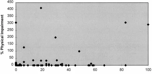
4.12 Correlation of Quay of Life lmpairment Score with Pbysical lmpairmeat Score 5. DISCUSSION S.1 Strengtbs of Stady 21 21 23 24 2S 2S 26 27 27 28 28 30 30 31 32 32 33
34
3" 35 35 36 37 38 40 42 43 44 45 4748
48S.l umts of Stady 49
S.3 Ilia 51
S.4 Evalutioa of Disease Impact 52
S.S Aaalysts of Rads 54
6. CONCLUSIONS
58
7. RESEARCH TEAM:
62
7.1 Pa lsciplll Raeardaen:: 62 7.l Comllltaats: 628. ACKNOWLEDGEMENTS
62
9.REFERENCES
63
LIST OF TABLES
Number
rit1e
PageTable 1 Case Variables and Their Sources
22
Table 2 Number of Cases by
35
Year and Serogroup
Table 3 Age Distnbution
of
Study Population 36Table4 Sources of Information
38
Table 5 Diagnostic Classification of Cases
39
Table 6 Percentage of Survivors with Sequelae 42
Table 7 Frequency of Physical Sequelae by Type and
43
SerogroupTable 8 Types of Sequelae of Serogroup C disease 44 versus Total Severity
Table 9 Summary of Serogroup B Sequelae
45
byCase
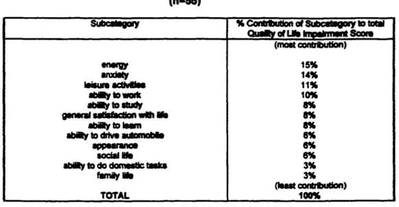
Table 10 Relative Importance of Subcategories in 46 Total Quality of Life lmpairment Score
LIST OF FIGURES
Number Tltle Page
F19ure 1 Status of Eligible Cases 37
Figure 2 Clinical Presentation of Serogroup C Cases 39 F19ure 3 Clinical Presentation of Serogroup B Cases
40
F19ure4 Evolution of Serogroup C Disease
by
41AgeGroup
F19ure 5 Evolution of Serogroup B Disease
by
41AgeGroup
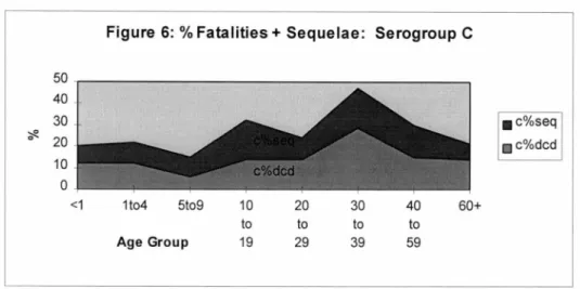
F19ure 6 Percentage of Fatalities and Sequelae 44 of Serogroup C Disease by Age Group
F19ure 7 Percentage of Physical lmpairment versus
47
Percentage Quality of.
Life
lmpairment ScoresF19ure 8 Various Levels of Disease Impact
52
LIST OF APPENDICIES
Number Trtle
Appendix 1 Questionnaire
Appendix 2 Letterof lntrodudion
Appendix 3 Consent Form
Appendix4 List of Participating Hospitals
Appendix 5 List of Variables
CIQ MSSS FRSQ CSF DIC LSPQ MADO CRC CUSE CSST ROS ARDS LPS
LIST OF ABBREVIATIONS
(in
order of appearance)
Comité d'immunisation du Québec
Ministère de la santé et des services sociaux du Québec
Fonds de recherche en santé du Québec Cerebrospinal Fluid
Disseminated lntravascular Coagulation Laboratoire de Santé Publique du Québec Maladies
à
déclaration obligatoireCentre de recherche clinique
Centre universitaire de santé de l'Estrie
Commission de la santé et de la sécurité du travail du Québec
Respiratory Distress Syndrome Adult Respiratory Distress Syndrome Meningococcal lipopolysaccharides
Neisseria meningitidis, a Gram-negative bacteria (commonly referred to as the meningococcus) causes both endemic and epidemic disease, principally meningococcal septicemia (meningococcemia) and meningitis (Centres for Disease Control and Prevention, 1985). Meningitis involves infection of the surface of the brai.n, while meningococcemia involves dissemination of meningococci in the bloodstream. Either condition
is
potentiallyfatal
andrequires
prompt treatment These infections are a serious public health problem worldwide. Resulting from the control of Haemophilus injluenzae type b infections, N. meningitidis bas become the leading cause ofbacterial meningitis in children and young adults in the United States (Centers forDisease
Control and Prevention, 1993). Surprisingly, meningococcal bacteria are commonly present in the upper respiratory tract of asymptomatic bumans (Caugant et al., 1994). This coloniz.ation is occasionally followed by the development of meningitis and/or acute septicaemia (Schwartz, 1989). 'Meningococcaldisease'
is therefore an appropriate term for these invasivemeningococcal infections which progress beyond the carrier state.
1.1 ConfUt
of
the StudyTwo serogroups of Neisseria meningitidis, B and C, each account for approximately 45% of cases in the United States (Centers for Disease Control and Prevention, 1993). In Canada, serogroup B strains have produced a relatively stable
number of cases of meningococcal
disease
in thelast
twenty years with a maximal incidence of infection in young infants over three months of age (V arughese et al., 1989). Outbreaks of group C meningococcal disease are common in North America,affecting
disproportionatelyschool
aged children and young adults (Jackson et al.;, 1995).An increase in
incidence
of meningococcal disease beganin
the province of Québec in 1990 and is associated with theemergence
of the serogroup C, serotype 2a strain of meningococci (Ashton et al., 1991). Similar strains were responsiblefor outbreaks in other areas in North America (Wbalen et al., 1995 ; Jackson et al., 1995) and Europe (Albertoni et al., 1987 ; Krizova & Musilek, 1995). A Canadian study demonstrated the predominance of a
new
strain ofthis
serotype, knownas
''ET-15". This new strain has been found to be particularly virul~t in terms of
case-fatality rate (Wbalen et al., 1995).
In
1992, many local and regional immunization campaigns against serogroup C meningococciwere
undertaken in regions of the province of Québec with outbreaks of type C meningococcal disease. These small scale campaigns eventually culminated with the decision to undertake a mass vaccination campaign of the entiie provincial population aged 6 months to 20 years (l.8 million persans) in 1992 (Direction de la santé publique, 1994). Tetravalent (serogroups A, C, Y, W135) or bivalent (serogroups A and C) vaccines were employed. This vaccination campaignwas
completed in the spring of 1993, reaching 84% of the target population, at an estimated cost of 25.7 million Canadian dollars (Buteau et al., 1997). Subsequent analysis of the incidence of meningococcal disease determinedtbat at least 37
cases
bad beenprevented
during thetirst
year following themass
vaccination campaign. The incidence of the disease droppedmarlcedly
in the unvaccinated fraction of the target population, while it remained unchanged among persons aged more than 20years.
This suggests that additional cases were avoided due to the contribution ofherd immunity (DeWals et al., 1996). During the1990-1994 period, serogroup B meningococcal disease remained endemic.
A thorough analysis of the overall impact on health of meningococcal
disease
is required to evaluate actual and potential benefits of immnnization and to guide public health policy. In additio~ this area of research bas been identified as aresearch priority by the Committee of Immunisation of Québec (CIQ) and the Québec Ministry of Health and Social Services (MSSS).
1.2 Study ObjectlvM
The goal of this project was to collect a maximum of information on complications, deaths, and sequelae among cases of serogroups B and C meningococcal diseasei
reported
in the province of Québec from 1990 to 1994. The study also questioned survivors on physical sequelae, treatments, assistance andservic:et
required in the long ~ and their impressions of the impact of thedisease on their quality of life. The results should enable a global evaluation of benefits of the type C meningococcal vaccination campaign completed in 1991-93. In addition, results will provide the data for decision making regarding future vaccination campaigns. This project bas been funded by the Québec ministry of
Health (MSSS), and the scientitic content of this project bas been given recognition at 100°.4 by the "Fonds de la recher~he en santé du Québec" (FRSQ) [ see appendix 6].
2. LITERATURE REVIEW
2.1
Nature of PathogenMeningococcal bacteria (Neisseria meningitidis) are diplococcal, Gram-negative bacteria. They are classified into serogroups by the antigenic nature of their polysaccharide capsule (AJ3,C,Y, and others). They can be further subclassified into serotypes, subtypes, and electrophoretic types (i.e. « ET-1 S » ). Although more than 13 serogroups have been identiti~ almost ail invasive disease is accounted for by the serogroups AJ3,C,Wl35, and Y. These bacteria have other somatic antigens, including a nucleoprotein, and a carbohydrate and lipid cell wall. The protein antigen and the polysaccharide capsule are very important for the virulence of meningococci.
2.2
T,.,,.,,,,..lon
Infection by meningococci begins with coloni7.ation of the upper respiratory tract. The resulting rhinopbaryngitis is almost always so subtle that it escapes detection upon clinical examination. Transmission is from a carrier
(usually asymptomatic) to another persan by airbome droplets containing meningococci which are mainly deposited on the mucosal surfaces of the nasopharynx and tonsils (Brandtzaeg, 1995; Pollock 1988). Overcrowding (and therefore low socioeconomic status) are risk factors for transmission (Voss et al., 1994 ). This is considered as a contributing factor to the high attack rate among new military recruits (Berild et al., 1980).
2.3 Preva/ence
of ltlenlngococcalC.rrlage and Infection
Approximately 100/o of the general population bas meningococci present in the nasopharyngeal region (Cartwright et al., 1987; Caugant et al., 1994); these individuals are "healthy carriers" of meningococci. ln
certain
situations of recrudescence of the disease, the rate of meningococcal carriage can climb to up to95% (Goldschneider et al., 1969). Tbere is not necessarily a strong correlation between the percentage of carriers and the number of individuals who develop invasive infections (meningococca1 disease). For example, in military cam~ or in prisons, carrier rates can be over 50% with very few cases of invasive infection (Fraser et al., 1973). However, in Québec en 1991 a large number of cases of meniqococcal disease
was
observed despite a low carrier rate (De W ais et al.,1994).
Overall, the most important factor in determining rates of disease is the presence of susceptible individuals in the population. The virulence of the
predominant serogroup is as important as the carrier rate in the occurrence of the disease.
2.4 Epidemiology of
Afen/ngococca/ O/aeaseThe incidence of meningococcal disease typically peaks in late winter. Cenain medical conditions increase risk of infectio~ such as terminal complement
deficienci~ aspleni~ and other diseases associated with immunosuppression
(such as .EilV or Streptococcus pneumoniae infection). Previously, military recruits in the United States (and elsewhere) had high rates of meningococcal
disease,
( especially serogroup C disease ), but these rates have declined since routine vaccinationwas
established (Brundage and Zollinger, 1987).2.5
Pathogenesl• of Meningococcal
D/seaseInfection typically consists of three steps. The first step is the coloniz.ation of the nasopharyngeal region by meningococci. Pathogenesis can follow in which meningococci succeed in entering the bloodstream. Finally, metastasis of meningococci can occur to the meninges or other locations in the body.
Mcningococci attach to receptors on mucosal epithelial cells via pili. Afterwards, they must cross the epithelium to enter the bloodstream. The presence of bacteria in the bloodstream is called bacteremia. The bacteria can then multiply in the bloodstream. The spread of a pathogenic germ in the bloodstream is called
bacteremia
This
condition can be present with or without the presence of cerebrospinal meningitis.2.6
Acute Men/ngococcemla:The tenn lllDlingoeo«aù refers to a meningococcal septicemia Acute meningococcemia involves brutal apparition of fever, c~ vomiting and skin
eruptions. ·This syndrome can lead rapidly to hypotension, vascular collapse, sbock, coma, and sometimes death. Endotoxin liberated from meningococci plays an important role in the pathogenesis of meningococcemia (Brandtz.aeg et al., 1989). Cytokines are activated, wbicb can cause hypotension, myocardial depression, and increased endothelial permeability (Monsalve et al., 1984). Other inflammatory mediators (platelet activating factor, leukotrienes, prostaglandins) can be produced causing tissue damage via enhanced granulocytic function and generation of intravascular clotting and thrombosis (Jafari and McCracken, 1992). Skin lesions are the result of microthromboses and/or acute vasculitis of small blood vessels, followed by suppuric necrosis. Meningococci can
sometimes
be cultun:d from these skin lesions.2. 7 Metastasia
of MenlngococciMeningitis is the most common presentation, but meningococci can cause local infections in cardiac valves, the pericardium, joints. lungs. or almost any
other tissues. The
nœninga
are the membranes surrounding the brain and thespinal
cord.Meningitis
is the generic name for chronic or acute inflammations of the meninges, whether microbial, viral, or chemical in nature. Clinical signs include perturbations in the cerebrospinal tluid (CSF), suchas
a reduction in the glucose/protein ratio or turbidity due to the presence of cells in the CSF.2.8 Clin/cal Forma of Men/ngococca/ Dlaeae:
A
bacteremia
probablyoccurs
in the majority ofcases
ofmeningococcal
disease (Apicella, 1995). Pneumonia
can
occasionallyoccur as
bacterial dissemination via endotracheal and branchial regions is possible. Nonnally, abacteremia
causesa
systemic reactionof fever, and therefore becomes a
septicemia. This fever reaction
can
be attenuated or even nonexistent in immunosuppressed patients (lymphoma, old age). Septicemia is usually accompanied by a petechial skin eruptio~ which can sometimes escape clinical examjnation (especially early in thecourse
of thedisease).
Classical complicationsof
septicemiaare
cardiovascularshock
anddisseminated
intravascular coagulation (DIC). Meningococci can multiply in different organs, although they usually do this inthe mcninges of the
central nervous system.2.9
Pathology of Menlngltls
Infection of the cerebrospinal membranes- , the cerebrospinal fluid and the surface of the brain generally
occurs
following bacteremia. An exception would be individuals with a CSF leak or fistula (often caused by head injury). Lymphatic disseminarion of bacteria from the nasopharyngeal mucosa is not thought to be a factorin
the pathogenesis of meningitis. The surface of the brain and the spinal cordmay
appear opaque, swollen and congested. The subarachnoid region contains cellular exudates. In certain cases, purulent trails can follow blood vessels. In fülminant infection. cellsinfiltrate
these vessels, producing vasculitis. Thrombotic occlusion of vessels can producea
cerebral infarctus, cortical destruction. and hemorrbage of the underlyingwhite
matter.2.10 Pathology of
Seque/aeSeqiula
are detined asany
residual pathology caused by the meningococcal infection and noted ina
subsequent medical exam. Many complex mechanisms exist wbich can produce sequelae, including, endotoxemia. increasedcortex
tb.is
can cause epileptic seizures ), septic shock, and disseminated inttavascular coagulation (DIC) (wbich can lead to necrosis and gangrene of extremities). Most common sequelae include skin necrosis, hearing loss, motor deficits, paralysis, and behavioral changes.2.10.1 Pathology
of
Sequelae of Meningococcemialn
meningococcal septicemia, many organs and areas may be affected. Persistent circulatory collapse is the single most important cause of death in fulminant meningococcemia (Brandtz.aeg, 1995). Capillary leakage syndrome can also occur, resulting in excessive extravascular tluid accumulation.Reduced renal function can occur as a sequela of meningococcemia Renal failure can occur as a complication of septic shock. This is associated with high levels of meningococcal lipopolysaccbarides (LPS). Peritoneal or haemodialysis or continuous hemofiltration are often used in the acute phase. Extremely high serum creatinine levels are a marker of severity and are thought to be cansed by a combination of renal failure, reduced excretion, and possibly increased breakdown of muscle proteins (Branttaeg çt al., 1989). A minority of shock patients never regain full kidney function, and transplantation is sometimes necessary. Although little research bas been performed to examine the pathological mechanism resulting in endotoxin-induced
rena1
failure in septic shock, widespread thrombosis in glomeruli and tubular necrosis bave been observed in primates with septic shock (V oss et al., 1991 ).Impaired 111111 fonction can also occur following septic shock. Adult respiratory distress syndrome (ARDS), an acute inflammatory capillary lealc syndrome of the lungs, can occur. Pulmonary compliance is reduced, and neutrophils bave been implicated in the pathogenesis (Repin~ 1992). High levels of LPS are also associated with ARDS (Brandttaeg et al., 1989). The exact molecular mechanisms leading to lung capillary leakage are unknown. A complex
interplay of LPS, cytokines, other inflammatory mediators and leukocytes may combine to alter the integrity of the lung capillary endothelium (Brandttaeg, 1995). Post-necrotic
skia scarring
is another common sequela ofmeningococcemia. The classical description states
that
« The fundamental pathologie lesion in the skin in meningococcemia is diffuse vascular damage». Fibrin and platelet thrombi with signs of leukocyte infiltration occlude vessels (Hill and Kinney, 1947). Meningococci can be present around the vessel wall of the dermis in the bacteremic state. Local endothelial cells are damaged which facilitates thrombus formation. Ecchymoses are related to consumption coagulopathy. This state can proceed to hemorrbage, ulceration and even gangrene in severe cases. This can result inscan,
and amputation of extremities.Decreased joint mobility is a rare sequela of arthritis caused by meningococcemia. Arthritis. can occur in various joints (knees, elbows, ankles, fingers) in either the septic fonn (with the presence of bacteria in the joint fluid), or the reactive fonn secondary to the inflammatory response. This can cause damage to the joint such as soft tissue swelling. Although this usually only causes temporary problems, they can be permanent (i.e. reduced range of motion of knee). Erosion of the
distal
femoral epiphyseal centre bas been reported (Olivieri et al., 1986), as bas periosteal elevation and demineralization of the distal humerus (Hammerschlag and Baker, 1976), and ankylosis (Scbaad, 1980).Pericarditis, endocarditis, and ocular complications are rare and transitory in documented cases and do not result in sequelae (Steven and Wood, 1995).
2.10.2
Pathology of sequelae of meningitis
Increasing levels of
LPS
or bacteria in theCSF
are associated with increasing disease severity,and
elicit cytokine release. The effects onthe
cerebral vasculature result in protein and leukocyteinflux,
altered brain glucose metaboli~ brain oedema and impaired cerebral circulation. The resulting cerebral oedema and ischemia can cause focal neurologie sequelae or death.Sensorineanl hearing loss is the classic example of a neurologie sequelae of meningitis. lt is thought
that
this is caused by inflammaifon ofthe
auditory nerve. This is supported by observations of impaired auditory nerve function as assessed by brainstem auditory evoked potentials. lmpaired langaage function and learning deficits mayresult.
An l!.mteady gait
bas
been proposed to be caused by vestibular damage in survivors ofmeningococcal meningitis (Farmer, 1945).Patients bave developed persistent bydrocepbalus and dementia , as well as poor concentration and emotional lability due to damage caused by meningeal inflammation by meningococci (Edwards and Baker, 1981 ).
Many other focal manifestations of meningitis can occur, such as epileptiform activity, cerebellar ataxia, sixth nerve lesion with diplopia, ~ seventb, and ninth nerve palsies, paralysis of facial muscles, and ipsilateral loss of taste sensation. However, in documented cases (Farmer, 1945) these manifestations are rare and virtually always transient. regressing completely (thus leaving no sequelae).
2.
f
1 Epldemlology ofSequelae
Many
published studies exist on sequelae of meningococcal disease, but none study specifically serotype 2a, serogroup C meningococcal disease. Frequency of sequelae as reported in the literature is variable.A meta-analysis of ail English language reports published from 1955 to 1991 (Baraff et al., 1993) reported a sequelae rate of 100/o for
the
meningitic form of diseasecausecl
by Neisseria meningitidis (ail serogroups combined).In a series of 562 patients in the Netherlands in 1989-1990, 8.5% of survivors of meningococcal disease were reported as baving serious sequelae (Schildkamp et al., 1996). ln Norway
in
1981-1982 lSoAt ofvictims of serogroup B meningococcal diseasewere
noted to have various sequelae (Djupesland et al., 1983).Finally
in Sweden an 11 % sequelae rate was reportedin
a series of predominantly serogroup B meningococcal disease from 1975 to 1989 (Berg et al .•1992). Although anecdotal reports mention
a
high prevalence of sequelae among survivors of meningococcal disease in Québec, there bave been no systematic studies of this subject.Hearing loss seems to
be
the most common of sequelae of bacterial meningitis, aftlicting 6% to CJOAt of meningococcal disease survivors (Voss et al., 1989; Dawson et al., 1990). Hearing losswas
reported in 9.5% of a predominantly serogroup B case series in Houston (United States) in 1977-1979 (Edwards and Baker, 1981 ). However, in a systematic study of 66 survivors of serogroup B meningococcal disease in Great-Britain, hearing losswas
noted in 5% of casesversus 3%
in
a
controlgroup
andno
other physical or psychologicalwere
detected (Moss, 1982). This demonstrates the facttbat
detected hearing loss may not actually have beencaused
by the meningococcal infection. Prevalencr. of hearing loss among survivors should therefore be interpreted with caution to avoid overestimation of hearing loss caused by meningococcaldisease.
Overall, there
are
significant differences in reported sequelae rates among studies. Many different factors can explain the variability of these observations,such as
definitions used for sequelae,the
smallnumber
ofcases
ineach
study, variations in the age of infected individuals, delays in treatment, length of follow-up, systematicuse
of various diagnostic tests, and finally thevirulence
of meningococci which can determine the relative frequency of the different clinical forms of meningococcal disease (meningitis vs. septicemia).2.12 Mortality
Rates _
ln the United States, the case-fatality rate is 13% for the meningitic form of the disease ( N. meningitidis isolated from the cerebrospinal tluid) and 11.5% for persans who bad N. meningitidis isolated from the blood (Centers for Disease Control
md
Prevention, 1993).Clinicians have noted that serogroup C meningococcal disease is particularly serious, with a high mortality rate. The Québec provincial surveillance system reported a mortality rate of 14% for serogroup C infection and 7% for serogroup B infection in confirmed cases from 1990 to 1994. This mirrors very
closely
reported
national mortality ratesin
Canada (1985 to 1992) of 14.5% for serogroup C and 6.00/o for serogroup 8 (Whalen et al.. 1995). A regional surveillance program in Lati~ Italy reported a mortality rate of 29% (26 deaths in 91 cases) for a series composed predominantly of serogroup C, serotype 2a meningococcal disease (Albertoni et al., 1987). In a meta-analysis of ail English-languagereports
of outcomes ofbacterial nieningitis published since 1955 (Baraff etal.,
1993), an overall mortality rate of 7.5% for Neisseria meningitidiswas
calculated (ail serogroups combined). Another study reviewing 261 cases of meningococcal disease in an American hospital (from 1957 to 1987) founci an overall case fatality rate of 10% (Havens et al., 1989). This rate varied widely over time (90/o for the period 1957-1963 versus 16% for the period 1980-1987). The severity of cases baving severe
disease
rose from 14% for the period 1957-1963 to 38% for the period 1980-1987. However, case-fatality rates did not vary with time when stratified by disease severity. This shows that crude case-fatality rates can be misleading if disease severity is not considered.2. 13 Meningococcal Vaccines
Pœvention of meningococcal disease through immunoprophylaxis
bas
been practiced for nearly 100 years (Frasc~ 1995). Polysaccharide vaccines against serogroup C, Wl35 and Y bave existed for 30 years (Goldschneider et al., 1969). The current tetravalent vaccine containing theA.
C, Y, and W-135 polysaccharides was licensed in the United States in 1981, and is also manufactured in Belgium andFrance. This vaccine (Menomune® -A, C, Y, W-135, Connaught Laboratories lnc.)
is
administered subcutaneously.2.13.1 Polysaccharide vaccine efficacy
Protection against meningococcal disease is due to serum bactericidal
antibodies (Goldschneider et al., 1969). This antibody response to meningococcal polysaccharide is age dependent. The peak antibody response following immunizarion with serogroup A polysaccharide increases in a linear mannet with the logarithm of age between 7 months and 21 years (Gold, 1979). There are large differences in the age-related acquisition of
natura1
antibodies to serogroup A andserogroup C polysaccharides. Serogroup A polysaccharide can induce antibody production in children as young as 3 months of age, wbile the serogroup C polysaccharide is poorly immunogenic for those aged under 2 years (Peltola et al.,·
1978, Gold et al., 1979). A majority of children have levels of anti-serogroup A antibodies of over 2µg/mL at age five, wbile similar levels for anti-serogroup C antibodies are not reached until adolescence (Gold, 1979). Clinical efficacies of 85%-100% are estimated for the serogroup A and C vaccines in older children and adults (Pinner et al., 1992 ; Sippel, 1981 ; Cochi et al., 1987 ; Rosenstein et al., 1996). Measurable levels of antibodies against group A and group C polysaccharides decrease markedly during the three years following a single vaccine dose, especially in younger children (Gold et al., 1979).
2.13.2 Recommendations for current vaccine usage
The lmmuniz.ation Practices Advisory Committee (ACIP) of the US Public Health Service does not currently recommend routine vaccination (of civilian populations in industrialized countries) with the quadrivalent meningococcal polysaccharide vaccine.
This
is due to its relative ineffectiveness in cbildren youngerthan
2years
of age, for whomthe
risk for endemic disease is highest. However,this
vaccine is recommended for controlling serogroup C meningococcal outbreaks, anda1so
for persons withterminal
complement component deficiencies and those who have anatomie or functional asplenia (Centers for Disease Control and Prevention, 1997). The American Academy of Pediatrics also advocates vaccinating children aged 2years
or older who are in high risk groups, including those with asplenia and those withterminal
complement defitjencies (Peter et al., 1991 ). Inclusion of meningococcal polysaccharide vaccines are not recommended for inclusion in routine World Health Organization recommended i.mmunization programs for several reasons including high cost for developing countries, short duration of protection, the irregularity of epidemics, and the changing importance of serogroups causingdisease
(Galazka. 1982). Vaccination is recommended for laboratory personnel who are routincly exposed to N. meningitidis in solutions that maybe
aerosoliz.ed.Finally,
vaccination is also sometimes recommended to individuals traveling to countries in wbich N. meningitidis is hyperendemic or epidemic, such as the « meningitis belt » of sub-Saharan Africa (Centers for Disease Control and Prevention, 1997).2.13.3 Development of new Meningococcal Vaccines
Current vaccines have the limitation of being poorly effective in young children, the age group at greatest risk of meningococcal disease. Despite their effectiveness in outbreak control, these vaccines do not offer long-term protection. lbis problem can be overcome by conversion of serogroup A and serogroup C polysaccharides (T-cell independent immunogens) into T-cell dependent immunogens via covalent linkage to carrier proteins. This approach
bas
been highly successful for Haemophilus injluenzae vaccines, and bas led to development and clinicaltrials
of serogroup A and C meningococcal vaccines employing the same technique (Constantino et al., 1992 ; Fairley et al., 1994). Preliminaryreports
indicate greater functional activity of
this
vaccine as compared to the polysaccharide vaccine (Liebennan et al_., 1996). Results of these clinical studies are expected to parallel earlier results with the haemophilus conjugate vaccines (Frasch, 1995).Serogroup B polysaccharide and conjugate vaccines are poorly immunogenic in hnmans (Centers for Disease Control and Prevention, 1997). A promising approach is the development of vaccines using outer membrane proteins as potc:ntial immunogens. Recently, a vaccine prepared from extemal membrane proteins of serogroup B meningococci bas been
tested
with an overall efficacy of SS°At (Cassio de Moraes et al., 1992). Another study found an overall efficacy of SOOAt, however therewas
no protection in younger children (Zollinger et al., 1991) . Overall, the development of these new vaccines, their efficacies, and their eventual selling prices are all subjects to be followed closely in coming years.3. METHODS
3.1 Type of Study
This
was
a retrospective study of cases of serogroup B and serogroup C meningococcal disease reported among the population residing in the province of Québec. Multiple sources of information were used, including the provincial public health reference laboratory (LSPQ), regional andprovincial
public health reco~ review of hospitalization records, a self-administered postal questionnaire, and telephone interviews of survivors.3.2 Case
Ascertainment
Meningococcal disease is a notifiable disease in Québec, and since 1990 cases reported by physicians and laboratories are entered into a provincial central registry (MADO). Regional health authorities collect additional information on reported
cases,
including the date of occurrence of the disease, laboratory confinneôon of the diagnosis, and the serogroup of the bacteria. In 1993, the amount of information routinely collected on caseswas
expanded to include symptoms, clinical diagnosis and evolution of the disease (including sequelae).In a majority of cases confirmed by bacterial culture, a specimen is forwarded to the provincial
public
health reference laboratory (LSPQ) forconfirmation and more detailed analysis and classification of the bacterial
strain
present. When a
strain
isolatedfrom
a patient is received at theLSPQ,
thecase
is notified to the regiooal health authority concemed.3.3
Study PopulationThe central provincial registry (MADO)
was
reviewed to identify all cases of cultW'e-proven serogroupB and C
meningococcal disease in the population residing in the province of Québec,occurring
between January 1 1990 and December 31 1994. Presence of meningococciin
normally sterile sites suchas
blood, cerebrospinal
fluid (CSF),
or synovial (joint)fluid
was required for inclusion in thestudy.
In certain cases
CSF
samples tested positive for meningococcal antigens. However, there were no cases for which the diagnosiswas
solely made on the basis of antigen detection (ail ofthesecases
bad at least one positive culture).Reported
cases for which the meningococcal serogroup was unspecified were excluded.In
addition, cenain cases bad only throat cultures positive for meningococci, and thus did not meet the criteria for invasive infection. These cases3.4
Data Collection
3.4.1 Categories of Cases
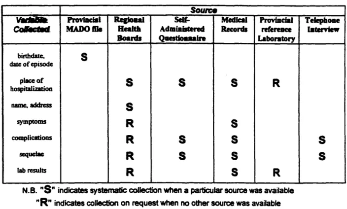
There were several different types of cases in this study, including survivors who were successfully contacted, untraceable survivo~ and deceased individuals. Data collection
was
different for each of these groups in tenns of data sources used due to the absence of the questionnaire and the telephone interview for untraceable and deceased individuals.3.4.2 Sources of Data
Multiple sources were
used
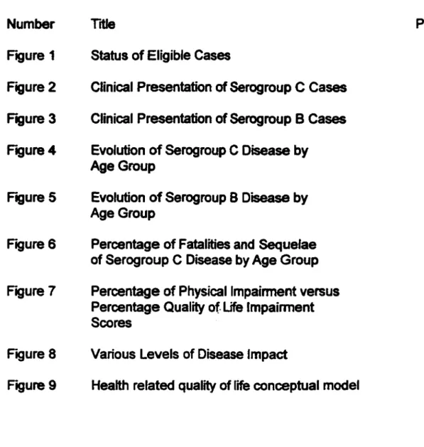
to obtain information on cases meeting the inclusion criteria (Table 1). Survivors (or their parents or guardians) were contacted by mail to obtain written consent to consult their hospital medical record and to complete the self-administered written questionnaire. Familles of deceased persans were neither contacted by mail nor by telephone, as the questionnaire on sequelae was not pertinent in their case.Table 1: Case Variables and tlaeir Sources
Soun:e
ProYblclal
Re&ioul
Sell- Medlcal Provbldal Telepboae Colllcl11f MADOftle Realtla AdmiaisCered Records refereace lllteniewBoards Qaestlouain Laban tory bitthdaœ.
s
dalc of episode place ofs
s
s
R
hospttalization name. addresss
symptomsR
s
complicationsR
s
s
scquefaeR
s
s
lab rcsultsR
s
R
N.B.
"S"
indicates systematic coflection when a particular sourcewas
available"R"
indicates collection on request when no other source was availablePlaces of hospitalizarion
were
usually obtained by the location of the declaring laboratory in the MADOfile.
However, inmany cases transfers
were noted upon consultation of medicalfiles.
Whennecessary,
the provincial reference laboratory (LSPQ) was contacted for confumations ofplaces
of hospitaliz.ation. In addition, they were contacted to obtain information regarding cultures received(blood, CSF, synovial tluid, tbroat swab) and results.
Finally, in certain
cases
survivors were interviewed by telephone to obtain supplementary information on sequelae. This allowed confirmation when information in medicalfiles
was incomplete or unclear. Regional investigations of reported cases alro supplied valuable information in certain cases regarding clinicals
s
symptoms, diagnosis, and disease evolution when this information
was
unavailable or unclear in the hospitalization record.3.4.3 Regional Health Boards
The provincial case registry
from
which cases were originally identified did not include names, addresses and places of hospitalization, which were required to contact patients and hospitals.This
information is routinely collectedby
regional health boards in their investigations of reported cases. Therefore, directors of public health and infectious disease for each of the 17 regional health boards of Québec were contacted to request this information.A list of cases
was
prepared for each of the 17 provincial health regions. This list included a confidential reference number, acode
numberfrom
the provincial declaration system (MADO), the date and location of the episode, the birthdate of the patient, the evolution of the disease (recovery, death), and the serogroup. This informationwas
used by regional health boards to identify and extract pertinent information on these cases from their files.A11 but three regional health boards supplied this information for cases in their rqioa. For the
three
remaining regions, the following procedures were adopted. In Recïon 04 (Mauricie-Bois-Francs), subjects were telephoned by a public health nurse to explain the project and obtain permission to release their names and addresses to researchers.In
Region 06 (Montréal-Centre}, the regional health board contacted participants directly, forwarding the questionnaire, consentfonn and letter of introduction. A cover letter
was
added to· the original mailout. encouraging individuals to participate while stressing that participation was entirely voluntary.Finally,
in Region16
(Montérégie) the regional healthboard
contacted participants directly (due to their affiliation with this health board,
researchers
were able to contact subjects on their behalf).3.4.4 Contact of Survivors
A package consisting of a letter of introduction, a consent fonn (for access to medical records), and a questionnaire
was
sent to survivors by registered mail (appendix 1). The nature of the project was clearly explained as was the fact thatparticipation
was
entirely voluntary. AU materials wereprinted
dou~le-sided inboth English and
French.
When individuals received the package but did not respond after two weeks, they were contacted by telephone to invite them to participate and answer an.y questions. If there was still no
response after
an additional week, the entire packagewas
remailed (including another questionnaire, a letter of introduction and a consent fœm). Certain subjects wished to participate but did not bave time to fill out dl8questionnaire.
These individuals were encouraged to sign and retum the consent form to allow consultation oftheir hospitali:zation record.In certain ~ envelopes were retumed due to change of address. Attempts were made to contact these individuals by telephone. When efforts to
retrace subjects
by
current telephone directories anddirectory
assistance were unsuccessful, they were classified as being "untraceable".3.4.5 Postal Questionnaire
A self-administered postal questionnaire
was
developed for this project to be mailed to disease survivors (appendix l ). There were five main sections to be completed in this questio~ including information regarding hospital stays, disease impact onhealth
status, health care/ services received. persona! costs incurred and disease impact on quality of life. Descriptions of complications and sequelae were obtained, as well as costs which were incurred as a result of the disease.The pre-test consisted of consultation of experts for revisions in questionnaire content and administration to individuals from various milieus to assess and improve clarity.
The 8-page bilingual questionnaire, (printed in English and French)
was
mailed out with a letter of introduction explaining the project (appendix 2), and a consent form (appendix 3). For subjects younger than 14 years old, parents (or guardims)
wcre
invited to complete the questionnaire.3.4.6 Assessment of Disease Impact on Quality of Lite
In addition to assessment of physical sequelae, this study aimed to explore the impact of meningococcal disease on survivors' quality oflife. Current literature .
and available instruments for quality of life measurement were reviewed
in
search of an appropriate validated questionnaire. A brief, simple scale which could be integrated witb the other sections of the questionnaire on sequelaewas
required. Existing instruments were eitber too lengthy or inappropriate for assessment of theimpact of meningococcal
disease
on quality of life. Due to the lack ofan
appropriate instrument, a brief
12
question scalewas
developed wbich included broad quality of categories in areas which could be atfected by meningococcaldisease. These
categorieswere
reduction ofenergy,
increase ofanxiety,
reduction of social and leisure activities,reduced
quality of family life, poorer physical appearance, reduction in ability to leam and solve problems, and reduction in general satisfaction witb life. When applicable, subjectsa1so
noted any reduction in tbeir capacity to do house~ork, to drive and automobile, to work, and to study. Weil established metbodological principles were used in the development of such an instrument (Guyatt et al., 1986 ).3.4. 7 Refusais
People refusing to participate by mail or by phone, were removed from the active case list and no further inquiries were made. Reason for refusai
was
noted when specified by the individual.3.4.8 Deceasect and Untraceable lndividuals
For deceased persans,
theself-administered questionnaire
onsequelae was
not pertinent Therefore, familles of deceased persans were not contacted. When persans were untraceable, the questionnaire and telephone interview could not
be
administered. For these cases, permission to consult medical files
was
requestedfrom the
DirectorOf
Professional Services of the hospital concemed. Thiswas
in accordanœ with project approval fiom the provincial Access to Information Commimon.3.4.9 Hospitals
Access to hospital medical records
was
requested for individuals acceptingto participate (with written consent), for untraceable survivors, and for deceased individuals. In total, 91 different hospitals in Québec (84), Ontario (6) and New Brunswick (1) had admitted cases in the study (appendix 4).
A description of the research project as well as a copy of our project approval
from
theprovincial
Commisrion of Access to Information were sent tothe Director of Professional Services of each hospital conœmed, including a requat
for
acœss to medical files of cases in each institution. Severa! hospitalsalso submitted
this request to their respective Ethics Committees for approval. One hospital refused to grant access to medical files of five untraceable individuals, resulting in their exclusionfrom
this project.3.4.1 O Review of Hospitalisation Record
For each individual, the complete hospitali7.ation record and medical records of subsequent follow-up treatment were reviewed to determine disease evolution and outcome. The length of hospital stays were noted, as well as special treatments and tests received. Clinical symptoms present as well as results of
laboratory and other diagnostic tests were noted, as well as audiological evaluation
and other follow-up tests. Details on complications present during the course of hospitalization were also noted.
Follow up after hospital discbarge
was
also reviewed. Evolution of problems present upon hospital discbarge were noted, as were additional tests, treatments, and support in readaptation, surgery or physical therapy.Transfers to other hospitals were noted and medical files from these hospitals were also reviewed.
Whenever possible, hospitals were visited in person to review medical files. This included hospitals in and around the major urban areas of Québec (Montréal, Laval, Québec, Sherbrooke, Trois-Rivières, Gatineau-Hull-Ottawa), for a total of 48 hospitals visited. The remajning hospitals (n=43) were contacted to obtain a photocopy of the medical record ( appendix 4 ).
3.4.11 Classification of Cases
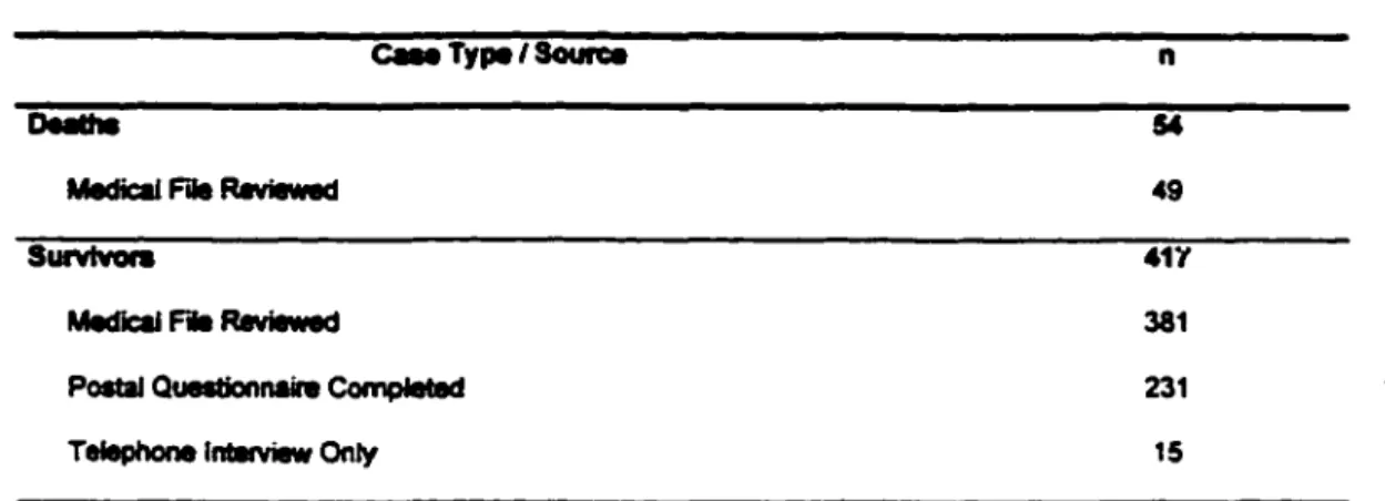
Ail included cases required presence of Neisseria meningitidis in a nonnally sterile site (blood, CSF, synovial fluid). Cases were classified into
different clinical categories on the
basis
of combined clinical and laboratory data. Due to the retrospective nature ofthis
study, it_was
impossible to apply rigid, uniform criteria to detine different clinical states. A pragmatic approach was used, based on the best available data. Lab results were combined with specific clinical signs of meningitis, septicaemia, and septic arthritis to classifyeach
case. Any of these clinical forms can occur alone or in combination.• Meningitis
was
defined by a CSF culture positive for N.meningitidis, orpresence
ofmicroscopie
or biochemical signs ofinflammation
in
the CSF, or distinct specific clinical signs of cerebromeningeal irritation (stiffneck, Kemig or Bmdzinslci signs, focal neurological deficits, coma, paralysis ).• Septicemia
was
defined by distinct clinical signs [ septic shock, purpura.petechiae,
disseminated intravascular coagulation (DlC)]• Septic Arthritis
was
defined by positive synovial fluid cultures.• Major complications were defined as
disseminated
intravascular coagulation (DIC), coma, cardiorespiratory arrest, respiratory distress syndrome (RDS), extensive necrosis or gangrene, and pericarditis.• Minor complications were defined as anemia, septic arthritis,
3.4.12 Telephone Interview
Wben questionnaire information was inconsistent with the medical file.
when additional details were needed, or when subjects simply failed to retum the
questionnaire, they
werecontacted by telephone to obtain the required information.
Details
not available
inmedical files could
beobtaincd, such as si7.e and evolution
of
skinscars, and evolution of other sequelae. For subjects who did not
retumthe
questionnaire,
thetelephone interview often confirmed the absence of sequelae.
3.5
Eth/calConalderatlona
1bis project
wasapproved by the provincial Access to Information
Commission, and the conditions of this approval werc respected. Project approval
was also granted by the Ethics Committee of the Centre of Clinical Research
(CRC) of the University Health Centre of Estrie (CUSE), the hospital with which
the researchers are affiliated.
For each regional health authority, conditions of approval granted by the
Director of Public
Healthand/or the Director of Infectious Disease and/or
thepersonnel
incharge
were respected.This includes special arrangements in the
threeregiom-of Mauricie-Bois-Francs, Montréal-Centre, and Montérégie, conceming
thecontact of survivors. Conditions of the Directors of Professional Services of the 91
hospitals involved
werealso
respectedas were those of each department of medical
records regarding consultation and photocopies of medical records. Conditions of
hospital ethics committees in certain hospitals were also met.
Informed consent
was
obtained from subjects, explaining clearly the project
an~empbasizing that participation
wasentirely voluntary. Refusais were dropped
from the case list and not pursued further.
Protection of confidentiality
was
a major ethical consideration
inthis
project. This
was
ensured by mailing questionnaires
byregistered mail to avoid
reœption by another
party inthe case of a change of address. Lists of names and
addresses ofpersons included
inthe study
were keptunder Iock
andkey,
withonly
the
mainresearcher having access.
Questionnaires were identified only by a confidential code number. When
medical files were
review~ ailinformation was transcribed onto a form identified
only by the confidential reference number. Wben possible,
personalinformation
was hidden before making photocopies of medical files. Wben this was impossible,
any identifying information was removed from photocopies and destroyed.
Finally,
a special effort
was
made torespond to questions of participants
conceming the disease and their
medicalcare.
3.6
Revlalon of :=ata/ltyRates
Medical files of deceased individuals were reviewed to confum the cause
of death for each case.
In certaincases, death was due to a pathology other
thanmeningococcal
disease(i.e. lymphoma). These
cases
were excluded from the total
number of deaths for each serogroup and the fatality
rateswere recalculated.
3.
7
Evaluation of
Seque/H
Information collected from medical file review, questionnaires and telephone interviews was combined to determine the presence of sequelae.
3.8 Claalflcatlon
of Physlcal Sequel•
Information collected from review of medical reco~ self-administered
questionnaires,
and
telepboneinterviews
of survivors were reviewed to determine the existence of physical sequelae of meningococcal disease.The medical
files
consulted bad variable degrees of completeness, organization, and legibility. Of the 45 individuals with identified sequelae, 11 were not successfully located, therefore medical recordswere
the only source of information for determination of the physical impairment scores. Results of diagnostic tests (audio~ renal function)as
wellas
objective clinical states (amputations, skin grafts,severe
scarring) were used to evaluate health status of these individuals.It
was preferable to include these individuals despite the lack of precicsion in esfimaring their health status tban to exclude them from the study.The "Annotated Scale of Bodily Injuries Regulation" (CSST, 1987) was used to provide a global assessment of severity and impact of sequelae. This is a scale used by the
Québec
Occupational Health and Safety Commission for compensation of occupational injuries. The scale is widely used in the province and is comprehensive, taking account of anatomie and physiologie deficits. disfigurement, and suffering or loss of enjoyment of life resulting from the deficitor disfigurement The scale
was
used
to calculate an overall percentage ofpermanent
mentaVphysical impainnent for each individual with identified physical sequelae (hereafter referred to as the 'CSST impainnent score'). An impairment score of l 00%. normally provides a compensation equivalent to full salary.For the sake of uniformity, one researcher performed a1l calculations of percentages of physical/mental impairment. It is important to note that for certain types of sequelae there is a tbreshold which must be surpassed to have a value for percentage of physical/mental impairment greater
tban
i.ero. For sensorineuralhearing Joss, the CSST threshold for compensation (average threshold of 30 dB, CSST, 1987) was used. Certain individuals had
very
minor sequelae which produced a percentage of 00/o.These
includedvery
minor (non-apparent) skin scars, andvery
minor hearing105:S.
For our study, cases with physical sequelae were defined as those for which a percentage of physical impairment greated than zero was calculated using the above scale.3.9
Scorlng of Questionnai,.: Reductlon of Quallty of
LifeSubjects
were
asked about the extent of their recovery from meningococcaldises•
Thœe
considering their recovery to be incomplete were asked to complete a section of the questionnaire conceming the impact of the disease on their quality of life. The twelve questions consisted of a series of statements (see appendix 1). Subjects were asked to note their degree of agreement with each statement on a five-point Likert scale (ranging from "not at ail" to "a lot"). A final score out of1 OO
pointswas
then calculated.This
was
doneby
calculating the total number of points of reduction of quality of life noteddividcd by
the total number of points possible for applicable questions (four of the twelve questions were not applicable to certain subjects ). The final score expressed as a percentage represents the percentage of a maximum impainnent score on the questionnaire in terms of reduction of quality oflife.
4. RESULTS
4.1 Number of
Cu•
by Yur and SerogroupA total of 4 71 eligible cases were identified during the years 1990 to 1994. 65% of these cases were of serogroup C infection, and the remaining 35% were of serogroup B infection (Table 2). A dramatic drop in the number of serogroup C cases occurred in 1993 following the provincial vaccination campaign which was completed in March 1993. Finally, in 1994 there were actually more cases of serogroup B discase (41 cases) than of serogroup C disease (35 cases).
Table2
Number of C...
bf
Yar and Serogroupv ...
~·
SerograupC TolllC..c...
c..
1990 12 37 49 1991 28 91 119 1992 47 94 141 1993 39 47 88 1994 41 35 7S TOTAL 187 304 4714.2 Subtyplng of Serogroup
C
stralna
Subtyping of
bacterial
strainswas
examined
for cases ofserogroup
C disease. Among serogroup C strains tbatwere
cbaracteri7.ed at the reference laboratory, 93% were of serotype 2a (2071223) , and 8<J0.4 were of electrophoretic type1
s
(2011226).
4.3 Characterlat/cs of Study Population
For both serogroups, 49 % of subjects
wcre
male. The average age of cases was13
.5 years
for serogroupB
and17 .6 years
for serogroupC. This
difference was statistically signi.ficant (p<0.05, t test). The distribution across age groups was different between serogroups B and C, notably inthat
the maximum number of cases (mode)was
observed in the l 0-19 year agegroup
for serogroup C, while thismaximmn
was
observed in the under 1 year agegroup
for serogroup B cases (referTable 3
Age Distribution of Study Population
AgeGfoup TOTAL <1
ll:
28•
1~ 32 57a
5-1 7 32 31 10-11 18W.:
117 20-ll 30 74 1CM...
10 14 Z4 TOTAL 117..
471·-mode
4.4 Participation
Rate
Of the study population, 11.5% (54/471) died from meningococcal disease. Among disease survivors, 35% (144/417) were not
successfully
located due toaddress change. In additio~ five individuals were no longer living at the time of the study. Of contacted individuals, 96% participated
in
the study, and 4% refused to participate or did not respond. On the average, participants completed the questionnaire 37.8 months after disease occurrence (minimum delay 9 months, maximum delay 72 months, median 39 months).Ellglble Caan n8'71
Contadlld Not Contadlld
n-298 na149
0. 111 ;:I Untnleubl9
(Vlfiaus cal-) ns144
na5
Most individuals who did not
initially
retumthe
questionnaire claimed tohave done so because they had no sequelae and therefore no problems to mention in the questionnaire. They were often under the impression that their participation