The X-linked trichothiodystrophy-causing gene
RNF113A links the spliceosome to cell survival
upon DNA damage
Kateryna Shostak
1,2,11
, Zheshen Jiang
1,2,11
, Benoit Charloteaux
1,3,10,11
, Alice Mayer
1,3
, Yvette Habraken
1,4
,
Lars Tharun
5
, Sebastian Klein
5
, Xinyi Xu
1,2
, Hong Quan Duong
1,2,6
, Andrii Vislovukh
1,2
, Pierre Close
1,7,8
,
Alexandra Florin
5
, Florian Rambow
9
, Jean-Christophe Marine
9
, Reinhard Büttner
5
& Alain Chariot
1,2,8
✉
Prolonged cell survival occurs through the expression of speci
fic protein isoforms generated
by alternate splicing of mRNA precursors in cancer cells. How alternate splicing regulates
tumor development and resistance to targeted therapies in cancer remain poorly understood.
Here we show that RNF113A, whose loss-of-function causes the X-linked trichothiodystrophy,
is overexpressed in lung cancer and protects from Cisplatin-dependent cell death. RNF113A is
a RNA-binding protein which regulates the splicing of multiple candidates involved in cell
survival. RNF113A de
ficiency triggers cell death upon DNA damage through multiple
mechanisms, including apoptosis via the destabilization of the prosurvival protein MCL-1,
ferroptosis due to enhanced SAT1 expression, and increased production of ROS due to altered
Noxa1 expression. RNF113A deficiency circumvents the resistance to Cisplatin and to BCL-2
inhibitors through the destabilization of MCL-1, which thus defines spliceosome inhibitors as
a therapeutic approach to treat tumors showing acquired resistance to specific drugs due to
MCL-1 stabilization.
https://doi.org/10.1038/s41467-020-15003-7
OPEN
1Interdisciplinary Cluster for Applied Genoproteomics (GIGA), University of Liege, CHU, Sart-Tilman, Liège, Belgium.2Laboratory of Medical Chemistry,
University of Liege, CHU, Sart-Tilman, Liège, Belgium.3GIGA Genomics Platform, University of Liege, CHU, Sart-Tilman, Liège, Belgium.4Laboratory of Gene
Expression and cancer, University of Liege, CHU, Sart-Tilman, Liège, Belgium.5Institute for Pathology-University Hospital, Cologne, Germany.6Center for
Molecular Biology, Institute of Research and Development, Duy Tan University, 03 Quang Trung, Danang, Vietnam.7Laboratory of Cancer Signaling,
University of Liege, CHU, Sart-Tilman, Liège, Belgium.8Walloon Excellence in Life Sciences and Biotechnology (WELBIO), Wavres, Belgium.9Laboratory for
Molecular Cancer Biology, VIB Center for Cancer Biology and KULeuven, Department of Oncology, 3000 Leuven, Belgium.10Present address: Department of
Human Genetics, CHU of Liege, University of Liege, Sart-Tilman, 4000 Liège, Belgium.11These authors contributed equally: Kateryna Shostak, Zheshen Jiang,
Benoit Charloteaux. ✉email:[email protected]
123456789
S
pliceosomes are dynamic Ribonucleoprotein (RNP)
com-plexes required for pre-mRNA splicing, i.e., the removal of
non-coding intervening sequences (introns) from
pre-mRNAs and for the ligation of coding sequences (exons) to
generate mature mRNAs
1–3. Intron excision occurs thanks to
short sequence motifs in the pre-mRNA, at boundaries between
the upstream exon and intron (the 5′ splice site) and the intron
and downstream exon (the 3′splice site)
1,2. protein,
RNA-RNA and protein–protein interactions are critical for the proper
recognition of splice sites. These interactions are dynamic during
the splicing cycle and are critical for the formation,
rearrange-ment and dissociations of the spliceosomal complexes
4.
Alternate splicing of mRNA precursors allows more than 95%
of human genes to generate a variety of RNA species and distinct
proteins from a single gene. Protein isoforms are selected by
cancer cells to sustain survival and to promote tumor
develop-ment and resistance to therapies
5. Cancer cells exhibit global
splicing deregulations due to mutations within mRNA sequences
(“cis-acting mutations”), and in splicing factors (“trans-acting
mutations”) or to changes in expression levels of splicing
factors
6,7. As a result, the spliceosome has been defined as a
promising therapeutic targets, as least for aggressive Myc-driven
tumors and for malignant glioma
8,9. Yet, transcripts whose
spli-cing is deregulated in cancer cells only start to be identified
10.
The DNA damage response (DDR) helps the body to face
thousands of DNA lesions
11,12. These lesions, if not repaired, lead
to mutations or genomic aberrations that threaten viability. DNA
damage is induced by multiple sources ranging from byproducts
of cell metabolism and oxidative damage to ionizing radiation
and chemotherapeutic agents. Nucleotide excision repair (NER)
repairs single-stranded DNA damage by removing
helix-distorting DNA lesions induced by UV-light, ROS-induced
cyclopurines and intrastrand crosslinks (ISCs) generated by
chemotherapeutic drugs such as Cisplatin
13. These ISCs activate
several signal transduction pathways/kinases such as ATR
14. ATR
targets substrates including Chk1 in order to help tumor cells
survive the DNA damage.
The double-strand breaks (DSBs) do not occur as frequently as
DNA single-strand breaks (SSBs) but are more difficult to repair
and therefore extremely toxic. The DDR includes the sensing of
the broken DNA molecule, the activation of specific signaling
pathways and
finally, the repair of the DNA lesion
11. DSB repair
involves two main pathways, the homologous recombination
(HR) and the non-homologous end-joining (NHEJ)
15,16. In
NHEJ, DSBs are sensed by the Ku70/80 heterodimer that recruits
the DNA dependent protein kinase catalytic subunit
(DNA-PKcs), Paralog of XRCC4 and XLF (PAXX) and end-processing
enzymes leading to repair by the DNA ligase IV/DNA ligase
IV-X-ray cross complementing protein 4 (XRCC4)/XRCC4-like
factor (XLF, also referred to as Cernunnos) complex
11,17,18. The
recruitment of DNA-PKcs to DSBs occurs within seconds
fol-lowing DNA damage
19. DSBs trigger DNA-PKcs
autopho-sphorylation on the PQR cluster that includes Ser2056 and on the
ABCDE cluster of 6 amino acid residues between Thr2609 and
Thr2647, which is also targeted by the kinase ATM upon DNA
damage
20–22. These phosphorylations alter the affinity of
DNA-PKcs for DNA and inactivate its kinase activity to promote
NHEJ
23–25.
Cancer cells show genomic instability
26,27. Although the DDR is
commonly activated in early neoplastic lesions as a protective
mechanism against malignancy, cancer cells overcome this barrier
through the mutational or epigenetic inactivation of DDR
com-ponents to enhance cell proliferation and survival, despite increased
genomic instability
28–31.
We show here that RNF113A, also referred to as Cwc24 in yeast,
whose deficiency causes a novel X-linked trichothiodystrophy
(TTD) and enhances sensitivity to Interstrand DNA Crosslinking
agents in C.elegans
32,33, is aberrantly expressed in pulmonary
adenocarcinomas. RNF113A promotes cell survival in
Cisplatin-treated lung cancer-derived cells as a subunit of the spliceosome.
We characterized all transcripts whose splicing relies on RNF113A
and also established a link between RNF113A and MCL-1
stabili-zation, with important consequences for the treatment of lung
cancer cells showing some acquired resistance to BCL-2 inhibitors.
Therefore, our data define RNF113A as a promising therapeutic
target.
Results
RNF113A is increased in pulmonary adenocarcinomas. In a
search for E3 ligases overexpressed in cancer, we got interested in
RNF113A which is detected in all human cases of haematological
or solid tumors (
http://www.proteinatlas.org/ENSG00000125352
–RNF113A/cancer
). RNF113A expression was higher in our
clinical cases of pulmonary adenocarcinomas than in tumor-free
lung parenchymas, independently of the K-RAS or EGFR
muta-tional status (Fig.
1
a and Supplementary Data 1). A majority of
lung malignancies showed a mostly nuclear staining of RNF113A
(Fig.
1
b). A weak and almost exclusively nuclear staining of
RNF113A was detected in normal lung epithelium cells
(Sup-plementary Fig. 1a). Importantly, patients with high levels of
RNF113A showed a shorter survival rate (Fig.
1
c). RNF113A
expression increased at both mRNA and protein levels in
Cisplatin-treated A549 and BZR-T33 adenocarcinoma-derived
cells (Fig.
1
d, e). Consistently, RNF113A expression was barely
detectable in untreated A549 cells but was detected in
Cisplatin-treated cells by immunofluorescence analyses (Fig.
1
f).
Camp-tothecin, a topoisomerase I inhibitor, also increased RNF113A
expression in both A549 and BZR-T33 cells while Etoposide, a
topoisomerase II inhibitor, did not (Fig.
1
g, h and Supplementary
Fig. 1b, respectively). RNF113A expression was also weakly
induced by
γ-irradiation (Supplementary Fig. 1c). RNF113A
expression was also induced by Cisplatin in normal human
der-mal
fibroblasts (Supplementary Fig. 1d). Therefore, RNF113A
expression is induced by some DNA-damaging signals and is
increased in lung cancer.
CCAAT/enhancer-binding protein beta (C/EBPβ) expression is
induced by DNA-damaging agents
34. Therefore, we investigated
whether C/EBPβ is required for the induction of RNF113A
expression by Cisplatin. C/EBPβ deficiency interfered with
Cisplatin-dependent induction of RNF113A in A549 cells (Fig.
1
i).
Consistently, we found C/EBPβ binding sites on the RNF113A
promoter using the TFbind software (
http://tfbind.hgc.jp/
)
(Fig.
1
j). C/EBPβ was recruited on site 1 in unstimulated A549
cells and on sites 1 to 4 in Cisplatin-treated cells (Fig.
1
j). p53 was
dispensable for RNF113A expression as the incubation of A549
cells with Nutlin, which disrupts the interaction of the E3 ligase
MDM2 with p53, or with JNJ26854165, a MDM2 inhibitor
35,
did not impact on RNF113A expression (Fig.
1
k). Therefore,
Cisplatin induces the expression of RNF113A through a
C/EBPβ-dependent but p53-inC/EBPβ-dependent pathway.
RNF113A protects from Cisplatin-dependent cell death. We
next explored whether RNF113A is involved in the DDR.
Enhanced RNF113A expression in A549 cells interfered with
Cisplatin-dependent DNA-PKcs phosphorylation on Ser2056, a
marker of DNA damage (Fig.
2
a). RNF113A overexpression
protected A549 cells from Cisplatin-induced death (Fig.
2
b). On
the other hand, RNF113A deficiency enhanced cell death in
Cisplatin-treated lung cancer A549 and BZR-T33 cells (Fig.
2
c
and Supplementary Fig. 2a). RNF113A deficiency did not impact
on p53 phosphorylation in BZR-T33 cells triggered by Cisplatin
(Fig.
2
d). Cisplatin-dependent DNA-PKcs phosphorylation on
S2056 was increased upon RNF113A deficiency in BZR-T33,
A549 and HT1975 cells showing distinct p53 status (Fig.
2
d,
Supplementary Fig. 2b and Supplementary Fig. 2c). Accordingly,
RNF113A deficiency enhanced the number of both
phospho-H
2AX (pH
2AX) and phospho-DNA-PKcs (pDNA-PKcs) positive
BZR-T33 cells, suggesting that these cells fail to repair DNA
(Fig.
2
e, f). RNF113 overexpression also protected A549 cells
from cell death induced by Etoposide and limited DNA-PKcs
phosphorylation on serine S2056 (Supplementary Fig. 3a).
Con-sistently, cell death triggered by Etoposide was more pronounced
upon RNF113A deficiency in A549 cells (Supplementary Fig. 3b).
80 – 80 – 80 – 10 – 40 – 80 85 – 40 60 – 40 – 30 – 30 – 15 – T N T N T N T N T N T TN T T N T N T N T N T N RNF113A β-Actin Tumor (%) 1 2 3 4 5 6 7 8 9 10 11 12 13 14 15 16 17 18 19 20 21 22 23 24
a
b
Cy to pl a s m ic Mostly nuclear Mo stl y nucl e a r 50 μm pH2AX pDNA-PKcsDAPI Merge DAPI DAPI RNF113A
Untreated
c
d
BZR-T33 cells 0 2 4 8 12 16 20 24 1 2 3 4 5 6 7 8 A549 cells 0 2 4 8 12 16 20 24 1 2 3 4 5 6 7 8 0 2 4 8 12 16 20 24e
f
h
RNF113A HSP90 DNA-PKcs (h) F old induction (% ) BZR-T33 cells pDNA-PKcsS2056 0 2 4 8 12 16 20 24 0 2 4 8 12 16 20 24 1 2 3 4 5 6 7 8 1 2 3 4 5 6 7 8 (h) Cisplatin 25 μM (h) Cisplatin 25 μM (h) Cisplatin (25 μM, 24 h) Cisplatin 25 μM, 24 h F old induction (% ) RNF113A HSP90 DNA-PKcs pDNA-PKcsS2056 F old induction (% ) F o ld induction (% ) Camptothecin 25 μM (h) 0 2Camptothecin 25 4 8 12 16 20 24μM (h) RNF113A DNA-PKcs Ku80 (h) (h) pDNA-PKcsS2056 Kaplan–Meier plot Days Pr obabilityg
A549 cells UntreatedJNJ26854165Nutlin 1 2 3 RNF113A + + + ShRNAs Control C/EBPβ#1 C/EBPβ#2 1 2 3 4 5 6 C/EBPβ C/EBPβi
j
High n = 79 Low n = 125 315 315 90 55 315 315 90 55 315 315 90 90 55 315 315 90 90 55 44 55 44 55 90 55 55 90 90 44 (Long expo.) (Short expo.)k
START +1 RNF113A promoter SCORE 91.6 SCORE 87.3 SCORE 82.4 SCORE 84.3 –537 –552 –1099 –1113 –1275 –1286 –1403 –1417 Site 1 Site 2 Site 3 Site 4 0 2 4 8 12 16 20 24 0 2 4 8 10 16 20 24 0 200 400 600 A549 cells 0 200 400 600 800 1000 0 100 200 300 0 100 200 300 400 500 0 10 20 30 40 Fold enrichment to IgG
Site 1 Site 2 Site 3 Site 4
IgG non treated Non treated IgG Cisplatin 24 h Cisplatin 24 h *** * ** * ** *** HSP90 200 μm 0 0.4 0.8 0 500 1000 1500 2000 2500 3000 3500 Merge Merge HSP90 RNF113A HSP90 DNA-PKcs Ku80 pDNA-PKcsS2056 RNF113A HSP90 p53 MDM2
If cells are allowed to resume proliferation after being stimulated
with Cisplatin for 16 h, ATR activation assessed through
phos-phorylation of its target Chk1, was also defective upon RNF113A
deficiency in A549 cells (Fig.
2
g). RNF113A-depleted cells
underwent Caspase 3-dependent cell death upon DNA damage
(Fig.
2
g). The ability of control versus RNF113A-deficient
BZR-T33 cells to undergo DNA repair was assessed with the comet
assay. RNF113A-deficient cells showed more DNA damage,
especially after Cisplatin treatment, as assessed through the
quantification of the tail moment (Fig.
2
h). Thus, RNF113A
promotes DNA repair.
RNF113A is recruited on some DNA damage-induced foci. We
next explored whether RNF113A is recruited on DNA damage
foci, using a RNAse A-based extraction protocol to visualize the
formation of Cisplatin-induced foci on damaged DNA. RNF113A
colocalized with phosphorylated DNA-PKcs but not with
phospho-H
2AX foci in Cisplatin-treated cells (Supplementary
Figs. 4a, b, respectively). Consistently, RNF113A was found in
chromatin fractions in BZR-T33 cells treated to Cisplatin
(Fig.
3
a). RNF113A-deficient cells also had more Ku70/80 and
DNA-PKcs recruited to chromatin fractions upon Cisplatin
treatment (Fig.
3
b). Conversely, RNF113A-overexpressing A549
cells had less Ku70/80, DNA-PKcs and DNA ligase IV in
chro-matin fractions when treated with Cisplatin (Fig.
3
c and
Sup-plementary Fig. 4c), suggesting that RNF113A controls the
recruitment of DNA repair factors on chromatin. To explore
whether RNF113A regulates the presence of phospho-H
2AX on
DSB sites, we used the DIvA U2-OS cell line, which stably
expresses the restrictase AsiSI under ER promoter. These cells
generate several randomly distributed and sequence-specific
DSBs
36. Treatment of this cell line with 4-hydroxy tamoxifen
(4OHT) generated DSBs since multiple pH
2AX
+cells were
detected by immunofluorescence (Supplementary Fig. 5). We
therefore generated control and RNF113A-depleted cells
(Sup-plementary Fig. 5). ChIP assays were conducted to assess the
presence of pH
2AX on AsiSI sites in both control and
RNF113A-depleted cells using appropriate primers
36. pH
2
AX on H
2AX-associated AsiSI sites using primers 183, 906, 307 and 221
36was
defective upon RNF113A deficiency (Fig.
3
d). As negative
con-trols, we also conducted these experiments using primers 811 and
903, which are not H
2AX-associated AsiSI sites (Fig.
3
d)
36.
Therefore, RNF113A controls the pool of NHEJ factors recruited
to damaged DNA.
In support with a nuclear localization of RNF113A, wild type
RNF113A and a mutant lacking the
first 30 N-terminal amino
acids (“ΔN30”) or the RING domain (“ΔRING”, which lacks
amino acids from 262 to 300) were mostly found in the nucleus
and colocalized with DNA-PKcs when transfected in A549 cells
(Fig.
3
e). On the other hand, a RNF113A construct lacking the
first 60 or 90 N-terminal amino acids (“ΔN60 and ΔN90”) mostly
showed both nuclear and cytoplasmic localizations (Fig.
3
e). A
predicted nuclear localization signal (NLS) was found on
RNF113A, using the SeqNLS algorithm
37(Fig.
3
e). Despite the
fact that RNF113A was found in the nucleus of lung cancer cells,
Cisplatin enriched the cytoplasmic pool of RNF113A (Fig.
3
a).
This was also true in BEAS-2B cells, which are derived from a
normal bronchial epithelium (Supplementary Fig. 4d).
Consis-tently, cells showing DNA damage (i.e., positive for pH
2AX) and
undergoing apoptosis (i.e., in which Cyt. C was released) showed
a cytoplasmic staining of RNF113A (Fig.
3
f). Therefore, DNA
damage and cell apoptosis triggered by Cisplatin correlates with
the shuttling of RNF113A to the cytoplasm.
RNF113A acts as a subunit of the spliceosome. As RNF113A is
a spliceosome subunit
4,38, we carried out RNA
immunopre-cipitation (RIP) experiments in A549 cells and looked for
spliceosome subunits. We
first fixed cells with
paraformalde-hyde (PFA) to trap large protein complexes onto RNAs.
RNF113A, SF3B1 (another spliceosome subunit) and the
ribosomal protein RPL7 but not HSP90 were found on RNAs
(Fig.
4
a). Next, we cross-linked our cells with UV to
dis-criminate direct versus indirect bindings of spliceosome
sub-units to RNAs. In those circumstances, RNF113A bound
mRNAs in unstimulated cells which were not treated with
RNAse and this binding was negatively regulated by Cisplatin,
which
fits with our previous data showing that a pool of
RNF113A moves into the cytoplasm upon DNA damage
(Fig.
4
b). As expected, SF3B1 as well RPL7 were also found
associated to RNAs and SF3B1 binding to RNAs was also
impaired upon Cisplatin stimulation (Fig.
4
a, b). Therefore,
RNF113A directly binds RNAs.
Because a displacement of spliceosomes causes the
accumula-tion of three-strand nucleic acid structures formed by an RNA:
DNA hybrid plus a displaced DNA strand (ssDNA) and referred
to as
“R-loops”
39, we next explored whether RNF113A deficiency
leads to the accumulation of R-loops. RNF113A-depleted A549
cells indeed showed more R-loops, as judged by
immunofluores-cence analyses using an antibody that specifically detects these
R-loops (Fig.
4
c). Protein levels of the spliceosome factor SF3B2 were
decreased upon RNF113A deficiency in A549 cells (Fig.
4
d)
4.
Moreover, protein levels of DNA repair factors such as RNF8 and
Rad51, which are downregulated upon impaired splicing
40, were
also decreased in RNF113A-depleted A549 cells (Fig.
4
d).
Although SF3B2 levels did not decrease upon RNF113A
deficiency in BZR-T33 cells, in contrast to A549 cells, SF3B2
Fig. 1 RNF113A expression in lung cancer and upon Cisplatin treatment. a RNF113A expression in lung cancer. Western blot (WB) analyses with humanlung tumors (“T”) and corresponding normal adjacent tissues (“N”). Numbers refer to the percentage of tumor cells. b. Subcellular localizations of RNF113A
in lung adenocarcinomas. Anti-RNF113A Immunohistochemistry analyses were conducted on a Tissue-MicroArray (TMA) (×40 objective). Stromal cells
served as internal negative control.c Patients with high levels of RNF113A mRNAs show a shorter survival rate (Kaplan-Meir plot). d, e RNF113A expression
is induced by Cisplatin in A549 (d) or BZR-T33 (e) cells. On the top, RNF113A mRNA levels in unstimulated cells is set to 100% and levels in other
experimental conditions are relative to that after normalization withβ-actin. Data from two independent experiments performed in triplicates (means ± SD)
are shown. At the bottom, WB analyses. The anti-pDNA-PKcs (S2056) antibody was used to prove DNA damage.f Induction of RNF113A expression by
Cisplatin in A549 cells (immunofluorescence analyses). g, h RNF113A expression is induced by Camptothecin in A549 (g) or BZR-T33 (h) cells. On the top,
data from two Real-time PCR (independent) analyses performed in triplicates (means ± SD) are plotted as described in (d). At the bottom, WB analyses.
i RNF113A induction by Cisplatin occurs through a C/EBPβ-dependent pathway. Control or C/EBPβ-depleted A549 cells were treated or not with Cisplatin
(25μM) for 24 h and WB analyses were done. Expo. = exposure. j C/EBPβ is recruited on the RNF113A promoter. C/EBPβ binding sites were identified
(Tfbind software) and ChIP assays using an anti-C/EBPβ antibody were carried out. Histogram show recruitment C/EBPβ on indicated sites with or without
treatment (IgG antibody was used as negative control). RNF113A promoter is lacking a TATA box. Results of two independent experiments (means ± SD,
Studentt-test, ***p < 0.001, **p < 0.01, *p < 0.05) are shown. START = start of transcription. k RNF113A expression is not regulated by p53. A549 cells
binding to SF3B1 was nevertheless impaired in both cell lines,
suggesting that the assembly of spliceosome subunits relies on
RNF113A (Fig.
4
e, f). Consistently, R-loops accumulated in
SF3B2-depleted A549 cells (Fig.
4
g). Moreover, SF3B2 deficiency
also led to more DNA damage, as evidenced by enhanced
phosphorylated levels of DNA-PKcs and to enhanced cell death in
A549 cells treated or not with Cisplatin (Fig.
4
h, i, respectively).
Finally, both SF3B1 and SF3B2 were found in the cytoplasm and
in the nucleus of A549 cells but Cisplatin promoted their
disengagement from chromatin, as previously seen for RNF113A
(Fig.
4
j). Therefore, the spliceosome subunit SF3B2 promotes cell
survival upon DNA damage, similarly to RNF113A.
c
d
RNF113A HSP90 20 0 1 2 3 4 5 6 Cisplatin (25 μM, 20 h) 104 103 102 101 100 104 103 102 101 100 104 103 102 101 100 104 103 102 101 100 104 103 102 101 100 104 103 102 101 100 104 103 102 101 100 104 103 102 101 100 0.85% 0.82% 10.43% 14.61% 3.47% 6.64% Percentage of cells in late apoptosis20 0
a
FLAG-EV FLAG-RNF113A 1.08% 0.88% Untreated Cisplatin 25 μM 20 h Cisplatin 25 μM 20 h FLAG-EV FLAG-RNF113A Cisplatin 25 μM (h) A549 cells 27.63% 14.02% 23.56% 15.97% 5.27% 8.24% 9.63% 3.54% 13.73% 6.87% 3.34% 0.52% UntreatedShRNA RNF113A#1 ShRNA RNF113A#2 ShRNA CTRL
Percentage of cells in late apoptosis
20 0 Cisplatin 25 μM 20 h ShRNA RNF113A#1 ShRNA RNF113A#2 ShRNA CTRL A549 cells HSP90 RNF113A (Short expos.) pDNA-PKcsS2056 Cisplatin 25 μM (h) 1 2 3 4 5 6 7 8 9 10 11 12
b
DNA-PKcs 0 2 4162024 0 2 4162024 FLAG-EV FLAG-RNF113A RNF113A p53 HSP90 DNA-PKcs 1 2 3 4 5 6 pp53S15 pDNA-PKcsS2056e
0 3 6 9 12 15 18 *** *** * *** Tail moment 0 20 0 20 0 20 Cisplatin 25 μM (h) CTRL ShRNA RNF113A #2 Cisplatin 25 μM (h)f
ShRNAs RNF113A#2 CTRL 0CTRLRNF113A#1RNF113A#2CTRLRNF113A#1RNF113A#2 shCTRLshRNF113A#1shRNF113A#2shCTRLshRNF113A#1shRNF113A#2 0 0 20 20 20
g
315 315 90 55 315 315 90 55 55 55 90 55 CTRL CTRL RNF113ADAPI pH2AX Merge
DAPI pH2AX Merge
DAPI pH2AX Merge
DAPI pH2AX Merge
RNF113A Cisplatin DAPI DAPI pDNA-PKcs DAPI DAPI CTRL CTRL RNF113A Merge RNF113A ** *** *** *** ** ** *** *** pDNA-PKCs 0 10 20 30 40 pH2AX 0 10 20 30 40 50 % of positive cells ShRNAs CTRL RNF113A Cisplatin CTRL RNF113A Cisplatin ShRNAs CTRL RNF113A Cisplatin CTRL RNF113A Cisplatin % of positive cells RNF113A 55 20
h
pChk1 S345 Cisplatin (25 μM, 16 h) RNF113A HSP90 Cleaved Caspase 3 Control RNF113A Chk1 + + + + + + + + + + Release (h) 8 1 2 4 1 2 4 8 ShRNAs 1 2 3 4 5 6 7 8 9 10 11 12 17 90 55 55 55 Cisplatin CisplatinCisplatin Cisplatin Cisplatin
Cisplatin Cisplatin Cisplatin
Cisplatin Cisplatin pDNA-PKcs pDNA-PKcs pDNA-PKcs Merge Merge Merge 0 10 20 30 40 *** ** *** *** Cisplatin (25 μM) 0 5 10 15 *** ns Annexin V PI Annexin V PI Cisplatin 100 μm 100 μm 20 μm 104 103 102 101 100 104 103 102 101 100 104 103 102 101 100 104 103 102 101 100 104 103 102 101 100 104 103 102 101 100 104 103 102 101 100 104 103 102 101 100 104 103 102 101 100 104 103 102 101 100 104 103 102 101 100 104 103 102 101 100
To investigate the impact of RNF113A depletion on both gene
expression and splicing events, we
first concentrated on
candidates such as Rad51 and RNF8 which are regulated by
splicing
40. RNF113A-depleted cells accumulated Rad51 and
RNF8 pre-mRNAs in which intron 1 is retained, especially after
Cisplatin stimulation (Supplementary Figs. 6a and b,
respec-tively). Therefore, RNF113A is required for the splicing of both
Rad51 and RNF8. SF3B2-depleted cells also accumulated Rad51
pre-mRNAs, especially after Cisplatin treatment (Supplementary
Fig. 6a). As RNF113A-depleted cells undergo cell death upon
Cisplatin stimulation, we next wondered whether Caspase 3
activation is the causal event rather than the consequence of the
splicing deregulations seen upon RNF113A deficiency. To address
this issue, we pretreated A549 cells with ZVAD, a caspase
inhibitor, and assessed Rad51 and RNF8 splicing upon Cisplatin
stimulation in both control and RNF113A-depleted cells. ZVAD
did not impact on the accumulation of both Rad51 and RNF8
pre-mRNAs upon RNF113A deficiency in Cisplatin-treated cells
(Supplementary Fig. 6a, b, respectively). Therefore, apoptosis is
the consequence rather than the cause of the splicing
deregula-tions seen upon RNF113A deficiency in cells treated with
Cisplatin.
We expanded this analysis genomewide by carrying out
high-throughput RNA sequencing on total RNA extracted from
control and RNF113A-depleted A549 cells treated or not with
Cisplatin. Having performed two sequencing runs on the pooled
mRNA libraries to obtain a minimum of 45 millions of
paired-end reads per sample, we had sufficient coverage of lowly
abundant transcripts to perform analyses of the resulting dataset
at several levels: (i) a differential gene expression analysis to
evaluate the expression changes induced by the different
treatments (RNF113A depletion and/or Cisplatin) at the gene
level; (ii) a differential gene splicing analysis assessing individual
splicing events categorized by type (i.e., skipped exon, retained
intron, alternative 5′ splicing site, alternative 3′ splicing site, and
mutually exclusive exons); and (iii) a global analysis of intron
retention at the gene level.
Differential expression analysis showed that the expression
level of many genes is affected by Cisplatin treatment and/or by
RNF113A depletion (Supplementary Fig. 7a, b, and
Supplemen-tary Data 2). For the majority of these candidates, expression
changes can mostly be explained by an additive model where
Cisplatin treatment and RNF113A depletion have independent
effects, with 3703 genes whose mRNA expression levels were
significantly changed upon Cisplatin treatment (1722 upregulated
and 1981 downregulated; FDR q-value < 5% and twofold change
minimum) and 1816 genes significantly affected by RNF113A
depletion (866 upregulated and 950 downregulated; FDR q-value
< 5% and twofold change). For a few genes (293), the interplay of
the two treatments is more complex with a contrasted impact of
the depletion between Cisplatin-treated and untreated cells. This
was the case for instance for NUPR1 for which RNF113A
depletion drastically reduced the increase in mRNA expression
level induced by Cisplatin. Although NUPR1 mRNA expression
level increased by 5.6 fold in control cells treated with Cisplatin,
this increase was only of 1.5 fold in RNF113A-depleted cells
(Supplementary Data 2).
To verify the validity of our RNAseq dataset, we performed a
gene set enrichment analysis (GSEA). As expected, this analysis
highlighted a significant enrichment in signatures typical of
apoptosis and DNA repair (FDR q-value < 1%) (Supplementary
Fig. 7c). We also confirmed in independent Real-Time PCR
experiments the expression changes observed for a few candidates
highlighted in the systematic differential expression analysis.
TRIM29 and CEACAM5 expression were increased upon
RNF113A deficiency while GFRA1 expression was decreased
(Supplementary Fig. 7d).
We then focused on the role of RNF113A in splicing using
rMATS (replicate Multivariate Analysis of Transcript Splicing), a
computational method dedicated to the detection of differential
splicing events from replicate RNAseq data
41. Using a
hierarch-ical model to account for uncertainty in each replicate and
variability between replicates, rMATS assesses individual splicing
events from
five different categories: skipped exons, retained
intron, alternative 5′ splicing site, alternative 3′ splicing site, and
mutually exclusive exons (Fig.
5
a).
To evaluate the impact of Cisplatin and/or RNF113A depletion
on splicing, we performed four different comparisons, each
evaluating the impact of one factor, the other one kept constant
(Fig.
5
a, b). The most frequent categories of splicing events
evaluated by rMATS involved exons (Skipped exons and mutually
exclusive exons) but several thousands of events were also
evaluated for each one of the other categories (Fig.
5
b,
Supplementary Fig. 8a and Supplementary Data 3-8). Significant
alternative splicing events (FDR q-value < 5% and difference in
inclusion level > 20%) were detected for all comparisons and for
all categories of events. Cisplatin treatment in RNF113A-depleted
cells was associated with the highest number of significant exon
skipping events (1342 exons show significantly higher inclusion
levels when depleted cells are treated with Cisplatin). RNF113A
depletion was shown to induce smaller changes on exon
retention, although hundreds of events were significantly affected.
Fig. 2 RNF113A limits Cisplatin-dependent cell death. a RNF113A overexpression interferes with DNA-PKcs phosphorylation upon Cisplatin treatment.Control or RNF113A-overexpressing A549 cells were stimulated or not with Cisplatin and WB analyses were done.b RNF113A overexpression limits
Cisplatin-dependent cell death. Control or RNF113A-overexpressing A549 cells were untreated or stimulated with Cisplatin. The percentage of cells in early (Annexin V positive and PI negative) or late apoptosis (Annexin V positive and PI positive) was assessed by FACS. On the left, FACS data from one
representative experiment. On the right, the histogram from two independent experiments (Studentt-test, p-values: ***<0.001). ns = non significant.
c RNF113A deficiency enhances Cisplatin-mediated cell death. Extracts from control (“ShCTRL”) or from RNF113A-depleted (“ShRNF113A#1 and
ShRNF113A#2”) A549 cells were subjected to WB analyses. Cell survival upon Cisplatin treatment was assessed by FACS (right panel). On the left, FACS
data from two independent experiments are illustrated in the histogram (Studentt-test, ***p < 0.001). d RNF113A negatively regulates Cisplatin-induced
DNA-PKcs phosphorylation. Control or RNF113A-depleted BZR-T33 cells were untreated or stimulated with Cisplatin and WB analyses were done.
e, f. RNF113A deficiency enhances the number of pH2AX (S139) (e) and pDNA-PKcs (S2056)+(f) cells upon Cisplatin treatment. Control and
RNF113A-deficient A549 cells were treated with Cisplatin (25 μM) for 4 h and immunofluorescence analyses were done to quantify the number of pH2AX+or
pDNA-PKcs (S2056)+cells. The corresponding histogram represents 10 blindly takenfields containing at least 150 nuclei per field (Student t-test,
***p < 0.001, **p < 0.01). g RNF113A deficiency impairs ATR activation upon DNA damage. Control or RNF113A-depleted A549 cells were treated or not
with Cisplatin and then allowed to grow in a fresh media for the indicated periods of time. Chk1 phosphorylation was assessed by WB.h RNF113A promotes
DNA repair. Control versus RNF113A-depleted BZR-T33 cells were treated with Cisplatin and an alkaline Comet Assay was done. Fifty images per conditions were analyzed by OpenComet in ImageJ. The tail moment of every cell was calculated in control versus RNF113A-depleted BZR-T33 cells. Data
C
a
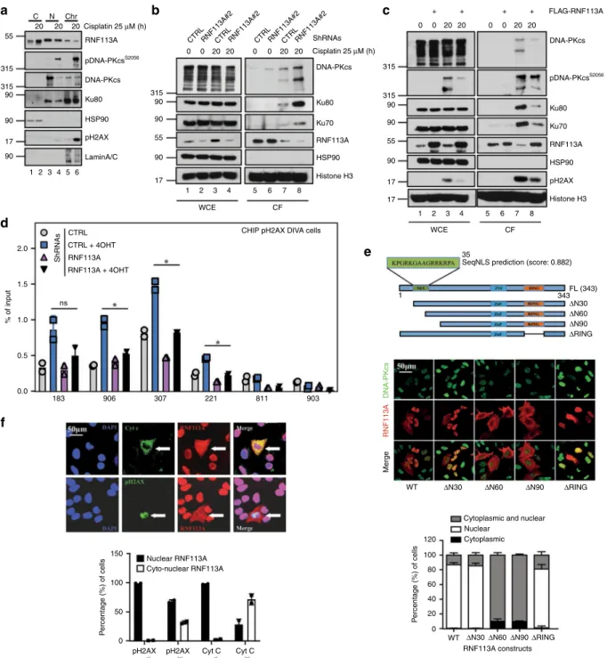
Nb
20 20 55 315 315 90 90 90 1 2 3 4 5 6 17 315 90 90 55 90 17 20 Cisplatin 25 μM (h) RNF113A pDNA-PKcsS2056 DNA-PKcs Ku80 Cisplatin 25 μM (h) DNA-PKcs DNA-PKcs pDNA-PKcsS2056 ShRNAs Ku80 Ku70 RNF113A HSP90 Histone H3 Ku80 Ku70 RNF113A HSP90 pH2AX Histone H3 HSP90 pH2AX LaminA/CCTRL CHIP pH2AX DIVA cells
2.0 1.5 1.0 % of input 0.5 0.0 183 100 P e rcentage (%) of cells 50 pH2AX negative pH2AX positive Cyt C negative Cyt C positive 150 P e rcentage (%) of cells 100 80 60 40 20 120 0 Nuclear RNF113A
Cytoplasmic and nuclear Nuclear Cyto-nuclear RNF113A 0 906 307 221 811 903 CTRL + 4OHT ShRNAs RNF113A RNF113A + 4OHT ns Chr 1 2 3 4 WCE CF 5 6 7 8 1 2 3 4 35 1
SeqNLS prediction (score: 0.882)
FL (343) 343 ΔN30 ΔN60 ΔN90 ΔRING ΔN30 WT ΔN60 ΔN90 RNF113A constructs ΔRING ΔN30 ΔN60 ΔN90 ΔRING WT Merge RNF113A DNA-PKcs CF WCE 5 6 7 8 315 0 0 20 20 + + 0 0 20 20 FLAG-RNF113A 315 90 90 90 55 17 17 + + Cytoplasmic 0 0 CTRLRNF113A#2RNF113A#2CTRLCTRL RNF113A#2RNF113A#2CTRL 20 20 0 0 20 20
c
d
e
f
Fig. 3 RNF113A is recruited on DNA damage-induced foci. a RNF113A is in both the cytoplasm and the nucleus. A549 cells were treated or not with
Cisplatin and WB analyses were carried out with cytoplasmic, nuclear and chromatin-enriched extracts.b, c RNF113A controls the recrutment of NHEJ
factors on chromatin upon DNA damage. Control versus RNF113A-depleted A549 cells (b) or control versus RNF113A-overexpressing A549 cells (c) were
treated or not with Cisplatin and WB analyses were carried out on chromatin fractions after pre-extraction with the CSK+ RNase A buffer. d RNF113A
controls the recruitment of pH2AX on DSBs. ChIP assays were conducted with extracts from control and RNF113A-depleted DIvA U2-OS cells treated or
not with 4-hydroxy Tamoxifen (TAM). Primers 183, 906, 307, and 221 are pH2AX-associated AsiSI sites while primers 811 and 903 are non-associated and
serve as negative control36. Immunoprecipitations using anti-IgG antibody served as negative control. The histogram shows recruitment of pH
2AX on
indicated sites. Results of two independent experiments (means ± SD, Studentt-test, *p < 0.05) are shown. e A N-terminal nuclear localization signal (NLS)
controls the nuclear import of RNF113A. The human RNF113A sequence was analyzed using the online NLS prediction algorithm SeqNLS (http://mleg.cse.
sc.edu/seqNLS/) and the identified NLS (residues 21–35) is shown in green with a possibility score of 0.882. The subcellular localization of RNF113A
constructs was analyzed by immunofluorescence in A549 cells, using DNA-PKcs as a nuclear marker. The percentage of cells showing a cytoplasmic and/
or nuclear localization of RNF113A constructs is illustrated in the histogram below.f RNF113A moves in the cytoplasm of apoptotic cells showing some DNA
damage. Anti-RNF113A Immunofluorescence analyses were conducted in Cisplatin-treated A549 cells. Cells showing some DNA damage or undergoing
cell apoptosis were identifed through anti-pH2AX and Cyt C stainings, respectively. The histogram show the percentage of cells showing a nuclear or a
Depletion of RNF113A in Cisplatin-treated cells was associated
with much higher intron retention levels (1397 introns with
significantly higher inclusion levels in depleted samples).
When normalizing for the number of events evaluated in
each category, intron retention was by far the most frequently
affected category (Fig.
5
c). The most frequent differences were
observed upon RNF113A depletion, with 22% and 34% of the
events showing a significant intron retention increase in
untreated and in Cisplatin-treated cells, respectively. Although
cisplatin induced only a few changes in intron retention level
in control cells, the same treatment induced more intron
retention in RNF113A-depleted cells, consistent with a role of
RNF113A in intron splicing. Almost all events significant
upon depletion were associated with an intron retention level
higher in depleted than in non-depleted cells (Fig.
5
d, and
Supplementary Fig. 8a). In contrast, the impact of Cisplatin on
intron retention was more balanced with both higher and
lower intron inclusion levels depending on the event
considered, especially in control cells (Fig.
5
d and
Supple-mentary Fig. 8a).
c
d
Rad51 CTRL RNF113A 2 3 4 5 6 7 8 9 10 11 12 01216 1820 24 01216 1820 24 HSP90 SF3B2 RNF8 ShRNAs A549 cells Cisplatin 25 μM (h) 1f
WCE IP I P CTRL RNF113A + + + BZR-T33 cells HSP90 RNF113A SF3B1 SF3B2 SF3B2 1 2 3 ShRNAs Cisplatin (25 μM, 24 h) + + + + RNase A + + + + HSP90 RPL7 SF3B1 RNF113A 1 2 3 4 5 6 7 8 Input OligodT IPa
55 150 90 34 150 34 90 55 90 55 150 90 150 150b
HSP90 RPL7 RNF113A Input OligodT IP + + + + RNaseA + + + + SF3B1 1 2 3 4 5 6 7 8 Cisplatin (25 μM, 24h) 55 150 90 34 Sh CTRL Sh RNF113A DAPI S9.6 DAPI S9.6 Sh RNF113A + RNase H DAPI S9.6 R-loopsSpeckles per nucleus
ShRNAs CTRL RNF113A RNF113A
+ RNase H *** *** *** 0 10 20 30
g
CTRL shRNA SF3B2 shRNA#1 SF3B2 shRNA#2 Cisplatin 0 h Cisplatin 24 h 0.72% 1.19% 1.91% 2.04% 8.63% 5.93% 20.77% 15.58% 6.65% 5.00% 10.26% 14.95% 100 100 101 101 102 102 103 103 104 100 101 102 103 104 100 101 102 103 104 100 101 102 103 104 100 101 102 103 104 100 101 102 103 104 104 100 101 102 103 104 100101 102 103 104 100101102103 104 100 101 102 103 104 100 101 102 103 104 ShRNAs CTRL SF3B2#1 SF3B2#2Percentage of apoptotic cells
*** *** Cisplatin (h) 0 24 Annexin V PI SF3B2 SF3B1 HSP90 DNA-PKcs Cisplatin + + + CTRL SF3B2#1 + + + + SF3B2#2 + + ShRNAs 1 2 3 4 5 6 pDNA-PKcsS2056 150 90 315 315 150 RNF113A SF3B2 SF3B1 pH2AX Histone H3 α-tubulin + + C N 55 17 55 17 150 150
i
1 2 3 4 Sh SF3B2 Sh SF3B2 + RNaseH DAPI S9.6 DAPI S9.6 Sh CTRL DAPI S9.6 *** *** *** 0 10 20 30 40Speckles per nucleus
ShRNAs CTRL SF3B2 SF3B2 + RNase H R-loops 0 5 10 15 20
h
j
Cisplatin (25 μM, 24 h) 20 μm 20 μm IgG SF3B1e
RNF113A IP IgG SF3B1 CTRL RNF113A + + + + + A549 cells WCE IP Cisplatin + + HSP90 RNF113A SF3B1 SF3B2 SF3B2 SF3B1 ShRNAs 150 150 150 150 55 90Athough the analysis with rMATS clearly shows an impact of
RNF113A depletion on all types of splicing events and most
notably on intron retention, there are two potential caveats in this
analysis. First, rMATS evaluate various number of events per
gene. Second, only a small fraction of intron retention events is
analyzed as introns are generally not described in genome
annotations. To verify that the above results are not affected by
these caveats, we designed an alternative strategy measuring
specifically the coverage of intronic versus exonic regions for all
transcribed protein-coding genes detected in our transcriptomic
profiles. Strikingly, RNF113A depletion induced an increase of
the relative fraction of reads mapping to intronic regions for most
genes and the effect was even more pronounced in
Cisplatin-treated cells (Fig.
5
e, f). This increase was significant when
comparing the distributions of all genes (Supplementary Fig. 8b)
or global measures for the entire transcriptome (Fig.
5
f).
Although this effect was less obvious for genes with lower
expression levels (Supplementary Fig. 8c), the impact of
RNF113A depletion appeared to be largely unspecific and to
affect most genes. Therefore, beyond an expected impact on the
gene expression, the splicing is globally affected. Although some
specific exon skipping events are significant upon RNF113A
silencing, the main impact of the knockdown is a slight but
significant increase of intron retention. These observations are
consistent with the reported role of RNF113A as a spliceosome
subunit.
RNF113A controls the splicing of pro-survival candidates. We
next concentrated on differential splicing of candidates found in
our RNA-Seq analyses. SAT1 was a candidate impacted by the
treatments both at the transcriptional and at the splicing level.
Indeed, SAT1 expression level significantly increased upon
Cis-platin treatment (Log2 fold change
= 1.5; adjusted p-value = 1.1E
−23; Supplementary Table 2) while its splicing was affected
specificially upon RNF113A deficiency in Cisplatin-treated cells.
SAT1 gene has 7 exons. Exon 4 contains a premature STOP
codon and need to be skipped to give rise to a mRNA coding for a
functional protein (Fig.
6
a). The analysis performed with rMATS
showed that in control cells treated or not with Cisplatin and in
untreated RNF113A-depleted cells, exon 4 is included in roughly
half of the transcripts (Fig.
6
b, left panel). In contrast, the
inclusion level of exon 4 was significatively lower in
RNF113A-depleted cells treated with Cisplatin (Fig.
6
b, left panel).
Com-bined with the effect of Cisplatin on SAT1 expression, the
mod-ifications in exon 4 inclusion level leads to major differences in
abundance of the two corresponding transcripts between the four
sets of conditions (Fig.
6
b, middle and right panels). Cisplatin
increased the expression of the SAT1-encoding mRNA in both
control and RNF113A-depleted cells (Fig.
6
b, right panel). While
Cisplatin also induced an increase in exon 4-containing mRNAs
in control cells, this increase was however abolished in
RNF113A-depleted cells (Fig.
6
b, middle panel). Quantification of SAT1
mRNAs by RT-PCR confirmed the increase of the SAT1-coding
transcript in the RNF113A-depleted cells treated with Cisplatin
(Fig.
6
c, lower panels). Apoptosis was not the causal event of this
process as RNF113A-depleted cells treated with Cisplatin in
which Caspase 3 activation was prevented by ZVAD still showed
elevated levels of the SAT1-coding transcript (Fig.
6
c). SF3B2
deficiency also caused an increase of the SAT1-coding transcript
upon treatment with Cisplatin, demonstrating again that SF3B2
and RNF113A deficiencies share many similarities (Fig.
6
d).
Moreover, ZVAD did not prevent these splicing deregulations seen
upon Cisplatin treatment of SF3B2-depleted cells (Fig.
6
d). As a
result of this increase of the SAT1-coding transcript, protein levels
of SAT1 dramatically increased in RNF113A-depleted A549 cells
treated with Cisplatin (Fig.
6
e). As SAT1 promotes ferroptosis
42, we
reasoned that RNF113A-depleted cells may undergo ferroptosis
upon stimulation with Cisplatin. RNF113A deficiency indeed
potentiated the production of lipid ROS, which accumulate in cells
undergoing ferroptosis, upon Cisplatin stimulation, as evidenced by
the quantification of BODIPY-C11 staining (Fig.
6
f).
If NUPR1 expression is increased by Cisplatin in control cells,
this induction is severely defective in RNF113A-deficient cells
(Fig.
7
a, b). This effect on NUPR1 mRNA expression level was
observed both in our high-throughput RNA sequencing data and
by real-time PCR (left and right panels, respectively). To check
for a specific impact on NUPR1 splicing, we designed a RT-PCR
experiment with primers targeting regions in exons 1 and 2. We
detected two NUPR1 transcripts, a
first transcript properly spliced
(all intron removed) and translated into the NUPR1 protein, as
well as a second transcript in which intron 1 was not removed,
which presumably does not translate into any polypeptide and
undergo NMD (Fig.
7
b). The longer transcript was preferentially
detected upon RNF113A deficiency in Cisplatin-treated cells,
indicating that the spliceosome removes intron 1 to generate the
NUPR1-encoding transcript. Moreover, SF3B2-deficient cells also
failed to induce NUPR1 expression upon Cisplatin treatment
(Supplementary Fig. 9). Conversely, RNF113A overexpression in
A549 cells enhanced NUPR1 expression after Cisplatin treatment
(Fig.
7
c). We also analyzed TCGA data and observed that both
RNF113A and NUPR1 expression were indeed positively
correlated in lung cancer (Fig.
7
d). Therefore, RNF113A, and
by extension the spliceosome, promotes NUPR1 expression upon
DNA damage through the proper splicing of its transcript. In
Fig. 4 RNF113 is a RNA-binding protein. a, b RNF113A is recruited on RNAs. Unstimulated or Cisplatin-treated A549 cells were treated withparaformaldehyde tofix larger complexes (a) or irradiated with UV to covalently crosslink direct RNA-protein interaction (b), incubated or not with RNase
H and RNA immunoprecipitations using OligodT magnetic beads were done followed by WB analyses (left lanes). Cell extracts before RNA
Immunoprecipitations were also subjected to WB analyses (“Input”). c depleted lung cancer cells accumulate R-loops. Control or
RNF113A-depleted A549 cells were subjected to immunofluorescence analyses using the anti-R-loops antibody (63X objective lens). A quantification of DNA-RNA
hybrids is illustrated. For quantification, nuclei specles in a total of 50 nucleus in each experimental condition were counted (Student t-test, ***p < 0.001).
d RNF113A promotes SF3B2, Rad51 and RNF8 expression. Control or RNF113A-depleted A549 cells were untreated or stimulated with Cisplatin and WB
analyses were done.e, f RNF113A is required for the integrity of the spliceosome. Control or RNF113A-depleted A549 (e) or BZR-T33 (f) cells treated or not
with Cisplatin were subjected to anti-IgG (negative control) or -SF3B1 immunoprecipitations followed by anti-SF3B2 WBs. Cell extracts were also subjected
to WBs (lower panels).g SF3B2-depleted lung cancer cells accumulate DNA-RNA hybrids. Control or SF3B2-depleted A549 cells were subjected to
immunofluorescence analyses as described in (c). h SF3B2 deficiency enhances DNA-PKcs phosphorylation upon Cisplatin treatment. Control and
SF3B2-depleted A549 cells were treated or not with Cisplatin (25μM for 24 h) and WB analyses were done. i SF3B2 deficiency enhances cell death upon DNA
damage. On the left, cell survival upon Cisplatin treatment in control and SF3B2-depleted A549 cells was assessed by FACS. The percentage of cells in
early or late apoptosis is quantified. On the right, FACS data from two independent experiments are illustrated (Student t-test, ***p < 0.001). j SF3B1,
SF3B2, and RNF113A moves from the nucleus to the cytoplasm of lung cancer cells upon DNA damage. A549 cells were treated or not with Cisplatin and WB analyses were done (cytoplasmic and nuclear extracts).
agreement with the fact that the genetic inactivation of NUPR1
triggers senescence
43, the depletion of RNF113A in A549 cells led
to senescence, as evidenced by an increase of
β-galactosidase-positive cells (Fig.
7
e). Therefore, RNF113A expression prevents
senescence, at least by maintaining NUPR1 expression in lung
cancer cells.
The NADPH oxidase activator 1 Noxa1 catalyses the
produc-tion of intracellular and extracellular superoxide (O
2−) from O
2and NADPH
44. Interestingly, multiple transcripts of Noxa1 were
detected in A549 cells (Fig.
8
a). The 483 amino acids long Noxa1
protein is generated from the translation of a transcript lacking
both introns 9 and 10 (Fig.
8
a). Two additional transcripts, which
Before splicingInclusion (2 splicing events)
Skipping (1 splicing event)
SE = skipped exon
Before splicing
Inclusion (0 splicing event)
Skipping (1 splicing event)
RI = retained intron
Before splicing
Inclusion (1 splicing event)
Skipping (1 splicing event)
A5SS = alternative 5’ splicing site
Before splicing
Inclusion (1 splicing event)
Skipping (1 splicing event)
A3SS = alternative 3’ splicing site
Before splicing
Inclusion = first exon (2 splicing events)
Skipping = second exon (2 splicing events)
MXE = mutually exclusive exons
a
b
c
d
Cisplatin treated control cells
Untreated control cells Untreated control cells Cisplatin treated control cells Untreated RNF113A-depleted cells 1.00 0.75 0.50 0.25 0.00 1.00 0.75 0.50 0.25 0.00 0.00 0.25 0.50 0.75 1.00 1.00 0.75 0.50 0.25 0.00 1.00 0.75 0.50 0.25 0.00 1.00 0.75 0.50 0.25 0.00 1.00 0.75 0.50 0.25 0.00 1.00 0.75 0.50 0.25 0.00 Fraction of transcripts with retained intron
Intron/exonic read ratio
in RNF113A-depleted A549 cells (log2)
Intron/exonic read ratio in control A549 cells (log2) 0 5 –5 –10 –15 –10 –5 0 5 –15 –10 –5 0 5 Untreated Cisplatin
e
Fraction of reads covering introns versus exons (%)
0 5 2.5 Untreated Cisplatin
f
15 5 10 Number of exonic reads (log2) CTRL shRNA RNF113A shRNA*
*
**
7.5S1 S2 Of On SE RI A3SS A5SS MXE
C Ccis Cisplatin Control cells 68,403 (363:899) 4111 (120:206) 5653 (43:83) 3854 (50:60) 14,456 (53:111) C D Depletion Untreated cells 68,784 (314:290) 4059 (43:906) 5587 (23:81) 3834 (30:75) 14,517 (28:54) Ccis Dcis Depletion Cisplatin-treated cells 69,112 (345:578) 4153 (37:1397) 5697 (35:218) 3913 (41:169) 14,505 (52:109) D Dcis Cisplatin Depleted cells 71,063 (534:1342) 4116 (116:601) 5673 (48:156) 3916 (64:169) 15,019 (104:148)
Samples Evaluating the effect Number of alternative splicing events :
Fraction of analysed
splicing events
shRNA: Cisplatin:
SE RI A5SS A3SS MXE
CTL CTL CTL CTL RNF-113A RNF-113A RNF-113A RNF-113A shRNA: Cisplatin: S1 S2
Not significantly different Significantly lower Significantly higher Level of inclusion in S2 – + + + – – + – CTL CTL CTL CTL RNF-113A RNF-113A RNF-113A RNF-113A – + + + – – + – CTL CTL CTL CTL RNF-113A RNF-113A RNF-113A RNF-113A – + + + – – + – CTL CTL CTL CTL RNF-113A RNF-113A RNF-113A RNF-113A – + + + – – + – CTL CTL CTL CTL RNF-113A RNF-113A RNF-113A RNF-113A – + + + – – + – 1.00 0.75 0.50 0.25 0.00 Untreated RNF113A-depleted cells Cisplatin treated RNF113A-depleted cells Cisplatin treated RNF113A-depleted cells
includes intron 9 and/or 10, were also detected and encode
shorter 318 or 283 amino acids long Noxa1 proteins, respectively
due to the appearance of premature stop codons (Fig.
8
a).
RNF113A deficiency had dramatic consequences on Noxa1
expression as the smaller 318 amino acids protein generated
from the translation of the transcript which includes intron 10
(“Noxa1 ΔSH3”) accumulated in RNF113A-depleted cells
(Fig.
8
b). This smaller isoform lacks the C-terminal SH3 domain
and more efficiently activates NOX1
45(Fig.
8
a, b). The
accumulation of Noxa1
ΔSH3 was also seen in SF3B2-depleted
A549 cells (Fig.
8
c). Consistently, the depletion of RNF113A in
A549 cells potentiated ROS production in both unstimulated and
Cisplatin-treated cells (Fig.
8
d). Moreover, the NADPH/NADP
ratio, which reflects NOX1 activity, was increased upon RNF113A
deficiency in Cisplatin-treated cells (Fig.
8
e). Therefore, RNF113A
expression limits the production of ROS, at least through
Noxa1 splicing.
RNF113A stabilizes MCL-1. ROS production destabilizes
MCL-1
46. Moreover, MCL-1 limits cell death triggered by
Cis-platin in lung cancer cells
47. Therefore, we investigated whether
RNF113A controls MCL-1 expression. MCL-1 protein levels
decreased in RNF113A-depleted A549, BZR-T33, H1975 or
Calu-6 cells subjected to Cisplatin and undergoing Caspase
3-dependent apoptosis (Fig.
9
a, Supplementary Fig. 10a, b and
Fig.
9
b, respectively). MCL-1 phosphorylation by GSK3 similarly
decreased in RNF113A-depleted A549 cells, suggesting that a
deregulation of MCL-1 phosphorylation, which triggers its
degradation
48, was not responsible for MCL-1 disappearance
from the cytoplasm upon Cisplatin treatment of
RNF113A-depleted cells (Fig.
9
a). On the other hand, RNF113A deficiency
in Caspase 3-negative HCC827 cells did not impact on MCL-1
levels upon Cisplatin treatment but nevertheless triggered more
Caspase 8 activation and more cell death (Fig.
9
b and
Supple-mentary Fig. 10c). On the other hand, RNF113A-overexpressing
cells showed elevated MCL-1 protein levels when treated to
Cisplatin and MCL-1 half-life was enhanced in Cisplatin-treated
and RNF113A-overexpressing A549
cells (Supplementary
Fig. 10d and Fig. 9c, respectively). MCL-1 stabilization was due
to a defective proteasome-dependent degradation as the
pro-teasome inhibitor MG132 restored MCL-1 protein levels in
RNF113A-depleted A549 cells treated with Cisplatin (Fig.
9
d).
Moreover, polyubiquitin chains were also detected in a TUBE
assay from RNF113A-depleted A549 cells treated with Cisplatin
in which the proteasome was blocked (Fig.
9
e). We next looked
at expression levels of regulators of MCL-1 polyubiquitination
and focused on USP9X, which deubiquitinates and stabilizes
MCL-1
49. USP9X levels were decreased in RNF113A-depleted
cells, which may contribute to the enhanced degradative
poly-ubiquitination of MCL-1 (Fig.
9
f). Our data has some clinical
relevance as RNF113A and MCL-1 protein levels positively
correlated in clinical cases of lung cancer (Fig.
9
g).
SF3B2-depleted cells, which express less RNF113A, also had less
MCL-1, at least because of decreased USP9X levels (Fig.
9
h).
Given the central role of BCL-2 in cell survival, BH3 mimetics/
BCL-2 inhibitors such as ABT-737 were designed and indeed
causes regression of established tumors
50. Nevertheless, MCL-1
promotes resistance to ABT737
51. As RNF113A stabilizes MCL-1,
we reasoned that RNF113A deficiency may enhance
ABT737-dependent cell death through MCL-1 downregulation. Indeed,
cell death upon treatment with ABT737 was more pronounced
upon RNF113A deficiency in A549 cells (Fig.
9
i). Therefore,
RNF113A acts as a pro-survival candidate, at least by maintaining
MCL-1 protein levels.
RNF113A de
ficiency circumvents resistance to Cisplatin. As
MCL-1 promotes resistance to Cisplatin and because MCL-1
expression relies on RNF113A, we next explored whether
RNF113A also contributes to resistance to Cisplatin in lung
cancer cells. We generated Cisplatin-resistant A549 cells by
cul-turing parental A549 cells (“A549/P”) with increasing
con-centrations of Cisplatin. Resistant cells (“A549/CR (4.5)”) did not
dramatically undergo cell death when subjected to increasing
concentrations of Cisplatin and underwent epithelial to
mesenchymal transition (EMT), as judged by an elongated
morphology and by lower levels of E-cadherin (Fig.
10
a).
RNF113A deficiency sensitized these resistant cells to cell death
triggered by Cisplatin (Fig.
10
b). Mechanistically, RNF113A
deficiency triggered Caspase 9 and 3 activation upon treatment
with Cisplatin, at least due to enhanced DNA-PKcs
phosphor-ylation and to decreased MCL-1 protein levels (Fig.
10
c). The
defective expression of SF3B2, Rad51, and RNF8 seen upon
RNF113A deficiency in parental A549 cells was also observed in
Cisplatin-resistant cells (Fig.
10
d). As a result, cell death triggered
by ABT737 was more pronounced upon RNF113A deficiency in
Cisplatin-resistant A549 cells (Fig.
10
e). RNF113A deficiency also
triggered tumor regression in vivo upon treatment with Cisplatin
when resistant A549 cells were transplanted into
immunodefi-cient mice (Fig.
10
f). Therefore, RNF113A promotes
chemore-sistance to Cisplatin in lung cancer cells, at least by stabilizing
MCL-1 levels.
Discussion
We show here that RNF113A promotes cell survival upon
DNA damage as a spliceosome subunit. Splicing targets of
Fig. 5 Increased intron retention in mRNAs upon RNF113A deficiency, especially in Cisplatin-treated lung cancer cells. a–d Analysis of alternative
splicing (AS) at the level of individual splicing events with rMATS (replicate Multivariate Analysis of Transcript Splicing).a Schematic representation of the
different types of AS events analyzed by rMATS. Inclusion and skipping forms are quantified using reads overlapping junctions and reads unique to the
inclusion form.b Number of AS events found in RNF113A-depleted and control A549 cells treated or not with Cisplatin. Each entry in the table has three
values: Total number of events evaluated in that comparison (Inclusion significantly lower in S2: Inclusion significantly higher in S2). Samples are as follow:
C= control shRNA—no drug, Ccis = control shRNA + Cisplatin, D = RNF113A shRNA—no drug, Dcis = RNF113A shRNA + Cisplatin. Significant events are
defined as FDR<5% and delta inclusion level (|ΔΨ|) of at least 20%. c Fraction of analyzed AS events with inclusion level significantly lower or higher upon
treatment (four comparisons) for each category of AS events.d Comparison of fraction of transcripts with retained intron in RNF113A-depleted and control
A549 cells treated or not with Cisplatin. Each dot represents an intron retention event. Events with statistically significant difference between conditions
are represented in red. Black broken line: identity axis.e-f Analysis of Intron Retention at the level of individual genes. e Comparison, for each
protein-coding gene, of the ratio of mRNAseq reads in intronic versus exonic regions (average of three replicates) between RNF113A-depleted and control A549
cells treated or not with Cisplatin. Black broken line: identity axis. Shift towards higher ratios upon RNF113A depletion are significant (p-value < 2.2 × 10⁻¹⁶;
Wilcoxon signed rank tests (paired tests); see Supplementary Fig. 8c).f Total number of reads covering introns versus total number of reads restricted to
annotated exons for both RNF113A-depleted and control A549 cells treated or not with Cisplatin. Data were obtained from three replicates for each
RNF113A include multiple pro-survival candidates. Interfering
with RNF113A triggers cell apoptosis, at least through MCL-1
destabilization, which circumvents the acquired resistance to
BCL-2 inhibitors. RNF113A deficiency also triggers ferroptosis,
at least through SAT1 expression and enhances ROS production
upon DNA damage. SF3B2 deficiency leads to very similar
consequences as the loss of RNF113A, which suggests that both
proteins have similar functions. Therefore, the spliceosome is a
major actor of cell survival in lung cancer and also define
RNF113A as a promising anti-cancer target to
fight the acquired
resistance to BCL-2 inhibitors.
The mechanisms by which RNF113A promotes MCL-1
stabi-lity may be cell-type dependent. Indeed, Cisplatin-resistant cells
depleted for RNF113A show appearance of the short and
a
HSP90 RNF113A SAT1 CTRL RNF113A Cisplatin (25 μM, 24 h) + + ShRNAsc
f
100 80 60 40 20 0 102 103 104 0 ShRNA control + Cisplatin GFP-A ShRNA RNF113A + Cisplatine
ShRNAs CTRL RNF113A Untreated Cisplatin (24 h) *** *** Mean events 26 55 90 SAT1-encoding mRNA STOP* Coding sequence ATG Primer #1 Primer #2SAT1 mRNA undergoing NMD 7 Exons: STOP* Cisplatin ShRNAs CTRL RNF113A Untreated Cisplatin (24 h) 3 4 5 3 5 60 40 20 0 ShRNA: CTRL RNF113A
Exon 4 Inclusion level (%)
*** ***
CTRL RNF113A
Reads overlapping exons 3 & 5
(normalised read count)
1000 0 2000 * 6000 0 Reads mapping to exon 4 (normalised read count)
CTRL RNF113A 4000 2000
b
0 200 400 600 800 1000 0 1 2 3 4 ShRNAs CTRL RNF113ARatio coding/non coding
Untreated Cisplatin (24 h) ZVAD 250 bp 500 bp SAT1 transcripts ZVAD HSP90 Cleaved caspase 3 RNF113A Control RNF113A Cisplatin ZVAD ShRNAs 55 90 17 1 2 3 4 5 6 7 8 Cisplatin ShRNAs CTRL SF3B2 250 bp 500 bp SAT1 transcripts ZVAD STOP 1 2 3 4 5 6 + + + + + + + + + + 1 2 3 4 + + + + + +
d
0.9 1.0 1.1 1.2 1.3 1.4 Untreated Cisplatin (24 h) ZVADRatio coding/non coding
pro-apoptotic form of MCL-1 and lower levels of the long and
pro-survival form of MCL-1, suggesting that MCL-1 is a direct
target of the spliceosome in these cells. We did not
find any
evidence that MCL-1 splicing was regulated by RNF113A in
parental cells. Yet, MCL-1 degradative polyubiquitination was
enhanced upon RNF113A deficiency in these cells. USP9X, which
stabilizes MCL-1 by promoting its deubiquitination
49, is less
expressed upon RNF113A deficiency. This mechanism may
contribute to the destabilization of MCL-1 seen in
RNF113A-depleted cells. Alternatively RNF113A may directly inhibit the
function of any E3 ligase such as MULE that targets MCL-1 for
degradation upon DNA damage
52. Another molecular
mechan-ism may involve ROS production. Indeed, ROS, which are more
produced upon RNF113A deficiency, destabilize MCL-1 through
a poorly characterized pathway
46,53.
RNF113A deficiency leads to multiple types of cell death in
addition to apoptosis upon DNA damage. Indeed, Caspase
3-deficient lung cancer cells still undergo cell death upon RNF113A
deficiency when treated with Cisplatin. We actually show that
ferroptosis occurs when RNF113A-depleted cells are subjected to
a DNA damage signal.
The repair of DNA alkylation damage involves the alkylation
repair complex ASCC (activating signal cointegrator complex)
which relocalizes to specific nuclear foci with spliceosome
pro-teins and basal transcription factors upon exposure to alkylating
agents
54. This recruitment to nuclear foci requires the sensing of
polyubiquitin chains by the CUE (coupling of ubiquitin
con-jugation to ER degradation) of ASCC2. RNF113A is the E3 ligase
that catalyses the formation of these K63-linked non degradative
polyubiquitination chains on BRR2, an ASCC2-interacting
pro-tein
54. RNF113A is relocalized to ASCC2-enriched foci upon
DNA damage, which appears to be distinct from pH
2A.X S139
+foci, a
finding that we also report in our study. The N-terminal
domain of RNF113A is critical for the binding to BRR2. We show
here that this domain includes the NLS of RNF113A, which is
consistent with the idea that all functions of RNF113A in the
nucleus critically relies on its N-terminal domain. Interestingly,
we demonstrate that Cisplatin triggers the cytoplasmic shuttling
of RNF113A. SF3B2 also disengages from the chromatin upon
DNA damage. This observation is not in contradiction with the
fact that splicing events occur in the nucleus as a pool of both
nuclear RNF113A and SF3B2 can still be found in lung cancer
cells showing some DNA damage. Spliceosome subunits such as
SR proteins are actually dephosphorylated to facilitate the export
of spliced mRNAs to the cytosol in order to enhance
transla-tion
55. Whether post-translational modifications of RNF113A
regulates its cellular localization remains unknown. In any case,
this strongly suggests that RNF113A moves into the cytoplasm,
presumably with some spliced transcripts upon DNA damage.
Let’s note however that the pool of cytoplasmic SF3B2 did
not increase upon Cisplatin treatment, which differs from
RNF113A. This is the only property that distinguishes SF3B2
from RNF113A.
It is unclear whether RNF113A exclusively works as a
spli-ceosome subunit. As SF3B2 deficiency mimics the phenotypical
alterations seen in RNF113A-depleted cells, this suggests that
RNF113A works as a spliceosome subunit. Yet, the fact that
RNF113A polyubiquitinates proteins such as BRR2, which does
not regulate RNA splicing
4, indicates that some
spliceosome-independent functions of RNF1113A may also occur. Moreover,
the co-localization of RNF113A with DNA-PKcs in
Cisplatin-treated A549 cells and the defective engagement of pH
2AX to
DSB sites in depleted DIvA cells suggests its recruitment at the
extremities DNA DSBs where it could act as an E3 ligase to
directly promote DNA repair. Alternatively, RNF113A may
indirectly promote this process as a spliceosome subunit. We
actually demonstrate that RNF8, which promotes histone
poly-ubiquitination and the recruitment of 53BP1 and BRCA1 repair
proteins to double-strand breaks
56, is a target of RNF113A as a
spliceosome subunit. Therefore, RNF113A is involved in DNA
repair through both direct and indirect mechanisms. The absence
of strong ATR activation observed in RNF113A-depleted cells
allowed to resume growth in a Cisplatin-free media, indicates
either a deficient replication-stress response or more likely the
persistance of damage-induced cell cycle arrest due to the high
level of damage present. All experiments were conducted after 24
h of permanent contact with Cisplatin. Only one experiment was
performed with a recovery period in order to best visualize ATR
activation, which requires replication.
Our data demonstrate that the spliceosome contributes to the
acquired resistance to Cisplatin, at least by promoting MCL-1
stability. Therefore, targeting RNF113A or SF3B2 is a strategy to
circumvent the acquired resistance of lung cancer cells to
Cis-platin. Patients suffering from lung cancer and showing some
acquired resistance to ABT737 may also benefit from the
inhi-bition of the spliceosome. To conclude, it is tempting to speculate
that any tumors showing some MCL-1 stabilization may benefit
from spliceosome inhibitors targeting RNF113A or SF3B2.
Methods
Cell lines, antibodies, plasmids, and treatments. The human adenocarcinoma A549 (CCL-185) cell line, H1975 (CRL-5908) and 293 (CRL-1573) cells as well
as normal human dermalfibroblasts (PCS-201-012) were purchased from the
American Type Culture Collection (ATCC, Manassas, VA, USA). BZR-T33 cells were kindly provided by Dr. Christine Gilles (GIGA-Cancer, University of Liege, Belgium). The Lenti-X 293T cell line was obtained from Clontech Laboratories (catalog number 632180) (Palo Alto, CA, USA). All cell lines (including normal
human dermalfibroblasts) were tested for mycoplasma contamination and were
maintained in Dulbecco’s Modified Eagle’s Medium (DMEM) (Lonza, Basel,
Fig. 6 RNF113A regulates the splicing of SAT1. a–c RNF113A promotes SAT1 splicing. a Representation of SAT1 transcripts. The coding and non-coding
sequences are illustrated as green and blue rectangles, respectively. A defective splicing of exon 4 (red rectangle) leads to a premature STOP codon.
b rMATS results for the analysis of exon 4 skipping inSAT1 transcripts. The first barplot on the left represents the inclusion level of exon 4 in SAT1
transcripts in RNF113A-depleted or control cells, treated or not with Cisplatin. The middle and right plots represent the number of reads specific to
transcripts with or without exon 4, respectively. Read counts have been normalized for differences in sample sequencing depth using DESeq2 size factor
(median ratio method). Data were obtained from 3 replicates for each experimental condition (mean ± SD; Left panel: ***= FDR q-value < 0.001 from
rMATS; Middle and right panels: *p < 0.05, two-sided T-test on normalized read counts). c, d RNF113A and SF3B2 deficiencies share common defects in
SAT1 splicing. Transcripts are detected by RT-PCR experiments, using both primers depicted in a. Data for both control and RNF113A or SF3B2-depleted
cells are shown (c and d, respectively). A quantification of all signals is illustrated for both experiments. On the top (c), WB analyses were done with
control or RNF113A-depleted cells treated or not with Cisplatin (25μM for 24 h) and treated or not with ZVAD (20 μM for 24 h). e Enhanced SAT1 protein
levels in Cisplatin-treated and RNF113A-deficient lung cancer cells. Control and RNF113A-depleted A549 cells were treated or not with Cisplatin (25 μM for
24 h) and WB analyses were done.f RNF113A deficiency triggers ferroptosis upon Cisplatin stimulation. Control and RNF113A-depleted A549 cells were
treated or not with Cisplatin (25μM) and lipid ROS were quantified by BODIPY-C11 staining, using a flow cytometer. Data from two experiments performed