Université de Montréal
Influence du chondroitin sulfate (CS)
sur l’activité et l’expression de plusieurs isoformes du Cytochrome
P450
et de la NADPH P450 réductase
Par
Mirela Onita Iovu
Département de pharmacologie
Faculté de Médecine
Mémoire présenté à la Faculté des études supérieures en vue de l’obtention du grade de
Maître en Sciences (M. Sc)
en Pharmacologie
Avril 2009
Université de Montréal Faculté des études supérieures
Ce mémoire intitulé :
Influence du chondroitin sulfate (CS)
sur l’activité et l’expression de plusieurs isoformes du Cytochrome P450
et de la NADPH P450 réductase
Présenté par : Mirela Onita Iovu
a été évalué par un jury composé des personnes suivantes :
Dr.Pierre Haddat, PhD Président-rapporteur Dr. Patrick du Souich, M.D., PhD Directeur de recherche Dr. Jean Spénard, PhD Membre du Jury
3
ABSTRACT
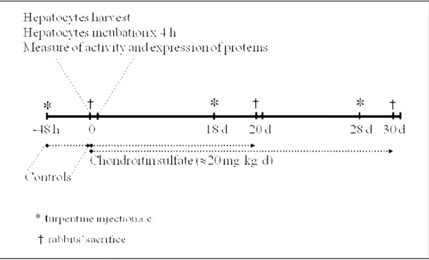
In rabbits, an acute inflammatory reaction induced by the injection of turpentine causes a decrease in cytochrome P450 (CYP) isoforms activity and expression. Chondroitin sulfate (CS) is a Symptomatic Slow Acting Drug for OsteoArthritis (SYSADOA) that elicits anti-inflammatory effects. Since patients take CS over long periods, it was of interest to assess whether CS modulates the activity of cytochrome P450 isoforms. In order to determine the effect of CS on the cytochrome P450, CS was administered in vivo to two animal models, e.g. chronic intake of CS in control rabbits, and chronic intake of CS in rabbits with a CYP down-regulated by an inflammatory reaction (IR). We used six groups of five rabbits: three to assess the effect of CS on cytochrome P450, one without CS and two receiving orally about 20 mg/kg/day CS for 20 and 30 days; and the remaining three groups of rabbits received turpentine s.c. to generate an aseptic IR (AIR) 48 h before their sacrifice, e.g. days -2, 18 and 28, while exposed to CS for 0, 20 or 30 days, respectively.
In order to verify the presence of inflammation we measured the seromucoids in serum of rabbits with an AIR. Another marker of inflammation, e.g. nitric oxyde (NO.) production, was assessed in control hepatocytes (Hcont) and in hepatocytes from rabbits with an AIR (Hinfla). In addition, the effect of CS on the nuclear translocation of NF-κB was studied by fluorescence in hepatocytes. Finally, in hepatocytes (both Hcont and Hinfla) the CYP3A6, CYP1A2 and NADPH P450 reductase (NADPH) activity, expression and mRNA were measured. In vitro, the effect of different concentrations of CS, 4S-, 6S- and 4,6S-sulfated disaccharides of CS on the cytochrome P450 was documented.
Compared with control rabbits, 20 and 30 days CS did not affect the activity of CYP3A6 and CYP1A2. The AIR increased seromucoids from 8.4±1.6 mg/dl in controls to 95.1±5.7 (p<0.05), as
4
well as the nuclear translocation of NF-κB, and nitric oxide concentrations. The AIR reduced CYP3A6 activity by 62% and CYP1A2 activity by 54%, decrease associated to a reduction in protein expression and in mRNA, e.g. pre-transcriptional down-regulation.
The nuclear translocation of NF-κB was prevented by the administration of CS to rabbits with an AIR, moreover CS impeded the increase of the concentrations of nitric oxide; however CS did not prevent the increase in seromucoids. CS did not prevent the down-regulation of CYP1A2 produced by the inflammatory reaction.
CS prevented the time-dependent down-regulation of CYP3A6 in control rabbits and in rabbits with an inflammatory reaction. In this last group, CS restored the amounts of CYP3A6 protein to levels observed in control rabbits, however this increase was independent of the mRNA that remained very depressed. It is noteworthy that even if CS increased CYP3A6 protein, its activity was not recovered. CS did not affect NADPH activity or expression.
Finally, in vitro, CS, 4S-, 6S and 4,6S-sulfated disaccharides of CS did not change the activity and expression of the two isoforms of CYP, and of NADPH.
It is concluded that CS does not affect the activity or expression of CYP1A2, nor prevents CYP1A2 AIR-induced down-regulation. However, CS prevents the down-regulation of CYP3A6 time dependently and following the AIR but does not prevent the decrease of catalytic activity.
5
RESUME
Le CS fait partie de la famille des SYSADOA (SYmptomatic Slow Acting Drugs for OsteoArthritis) et est utilisé par les patients avec de l’ostéoarthrose de façon chronique pour ses propriétés anti-inflammatoires. Étant donné que ces patients reçoivent d’autres médicaments, il était intéressant de documenter les effets du CS sur le cytochrome P450 et la NADPH-réductase (NADPH).
Pour cette étude, deux modèles ont été utilisés: des lapins témoins (LT) et des lapins avec une réaction inflammatoire (LRI) afin de diminuer l’activité et l’expression du CYP. Six groupes contenant chacun cinq lapins ont été utilisés: un groupe sans CS et deux groupes qui ont pris oralement dans l’eau approximativement 20.5 mg/kg/jour de CS pendant 20 et 30 jours; les lapins des trois groupes restants ont pris du CS comme décrit plus haut, mais ont reçu 5 ml sous-cutanées de térébenthine afin de produire une réaction inflammatoire aseptique (RIA) deux jours avant leur sacrifice, c’est-à-dire aux jours -2, 18 et 28. Les hépatocytes ont été isolés pour évaluer l’activité et l’expression du CYP3A6, CYP1A2 et NADPH et aussi le ARNm de ces protéines. In vitro, nous avons étudié l’effet de différentes concentrations de CS-disaccharides sulfatés, 4S, 6S, et 4,6S de CS, sur l’activité et l’expression du CYP1A2 et du CYP3A6. Pour documenter la présence de la réaction inflammatoire, nous avons mesure les mucoprotéines, dans le sérum des lapins avec une réaction inflammatoire. Aussi nous avons mesuré la présence de l’oxide nitrique (NO) chez les hépatocytes de lapins contrôles et chez les hépatocytes des lapins avec une réaction inflammatoire. La translocation nucléaire du NF-κB a été etudiée par fluorescence chez les hépatocytes.
Par comparaison aux lapins témoins, l’administration du CS pendant 20 et 30 jours n’affecte pas l’activité du CYP3A6 et du CYP1A2. La RIA a augmenté les mucoprotéines à 95,1±5,7 vs 8,4±1,6 mg/dl dans les lapins témoins (p<0,05). La RIA a diminué l’activité du CYP3A6 de 62% et l’activité
6
du CYP 1A2 de 54%. Le CS n’empêché pas la diminution du CYP1A2 produite par la RIA. Par ailleurs, le CS n’affecte pas l’activité ni l’expression de la NADPH.
La translocation nucléaire de NF-κB a été empêche par l’administration chronique de CS aux lapins avec RIA; en plus, la concentration de l’oxide nitrique n’a pas démontré une augmentation en présence de CS; par contre, CS n’empêche pas l’augmentation des séromucoïdes.
Au contraire, CS affecte la diminution du CYP3A6 en fonction de temps et secondaire à la RIA. Dans ce group, CS a rétabli le niveau des protéines du CYP3A6 observé dans le group de lapins témoins. Pourtant cette croissance été independante de mRNA qui garde un niveau trés bas. Le plus remarcable a été la manière dont CS a augmenté la protéine du CYP3A6, sans avoir rétabli l’activité de cet isoforme. Finalement, in vitro, CS et ses trois disaccharides sulfatés (4S, 6S et 4,6S) n’affectent ni l’activité ni l’expression de CYP1A2, CYP3A6 et de la NADPH.
En conclusion, l’administration chronique de CS n’affecte pas l’activité ni l’expression du CYP1A2, ou la diminution du CYP1A2 produite par la réaction inflammatoire. Le CS n’affecte pas l’activité ni l’expression du NADPH. Cependant, CS empêche la diminution du CYP3A6 en fonction de temps et secondaire à la RIA.
7
TABLE OF CONTENTS
ABSTRACT ...3 ACKNOWLEDGEMENTS...14 I. INTRODUCTION...1 PART I: CYTOCHROME P450S ...16PART II: INFLAMMATION AND CYTOCHROME P450 ...41
PART III: OSTEOARTHRITIS ...47
II. HYPOTHESIS AND STUDY OBJECTIVE...1
III. MATERIALS AND METHODS ...1
EXPERIMENTAL PROTOCOL ...65
IV. RESULTS...1
CYTOCHROME P450 AND NADPH-REDUCTASE ACTIVITY...81
CYTOCHROME P450 AND NADPH-REDUCTASE EXPRESSION ...86
EFFECT OF CS ON CYP3A6 MRNA ...92
MARKERS OF INFLAMMATION ...93
EFFECT OF CS AND THE ∆DI-4S, ∆DI-6S AND ∆DI-4,6S DISACCHARIDES ON CYP1A2, CYP3A6 AND NADPH-REDUCTASE ACTIVITY AND EXPRESSION IN VITRO ...98
V. DISCUSSION...1
8
FIGURE LIST
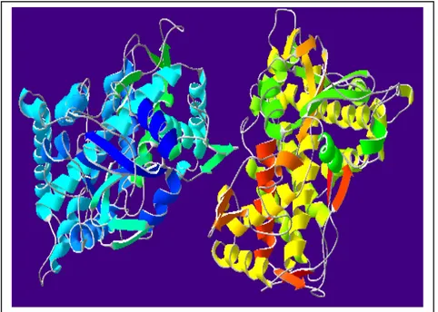
Figure 1 Secondary and tertiary structure of cytochrome P450...18
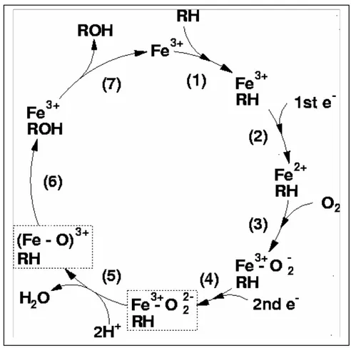
Figure 2. Catalytic cycle of cytochrome P450. ...20
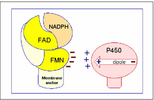
Figure 3. Electron transfer to cytochrome P450...39
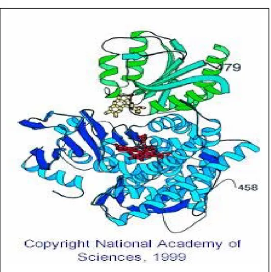
Figure 4. Three-dimensional structure of FMN domain of CPR...40
Figure 5. Standing anteroposterior (A) and standing flexed postero-anterior (B) views of the right knee.a...48
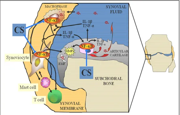
Figure 6. Effect of CS disaccharides sulphated in position 4 and/or 6 (∆di-4S, ∆di-6S, ∆di-4,6S) on Il-1β-induced NF-κB nuclear translocation ...56
Figure 7. Diagram depicting the potential sites of effect of CS and/or its disaccharidesa...60
Figure 8. Protocol representation ...66
Figure 9. (A) CYP1A2 activity in hepatocytes from rabbits pre-treated with CS for 20 and 30 days, with or without an inflammatory reaction...1
Figure 10. Protein expression at O Days, 20 Days and 30 Days for (A) CYP1A2, (B) CYP3A6, (C) NADPH-reductase. ...89
Figure 11. Effect of the administration of CS for 20 and 30 days on ...90
Figure 12. (A) CYP3A6 mRNA (expressed as percentage of controls). ...93
Figure 13. Fluorescent micrographs representing p65–NF-κB immunopositive nuclei in hepatocytes. ...96
Figure 14. Effect of chondroitin sulphate (CS) on NF-κB nuclear translocation in rabbits hepatocytes, in vivo...97
Figure 15. Activity and expression of CYP1A2, 3A6, and NADPH-reductase in the presence of different concentrations of CS and its disaccharides (∆di-4S, ∆di-6S and ∆di-4,6S), in vitro. ...98
Figure 16. Production of an inflammatory reaction. Infections by means of pathogen-associated molecular patterns (PAMPs) activate the pattern recognition receptors (PRRs), including toll-like receptor (TLR), in polymorphonuclear cells (PMNs) and other immune cells...103
9
TABLE LIST
Table 1. NADPH-reductase, CYP1A2, CYP3A6 activity assessed in hepatocytes from control rabbits (Hcont) and in hepatocytes from rabbits with an inflammatory reaction (Hinfla). *p <0,05 compared
with Hcont...81
Table 2. NADPH-reductase, CYP1A2, CYP3A6 activity assessed in the hepatocytes from control rabbits (Hcont) and in the hepatocytes from rabbits with an inflammatory reaction (Hinfla) following the administration of CS for 20 days. *p <0,05 compared with Hcont...82 Table 3. NADPH-reductase, CYP1A2, CYP3A6 activity assessed in hepatocytes from control rabbits (Hcont) and in hepatocytes from rabbits with an inflammatory reaction (Hinfla) following the administration of CS for 30 days. *p <0,05 compared with Hcont...83 Table 4 Seromucoid concentrations in serum from control rabbits and rabbits with a turpentine-induced inflammatory reaction (TIIR) at Day 0 and following the intake of approximately 20 mg/kg of chondroitin sulfate (CS) for 20 and 30 days. ...94 Table 5 Nitric oxide (NO•) concentrations in hepatocyte’s culture media from control rabbits and rabbits with a turpentine-induced inflammatory reaction (TIIR) at Day 0 and following the intake of approximately 20 mg/kg of chondroitin sulfate (CS) for 20 and 30 days...95
10
LIST OF ABBREVIATIONS
∆di-4S/6S/4,6S: (1-4)-O-(D-glucopyranosyluronic acid)–(1-3)-O-(2-N-acetamido-2-deoxy-D
-galactopyranosyl-4/6-sulfate) AhR: Aryl hydrocarbon receptor AIR: aseptic inflammatory reaction ALD: alcohol dehydrogenase AO: aldehyde oxidase
AP-1: activator protein
CAR: constitutive androstane receptor
CCAAT: cytidine-cytidine-adenosine-adenosine-thymidine COX: cyclooxygenase CPR: NADPH-cytochrome P450 reductase CS: chondroitin sulfate CYP: cytochrome P450 DFB: 3,4-difluorobenxyloxy-5,5-dimethyl-4-(4-methylsulfonyl phenyl)-(5H)-furan-2-one DNA: deoxyribonucleic acid
EM: extensive metabolizers
ERK: extracellular signal-regulated kinases FAD: flavin adenine dinucleotide
FGF: fibroblast growth factor FLS: fibroblast-like synoviocytes FMN: flavin mononucleotide
11 FN-f: fibronectin fragment
FXR: farnesoid X receptor
GAIT: Glucosamine/chondroitin Arthritis Intervention Trial GR: glucocorticoid receptor
HCONT: hepatocytes from control rabbits
HINFL: hepatocytes from rabbits with an inflammatory reaction HNF4: Hepatic Nuclear Factor 4
HRP: horse-radish peroxidase-conjugated secondary antibody IFN: interferon
IKK: IκB kinase IL: interleukin
IR: inflammatory reaction IκB: inhibitor of κB
JNK: Jun N-terminal kinase LPS: lipopolysaccharides LXR: liver X receptor
MAPK: mitogen-activated protein kinases MIP: macrophage inflammatory proteins MMP: metalloproteinases
mRNA: messenger ribonucleic acid
MROD: methoxyresorufin O-demethylation
NADPH: nicotinamide adenine dinucleotide phosphate NF-κB: nuclear factor kappa B
12 NO: nitric oxide
NOS: nitric oxide synthase
NSAIDS: non-steroidal anti-inflammatory drug OA: osteoarthritis
OPG: osteoprotegerin PGE: prostaglandin E
PI-3K: phosphatidylinositol- 3′-kinase PM: poor metabolizers
PPAR: peroxisome proliferator-activated receptor PRR: pattern recognition receptor
PXR: pregnane X receptor
RANKL: receptor activator of nuclear factor-kappa B ligand ROS: reactive oxygen species
RXR: retinoid X receptor
S/DMOAD: structure/disease modifying anti-osteoarthritis drug SAPK: stress-activated protein kinases
SNP: single nucleotide polymorphisms
SYSADOA: symptomatic slow acting drug in osteoarthritis TGF: tumor growth factor
TIIR: turpentine-induced inflammatory reaction TIMP: tissue inhibitors of metalloproteinases TLR: toll-like receptor
13 TR: thyroid hormone receptor
USP: ubiquitin specific protease
VEGF: vascular endothelial growth factor WME: William medium E
14
ACKNOWLEDGEMENTS
During the evolution of my project I have accumulated many debts, only a proportion of which I have the space to acknowledge here. I owe a great deal to colleagues, friends and members of my family who have helped me to extend my knowledge and who, through their own research, comments and questions have encouraged, supported and enlightened me.
First, I would like to thank to “THE BOSS”, Dr. Patrick du Souich, for his great patience in guiding me through the wonderful world of Cytochrome P450, and for his trust and wise advises, that are making a great difference in my life today. I would like to acknowledge the debt I owe to colleagues in the laboratory – Melanie and Marieve; I benefited greatly from working with them. Also, I am thankful to Lucie Héroux, which highlighted for me the importance of a rigorous work and who gave me excellent technical advices.
I offer my gratitude to my parents especially to my mother who spent her time with my precious daughter and for her continuous encouragements. Above all, I would like to thank my husband who always encouraged me to engage myself in this wonderful work. Thank you, Daniel for being there when I was tired and discouraged. Also, I would like to thank to my little daughter - Laurie, even it sounds weird. Every time I am looking in her eyes I am receiving the joy for life and strength to “climb the mountains”.
For all other friends that are too numerous to count it here and members of the extended family, I give you my gratitude. With you I had great times and I enjoyed every minute we’ve been together. Thank you for your praises and encouragements!
15
16
PART I: CYTOCHROME P450s
The purpose of this chapter is to review the main enzymes of the human cytochrome P450 (CYP) family and to understand its pivotal role in the metabolism of drugs.
1.1 The metabolism
The body is equipped with several mechanisms to ensure that the xenobiotics are effectively eliminated from the body. The small and non-polar molecules have a great affinity for membranes rendering them difficult to be eliminated. The role of metabolism is to promote excretion of these molecules by oxidizing a lipophilic and non-polar product in to a hydrophilic and polar one. Xenobiotic biotransformation is the principal mechanism for maintaining homeostasis during exposure of organisms to small foreign molecules and occurs predominantly in the liver, although biotransformation also occurs in the intestine, kidneys, lungs, placenta, nasal mucosa, and skin.
Generally, the reactions catalyzed by drug-metabolizing enzymes are divided into two groups, phase I and phase II reactions. Phase I reactions introduce a functional group that increases hydrophilicity and they can lead to either activation or inactivation of the drug. Phase I reactions are mediated by the cytochrome P450, flavin-containing monooxygenase, xanthine oxidase, prostaglandin H synthase, amine oxidase, alcohol dehydrogenase, aldehyde dehydrogenase, epoxide hydrolase, and esterase. Among of all these enzymes, the cytochrome P450s are by far the most common and the most important. Phase II reactions include glucuronidation, sulfation, methylation,
17
acetylation, glutathione conjugation, and amino acid conjugation. In general, these reactions, with the exception of methylation and acetylation, result in a large increase in xenobiotic hydrophilicity.
It is generally recognized that the expression of drug-metabolizing enzymes may be altered in response to development, aging, gender, genetic factors, nutrition, pregnancy, and pathophysiological conditions such as diabetes, long-term alcohol consumption, inflammation, and protein-calorie malnutrition.
1.2 Taxonomy of the cytochromes p450
The cytochrome P450 superfamily of enzymes comprises over 7700 known members, or distinct CYP gene sequences, across all organisms (http://drnelson.utmem.edu/CytochromeP450.html for latest count). The human genome encodes 57 different forms of CYP proteins, called isoforms or isoenzymes (Lewis, 2004; Guengerich, 2005). Of these, 15 or more are associated with drug and other xenobiotic metabolism in humans, including CYP1A1, CYP1A2, CYP1B1, CYP2A6, CYP2A13, CYP2B6, CYP2C8, CYP2C9, CYP2C18, CYP2C19, CYP2D6, CYP2E1, CYP3A4, CYP3A5, and CYP3A7 (Guengerich, 2003; Lewis, 2004). The nomenclature for CYP isoforms is derived from amino acid sequence similarity determined through gene sequencing. Usually, amino acid sequences with greater than 40% similarity are placed in the same family, designated by a number (e.g., CYP1), while those with greater than 55% similarity are grouped in the same subfamiliy, designated by a letter (e.g., CYP1A) (Danielson, 2002).
18 1.3. Structure and mechanism of action
The overall global structure of CYP enzymes is globular, composed of alpha and beta substructures, with several of these secondary motifs roughly coplanar to the prosthetic heme group (Danielson, 2002) (see Protein Data Bank for structures of CYPs: http://www.rcsb.org) (Figure 1). In eukaryote, the vast majority of these proteins are bound to the endoplasmic reticulum membrane.
Figure 1 Secondary and tertiary structure of cytochrome P450. Source: http://www.rcsb.org
Substrate reactivity can be altered by as little as a single amino acid difference, resulting in significant changes in substrate affinity and reaction regioselectivity and velocity (Danielson, 2002). The effects of such alterations may be observed in individuals possessing heritable genetic point mutations, or single nucleotide polymorphisms (SNPs), in their CYP genes, which may lead to reduced activity of the relevant isozyme (Parkinson, 2001). Interindividual allelic variations of this
19
nature can have undesirable pharmacological consequences, such as low blood clearance of a drug and exacerbation of toxic effects in poor metabolizers (Parkinson, 2001).
As a consequence, the CYP isoforms demonstrate differential affinity toward a myriad of potential substrates, as well as chemo- and region-selectivity toward reaction sites within these molecules. Active sites differences are found in substrate recognition sites (i.e., groups of amino acids in the active site that may determine reaction products by orienting the molecule via complementary chemical interactions) (Danielson, 2002).
1.4. General Properties and Mechanism of Action
The CYPs are moderately sized proteins having molecular weights that fall within the range of 48 to 53 kDa. The catalytic component of CYP is a heme cofactor, and the enzyme utilizes the redox chemistry of the Fe3+/Fe2+ couple to activate molecular oxygen to oxidize and chemically modify drug molecules. The complete functional system also involves a second enzyme, cytochrome P450 reductase. Cytochrome P450 reductase is a 190-kDa protein that has both flavin adenine dinucleotide (FAD) and flavin adenine mononucleotide (FMN) as cofactors that serve to sequentially transfer reducing equivalents from reduced nicotine adenine dinucleotide phosphate (NADPH) to cytochrome P450. NADPH cannot reduce cytochrome P450 directly; the heme Fe3+ of CYP can only accept electrons in discrete single electron steps, whereas reduction by a hydride (H–) ion from NADPH is a two-electron process. However, either the FMN or FAD cofactors of cytochrome P450 reductase can undergo a direct two-electron H– reduction by NADPH and then transfer the electrons to CYP in single one-electron steps. O2 is split into two oxygen atoms but only one atom is utilized in oxidizing the substrate (RH) while the second atom is reduced by two electrons to form water, in conformity with the following equation:
20
CYP + O2 + RH + 2e− + 2H+ → CYP + ROH + H2O
The cytochrome P450 catalytic cycle (1) is shown in greater detail in Figure 2.
Figure 2. Catalytic cycle of cytochrome P450. Source: http://www.tcm.phy.cam.ac.uk/~mds21
21 Step 1.
The substrate, RH, associates with the active site of the enzyme and perturbs the spin-state equilibrium. Water is ejected from the active site and the electronic configuration shifts to favor the high-spin form in which pentacoordinated heme Fe3+ becomes the dominant form-binding substrate. In this coordination state, Fe3+ is puckered out and above the plane in the direction of the sixth ligand site. The change in spin state alters the redox potential of the system so that the substrate-bound enzyme is now more easily reduced.
Step 2.
NADPH-dependent P450 reductase transfers an electron to heme Fe3+ to reduce it to heme Fe2+.
Step 3.
O2 binds to Fe2+, but can also dissociate. If it dissociates, the enzyme reverts to the heme Fe3+ resting state and generates superoxide radical anion in the process.
Step 4.
A second electron, via P450-reductase or in some instances cytochrome b5, is added to the system generating a heme-bound peroxide dianion formally equivalent to FeO2+.
Step 5.
H+ adds to the system generating a heme-bound hydroperoxide anion complex formally equivalent to heme FeO2H2+.
22 Step 6.
A second H+ is added. If H+ adds to the inner oxygen of heme, FeO2H2+ decoupling occurs, H2O2 is released, and the enzyme reverts to the heme Fe3+ form.
Step 7.
If the second H+ adds to the outer oxygen of heme FeO2H2+, water is formed and released. Residual heme FeO3+ bears an oxygen atom (oxene) complexed to heme Fe3+, a species considered to be analogous to compound 1, the reactive intermediate of the peroxidases. Decoupling can again occur via a two-electron reduction of FeO3+ plus the addition of two protons. This generates a molecule of water and the heme Fe3+ resting state of the enzyme. The degree to which this process occurs depends on the relative rates of heme FeO3+ reduction versus oxygen atom transfer to the substrate as outlined in the next step (2).
Step 8.
An oxygen atom is transferred from heme FeO3+ to the substrate forming the oxidized product, thereafter the product is released, and the enzyme reverts to its heme Fe3+ resting state.
23
1.5. Reactions catalyzed by cytochrome p450
The cytochrome P450 enzyme superfamily, one of the most important drug-metabolizing enzyme systems in humans, is responsible for the oxidative metabolism of a large number of endogenous compounds and xenobiotics (Nebert, 2002). Few enzymes are more striking in both versatility and in sheer number of substrates than the cytochrome P450 enzyme system. CYP detoxify harmful xenobiotics or, in some instance, bioactivate them to reactive species, through biotransformation (Ortiz de Montellano, 2005; Parkinson, 2001). The ubiquitous presence of CYP enzymes, paired with their broad substrate selectivity, suggest that the biotransformations catalyzed by these enzymes were essential to an organism’s ability to adapt to its environment. CYP enzymes catalyze the majority (>80%) of drug-related metabolism in humans (Guengerich, 2005), as well as the biosynthesis or catabolism of numerous endogenous substrates such as steroid hormones, eicosanoids, vitamine D, etc (Lewis, 2001). Common CYP mediated reactions are: alkyl hydroxylation, oxidation, dealkylation, epoxidation, and dehydrogenation.
1.6. Variability and Polymorphism
The area of pharmacogenetics (now also known as — or expanded to —"pharmacogenomics") was facilitated by the identification of the CYP enzymes involved in the drug metabolism phenotypes, and particularly by the development of molecular biology, which allows the precise characterization of genetic differences between individuals. The majority of the allelic differences are single nucleotide polymorphisms (SNPs), or single base changes. As anticipated from previous knowledge of pharmacoethnicity, many of these SNPs and polymorphisms show racial linkage. A
24
polymorphism is generally defined as a 1% frequency of an allelic variant in a population; below this frequency, the terms "rare genetic trait" or "rare allele" are applied or, in the case of a very detrimental allele, a mutant or "inborn error of metabolism.” A nomenclature system has been set up for CYP alleles (using the suffixes *1, *2, *3... and is maintained by Oscarson at
http://www.imm.ki.se/Cypalleles/ (Ortiz de Montellano, 2005).
The first characterization of a monogenic variability in a human drug-metabolizing CYP was the work of Smith with debrisoquine (Mahgoub, 1997) to which was added the work of Dengler and Eichelbaum on sparteine (Eichelbaum et al., 1979). This polymorphism was first described in the context of extensive metabolizers (EMs) and poor metabolizers (PMs). The debrisoquine polymorphism is now understood in terms of CYP2D6 and has been a prototype for research in this area.
Though CYP2D6 and CYP2C19 are often mentioned as displaying polymorphism, allelic mutants have been described in most human CYP isozymes involved in exogenous biotransformation (Smith et al., 1998). The study of CYP polymorphisms has been instrumental in discovering the substrate affinities of several isozymes; for example, the metabolism of S-mephenytoin was originally thought to be catalyzed exclusively by CYP2C9. However, kinetic studies of several variants of CYP2C9, derived from SNPs, showed that its relative contribution to S-mephenytoin biotransformation is negligible, being predominantly metabolized by CYP2C19 (Smith et al., 1998).
It should be pointed out that several of the CYPs can be down-regulated by cytokines, and the result has practical significance in the impairment of drug metabolism in individuals with colds or flu, or who have received vaccinations (Renton and Knickle, 1990).
25 1.7. Human CYP Enzymes
The following section briefly outlines important aspects of each family of human CYP. Members of three families of cytochrome P450, CYP1, CYP2, and CYP3 dominate human drug metabolism, and the primary property that distinguishes one CYP from another is the difference in the spectrum of activity displayed by each individual isoform in their ability to discriminate between substrates. Further details, on each of the 57 human CYPs, can be found elsewhere (Crivori and Poggesi, 2006; Danielson, 2002; Ekins et al., 2001; Lewis, 2001; Ortiz de Montellano, 2005; Parkinson, 2001).
1.7.1. CYP1 family
The CYP1A subfamily contains the two members, CYP1A1 and CYP1A2, which are involved in drug metabolism and have sparked considerable interest because they also seem to be associated with the metabolic activation of pro-carcinogens to mutagenic species.
• CYP1A1
In humans, of the two members, CYP1A2 is the major player while CYP1A1 is a relatively minor extrahepatic isoform associated with the oxidation of polycyclic aromatic hydrocarbons like benzo[a]pyrene. Similarly, in test rodent species CYP1A1 is responsible for the generation of toxic intermediates and carcinogenic metabolites (Miners and Mckinnon, 2000).
26
• CYP1A2
CYP1A2 has been implicated in the activation of procarcinogenic species such as aflatoxin B1, 2-acetylaminofluorene, and other arylamines. It tends to favour aromatic substrates, both heterocyclic aromatic substrates like caffeine and aromatic substrates like phenacetin (Miners and McKinnon, 2000). In the case of caffeine, CYP1A2 is the major isoform catalyzing the N-demethylation at the three N-methyl sites. In this regard, the 3-N-demethylation of caffeine to generate paraxanthine can serve as a particularly good in vivo indicator of the presence and activity of CYP1A2. Differences in CYP1A2 activity have clinical relevance. For instance, low CYP1A2 activity toward phenacetin favours a potentially toxic secondary pathway, deacetylation followed by quinoneimine formation and methemoglobinemia (Fischback and Lenk, 1985). High levels of CYP1A2 activity have also been associated with ineffectiveness of theophylline therapy (for asthma) (Kappas et al., 1978). Another concern is the co-carcinogenic effect. In this regard, there is some epidemiological evidence that high CYP1A2 activity (measured as in vivo caffeine metabolism) is associated with enhanced risk of colon cancer, although the effect was not seen in the absence of high N-acetyltransferase activity and high consumption of charbroiled meat (Lang et al., 1994). Some drug interactions at the CYP1A2 level have been reported.
27
1.7.2. CYP2 family
The CYP2 family contains isoforms from at least five subfamilies, 2A, 2B, 2C, 2D, and 2E, which contribute significantly to drug metabolism.
• CYP2A6
The 7-hydroxylation of coumarin (Pelkonen et al., 2000) and the initial carbon hydroxylation of the α-carbon to the pyrrolidine nitrogen of nicotine, which upon further oxidation by aldehyde oxidase (AO) yields cotinine, are the defining metabolic activities associated with CYP2A6. CYP2A6 is also responsible for the stereospecific 3’-hydroxylation of cotinine to form trans-3’-hydroxycotinine (Nakajima et al., 1996), a major metabolite of nicotine in the human. CYP2A6 is polymorphic and its activity has a significant effect on smoking behaviour. People with reduced or deficient CYP2A6 levels demonstrate a significantly reduced dependency upon nicotine (Tyndale and Sellers, 2002). While CYP2A6 is the primary CYP responsible for nicotine metabolism, only a few other substrates have thus far been identified where CYP2A6 serves a similar role (Le Gal, 2003). The few that have been identified suggest that the active site of CYP2A6 favours small aromatic or heteroaromatic substrates, alkoxy ethers, and N-nitrosoalkylamines that are neutral or basic in character.
CYP2A6 expression has been reported to be induced during infection by (carcinogenic) liver flukes (Satarug et al., 1996) and down-regulated during infection by hepatitis A virus (Pasanen et al., 1997).
28
• CYP2B6
While generally accounting for significantly less than 1% of the total CYP present in human liver, CYP2B6 is also found in extrahepatic tissue, including brain, and it has been established as a major catalyst for the oxidation of several important drugs in current clinical use. For example, CYP2B6 catalyzes the 4-hydroxylation and the N-decholorethylation of the anticancer agents’ cyclophosphamide and ifosfamide (Chang et al., 1993), respectively, the 4-hydroxylation of the anaesthetic agent propofol (Oda et al., 2001), and the methyl group hydroxylation of the antidepressant and antismoking agent bupropion (Hesse et al., 2000). The O-deethylation of 7-ethoxy-4-trifluoromethylcoumarin has been the favoured substrate to probe for CYP2B6 activity (Yuan et al., 2002), but recent evidence indicates that it is not as selective for CYP2B6 as one would hope because both CYP1A2 and CYP2E1 also catalyze this reaction. A much better indicator of CYP2B6 activity appears to be the N-demethylation of (S)-mephenytoin, particularly at higher concentrations of (S)-mephenytoin.
• CYP2C9
CYP2C9 is the most abundant isoform of the CYP2C subfamily (CYP2C8, CYP2C9, CYP2C18, and CYP2C19) and one of the most extensively characterized of all the human CYPs. The active site has been explored with a variety of substrates, and computer-derived homology models that predict substrate affinity have been developed. The enzyme displays a distinct preference for acidic substrates with the defining substrates being warfarin, tolbutamide, and the nonsteroidal anti-inflammatory drugs (NSAIDS). Typical examples of the latter are flurbiprofen and diclofenac. In the case of warfarin, CYP2C9 stereoselectively catalyzes the 7-hydroxylation and 6-hydroxylation of (S)-warfarin to generate both (S)-7-hydroxywarfarin and (S)-6-hydroxywarfarin in a ratio of 3:1.
29
Together the two biologically inactive metabolites account for more than 80% of the clearance of (S)-warfarin from the body (Black et al., 1996). Since (S)-(S)-warfarin is responsible for most of the drugs anticoagulant activity [(S)-warfarin is five to eight times more potent an anticoagulant than (R)-warfarin], CYP2C9 effectively controls the level of anticoagulation by controlling the in vivo concentration of (S)-warfarin, a drug with a narrow therapeutic index. As a consequence, interference with CYP2C9 activity could be expected to have a major impact on anticoagulant response. Thus, if a second drug, in addition to warfarin, were present in vivo, and if the second drug were either a substrate and/or inhibitor of CYP2C9, a serious drug interaction could result. This indeed seems to be the case as a number of warfarin drug interactions have been shown to be caused by a second drug inhibiting CYP2C9 (Rettie et al., 1992), and the metabolic inactivation of (S)-warfarin as a direct consequence. In this regard, it is informative to note that while (R)-warfarin is not a substrate of CYP2C9, it is a reasonably potent inhibitor (Ki = 8 µM) of the enzyme, and does affect the elimination rate of (S)-warfarin (Km = 4 µM) when the drug is administered as a racemate, its normal mode of administration (Kunze et al., 1991). Therefore, while the two enantiomers of the drug have comparable affinities for the enzyme, one enantiomer is a substrate while the other is an inhibitor.
Hydroxylation of the benzylic methyl group of tolbutamide, the preferred site of oxidative attack by CYP2C9 (Veronese et al., 1991), generates hydroxytolbutamide. Hydroxytolbutamide is rapidly oxidized by other enzymes, presumably aldehyde oxidase and/or alcohol dehydrogenase (ALD), to form the major isolated metabolite, the benzoic acid analog.
The major CYP2C9-catalyzed transformation of (S)-flurbiprofen is formation of (S)-4`-hydroxyflurbiprofen (Tracy et al., 1995) and that of diclofenac is formation of 4`-hydroxydiclofenac (Leemann et al., 1993). A recently determined crystal structure of flurbiprofen-bound CYP2C9 indicates that the interaction of the carboxylate anion of flurbiprofen with a complex of
hydrogen-30
bonded residues, Arg-108, Asp-293, and Asn-289, orients the substrate for regioselective hydroxylation (Wester et al., 2004). Moreover, the identification of this anionic-binding site helps explain how CYP2C9, an enzyme that has a relatively large active site, is able to catalyze the regioselective hydroxylation of small molecules such as the NSAIDS with high catalytic efficiency.
• CYP2C19
While CYP2C19 is not a major human CYP, it does illustrate two features of this enzyme family that are worth highlighting. First, it is 91% structurally homologous with CYP2C9 and yet the two enzymes have distinct substrate selectivity (Rettie et al., 2000). It is not particularly active in metabolizing the substrates that characterize CYP2C9 nor does it favour anionic substrates. Defining substrates include the anticonvulsant, mephenytoin, and the proton-pump inhibitor, omeprazole, neither of which is a substrate for CYP2C9. This suggests that relatively limited structural changes can have profound effects on substrate selectivity despite the fact that all the CYPs utilize the same activated oxygen species. Indeed, a change as limited as a single amino acid in an enzyme that is comprised of as many as 500 amino acids can have a major effect. For example, the I359L allelic variant of wild-type CYP2C9 is much less effective in metabolizing (S)-warfarin, the pharmacologically active enantiomer of racemic warfarin. In vitro, kinetic analysis of CYP2C9 I359L indicated that the mutant metabolized (S)-warfarin with a fivefold lower Vmax and a fivefold higher Km than the wild-type CYP2C9 (Haining et al., 1996), suggesting that individuals who carried this mutant would be much more sensitive to the effects of the anticoagulant and require a much lower dose. This indeed has been found to be the case (Steward et al., 1997).
The second important feature of CYP2C19 is that it is the first isoform to illustrate the potential importance of mutant forms of the enzyme to therapeutic outcome using standard dosing. The
4-31
hydroxylation of (S)-mephenytoin is the major metabolic pathway leading to the elimination and termination of the anticonvulsant activity of (S)-mephenytoin. CYP2C19 is the cytochrome CYP that catalyzes this metabolic transformation. However, in early studies the ability to metabolize mephenytoin seemed to vary within the population such that two distinct groups could be identified: extensive metabolizers and poor metabolizers. It turns out that a defective mutant form of CYP2C19 is carried by 4% of Caucasians but a full 20% of Asians. Thus, it is clear that if effective therapeutics is to be achieved, particularly with drugs with a narrow therapeutic index, knowledge of the metabolism of the drug and the enzymes and possible enzyme variants that control its metabolism is critical.
• CYP2D6
CYP2D6 can be considered a major contributor to the metabolism of a significant number of potent drugs used in human therapy, even though the amount of CYP2D6 present in human liver is generally less than 10% (Shimada et al., 1994) of the total amount of the cytochrome CYP present in human liver. From the perspective of substrate preference, CYP2D6 prefers basic substrates. Since most active central nervous system drugs are bases, it is hardly surprising that CYP2D6 plays an important role in the metabolic processing of these agents. A recent compilation listed 56 drugs where CYP2D6 is the primary or one of the major cytochrome CYP responsible for their metabolism (Zanger and Eichelbaum, 2000). Typical examples include the benzylic hydroxylation of the antidepressive agent, amitriptyline, the O-demethylation of the analgesic, codeine, the N-dealkylation of the antipsychotic, haloperidol, and the 4-hydroxylation of the antihypertensive, propranolol. In contrast, the antiarrhythmic agent, quinidine (also a base), is a potent (sub-µM) inhibitor of the enzyme. This fact illustrates that while the basic properties of quinidine insure that it has affinity for CYP2D6, affinity does not guarantee that the substrate will properly orient in the active site of the
32
enzyme with respect to the active oxidant, FeO3+, for efficient metabolic transformation. Thus quinidine could be considered as a “silent substrate” of CYP2D6, i.e., a compound that is a highly effective inhibitor by virtue of its affinity for the enzyme but one that is a poor substrate by its failure to achieve an efficient catalytically susceptible orientation. Silent substrates are potentially important causes of drug interactions because if they are present in vivo with another drug whose metabolism is governed by an enzyme that they potently inhibit, an exaggerated pharmacological response would result (Uetrecht and Trager, 2007).
Like CYP2C19, CYP2D6 exhibits a common genetic polymorphism. In fact it was the first cytochrome P450 for which a genetic polymorphism was clearly established (Meyer and Zanger, 1997). Historically, the two drugs that defined the polymorphism and indicated that individuals within the European population could be categorized as either extensive metabolizers or poor metabolizers were the antihypertensive agent, debrisoquine, and the labor-inducing agent, sparteine. About 5–10% of this population was found to be poor metabolizers and has little capacity to convert either of these two drugs to their major metabolites, 4-hydroxydebrisoquine and 5-dehydrosparteine. While the clinical usefulness of both the drugs has been superseded by the development of better agents, they can still be effectively used as analytical tools to evaluate the catalytic activity of CYP2D6 in vivo in an individual or in vitro in a liver sample (Uetrecht and Trager, 2007).
• CYP2E1
Chronic exposure of rats to ethanol leads to enhanced cytochrome P450 activity. After discovery of the phenomenon, the enhanced activity was soon characterized as being primarily due to the induction of a single CYP. This enzyme was subsequently identified as CYP2E1 (Raucy and Carpenter, 2000). Later studies with the selective CYP2E1 substrate, chlorzoxazone, confirmed that
33
chronic ethanol ingestion also led to the selective induction of CYP2E1 in humans. Ethanol is both an inducer and substrate of CYP2E1. Indeed, CYP2E1 seems to be structurally geared to favour small volatile molecules such as ketones, aldehydes, alcohols, halogenated alkenes, and alkanes as substrates (Koop, 1992). Moreover, many of these same compounds, like ethanol, are inducers of the enzyme. A major mechanism by which this diverse group of compounds appears to initiate induction is by inhibiting normal enzyme degradation.
The apparent preference for small molecules suggests that CYP2E1 has a restricted active site. This simple observation is supported by the formation of aryl–iron complexes (Fe–Ar) in the reactions of human CYP2E1 with phenyldiazene, 2-naphthylhydrazine and p-biphenylhydrazine (Mackman et al., 1996).
Since a number of CYP2E1 substrates are industrial chemicals to which large numbers of people are exposed, induction has significant toxicological implications. It turns out that the structural properties of many CYP2E1 substrates can lead to the formation of chemically reactive metabolites upon enzyme-catalyzed oxidation. There is evidence that a number of these reactive metabolites are either carcinogenic or generate the expression of other toxicities. For example, chloroform is converted to phosgene, other halohydrocarbons can similarly be metabolized to acid chlorides or reductively transformed to reactive radicals, e.g., CCl4 to •CCl3, ethanol is converted to acetaldehyde, and alkenes are converted to epoxides, e.g., butadiene to butadiene monoepoxide. In addition, CYP2E1 generates methyl carbonium ion, a reactive methylating species capable of methylating DNA, subsequent to the demethylation of tobacco-generated nitrosoamines, e.g., N, N-dimethylnitrosoamine, to N-methylnitrosoamine, methyl carbonium ion, water, and nitrogen (Uetrecht and Trager, 2007).
34
1.7.3. CYP3 family
The CYP3 family is often considered the most important of the drug-metabolizing enzymes in humans, particularly CYP3A4, which metabolizes the majority of exogenous compounds that potentially enter the human body, in addition to a few endogenous substrates such as steroids (Lewis, 2001; Parkinson, 2001).
• CYP3A4
Out of all the cytochrome P450s involved in human drug metabolism, CYP3A4 could be considered to be the most important by virtue of the fact that at least 50% of marketed drugs that are metabolized by CYPs are metabolized by the CYP3A4 (Shimada et al., 1994). Generally, it is the most abundant CYP present in human liver, averaging 29% in a study that determined CYP content in 60 human liver samples. Like all CYPs, percent content of any specific CYP can vary between individuals. The variability of CYP3A4 between individuals can be as high as 20-fold (Wrighton and Thummel, 2000).
In addition to being the most abundant CYP in human liver, it is also the most abundant CYP in human intestinal mucosa averaging about 40% of what is found in liver. The high intestinal content of CYP3A4 can have a major effect on the bioavailability of orally administered drugs, because any orally administered drug must first pass through the intestinal mucosa before reaching the systemic circulation. Thus, a significant fraction of a first-pass metabolism effect might be due to the passage through the intestine and exposure to CYP3A4 before the drug reaches the liver via the portal vein, where it is again exposed to metabolism in the liver before it enters the systemic circulation (Uetrecht and Trager, 2007).
35
A number of substrates of CYP3A4 have been used as in vivo and/or in vitro markers of the enzyme activity to determine CYP3A4 content in human subjects or in liver or intestinal preparations. Examples include the N-demethylation of erythromycin, the ring oxidation of nifedipine, the 6β-hydroxylation of testosterone, and the 1-hydroxylation of midazolam. Out of these examples, the 1-hydroxylation of midazolam has properties that make it the method of choice, particularly as an in vivo probe. Midazolam is completely adsorbed, has a half-life of 60 to 90 minutes, the 1-hydroxylation process is specific to CYP3A4 at the concentrations used, and it appears not to be a substrate for p-glycoprotein, the efflux pump present in the intestinal mucosa. This means that, if desired, it would be possible to independently assess the CYP3A4 content in liver and intestine within a subject by simultaneously administering oral and intravenous doses (one dose being labelled with a stable or radioactive isotope to distinguish it from the other dose) of midazolam.
The implication of the effectiveness of CYP3A4 in catalyzing the biotransformation of so many drugs in current use implies that at least potentially clinically significant drug interactions might be associated with the use of these drugs. Clearly, one might expect to observe a drug interaction when a drug primarily metabolized by CYP3A4 is co-administered with another medication that is also either a substrate or inhibitor of this enzyme. In clinical practice, however, this turns out not to be the major problem that might have been expected. In order for a significant interaction to occur, the enzyme must be substantially inhibited and this generally requires a concentration of the inhibitor at the active site of the enzyme well in excess of its Ki. For many inhibitors, the in vivo concentration achieved at the active site of the enzyme is less than its binding constant, i.e., Ki. Thus, significant interactions generally arise from very potent competitive inhibitors, i.e., ones with a Ki in the low micromolar or sub-micromolar region, or time-dependent inhibitors, i.e., ones that covalently modify the enzyme (Uetrecht and Trager, 2007). Also, variations in levels of CYP3A4 can cause clinical
36
problems when the therapeutic window is narrow. For instance, low cyclosporine levels will not prevent organ rejection during transplant but high levels can cause renal toxicity, so adjustment of the dose must be done very carefully (Yee et al., 1984).
The effect of disease on CYP3A4 has been considered. CYP3A4 expression appears to be decreased as a result of liver cirrhosis or cancer (El Mouelhi, 1984). CYP3A4 levels were also decreased in celiac disease and reversed by a change in diet (Lang et al., 1996).
The influence of herbal medicines on CYP3A4 represents an important issue in herb-drug interacions (Zhou et al., 2003). One of the most studied issues is St. John’s wort, which induces CYP3A4 by virtue of being an agonist of PXR receptor (Moore et al., 2000). The induction of CYP3A4 by St. John’s wort has been responsible for the loss of the effectiveness of oral contraceptives (Henderson et al., 2002).
Another issue is the inhibition of CYP3A4 by grapefruit juice and other fruit juices, first reported by Bailey (Bailey et al, 1990). The effect was rather specific for grapefruit and a few other citrus fruits (not orange), and warning labels now include this contraindication for many drugs (Greenblatt et al., 2001).
37
1.7.4. CYP4 family
The CYP4 family of isozymes is important in metabolizing endogenous fatty acids. Thus, this family typically binds substrates with a carboxylic acid moiety at the terminus of aliphatic chains and facilitates the ω-hydroxylation (and ω – 1 hydroxylation) of long chain fatty acids (Lewis, 2001). These enzymes do not play a major role in xenobiotic metabolism (Parkinson, 2001).
1.8. NADPH-cytochrome P450 reductase
It is well known that NADPH P450 reductase is a necessary component in the monooxygenase cycle and that high levels of the reductase promotes the catalytic activity of CYP-enzymes by supplying electrons to the CYP-cycle (Nakajima et al., 2002; Schenkman and Jansson, 2003; Wu et al., 2005).
Cytochrome P450-mediated microsomal electron transport is responsible for oxidative metabolism of both endogenous compounds, including fatty acids, steroids, and prostaglandins, and exogenous compounds ranging from therapeutic drugs and environmental toxicants to carcinogens. It is mediated by a multicomponent monooxygenase system, in which reducing equivalents from NADPH ultimately are transferred to molecular oxygen
(Shen & Kasper, 1993).
In its simplest form, the monooxygenase sytem consists of NADPH-cytochrome P450 reductase (CPR; NADPH-ferrihemoprotein reductase) and one of many cytochrome P450 isenzymes (Williams & Kamin, 1962; Phillips & Langdon, 1962). Both CPR and microsomal cytochromes P450 are integral membrane proteins, and CPR is one of only two known mammalian enzymes containing both
38
FMN and FAD as prosthetic groups. Other physiological electron acceptors of CPR include microsomal heme oxygenase (Schacter et al., 1972), and cytochrome b5 (Enoch & Strittmatter, 1979) and, although non-physiological, CPR is capable of transferring reducing equivalents to cytochrome c (Horecker, 1950).
CPR accepts a pair of electrons from NADPH as a hydride ion, with FAD and FMN being the port of entry and exit, respectively, and transfers these electrons one at a time to cytochromes P450. Cytochromes P450, in turn, use these reducing equivalents for the hydroxylation of a variety of substrates. The redox potentials of each flavin half-reaction in the native enzyme have been determined by potentiometric titrations (Iyanagi et al., 1974; Vermilion & Coon, 1978). The enzyme cycles between 1e- and 3e- reduced levels (or 2e- and 4e-), with the one-electron-reduced semiquinone of the FMN being the highest oxidation state during catalytic turnover (Masters et al., 1965; Backes & Reker-Backes, 1988).
The enzyme has two functional domains, a hydrophobic N-terminal membrane-binding domain and a hydrophilic C-terminal catalytic domain that is comprised of several structural domains. The hydrophobic N-terminal domain (6 kD) serves to anchor the protein molecule to the endoplasmic reticulum and nuclear envelope (Figure 3) (Kasper, 1971), thus ensuring proper spatial interaction for electron transfer between the reductase and cytochromes P450.
39
Figure 3. Electron transfer to cytochrome P450.
Source : http://www.uky.edu/Pharmacy/ps/porter/CPR_enzymology.htm
Chemical cross-linking and modification studies have shown that CPR contains multiple carboxylate groups, presumably contributed by the acidic amino acids aspartate and glutamate (Nisimoto, 1986). These charge groups pair with basic amino acids (lysines, arginines) on the various electron acceptor proteins. In addition, cytochrome P450 forms a dipole across the molecule, with the positive charge at the proximal face of the protein where the heme makes its closest approach to the surface (Hasemann et al., 1995). This is thought to be the surface most suitable for electron transfer from CPR. While electrostatic forces may serve to connect and orient the pair, hydrophobic forces contributed by nonpolar amino acids (leucine, tryptophan, valine, and others) may be responsible for bringing the two proteins close enough together for electron transfer (Inano and Tamaoki, 1985).
Site-directed mutagenesis studies have identified two clusters of acidic amino acids in the FMN domain of CPR (consistent with its role as the electron donor flavin) that, upon mutation to nonacidic
40
amino acids, disrupt the interaction with cytochrome P450 and cytochrome c (Shen and Kasper, 1995). The 3-dimensional structure of the reductase FMN domain-P450 BM3 complex (shown below in figure 4) supports the above model for interaction of these proteins (Servrioukova et al., 1999).
41
PART II: INFLAMMATION AND CYTOCHROME P450
Inflammation (Latin, inflammatio, to set on fire) is the complex biological response of vascular tissues to harmful stimuli, such as pathogens, damaged cells, or irritants. It is a protective attempt by the organism to remove the injurious stimuli as well as initiate the healing process for the tissue. Inflammatory responses are complex, and occur in response to a number of pathological disorders, including infection, tissue damage, burns, trauma, tumors, and autoimmune disease. The entire process usually is initiated by the activation of inflammatory cells, such as macrophages or neutrophils, and is characterized by the release of cytokines, mediators, acute phase proteins, and hormones. The release of these agents results in the acute phase response, which includes the formation of a number of proteins. Because inflammation is a key component of many disease states, the alteration of drug biotransformation during any inflammatory process has to be taken into account in clinical therapeutics (Renton, 2001).
Samaras and Deitz have first documented that CYP was altered during activation of host defence mechanisms, in an abstract published in 1953 (Samaras and Deitz, 1953).They noted that the actions of pentobarbital were greatly exaggerated in rats that had been treated with a Tryptan blue particulate that had activated the immune system.
The concept that infections and inflammatory compounds could alter drug disposition originated 23 years later in the late Gil Mannering’s laboratory at the University of Minnesota (Renton and Mannering, 1976). Agents such as tryptan blue dye, symosan, dextran sulphate, and latex beads have been assumed to lower CYP by activating an inflammatory response within the liver macrophages (Morgan, 1997). Inflammation in sites outside the liver, such as the administration of irritants,
42
adjuvants, or vaccines, has similar effects. In a classic model of inflammation, the subcutaneous administration of turpentine causes a significant decrease in a number of CYP-dependent reactions (Chindavijak et al., 1987; Morgan, 1989; Barakat & du Souich, 1996; El-Kadi et al., 1997; El-Kadi & du Souich, 1998; Bleau et al., 2000).
The first report of altered drug disposition in humans with an infection showed that the clearance of theophylline was diminished during upper respiratory tract infections caused by influenza or adenovirus (Chang et al., 1978). Several reports followed indicating that the use of theophylline in children with infections resulted in the accumulation of the drug in plasma to dangerous levels (Fleetham et al., 1978; Clarke & Boyd, 1978; Walker & Middlekamp, 1982; Greenwald & Koren, 1990). The accumulation of theophylline to levels in brain causing convulsions in several asthmatic children occurred during influenza A epidemic in Seattle (Woo et al., 1980; Kraemer et al., 1982). This effect of influenza on theophylline disposition has been reported by others. For instance Koren and Greenwald reported that routinely monitored theophylline levels tended towards the toxic range during influenza epidemics (Koren and Greenwald, 1985).
Changes in drug clearance have also been observed in disease states that involve an inflammatory response. Inflammation following surgical procedures reduced CYP3A4 activity as measured by the erythromycin breath test (Haas et al., 2003). The enzyme activity gradually diminished over a period of three days to produce a significant decrease at that time. The reduction in CYP3A4 activity at all times after surgery correlated with the concentrations of IL-6 present in blood. This study suggests that the presence of acute inflammation after elective surgery may impact on the metabolism and clearance of a large group of commonly used drugs.
43
2.1. Mechanisms of cytochrome P450 down-regulation
The down-regulation of CYP concentrations during infection and inflammation is almost certainly a complex and multifaceted process that involves a number of possible factors and the formation of a chain of mediators, leading to an eventual effect at the level of enzyme expression or function. Many of the conditions and immune modulators that alter CYP expression elicit a large number of effects that are mediated by a variety of mediators and intermediates. Although some have suggested that a common mediator may be involved, the diversity of agents down-regulating a variety of CYP isoforms would make this unlikely. There is much evidence to support the idea that the enzyme form and the “immune” activator determine the spectrum of response and its time course (Morgan, 1997).
2.1.1. Pro-inflammatory cytokines (Il-1, Il-6, IFN, TNF)
Since first suggested in 1976 that interferons could trigger a down-regulation of cytochrome P450 enzymes (Renton & Mannering; Leeson & Biedenback), it has been a common finding that the production and release of cytokines play a key role in the intermediate pathways and signal transduction that leads to the loss in cytochrome P450 (Morgan, 2001; Renton, 2001; Morgan, 1997; Renton and Knickle, 1990). Recombinant interferons of the three major classes have been shown to depress most isoforms of the cytochrome P450 in rodents (Parkinson, et al., 1982; Singh, Renton and Stebbing 1982; Calleja et al. 1998; Carelli et al. 1996).
Other cytokines including IL-1α, IL-1β, IL-2β, IL-6, TNF-α, and TGF-β have also been shown to mimic infection and inflammation by depressing several cytochrome P450 isoforms in rodents and in hepatocyte cultures (Fukuda et al. 1992; Clark et al., 1995; Sanne & Krueger, 1995; Barker et al,
44
1992; Wright & Morgan, 1991; Nadin et al, 1995). A recent review and tabular data collection indicates that many of the cytochrome P450 isoforms that are involved in steroid synthesis pathways in different mammalian species are modulated by cytokines (Herrmann et al., 2002). IL-6 appears to have a widely differentiated response on cytochrome P450 isoforms and has a dose dependent sensitivity often absent with other cytokines (Chen et al, 1992; Chen et al, 1994).
2.1.2. Nitric Oxide
Another mediator that has received much attention is nitric oxide (NO) that is produced by NOS2 in response to inflammation. There is no doubt that considerable amounts of NO is produced in mammalian species in response to inflammatory stimuli that could reduce cytochrome P450 activity by decreasing mRNA and protein levels, by altering the protein or by binding to the heme moiety (Liaudet et al., 2000). Although a number of studies have demonstrated that inflammation-mediated reduction of cytochrome P450 correlate with NO production, and that this decrease can be blocked by NOS inhibitors, other studies have shown that inhibition of NOS has no effect in modulating the effect of inflammation on cytochrome P450 isoforms (Hodgson & Renton, 1995; Monshouwer et al, 1996; Sewer & Morgan, 1997).
45
2.1.3. Oxidative Stress
Several other mechanisms underlying the down-regulation and/or the decrease in activity of cytochrome P450 enzymes have been suggested, including oxidative stress and the production of reactive oxygen mediators (Renton, 2001; Morgan, 1997). However, these mechanisms may play a role only for some specific cases and at specific levels of signal transductions pathways and appears to have a minor role in the widely reported down-regulation.
2.1.4. Decrease in Gene Expression
At the level of cytochrome P450 isoform expression itself, it is fairly well accepted that for most of the enzyme forms there is a decrease in the specific mRNA and subsequent protein synthesis during inflammation, infection or following the administration of cytokines (Renton, 2001; Morgan, 2001). For most cytochrome P450s the reduction in mRNA precedes the decrease in enzyme and its activity, and the entire process is following other pattern as that of enzyme induction processes (Renton, 2004). The down-regulation of cytochrome P450 isoforms involves an intracellular protein intermediate, assumed to be a transcription factor (Moochhala & Renton, 1991).
2.1.5. Intra-cellular Signalling Pathways
The response to cytokines is closely associated to the activation of transcription factors such as NF-κB or C/EBP (CCAAT-enhancer binding protein) (Morel & Barouki, 1998; Iber et al, 2000). In addition, the nuclear receptors constitutive androstane receptor (CAR) and pregnane X receptor (PXR) play a role in the inflammation mediated decrease in CYP2B and CYP3A (Van Ess et al, 2002; Sachdeva et al, 2003; Beigneux et al, 2002). From all evidence to date, it is clear that the
46
decrease of most cytochrome P450 isoforms during inflammatory responses involves a decrease in transcription following changes of the expression and regulation of a variety of transcription factors. Changes in specific transcription factors are targeted to specific cytochrome P450 isoforms. The apparent lack of evidence for the involvement of a single common transcription factor for all cytochrome P450 forms is likely a good explanation to account for the differential responses of the various enzyme families to diverse inflammatory stimuli (Renton, 2004).
2.1.6. Post Transcriptional Mechanisms
A few studies have suggested that for some cytochrome P450 isoforms inflammation mediated depression is partly due to post transcriptional mechanisms (Morgan, 1989; Delaporte and Renton, 1997). While it is clear that the decrease in cytochrome P450 isoforms can occur at post transcriptional stages, this mechanism only accounts for a small proportion of the reduction that occur during inflammatory responses and it appears to involve more frequently the CYP3A family. The vast majority of reports indicate that the down-regulation of most enzymes occurs at the level of transcription (Renton, 2004).
At the practical level more evidence continues to emerge indicating that drug clearance can be significantly changed in humans during disease states that involve an inflammatory component. With the recognition that most isoforms of cytochrome P450 are modified during periods of inflammation or infection, there may be a need to alter drug therapy during these periods and to be particularly vigilant at monitoring for untoward drug responses (Renton, 2004).
47
PART III: OSTEOARTHRITIS
The definition of osteoarthritis (OA) has evolved over the past two decades and now recognizes OA as a syndrome with a complex aetiology rather than as a single disease entity. Osteoarthritis can be defined as a gradual loss of articular cartilage, combined with thickening of the subchondral bone, bony outgrowths (osteophytes) at joint margins, and mild, chronic nonspecific synovial inflammation. The difference between physiologic aging of the cartilage and OA cartilage is not sharp. However, three cartilage stages can be identified: stage I, normal cartilage; stage II, aging cartilage; and stage III, OA.
3.1. Normal Cartilage
Normal cartilage has two main components. One is the extracellular matrix, which is rich in collagens (mainly types II, IX, and XI) and proteoglycans (mainly aggrecan). Aggrecan is a central core protein bearing numerous glycosaminoglycan chains of chondroitin sulfate and keratan sulfate, all capable of retaining molecules of water. The second component consists of isolated chondrocytes, which lie in the matrix. The matrix represents 95% of the cartilage and the chondrocytes only 5%. The matrix components are responsible for the tensile strength and resistance to mechanical loading of the articular cartilage (Klippel, 2008).
48
3.2. Osteoarthritic Joints
Osteoarthritic joints have abnormal cartilage and bone, with synovial and capsular lesions (Kenneth et al., 2003). Macroscopically, the most characteristic elements are reduced joint space, formation of osteophytes (protrusions of bone and cartilage) mostly at the margins of joints, and sclerosis of the subchondral bone (Figure 5).
.
Figure 5. Standing anteroposterior (A) and standing flexed postero-anterior (B) views of the right knee.a
aIn (A), no significant narrowing of the joint is identified although osteophytes and subchondral sclerosis,
indicative of osteoarthritis, are evident. In (B), however, the standing flexed view demonstrates complete articular cartilage loss in the lateral compartment.
B A
49
3.3. Enzymes Involved in Cartilage Degradation
The main proteinases involved in the destruction of the cartilage in OA are the matrix metalloproteinases (MMPs) (Cawston, 1998). There are at least 18 members of this gene family of neutral Zn2+ metalloproteinases. Because they are active at neutral pH, the MMPs can act on the cartilaginous matrix at some distance from the chondrocytes. They can be synthesized by chondrocytes and synoviocytes under the influence of cytokines.
The activities of MMPs are strictly controlled by stoichiometric inhibition with specific inhibitors, the tissue inhibitors of metalloproteinases (TIMP1-4). Therefore, the balance between the amounts of MMPs and TIMPs in cartilage determines if cartilage is degraded (Dean et al., 1989). MMPs produced by the chondrocytes and released into the extracellular matrix are activated by an enzyme cascade involving serine proteinases (plasminogen activator, plasminogen, plasmin), free radicals, cathepsins, and some membrane-type MMPs. This enzymatic cascade is regulated by natural inhibitors, including the TIMPs and the inhibitors of the plasminogen activator. MMP-13 is elevated in OA joint tissues, particularly in articular cartilage, and colocalizes with type II collagen cleavage epitopes in regions of matrix depletion in OA cartilage (Dean et al., 1989).
The other enzymes that can degrade type II collagen and proteoglycans are the cathepsins. They are active only at low pH and include the aspartate proteinases (cathepsin D) and cysteine proteinases (cathepsins B, H, K, L, and S) that are stored in chondrocyte lysosomes and released into the pericellular microenvironment. Glycosidases also may be important, because proteoglycans are very rich in carbohydrate chains. Although hyaluronidases are not present in cartilage, other glycosidases may contribute to the degradation of proteoglycans (Cawston et al., 1998).
50
3.4. Cytokines
Although OA has often classified as a non-inflammatory disease, numerous studies have shown that inflammatory cytokines provide essential biomechanical signals that stimulate chondrocytes to release cartilage-degrading enzymes. Pro-inflammatory cytokines synthesized by chondrocytes and synoviocytes bind to specific receptors on chondrocytes. These bound cytokines trigger the transcription of the MMP genes, and the genes’ products are exported from the cell in an inactive form. It is generally accepted that IL-1 is the pivotal cytokine released during inflammation of the osteoarthritic joint (Jacques et al., 2006). Other cytokines are released, including chemokines (IL-8), growth-regulated oncogene (GRO) alpha, macrophage inflammatory proteins (1 alpha and MIP-1 beta). Some of these cytokines and chemokines may be regulatory [e.g., IL-6, IL-8, lymphocyte inhibitory factor (LIF)], or inhibitory (e.g., IL-4, IL-10, IL-13, IFN-γ). IL-1 receptor antagonist, IL-4, IL10, and IL-13 prevent the secretion of some MMPs, and may increase the synthesis of TIMPs. In a more general way, IL-4 and IL-13 counteract the catabolic effects of IL-1. Finally, IL-1 alters the quality of the cartilage matrix by decreasing the synthesis of type II and IX collagens, while increasing the synthesis of type I and type III collagens.