Identification of Protein Targets of Nevirapine
Reactive Metabolites using Click Chemistry and Mass
Spectrometry-Based Differential Proteomics
par Ghada Eloraby
Département de pharmacologie
Mémoire présenté à la Faculté de médecine et des sciences de la santé en vue de l'obtention du grade de Maître ès Sciences (M.Sc.)
en Pharmacologie
Sherbrooke, Québec, Canada Juillet 2016
i
ABSTRACT
Identification of Protein Targets of Nevirapine Reactive Metabolites using
Click Chemistry and Mass Spectrometry-Based Differential Proteomics
par Ghada Eloraby
Département de pharmacologie
Mémoire présenté à la Faculté de Médecine et des Sciences de la Santé en vue de l'obtention du grade de Maître ès Sciences (M. Sc.) en pharmacologie Faculté de médecine et des sciences de la santé, Université de Sherbrooke,
Sherbrooke, Québec, Canada, J1H 5N4
Adverse drug reactions (ADRs) are undesirable effects caused after administration of a single dose or prolonged administration of drug or result from the combination of two or more drugs. Idiosyncratic drug reaction (IDR) is an adverse reaction that does not occur in most patients treated with a drug and does not involve the therapeutic effect of the drug. IDRs are unpredictable and often life-threatening. Idiosyncratic reaction is dependent on drug chemical characteristics or individual immunological response. IDRs are a major problem for drug development because they are usually not detected during clinical trials.
In this study we focused on IDRs of Nevirapine (NVP), which is a non-nucleoside reverse transcriptase inhibitor used for the treatment of Human Immunodeficiency Virus (HIV) infections. The use of NVP is limited by a relatively high incidence of skin rash. NVP also causes a rash in female Brown Norway (BN) rats, which we use as animal model for this study. Our hypothesis is that idiosyncratic skin reactions associated with NVP treatment are due to post-translational modifications of proteins (e.g., glutathionylation) detectable by
MS. The main objective of this study was to identify the proteins that are targeted by a reactive metabolite of Nevirapine in the skin.
The specific objectives derived from the general objective were as follow:
1) To implement the click chemistry approach to detect proteins modified by a reactive NVP-Alkyne (NVP-ALK) metabolite. The purpose of using NVP-ALK was to couple it with Biotin using cycloaddition Click Chemistry reaction.
2) To detect protein modification using Western blotting and Mass Spectrometry techniques, which is important to understand the mechanism of NVP induced toxicity. 3) To identify the proteins using MASCOT search engine for protein identification, by
comparing obtained spectrum from Mass Spectrometry with theoretical spectrum to find a matching peptide sequence.
4) To test if the drug or drug metabolites can cause harmful effects, as the induction of oxidative stress in cells (via protein glutathionylation). Oxidative stress causes cell damage that mediates signals, which likely induces the immune response.
The results showed that Nevirapine is metabolized to a reactive metabolite, which causes protein modification. The extracted protein from the treated BN rats matched 10% of keratin, which implies that keratin was the protein targeted by the NVP-ALK.
Key words: Nevirapine, HIV, IDR, Hypersensitivity, Click chemistry, Brown Norway rat, Western blotting, danger hypothesis.
iii
Résumé
Les effets indésirables (EI) sont les effets indésirables causés après l'administration d'une dose unique ou une administration prolongée du médicament ou le résultat de la combinaison de deux médicaments ou plus. La Réaction idiosyncratique (IDR) est une réaction indésirable qui ne se produit pas dans la plupart des patients traités avec un médicament et qui ne comporte pas l'effet thérapeutique du médicament. IDR sont imprévisibles et peuvent mettre la vie du malade en danger. Cette réaction dépend des caractéristiques chimiques du médicaments et/ou de la réponse immunitaire individuelle du patient. IDR est un problème majeur pour le développement de médicaments car ils ne sont généralement pas détectés au cours des essais cliniques.
Dans cette étude, nous nous sommes concentrés sur la Réaction idiosyncratique de névirapine (NVP) qui est un inhibiteur de transcriptase inverse non nucléosidique utilisé pour le traitement du virus d'immunodéficience humaine (VIH). L'utilisation de NVP est limitée par une incidence relativement élevée d'éruption cutanée. NVP provoque également une éruption cutanée chez les rats femelles de souche Brown Norway. Notre étude vise à mieux comprendre les IDRs induites par l'administration de NVP chez l'animal. La présente étude vise à vérifier l'hypothèse que les problèmes cutanés associés à la prise de NVP soient attribuables à la modification post-traductionnelle de protéines détactable par spectrométrie de masse. Les principaux objectifs de ce projet étaient :
1) Déterminer si la Nevirapine alcynes (NVP-ALK), un analogue de la NVP peut développer la même éruption cutanée que la NVP. La NVP-ALK a été couplé avec de la biotine en utilisant la réaction chimique (click chemistry).
2) Détecter les modifications post-traductionelles des proteines par Western blot et des techniques de spectrométrie de masse, pour comprendre le mécanisme de la toxicité induite par la NVP.
3) Identifier les protéines modifiées en utilisant le moteur de recherche MASCOT pour l'identification des protéines, en comparant le les spectres de masse obtenus avec les spectres théoriques pour trouver une séquence correspondante de peptide.
4) Tester si la NVP et ses métabolites peuvent provoquer des effets nocifs, comme l'induction d'un stress oxydatif dans les cellules (par la mesure de la glutathionylation des protéines).
Les résultats ont montré que la névirapine est métabolisé en métabolite réactif ce qui provoque une modification de la kératine. Ainsi nos résultats suggèrent que la kératine est la cible des métabolites de la NVP-ALK.
Mots clés : Névirapine, VIH, IDR, Hypersensibilité, chimie Click, Brown Norway rat, Western blot, spectrométrie de masse et stress oxidatif.
v
TABLE OF CONTENTS
ABSTRACT ... i
RESUME ... iii
TABLE OF CONTENTS ... v
LIST OF FIGURES ... vii
LIST OF TABLES ... ix
Chapter 1: Introduction ... 1
1.1 HIV and AIDS ... 1
1.2 HIV and AIDS Treatment ... 2
1.3 Idiosyncratic Drug Reactions ... 3
1.3.1 Hapten hypothesis ... 6
1.3.2 Danger Hypothesis ... 8
1.4 Nevirapine and Nevirapine Alkyne ... 10
1.5 Animal Model ... 14
1.6 Click Chemistry ... 14
1.7 Mass Spectrometry ... 17
1.7.1 Uses of mass spectrometry ... 18
1.8 Protein Identification ... 19
1.9 Protein S-Glutathionylation ... 19
1.10 Hypothesis and Objectives ... 20
Chapter 2: Materials and Methods ... 22
2.1 Optimizing Click Reaction Conditions ... 22
2.2 Confirmation of Click Reaction using Erlotinib ... 23
2.3 Animal Care ... 24
2.4 Protein Extraction ... 24
2.5 Protein Quantification ... 25
2.7 In Gel Digestion ... 27
2.8 Analysis by LC-ESI-MS and Protein Identification: ... 30
2.9 Protein S-glutathionylation ... 32
Chapter 3: Results ... 37
3.1 Click Chemistry Reaction ... 37
3.2 Confirmation of Click Chemistry Reaction Conditions using Erlotinib ... 39
3.3 Detection of Click Chemistry Reactions using Mass Spectrometry ... 40
3.4 Detection of Proteins Modified by Nevirapine Metabolites by Western Blot ... 48
3.5 Identification of Modified Protein by Nevirapine Metabolite using Mass Spectrometry and Protein Identification Software ... 49
3.6 Detection of S-glutathionylated Protein ... 53
3.7 Identification of Glutathionylated Proteins ... 54
Chapter 4: Discussion ... 56
4.1 Nevirapine Alkyne (NVP-ALK) ... 57
4.2 Click Chemistry Reaction ... 58
4.3 Protein Identification ... 59
4.4 NVP induced protein S-glutathionylation ... 60
4.5 What are the potential consequences associated with Actin S-glutathionylation?61 Chapter 5: Conclusion and Perspective ... 63
5.1 Conclusion and Perspective ... 63
5.2 Future Studies ... 65
Acknowledgements ... 66
vii
LIST OF FIGURES
Figure 1: Hapten hypothesis ... 7
Figure 2: Hypersensitivity by the immune system ... 9
Figure 3: Structures of Nevirapine (NVP) ... 10
Figure 4: Chemical mechanism of NVP-induced skin rash (Sharma et al., 2013) ... 12
Figure 5: Structures of Nevirapine and Nevirapine alkyne (Bernard et al., 2009) ... 13
Figure 6: Click fishing principle of metabolites of 1 Nevirapine alkyne (Bernard et al., 2009) ... 13
Figure 7: Overview of the most common Click Chemistry reactions (Jena Bioscience, 2016) ... 15
Figure 8: The premier click chemistry reaction (Kolb and Sharpless, 2003) ... 16
Figure 9: Conceptual illustration of the mass spectrometer showing the major components of mass spectrometer (Watson and Sparkman, 2007). ... 18
Figure 10: A photo showing the stained gel and the cut bands ... 28
Figure 11: Basics of protein identification at the MS level ... 29
Figure 12: A photo showing the Q-TOF-2 mass spectrometer used in LCMS analysis ... 31
Figure 13: On-membrane tryptic digestion of proteins for mass spectrometry analysis ... 34
Figure 14: PVDF membrane after cutting bands of interest ... 35
Figure 15: Nevirapine-Alkyne and Biotin-PEG3-Azide click reaction and expected m/z for the conjugated product ... 38
Figure 16: Confirmation of click chemistry reaction conditions using Erlotinib molecule . 39 Figure 17: ESI-MS/MS Spectrum of Biotin-PEG3-Azide ... 40
Figure 18: ESI-MS/MS spectrum of Nevirapine–Alkyne ... 41
Figure 19: ESI-MS after click chemistry reaction. ... 42
Figure 20: ESI-MS after click chemistry reaction without TBTA ... 43
Figure 21: ESI-MS/MS after click chemistry reaction without TBTA ... 44
Figure 22: ESI-MS after optimizing the reaction conditions ... 45
Figure 23: ESI-MS/MS analysis after optimizing the reaction conditions ... 46
Figure 25: Extracted proteins from skin of rats treated with NVP-ALK after coupling with
Biotin-PEG3-Azide by click chemistry reaction and detection by streptavidin-HRP ... 48
Figure 26: Protein hits from mascot software ... 49
Figure 27: Peptide summary of injected sample ... 50
Figure 28: Fragmented peptide in the sample ... 51
Figure 29: Protein sequence coverage ... 52
Figure 30: Immunoblot detection of glutationylated proteins in skin fractions of rats treated or not with NVP-ALK (in non-reducing SDS-PAGE condition (absence of DTT)). ... 53
Figure 31: Glutathionylated protein identification by MS and sequence database searching ... 55
LIST OF TABLES
Table 1: Optimized Conditions for Click Chemistry Reaction ... 23 Table 2: Identification of Glutathionylated Proteins ... 54
LIST OF ABBREVIATIONS
1DGE One-Dimensional Gel Electrophoresis 1L-1β Interleukin-1 Beta
2DGE Two-Dimensional Gel Electrophoresis ACN Acetonitrile
ADRs Adverse Drug Reactions
AIDS Acquired Immunodeficiency Syndrome ART Antiretroviral Therapy
BCA Bicinchoninic Acid BN Brown Norway Rats BSA Bovine Serum Albumin
CuAAC Cu(I)-catalyzed Azide-Alkyne Click Chemistry reaction CYP Cytochrome P450
DMF Dimethylformamide DMSO Dimethyl Sulfoxide DTT Dithiothreitol
ECL Enhanced Chemiluminescence
ESI-MS Electrospray Ionisation Mass Spectrometry HCOOH Formic Acid
HIV Human Immunodeficiency Virus
IDILI Idiosyncratic Drug-Induced Liver Injury IDR Idiosyncratic Drug Reaction
IDU Injection Drug Use
IgE Antibodies Called Immunoglobulin E INSTI Integrase Strand Transfer Inhibitors LC Liquid Chromatography
m/z Mass-to-Charge Ratio
MudPIT Multidimensional Protein Identification Technology NH4HCO3 Ammonium Bicarbonate
NNRTI Non-Nucleoside Reverse Transcriptase Inhibitor NVP Nevirapine
NVP-ALK Nevirapine Alkyne
PBS Phosphate-Buffered Saline PIs Protease Inhibitors
PVDF Polyvinylidene Difluoride Q Quadrupole
QTOF Quadrupole Time-of-Flight ROS Reactive Oxygen Species
RTIs Reverse-Transcriptase Inhibitors TBST Tris-Buffered Saline Tween-20 TBTA Tris-(Benzyltriazolylmethyl) Amine TFA Trifluoroacetic Acid
TNF-α Tumor Necrosis Factor Alpha TOF Time-of-Flight
UNAIDS Joint United Nations Programme on HIV/AIDS WHO World Health Organization
CHAPTER 1
INTRODUCTION
1.1 HIV and AIDS
Acquired Immunodeficiency Syndrome (AIDS) is caused by the Human Immunodeficiency Virus (HIV). The HIV is a retrovirus single stranded RNA virus characterized by a long incubation period. It infects vital cells in the human immune system such as T-helper cells, macrophages, and dendritic cells (Cunningham et al., 2010). HIV causes damage to all attacked cells and makes it impossible for the cells to function normally, even though the cells remain alive for a period. Overtime, the virus can develop into AIDS if untreated.
According to the World Health Organization (WHO), an estimated 2.0 million individuals worldwide were infected with HIV in 2014. This number included over 220,000 children (less than 15 years). Most of these children live in sub-Saharan Africa and were infected by their HIV-positive mothers during pregnancy, childbirth or breastfeeding. By the end of 2014, there were approximately 36.9 million people worldwide living with HIV/AIDS, 2.6 million were children. In addition, according to the WHO, an estimated 34 million people have died from AIDS-related causes so far, including 1.2 million in 2014. Furthermore, despite advances in the scientific understanding of HIV and its prevention and treatment as well as the efforts of the global health community and leading government and civil society organizations, most people living with HIV or at risk for HIV do not have access to prevention, care, and treatment, and there is still no cure (AIDS.gov, 2015).
Typically, HIV and AIDS carriers can no longer fight off infection. This leaves the immune system open to harmful diseases such as cancer. Thus, global efforts have been mounted to reduce the HIV infection rates and address the HIV epidemic. In addition, the number of people with HIV receiving treatment in resource-poor countries has dramatically increased in the past decade. According to the Joint United Nations Programme on HIV/AIDS (UNAIDS, 2016), in June 2015, 15.8 million people living with HIV were accessing antiretroviral therapy (ART) globally. In fact, 73% of the estimated 1.5 million pregnant women living with HIV globally were able to access it to avoid transmitting the HIV to their children (UNAIDS, 2016).
According to Public Health Agency of Canada (2015), the HIV incidence, prevalence, and proportion undiagnosed in 2014, were estimated as follows:
There were approximately 1396 cases of infections in 2014 attributed to the men who have sex with men, representing 54.3% of all new infections.
There was an estimate of 270 new infections in 2014 attributed to injection drug use (IDU) exposure.
The number of new infections attributed to the combined heterosexual contact exposure categories (non-endemic and endemic) was 839.
1.2 HIV and AIDS Treatment
There is no cure for the common virus. However, one can use self-treatment methods to strengthen the immune system. It is important to get treatment immediately after detection. A consideration of the replicative cycle of the human immunodeficiency virus (HIV) can
lead to the identification of several steps that represent potential targets for antiretroviral therapy, and several substances that can inhibit the replication of HIV in vitro have already been identified (Yarchoan and Broder, 1987).
HIV can be treated with antiretroviral drugs. They work on different stages of virus life cycle and can be summarized in the following four stages:
1. Fusion inhibitors: inhibit virus entry/fusion.
2. Reverse-transcriptase inhibitors (RTIs): inhibit reverse transcription of viral RNA to viral DNA.
3. Integrase inhibitors (also known as integrase strand transfer inhibitors, INSTI): inhibit viral DNA integration with T-helper cell DNA.
4. Protease inhibitors (PIs): inhibit protein synthesis and new virus assembly.
The most significant issue about antiretroviral drugs is that they are associated with adverse side reactions. These reactions are known as idiosyncratic drug reactions.
1.3 Idiosyncratic Drug Reactions
Adverse drug reaction (ADR) is an injury caused by taking medication (Guideline for Good Clinical Practice, 1996). ADRs may occur following a single dose or prolonged administration of a drug or result from the combination of two or more drugs. The meaning of this expression differs from the meaning of "side effect", as this last expression might also imply that the effects can be beneficial (Nebeker et al., 2004). ADRs remain an
Factors induce adverse drug reaction are age, gender, polypharmacy (multiple drugs), pregnancy, breastfeeding and drug allergy.
ADRs are classified into two basic types: Type A and Type B (Pirmohamed et al., 1998). Type A adverse reactions are common, predictable, dose dependent, can be detected in clinical trials (phase I-II), and can be detected in animals if an model for the drug in question is available. In addition, Type A represents about 80% of adverse reactions. On the other hand, Type B adverse drug reactions are known as (IDRs), they are uncommon, unpredictable, in some cases they are immune mediated, variable and severe. Unfortunately, without a valid animal model, it is practically impossible to determine whether a specific reactive metabolite is responsible for a given IDR. Given their unpredictable nature, IDRs are generally not detected until the drug is released onto the market because clinical trials involve a limited number of subjects (Attia, 2010).
IDRs appear to not be concentration dependent (Uetrecht, 2003). A minimal amount of drug will cause an immune response, but it is suspected that at a low enough concentration, a drug will be less likely to initiate an immune response. However, in adverse drug reactions involving overdoses, the toxic effect is simply an extension of the pharmacological effect. On the other hand, clinical symptoms of idiosyncratic drug reactions are different than the pharmacological effect of the drug.
IDRs pose a significant clinical threat and hamper drug development. The idiosyncratic nature of these reactions has made mechanistic studies exceedingly difficult, and yet
without a better understanding of the mechanisms involved it is unlikely that much progress can be made in dealing with the problem.
It is worth mentioning that, unpredictable ADRs that occur in small percentage of patients, can be life-threatening. They may lead to death in some cases. Very little is understood about the mechanism of IDRs and their unpredictable nature makes them very difficult to study. There is a large amount of circumstantial evidence that most are immune-mediated and are caused by chemically reactive metabolites of the drugs involved rather than to the drug itself (Uetrecht, 2014).
There are some drugs known by their severe and unpredictable adverse reactions, as mentioned before, and their reactions differ from one host to another such as the antiviral drugs. The antiviral drugs, however, do not share the same adverse reactions. They lead to non-adherence significant morbidity and treatment failure, consequently, the IDRs remain a challenge.
Nevirapine (NVP), also marketed under the trade name Viramune (Boehringer Ingelheim), is a non-nucleoside reverse transcriptase inhibitor (NNRTI) used to treat HIV-1 infection and AIDS. The most common adverse effect of nevirapine is the development of mild or moderate rash (13%) (Viramune, 2014; Facts sheet from AIDS TDN, 2014). Severe or life-threatening skin reactions have been observed in 1.5% of patients, including Stevens-Johnson syndrome, toxic epidermal necrolysis and hypersensitivity.
Severe and life-threatening skin reactions, including fatal cases, have been reported. These have included cases of Stevens-Johnson syndrome, toxic epidermal necrolysis, and hypersensitivity reactions characterized by rash, constitutional findings, and organ dysfunction.
Women tended to be at greater risk of Nevirapine-associated rash. Rash was reported in about half of patients with symptomatic hepatic side effects.
1. Very common (10% or more): Rash (including maculopapular erythematous cutaneous eruptions, with or without pruritus).
2. Common (1% to 10%): Moderate or severe rash (including rash, maculopapular rash, erythema nodosum, erythematous rash, papular rash, skin reaction, Stevens-Johnson syndrome, drug rash with eosinophilia and systemic symptoms; up to 7%).
1.3.1 Hapten hypothesis
Hapten hypothesis states that a reactive metabolite binds to protein making it foreign by inducing an immune response that in some cases leads to an adverse reaction. Landsteiner and Jacobs first introduced the theory behind the hapten hypothesis in 1935 (Landsteiner and Jacobs, 1935). The reactive molecule that binds to protein leading to an immune response is referred to as a hapten (Figure 1). This theory is based on the fact that drugs are thought to be too small to stimulate an immune response. They would therefore need to bind to a protein (hapten hypothesis), or to be metabolized to a protein reactive metabolite (prohapten hypothesis) to sensitize the immune system (Park et al., 2001).
Figure 1: Hapten hypothesis
Hapten hypothesis states that drugs may convert to reactive metabolites, which can then bind to protein and cause protein modification. The modified proteins can then trigger an immune response and induce IDRs. One drug molecule that responded to this hypothesis was Penicillin. It was found that the β-lactam ring of penicillin reacts irreversibly with free amino and sulfhydryl groups of proteins. In some patients, this leads to an immune response against the penicillin–protein adduct, and if the antibody response generates sufficient IgE antibodies, a severe allergic reaction such as anaphylaxis can result (Parker and Thiel, 1963). There are also minor breakdown products of penicillin that can covalently bind to proteins and lead to an immune response (Warrington, 2012).
Another drug molecule that appears to link reactive metabolite formation and the prohapten hypothesis is halothane. Halothane is oxidized by hepatic cytochrome P450 to the reactive trifluoroacetyl chloride, which can cause hepatotoxicity (Njoku et al., 1997). Although the
1980) and this implies that modification of protein by the reactive metabolite has led to an immune response.
It is important to determine which proteins are modified and to identify the reactive metabolites causing these modifications. Although radiochemical methods can be used, it is more common to use immunochemical methods to determine, which proteins are modified. This involves synthesis of an immunogen (such as reactive metabolite adduct conjugated to protein), against which antibodies are raised via immunization of animals with the immunogen (Attia, 2010). The identification of the modified proteins as well as reactive metabolites that are implicated is essential for a better understanding of idiosyncratic drug reactions.
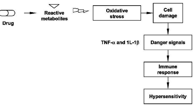
1.3.2 Danger Hypothesis
The danger hypothesis was first proposed by Matzinger in 1994 in response to discrepancies in the field of immunology as to what initiates and how an immune response is initiated (Matzinger, 1994). In this study, it was reported that the immunologists viewpoint that the immune system’s primary goal is to discriminate between self and non-self, may be changed. He presented the possibility that the immune system does not care about self and non-self; The primary driving force for this hypothesis being the need to detect and protect against danger (Matziger, 1994). The difference between the self and non-self in the danger hypothesis is that the major stimulus for initiation of an immune response comes from endogenous rather than exogenous signals and is controlled, not by cells of the immune system, but rather by the damaged tissue itself (Matziger, 2001).
The danger hypothesis affected the way immunologists view the immune response. In the danger hypothesis, a reactive metabolite causes cell damage or cell stress, which leads to upregulation of co-stimulatory factors on antigen presenting cells. Thus, the major determinant of whether an immune response is mounted against some agent is determined by whether that agent causes some type of cell damage. Assuming that most IDRs are immune-mediated, the danger hypothesis has the potential to explain whether drug (or more likely its reactive metabolites) causes cell damage (Li and Uetrecht, 2010). Damaged cells release danger signals, such as pro-inflammatory cytokines, TNF-α and 1L-1β. These signals can trigger an immune response, which leads to hypersensitivity as an adverse side effect (Figure 2).
1.4 Nevirapine and Nevirapine Alkyne
Treatment of HIV-1 infections with nevirapine is associated with skin and liver toxicity. These two organ toxicities range from mild to severe, in rare cases resulting in life-threatening liver failure or toxic epidermal necrolysis (Popovic et al., 2010). Nevirapine is one antiretroviral drug associated with IDRs. Figure 3 shows the structure of the Nevirapine. In this study we focused on studying Nevirapine induced hypersensitivity. The most common adverse effect of nevirapine is the development of mild or moderate rash (13%). Severe or life-threatening skin reactions have been observed in 1.5% of patients, including Stevens–Johnson syndrome which is a painful red or purplish rash that spreads on skins and mucous membranes, toxic epidermal necrolysis and hypersensitivity syndrome.
Figure 3: Structures of Nevirapine (NVP)
Nevirapine it is a non-nucleoside reverse transcriptase inhibitor used for the treatment of HIV infections. The use of Nevirapine is limited by a relatively high incidence of skin rash, some of which are life threatening and idiosyncratic drug-induced liver injury (IDILI). It also causes a rash in female Brown Norway rats (Shenton et al., 2003; 2004). The rash is clearly immune mediated in rats, and the characteristics of the rash in the rat model are very
similar to those in humans; therefore, it is likely that the mechanisms involved in the process are also similar (Shenton et al., 2005).
Drug metabolism plays an important role in developing IDR. Most drug metabolism occurs in the liver. However, liver-generated metabolites are unlikely to stimulate T-cell-mediated reactions in other organs, unless the metabolite avoids detoxification processes, and is stable enough to circulate in the blood (Ju and Uetrecht, 1999; Winter et al., 2004). Although the liver is the main site of drug metabolism, it is known that other organs express drug-metabolizing enzymes, such as the skin or immune cells themselves (Lavergne et al., 2008).
Chen et al. (2008) observed that substitution of the NVP methyl hydrogens with deuterium markedly decreased the formation of 12-OH-NVP as well as the incidence and severity of the rash. The treatment with a lower dose of 12-OH-NVP induced the same degree of skin rash as the treatment with NVP itself (see Figure 4). In addition, Sharma et al. (2013), using Brown Norway (BN) rat model, showed that the 12-hydroxylation pathway is involved in the induction of the skin rash and the most likely candidate for a reactive metabolite of 12-OH-NVP is the benzylic sulfate. Sharma et al. (2013) reported that Nevirapine is metabolized in the liver by cytochrome P450 (CYP) to Nevirapine metabolites, which induces the skin rash. They proposed the chemical mechanism of NVP-induced skin rash as a result from the covalent binding of 12-OH-NVP sulfate in the skin as shown in Figure 4.
Figure 4: Chemical mechanism of NVP-induced skin rash (Sharma et al., 2013)
Lavergne et al. (2008) stated that skin cells express less drug metabolizing enzymes than hepatocytes; however, skin is larger in terms of weight and surface area than the liver, and is also highly vascularized. Therefore, the skin could be as important as the liver in bioactivating certain drugs, and could play a specific key role in drug-induced skin reactions.
The ability of some metabolites, or sometimes the parent drug itself, to form protein adducts has been mainly investigated in vitro. In drug-induced immune-mediated liver diseases, such drug-protein adduct formation has been correlated with the development of hepatic injury (Aithal and Day, 2007). The formation of a metabolite-protein adducts could be cytotoxic by altering an important protein function.
Through the research activities in the Department of Pharmacology at the Faculty of Medicine and Health Sciences in University of Sherbrooke, Prof. Klaus Klarskov and his
research team designed a Nevirapine (NVP) analog in which the cyclopropyl group was replaced with an alkyne group. This compound (referred to as 1 Nevirapine alkyne) was synthesized following a seven-step sequence suitable for large scale production (Bernard et al., 2009). Figure 5 shows the structures of 1 Nevirapine alkyne in comparison with the Nevirapine. The terminal alkyne group is an appropriate site for reaction with azide conjugated probes using click chemistry adapted to biological media, as illustrated in Figure 6, to facilitate the detection of the modified protein by Nevirapine metabolite (Bernard et al., 2009).
Figure 5: Structures of Nevirapine and Nevirapine alkyne (Bernard et al., 2009)
Figure 6: Click fishing principle of metabolites of 1 Nevirapine alkyne (Bernard et al., Nevirapine alkyne (NVP-ALK)
1.5 Animal Model
If we could predict and prevent IDRs it would have a profound effect on drug development and therapy. Given their unpredictable nature, IDRs are generally not detected until the drug is released onto the market because clinical trials involve a limited number of subjects (Attia, 2010). Hypothesis testing requires valid animal models with characteristics similar to the idiosyncratic reactions that occur in patients. The best models, in which a rodent develops a clinical syndrome similar to what occurs in humans, appear to be penicillamine-induced autoimmunity in Brown Norway (BN) rats and nevirapine-penicillamine-induced skin rash in rats (Shenton et al., 2003). The nevirapine-induced skin rash in rats has characteristics very similar to the idiosyncratic reaction that occurs in humans. It is therefore perfectly suited for studying drug-induced idiosyncratic skin reactions.
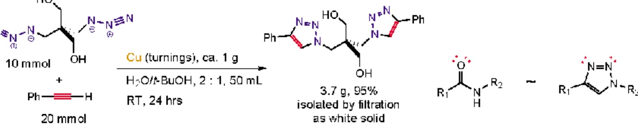
1.6 Click Chemistry
The “Click Chemistry” was introduced in 2001 (Kolb et al., 2001) to describe pairs of functional groups that rapidly and selectively react “click” with each other in mild, aqueous conditions. The concept of Click Chemistry has been transformed into convenient, versatile and reliable two-step coupling procedures of two molecules (Kolb et al., 2001). A click reaction must be of wide scope, giving consistently high yields with a variety of starting materials. It must be easy to perform, be insensitive to oxygen or water, and use only readily available reagents. Reaction work-up and product isolation must be simple, without requiring chromatographic purification (Kolb et al., 2001). The presence of water in these reactions is beneficial, not just for reactivity reasons, but also because water is the best heat capacity handling the enormous heat output when click reactions are performed (Kolb and Sharpless, 2003).
Most common click chemistry reactions (Figure 7) are:
1. Cu(I)-catalyzed Azide-Alkyne Click Chemistry reaction (CuAAC) 2. Strain-promoted Azide-Alkyne Click Chemistry reaction (SPAAC) 3. Tetrazine-Alkene Ligation
Figure 7: Overview of the most common Click Chemistry reactions (Jena Bioscience, 2016)
The Cu(I)-catalyzed Azide-Alkyne Click Chemistry reaction (CuAAC) (1) relies on the presence of Cu(I) ions whereas the Copper-free strain-promoted Azide-Alkyne Click
(Staudinger Ligation) is hampered by the instability of phosphines and slow reaction kinetics. Recent focus therefore shifted towards strain-promoted reactions with cyclooctynes and Tetrazine-Alkene Ligation, respectively (Jena Bioscience, 2016).
The copper-(I)-catalyzed 1,2,3-triazole formation from azides and terminal acetylenes is a particularly powerful linking reaction, due to its high specificity, and the bio-compatibility of the reactants. Pasini (2013) reported that Cu(I)-catalyzed Azide-Alkyne cycloaddition (CuAAC) is an established tool used for the construction of complex molecular architectures and given its efficiency, it has been widely applied for bioconjugation, polymer and dendrimer synthesis. Figure 8 shows the ‘perfect’ reaction as defined by Kolb and Sharpless (2003), which was made possible because of the dramatic rate acceleration of the Azide-Alkyne coupling event under copper-(I) catalysis (Rostovtsev et al., 2002; Tornøe et al., 2002), and the beneficial effects of water (Rostovtsev et al., 2002).
Figure 8: The premier click chemistry reaction (Kolb and Sharpless, 2003)
The copper-(I)-catalyzed coupling of azides and terminal acetylenes creates 1,4-disubstituted 1,2,3-triazole linkages, which share useful topological and electronic features with nature’s ubiquitous amide connectors. However, unlike amides, triazoles are not susceptible to cleavage (Kolb and Sharpless, 2003).
1.7 Mass Spectrometry
Mass spectrometry (MS) is an analytical technique that can be used selectively to detect and determine the amount of a given analyte. Accurate mass MS is also used to determine the elemental composition and the molecular structure of an analyte (Watson and Sparkman, 2007). It can provide both qualitative (structure) and quantitative (molecular mass or concentration) information on molecules after their conversion to ions and it can be used to identify unknown molecules and proteins (Ho et al., 2003).
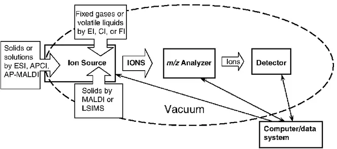
Electrospray ionisation mass spectrometry (ESI-MS) has been used in laboratories to provide a sensitive, robust, and reliable tool for studying, at femto-mole quantities in micro-litre sample volumes, non-volatile and thermally labile bio-molecules that are not amenable to analysis by other conventional techniques (Ho et al., 2003). Figure 9 shows a conceptual illustration of the mass spectrometer as provided by Watson, and Sparkman (2007). This conceptual illustration shows the major components of the mass spectrometer, which are: (i) sample inlets (dependent on sample and ionization technique); (ii) ion source (origin of gas phase ions); (iii) mass-to-charge ratio (m/z) analyzer (portion of instrument responsible for separation of ions according to their individual m/z values); (iv) detector (generates the signals that are a recording of the m/z values and abundances of the ions); (v) vacuum system (the components that remove molecules, thereby providing a collision-free path for the ions from the ion source to the detector); and (vi) the computer (coordinates the functions of the individual components and records and stores the data).
Figure 9: Conceptual illustration of the mass spectrometer showing the major components of mass spectrometer (Watson and Sparkman, 2007).
1.7.1 Uses of mass spectrometry
Mass spectrometry has become established as the primary method for protein identification from complex mixtures of biological origin. Almost without exception, protein identification is based on the analysis of peptides generated by proteolyic digestion. The most widely used enzyme is trypsin, which hydrolyzes the protein on the C-terminal side of lysine and arginine, unless the subsequent amino acid in the sequence is a proline (Baldwin, 2004).
The mass spectrometry (MS) may also be used for: (i) mass determination of biomolecules; (ii) identifying unknown compounds; and (iii) sequencing determination.
1.8 Protein Identification
Mass spectrometry (MS)-based proteomics is a highly sensitive method for protein identification and characterization (Aebersold and Mann, 2003) and it is a standard platform in proteomics (Delahunty and Yates III, 2005). The two commonly used MS-based proteomic approaches are multidimensional protein identification technology (MudPIT) and shotgun proteomics where peptides from in-solution digestion of the whole proteome are usually analyzed by multidimensional LC-MS/MS. The gel-based proteomics where peptides eluted from one-dimensional or two-dimensional gel electrophoresis (1DGE or 2DGE) (Görg et al., 2004) are normally analyzed by MALDI-MS or LC-MS/MS. The main difference between ESI and MALDI for protein identification is the ease of fragmentation of peptides. ESI normally produces multiply charged parent ions that require less energy for fragmentation compared to singly charged peptides, which are the dominant in MALDI (Ishihama, 2005).
1.9 Protein S-Glutathionylation
Glutathione (GSH) is an antioxidant in cells that prevents cell damages caused by reactive oxygen species such as free radicals reactive nitrogen species, peroxides, lipid peroxides and heavy metals.
Protein S-glutathionylation is a reversible mechanism for the global regulation of protein functions. It occurs through the formation of mixed disulfides between GSH and cysteine in proteins. In oxidative stress, glutathione is converted to its oxidized form, glutathione
stress (Brigelius et al., 1983). The S-glutathionylation significantly affects the protein structure and function (Holmgren et al., 2005; Dalle-Donne et al., 2007).
In several cell types, Reactive Oxygen Species (ROS) production catalyze glutathionylation of free thiol groups (-SH) on cysteine residues of proteins to form protein-glutathione mixed disufide adducts (Pr-SSG) (Hurd et al., 2005; Leichert and Jakob, 2006; Gallogly and Mieyal, 2007; Janssen-Heininger et al., 2008). Protein S-glutathionylation can affect the function of various proteins including actin, protein tyrosine kinases and phosphatases, Ras, and transcription factors such as NF-kB, and hence modulate cell signaling and function (Sakai et al., 2012).
The objective of using GSH antibody is to identify specific proteins that are modified by glutathionylation. It has been noted that the reactivity of the antibody depends upon the nature surrounding the epitope and that some glutathionylated proteins are not recognized or only weakly recognized by the antibody (Brennan et al., 2006). Nevertheless, the antibody technique allows detection of glutathionylated proteins in cells and tissues under conditions of oxidative or nitrosative stress (Hill et al., 2010).
1.10 Hypothesis and Objectives
Our hypothesis is that idiosyncratic skin reaction associated with NVP treatment are due to post-translational modifications of proteins (e.g., glutathionylation) detectable by MS. The specific objectives derived from the general objective were as follow:
1. To implement the click chemistry approach to detect proteins modified by a reactive NVP-Alkyne (NVP-ALK) metabolite. The purpose of using NVP-ALK was to couple it with Biotin using cycloaddition Click Chemistry reaction.
2. To detect protein modification using Western blotting and Mass Spectrometry techniques, which is important to understand the mechanism of NVP induced toxicity.
3. To identify the proteins using MASCOT search engine for protein identification, by comparing obtained spectrum from Mass Spectrometry with theoretical spectrum to find a matching peptide sequence.
4. To test if the drug or drug metabolites can cause harmful effects, as the induction of oxidative stress in cells (via protein glutathionylation). Oxidative stress causes cell damage that mediates signals, which likely induces the immune response.
CHAPTER 2
MATERIALS AND METHODS
2.1 Optimizing Click Reaction Conditions
The protocol that has been used in this work was introduced by Presolski et al. (2011) where ascorbate was used as reducing agent to maintain the required cuprous oxidation state. Three to five equivalents of copper-binding ligand TBTA was used to accelerate the reaction and serve as a sacrificial reductant, which protects the biomolecules from oxidation.
The following steps summarize a complete test that has been conducted for NVP-ALK click chemistry reaction. In an Eppendorf tube, 4 µl of 5 mM Biotin-PEG3-Azide in Dimethyl sulfoxide (DMSO) (Sigma) was added to 4 µl of 5 mM NVP-ALK in DMSO (Synthetized in Prof. Klaus Klarskov laboratory), then 4 µl of 50 mM CuSO4 in H2O
(Sigma) was pipetted to 4 µl of 50 mM ascorbic acid in H2O (Sigma). After that, 11.7 µl of
1 mM TBTA in DMSO (Sigma) and 168 µl of Phosphate-buffered saline (PBS) 1X were added. Thereafter, the mix was vortex and incubated at different temperatures for variable incubation periods. After completing the incubation, the analysis of samples was carried out by ESI–MS and tandem MS on a Micromass Q-TOF-2 (Waters Corp. ON, Canada) to verify if click reaction happened through detecting the mass of conjugated molecule.
Several assays were attempted to determine the optimized conditions for the click reaction, such as changing the temperature (room temperature to 95oC), incubation period (2 hr to 24
hr), concentrations of both molecules (NVP-ALK and Biotin-PEG3-azide), the use of ligand (with and without). Among these trials, the optimized conditions were obtained when both NVP-ALK and Biotin-PEG3-Azide molecules were conjugated together. This was achieved at 37oC for an incubation period of 24 hr with a TBTA ligand. The optimized conditions are listed in Table 1.
Table 1: Optimized Conditions for Click Chemistry Reaction
Temperature 37oC
Incubation period 24 hr
Ligand TBTA*
*TBTA =Tris-(benzyltriazolylmethyl) amine
2.2 Confirmation of Click Reaction using Erlotinib
This step was used to verify and confirm the occurrence of the click chemistry reaction. Erlotinib (a drug used for cancer treatment) was used as it has already an alkyne group in its structure. The ChemDraw software was used to draw the conjugated molecule of Biotin-PEG3-Azide and Erlotinib (see Figure 15). Thereafter the same procedure used for NVP-ALK was followed: In an Eppendorf tube 4 µl of 5 mM biotin-PEG3-Azide in DMSO was added to 4 µl of 5 mM Erlotinib in DMSO, and then 4 µl of 50 mM CuSO4 in H2O was
pipetted to 4 µl of 50 mM ascorbic acid in H2O. After that, 11.7 µl of 1 mM TBTA in
DMSO and 168 µl of PBS 1X were added. Thereafter, the mix was vortex and incubated at 37oC for 24 hr. After completing the incubation, the analysis of samples was carried out by
2.3 Animal Care
Animal model was developed and characterized by Daniel Defoy in Prof. Klaus Klarskov laboratory. This animal model of Nevirapine-Alkyne induced skin rash in female Brown Norway (BN) shares many characteristics of the rash that occurs in human. The rats were given Nevirapine-Alkyne for 6 weeks until they developed rashes. In addition, some rats were given Nevirapine and some others were not treated and were, therefore, used as controls in this study. Female BN rats (150−175 g; between 8 and 10 weeks of age) were obtained from Charles River (Montreal, QC).
Rats were housed in pairs in standard cages in a 12:12 hr light/dark cycles with access to water and powdered lab chow diet (Leis Pet Distribution, Inc. Wellesley, ON). Following a 1-week acclimatization period, rats were either maintained on control chow or started on drug-containing diet (treatment groups). Drug was mixed thoroughly with powdered lab chow; it was to be administered orally. The amount of drug administered to animals was 150 mg/kg/day calculated based on body weight of the rats and their daily food intake. Rats were exposed to Nevirapine and Nevirapine-Alkyne for another 3 weeks until they developed skin rash as an adverse effect.
2.4 Protein Extraction
Rats were sacrificed by Daniel Defoy via CO2 asphyxiation. At sacrifice, hair was removed
from the rats using an electric shaver, and the skin was cleaned of remaining hair using PBS (1× phosphate buffered saline, pH 7.4) and Kimwipes. Whole skin from the back was
removed homogenized in cell lysis buffer (Cell Signaling Technologies) with protease inhibitor (SIGMA) in a 10:1 ratio using a Polytron 2100 homogenizer (1.5 mL working cell lysis buffer per fraction). In order to clarify the crude fractions, samples were centrifuged at 13,000 rpm for 2 min. The supernatant was separated from insoluble debris and stored at −80 °C.
2.5 Protein Quantification
After protein extraction from treated and untreated rats, bicinchoninic acid (BCA) protein assay kit (Thermofisher) was used to determine protein concentration in each sample. BCA kit contains BSA (bovine serum albumin) and two reagents A and B. The BSA was diluted using distilled water; the dilutions were 2 mg/ml, 1.5mg/ml, 0.75 mg/ml, 0.5 mg/ml, 0.25 mg/ml, 0.125 mg/ml, and 0.025 mg/ml of BSA. Reagents A and B were combined together in 50/1 ratio just before protein quantification. Then, 25 µl of each BSA standard dilution and from the samples were pipetted into 96 microplate. After that, 200 µl of the mixture (reagents A and B) was added to each sample and standard. Thereafter, the plate was incubated at 37°C for 30 min. The absorbance was measured at 562 nm on a plate reader. Finally, we used the Excel software (Microsoft Inc.) to calculate the protein concentration.
2.6 Western Blot
Protein extracted from the untreated rats as control (CLT) was used. Thereafter the following procedure was followed:
mM biotin-PEG3-Azide in DMSO, and 4 µl of 50 mM CuSO4 in H2O. After that, 4 µl of 50
mM ascorbic acid in H2O was added to 11.7 µl of 1 mM TBTA in DMSO and 168 µl of
PBS 1X. Thereafter, the mix was vortex and sonicated for 15 min, and incubated at 37oC for 24 hr. This process was repeated for the control (CTL) samples.
Electrophoreses:
After 24 hr of incubation at 37oC, the following procedure was used to separate and to visualize proteins on a western blot:
Twenty µl of each sample was mixed with 10 µl 2X sample loading buffer (62.5 mM Tris-HCl, pH 6.8 (Sigma-Aldrich), 2% SDS (Sodium dodecyl sulfate), 25% glycerol, 0.01% bromophenol blue) with freshly prepared 5% Dithiothreitol (DTT) (Promega) were heated at 95oC for 5 min. After that, the samples were loaded on the gel (Precast gel, 4–20% Criterion Tris-HCl Gel, 18 well, 30 µl) (BIO-RAD). Then Electrophoresis was run at 20 mA then increased to 40 mA, after passing the stacking gel. The electrophoresis running buffer (Bio-Rad) consisted of 25 mM Tris, 192 mM glycine, and 0.1% SDS, pH 8.3. The electrophoresis was performed using PowerPac 1000 (BIO-RAD). After electrophoresis, the proteins were visualized using coomassie blue stain (BIO-RAD).
Transfer:
Protein was transferred onto a Polyvinylidene difluoride (PVDF)–membrane (Western blotting membrane) (Roche) as it has more affinity and binding capacity to protein than nitrocellulose membrane. Before protein transfer, the PVDF membrane was soaked in pure methanol. The transfer was performed using a Trans-Blot SD semi-dry transfer cell
(BIO-RAD) for 55 min. at 0.8 mA/cm2 as described by Methogo et al. (2005). A thick filter paper was soaked in transfer buffer (20% Tris–glycine running buffer).
After semi dry transfer, the membrane was stained with India ink (100 ml H2O + 1 ml
acetic acid + 100 µl of India ink + 50 µl TWEEN 20) for 24 hr. Then, washed twice with Tris-buffered saline Tween-20 (TBST) solution for 5 min. Membrane was then blocked in 5% non-fat milk blocking solution in TBST (5 g milk + 100 ml TBST) for 2 hr at room temperature. Then, it was washed in three changes of TBST.
Following that, the membrane was incubated with a 1:5000 dilution of Streptavidin HRP (Invitrogen) (50 ml TBS + 50 µl Tween 20 +10 µl streptavidin HRP) overnight. Thereafter, the membrane was washed 3 times for 20 min. with TBST, then incubated with enhanced chemiluminescence ECL (western lightening-ECL) (PerkinElmer) (1 ml reactive A + 1 ml reactive B, mix) for 1 min. The film was exposed in a dark room for 30 sec, 1 min., and 5 min.
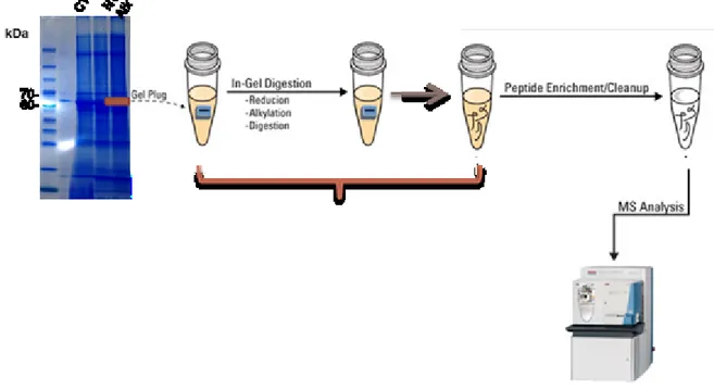
2.7 In Gel Digestion
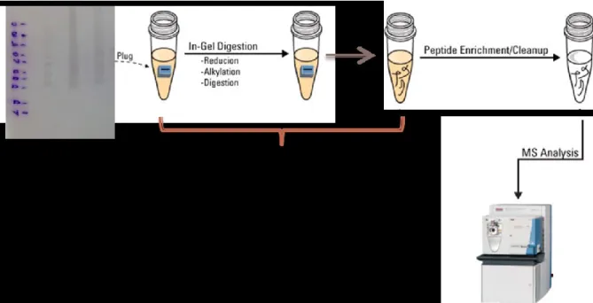
After detecting protein bands by Western blotting, another electrophoresis was performed as described previously (Section 2.7). Then the gel was fixed in 40% methanol / 10% acetic acid / 50% water for 30 min. After that, the gel was stained with coomassie blue stain (BIO-RAD) overnight, then destained by using distilled water until background became clear. The gel was placed over a glass tray then bands of interest were cut as shown in
Figure 10: A photo showing the stained gel and the cut bands
After cutting the bands of interest (as shown in Figure 10), the gel pieces (protein bands) was placed in Eppendorf tube and a shortly prepared 400 µl of 100 mM NH4HCO3 was
added then vortexed then left for 15 min. After that, solution was removed from the tube and 400 µl of acetonitrile (ACN) was added. After 15 min., ACN was removed from the tube then 50 µl of shortly prepared DTT 5 mg/ml in 200 mM NH4HCO3 was added then
incubated at 37oC for 45 min. then the DTT solution was removed after the incubation. After that, 70 µl of Iodoacitamide 25 mg/ml in 200 mM NH4HCO3 shortly prepared was
added, then incubated at room temperature for 30 min. in the dark. Iodoacetamide solution was removed. 400 µl of solution 100 mM NH4HCO3 containing 50% ACN was added and
incubated at room temperature for 30 min. on the mixer. Solution was removed then gel pieces were smashed (by using a clean tip) then dried using a vacuum dryer for 2-5 min. The Gel pieces were rehydrated and digested on ice with 25 µl of 100 ng/µl trypsin (Trypsin/Lys-C Mix, Mass Spec Grade, Promega) in 40 % Dimethylformamide (DMF) /100 mM NH4HCO3 for 15 min. Then excess solution was removed then 50 µl 50 mM
NH4HCO3 20% DMF were added and incubated at 37oC overnight. Samples were
centrifuged and incubated in an ultrasonic bath for 10 min., followed by another centrifugation. Finally, 5 µl of 10% formic acid HCOOH solution in water was added, sample was placed in ultrasonic bath for 15 min. then centrifuged again. Supernatant was collected, then placed in a clean Eppendorf (see Figure 11).
Pre-LC-MS-MS sample preparation:
Before running the analysis, 1 µl of 5M urea in water was added to each sample. After that, the sample was dried using a speed-vac.
2.8 Analysis by LC-ESI-MS and Protein Identification:
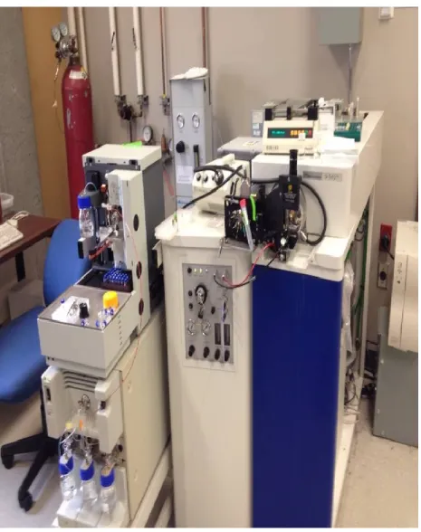
The method used for the analysis by LC-ESI-MS for protein identification was adapted from the work conducted by Prof. Klaus Klarskov (Dept. of Pharmacology and Physiology, University of Sherbrooke) detailed in Methogo et al. (2007). Peptides derived from digestion were analyzed by nanoliquid chromatography coupled on-line with tandem electrospray ionization time-of-flight mass spectrometry (nLC-ESIMS/MS) consisting of a capillary liquid chromatograph with autosampler and a QTOF 2E mass spectrometer (Waters, MA, USA) (Figure 12). Tryptic peptides were dissolved in 14 µl solvent A (1% acetonitrile, 1% isopropanol in 0.2% aqueous formic acid), loaded onto a 0.5 mm × 250 µm i.d. union packed with Magic C18 AQ (200 A, 5 µm; Michrom BioResources,Auburn, CA) and separated on a 100 µm i.d. × 10 cm capillary column packed with the same material. Each analysis was succeeded by injection of 10 µl 50% (v/v) aqueous isopropanol followed by running a blank gradient, which efficiently eliminated contamination from sample carry over.
Figure 12: A photo showing the Q-TOF-2 mass spectrometer used in LCMS analysis
The following linear gradient was used: from 10 to 70% B in 30 min., to 90% B in 5 min., and 10% B in 3 min. The flow rate was split approximately 10× to 0.25–0.30 µl/min. Survey scans were acquired at 1.9 s/scan. Depending on the charge state and intensity, doubly and triply charged parent ions were automatically selected for tandem mass spectrometry acquired at 1 s/scan with a preset maximum of five scans per selected parent
acquisition time on ions from background, impurities and detergents. Although a low threshold is not a guaranty of acquiring MS/MS spectra useful for protein identification, it ensures selection of maximum number of doubly and triply charged ions for MS/MS analysis, during the separation. Raw MS/MS spectra were processed automatically using the ProteinLynx software and converted into peak lists readable by the MASCOT search algorithm (http://www.matrixscience.com). The following search parameters were used: database; MSDB, species; Rattus, allowance of two missed tryptic cleavages, oxidation of methionine and cysteine carbamidomethylation, fragment mass tolerance; 0.25 Da.
2.9 Protein S-glutathionylation
To test this hypothesis, we extracted proteins from rats treated or not with NVP and NVP-ALK. Twenty microliters of each sample was added to 10 µl of sample loading buffer (62.5 mM Tris-HCl, pH 6.8, 2% SDS, 25% glycerol, 0.01% bromophenol blue) without DTT, and then heated at 95oC for 5 min. Then samples were loaded on the gel (4–20% Criterion Tris-HCl Gel, 18 well, 30 µl) (BIO-RAD). Electrophoresis were run at 20 mA then increased to 40 mA after passing the stacking gel (PowerPac 1000, BIO-RAD). Electrophoresis running buffer (BIO-RAD) consisted of 25 mM Tris, 192 mM glycine, and 0.1% SDS, pH 8.3.
Transfer:
After electrophoresis, the proteins were transferred onto a pre-soaked PVDF–membrane (Western blotting membrane) (Roche) in pure methanol for 15-30 min. The membrane wetting is an important step for proper protein binding. After electrophoresis, the gel was
immediately equilibrated in transfer buffer (2.93g glycine 200 ml methanol, up to 1 liter w/dH20) for 15 min. This step was conducted to remove the excess electrophoresis buffer and detergent from SDS-gel. The wetted blot paper was also soaked and the buffer and gel were placed between wetted blot papers. The cathode plate was placed carefully onto the stack. The transfer was performed using a Trans-Blot SD semi-dry transfer cell (BIO-RAD) for 55 min. at 0.8 mA/cm2.
After that, the membrane was stained with India ink (100 ml H2O + 1 ml acetic acid + 100
µl of India ink + 50 µl TWEEN 20) for 24 hr. Then, it was washed with TBST. Membrane was then blocked in 5% non-fat milk blocking solution in TBST (5 g milk +100 ml TBST) for 2 hr. at room temperature then washed twice with TBST for 5 min. each.
The membrane was incubated overnight at room temperature with the first antibody anti-glutathione monoclonal antibody (Virogen) diluted 1000x in TBST. Then membrane was washed (three changes) in TBST followed by a 90 min. incubation at room temperature in secondary antiserum Goat Anti-Mouse IgG H&L (HRP) diluted at 1: 10000 (5 µl antibody + 50 ml TBS). Then membrane was washed twice with TBST
After that, it was incubated with enhanced chemiluminescence ECL (western lightening-ECL) (PerkinElmer) (1 ml reactive A + 1 ml reactive B, mix) for 1 min. Films exposed in a dark room for 30 sec, 1 min., 5 min.
On membrane digestion:
For identification of glutathionylated proteins, detected protein bands were cut directly from the PVDF membrane and digested on the membrane as described by Methogo et al. (2005). Figure 13 summarizes the membrane digestion while Figure 14 shows the PVDF membrane after cutting bands of interest.
Figure 14: PVDF membrane after cutting bands of interest
Proteolytic cleavage, sample preparation.
After electro-blotting and immuno detection, protein spots for on-membrane digestion, were excised washed in 1 ml 20% methanol MeOH and reduced in 20 µl of 100 mM NH4HCO3 containing 5 g/l DTT for 45 min. at 37°C.
The carbamidomethylation was performed in the dark in 40 l of 200 mM NH4HCO3
containing 25 µg/µl of iodoacetamide for 30 min. Membrane pieces were rinsed four times in 400 l 100 mM ammonium bicarbonate containing 50% acetonitrile.
Digestion on the membrane was performed at 37°C for 3.5 hr in 8 µl of 50% DMF, 100 mM NH4HCO3 containing 20 ng/µl of trypsin. This buffer (NH4CO3/DMF) was prepared
The samples were centrifuged every 30 min. to prevent the membrane pieces from drying out. After digestion, 1 µl 5 M urea was added to the samples. The samples were sonicated for 2 min. and, followed by immediate transfer to Q-TOF vials then lyophilization. For analysis and protein identification, samples were sent to Phenoswitch bioscience (Sherbrooke).
CHAPTER 3
RESULTS
3.1 Click Chemistry Reaction
First step of the project was to perform a click chemistry reaction by coupling two molecules, which are Nevirapine Alkyne (NVP-ALK) and Biotine-PEG3-Azide using certain conditions. The NVP-ALK and Biotine-PEG3-Azide conjugated molecule is shown in Figure 15. Click chemistry reaction was the tool to detect proteins modified by reactive metabolites of the NVP-alkyne in the samples.
To determine if the click reaction occurred, the mass of each molecule and the mass of the conjugated molecule was calculated using the ChemDraw software and was measured by mass spectrometry (MS). The MS analysis of the samples revealed that NVP-ALK and Biotin conjugate are binding together under these circumstances (optimized conditions listed in Table 1) and a new molecule has a mass equal to the sum of the individual masses of NVP-ALK and Biotin conjugate (see result for ESI-MS analysis result). It should be mentioned that the Tris-(benzyltriazolylmethyl) amine (TBTA) was used to protect Cu(I) from oxidation.
Figure 15: Nevirapine-Alkyne and Biotin-PEG3-Azide click reaction and expected m/z for the conjugated product
3.2 Confirmation of Click Chemistry Reaction Conditions using Erlotinib
This step was used to verify and confirm the occurrence of the click chemistry conditions. Erlotinib was used as it has an alkyne group in its structure. The ChemDraw software was used to draw the conjugated molecule of Biotin-PEG3-Azide and Erlotinib. Figure 16 shows the individual molecules and the conjugated one as calculated by ChemDraw software.
3.3 Detection of Click Chemistry Reactions using Mass Spectrometry
The click chemistry reactions were conducted using the optimized working conditions, which were determined as detailed in Chapter 2 and the optimized conditions are listed in Table 1.
The structure and the molecular weight of Biotin-PEG3-Azide are shown in Figures 15 and Figure 16. As mentioned earlier, the drawings of the individual molecules before starting the click chemistry reaction was important to analyze each molecule to confirm their mass and to test the accuracy of the MS instrument.
Figure 17 shows ESI/MS/MS Spectrum of Biotin-PEG3-Azide, the mass on charge (m/z) was identical to calculated mass by ChemDraw software.
In addition, Figure 18 shows the ESI/MS/MS analysis for Nevirapine-Alkyne molecule before click chemistry reaction.
Figure 19 shows the ESI-MS of a mixture contained both molecules (Nevirapine-Alkyne + Biotin-PEG3-Azide) separately in the presence of copper sulfate as catalyst and ascorbic acid. The two molecules were detected separately, which means that cycloaddition reaction did not occur. Despite the fact that click chemistry reaction is a high yield reaction, it needed a lot of optimisation to determine the best condition in order to allow the cycloaddition reaction to occur. During the optimization, different experimental conditions such as varying the temperature (room temperature to 95oC), the time of incubation (2 hr to 24 hr), the concentrations of both molecules (NVP-ALK and Biotin-PEG3-azide) and employing or not TBTA (Cu(I)-stabilizing ligand) were tested. Figures 20 and 21 show some experimental conditions that were not successful. The optimized conditions were obtained when both NVP-ALK and Biotin-PEG3-Azide molecules were conjugated together. This was achieved at 37oC for an incubation period of 24 hr with a TBTA Cu(I)-stabilizing ligand (Table 1).
Figure 20 was obtained after ESI/MS analysis of click chemistry reaction without using TBTA as ligand. The figure shows that the reaction did not yield the m/z calculated. It yielded a molecule with m/z 593, what may have happened is that the conjugated compound fragmented easily. This means that, the reaction needed optimization to yield the conjugated molecule and to make it more stable and MS cone voltage should have been optimized to minimize in-source fragmentation.
The spectra shown in Figures 22 and 23 were obtained after analyzing the click chemistry reaction at condition at 37oC for 24 hr and after adding TBTA as a ligand to accelerate the reaction. The results confirmed that the chemistry reaction occurred and yielded the expected m/z of the conjugated molecule (NVP-ALK and Biotin-PEG3-Azide).
Click reaction conditions were tested with another alkyne containing drug, Erlotinib, a drug used for treating cancer. The same conditions used for NVP-ALK were applied. Figure 24 showed that, Erlotinib was effectively conjugated with Biotine-PEG3-Azide.
3.4 Detection of Proteins Modified by Nevirapine Metabolites by Western Blot
Figure 25: Extracted proteins from skin of rats treated with NVP-ALK after coupling with Biotin-PEG3-Azide by click chemistry reaction and detection by streptavidin-HRP
Figure 25 shows the immunoblot of rat skin untreated (CTR) and treated with NVP-ALK, after incubation with streptavidin HRP. Streptavidin HRP was used due to its high affinity to Biotin. The detected proteins appeared as a smeared band.
3.5 Identification of Modified Protein by Nevirapine Metabolite using Mass Spectrometry and Protein Identification Software
Figure 26: Protein hits from mascot software
Figure 26 shows that the recovered peptides from in-gel digestion analyzed by nLC-MS/MS. The corresponding proteins were identified in the Rattus protein database using MASCOT. Figure 26 shows a list of proteins identified despite that the band was a large smear band, still resulting in positive protein identification.
Figure 27: Peptide summary of injected sample
Figure 27 shows peptides summary from two proteins identified from samples from the BN rats treated with NVP-ALK. The high score indicates a very high probability of correct protein identification.
Figure 28: Fragmented peptide in the sample
Figure 29: Protein sequence coverage
Figure 29 shows that the identified protein was keratin. Peptides from samples covered 10% of keratin. Thus, keratin was identified as the main protein that may be modified by NVP-ALK metabolite. This result, however, still needs further investigation.
3.6 Detection of S-glutathionylated Protein
kDa
Figure 30: Immunoblot detection of glutationylated proteins in skin fractions of rats treated or not with NVP-ALK (in non-reducing SDS-PAGE condition (absence of DTT)).
CTR: untreated rats, NVP: treated rats with NVP and ALK: treated rats with NVP-ALK
In order to detect the proteins modified by glutathionylation, anti-glutathione monoclonal antibody (virogen) was used. It has been noted that the reactivity of the antibody depends upon the nature surrounding the epitope and that some glutathionylated proteins are not or only weakly recognized by the antibody (Brennan et al., 2006). Nevertheless, Western blot allows detection of glutathionylated proteins in cells and tissues under conditions of oxidative or nitrosactive stress, without the addition of reducing agents (DTT) (Hill et al., 2010).
3.7 Identification of Glutathionylated Proteins
Table 2: Identification of Glutathionylated Proteins
Name N Unused Total %Cov %Cov(50) %Cov(95)
Actin, cytoplasmic 2 OS=Rattus norvegicus GN=Actg1 PE=1 SV=1 1 18.59 18.59 47.99999893 40.0000006 38.13000023 Actin, cytoplasmic 1 OS=Rattus norvegicus GN=Actb PE=1 SV=1 1 0 18.59 47.99999893 40.0000006 38.13000023 Elongation factor 1-alpha 1 OS=Rattus norvegicus GN=Eef1a1 PE=1 SV=1 2 14 14 24.4599998 20.78000009 20.78000009 Protein LOC100360413 OS=Rattus norvegicus GN=LOC100360413 PE=4 SV=1 2 0 12 20.99999934 17.31999964 17.31999964 Elongation factor 1-alpha 2 OS=Rattus norvegicus GN=Eef1a2 PE=2 SV=1 2 0 12 19.22000051 15.54999948 15.54999948
Heat shock protein HSP 90-beta OS=Rattus norvegicus GN=Hsp90ab1 PE=1 SV=4 3 12.93 12.93 12.02000007 12.02000007 10.63999981 Protein Hist1h2bf OS=Rattus norvegicus GN=Hist1h2bf PE=4 SV=1 4 10 10 46.02999985 40.47999978 40.47999978
Table 2 shows a preliminary attempt to identify the glutathionylated proteins using on-membrane digestion, LC-ESIMS/MS and database searching (Peakview software (Sciex)). The identification of gutathionylated proteins (Table 2) has correctly identified several proteins in the gel band that were judged by WB to contain at least one glutathionylated protein. However, there was no MS evidence to determine which of the identified proteins were glutathionylated neither if there was more than one protein that was glutathionylated. In addition, from the results it was observed that the peptides from samples covered 38% of actin. Since it was not possible to obtain analytical evidence from the MS fragmentation of the glutathionylated peptide from actin or other proteins, further investigation is required to identify and confirm the glutathionylated protein(s).
Furthermore, Figure 31 shows peptide summaries from sample of BN rats treated with NVP-ALK. The molecular weight of actin is 40 kDa but we detected it at 140 kDa. This can be explained by the fact that actin can bind to other proteins as ABP to form a polymer. Further studies are required to confirm this hypothesis.