HAL Id: inserm-00344389
https://www.hal.inserm.fr/inserm-00344389
Submitted on 31 Aug 2009HAL is a multi-disciplinary open access
archive for the deposit and dissemination of sci-entific research documents, whether they are pub-lished or not. The documents may come from teaching and research institutions in France or abroad, or from public or private research centers.
L’archive ouverte pluridisciplinaire HAL, est destinée au dépôt et à la diffusion de documents scientifiques de niveau recherche, publiés ou non, émanant des établissements d’enseignement et de recherche français ou étrangers, des laboratoires publics ou privés.
Bernard Gibaud
To cite this version:
Bernard Gibaud. The DICOM standard : a brief overview. Lemoigne Yves, Caner Alessandra (eds). Molecular imaging: Computer reconstruction and practice, Springer, pp.229-238, 2008, NATO Science for Peace and Security Series. �inserm-00344389�
THE DICOM STANDARD: A BRIEF OVERVIEW
Bernard GIBAUD
Unit / Project VISAGES, INSERM/INRIA/CNRS/University of Rennes 1. 2, Avenue du Pr Léon Bernard, F-35043 Rennes. France Correspondence: Bernard Gibaud,
Abstract: The DICOM standard has now become the uncontested standard for the exchange and management of biomedical images. Everyone acknowledges its prominent role in the emergence of multi-vendor Picture Archiving and Communication Systems (PACS), and their successful integration with Hospital Information Systems and Radiology Information Systems, thanks to the Integrating the Healthcare Enterprise (IHE) initiative. We introduce here the basic concepts retained for the definition of objects and services in DICOM, with the hope that it will help the reader to find his or her way in the vast DICOM documentation available on the web.
Key words: Medical Imaging, PACS, DICOM
1.
INTRODUCTION
Exchanging images between various kinds of equipment has been an issue since the very beginning of digital medical imaging. The ACR/NEMA standard, issued in 1985 and 1988, did not provide a satisfactory solution and the medical imaging community had to wait until 1993 to have a real usable standard available. The publication of this standard was followed by several demonstrations in key international congresses, especially the RSNA in 1993, which convinced the whole community that the standard actually led to successful implementations, and that the manufacturers were willing to use it, and turn the page of proprietary formats and solutions. Since that time, the position of DICOM was significantly reinforced, with the creation of the DICOM Committee, and the collaboration with other standards
development organizations such as the Comité Européen de Normalisation (CEN) in the mid-nineties, and ISO TC 215 “Health Informatics” in 1999.
DICOM has now become the international standard in the field of biomedical imaging. Its influence was critical in the emergence of multi-vendor technical solutions for Picture Archiving and Communication Systems (PACS), and in providing appropriate solutions for the integration with the other information systems involved, especially the Hospital Information Systems and the Radiology Information Systems. These integration issues were addressed by the Integrating the Healthcare Enterprise Initiative (IHE) – initially in the USA in 1998, under the auspices of the RSNA and HIMSS, then world-wide, with the launching of IHE-Europe in 2001, and similar initiatives in Asia more recently.
2.
THE DICOM COMMITTEE
The DICOM committee gathers various kinds of contributors. All the big manufacturers of the field of biomedical imaging are members of the DICOM Committee. There are also many professional societies involved, in radiology, but also from other fields such as cardiology, dentistry, pathology, ophthalmology, as well as stakeholder institutions such as the American National Cancer Institute.
The scope is quite broad with currently 26 working groups addressing the various aspects involved (Table 1.) Among those working groups, one has to distinguish Working Group 6 (“Base standard”), whose function is to guarantee the overall technical consistency of the standard, and Working Group 10 (“Strategic advisory”), in charge of advising the DICOM Standard Committee on long term orientations that should be followed.
DICOM Committee Working Groups
WG-01: Cardiac and Vascular Information WG-14: Security WG-02: Projection Radiography and Angiography WG-15: Digital Mammography and CAD
WG-03: Nuclear Medicine WG-16: Magnetic Resonance WG-04: Compression WG-17: 3D
WG-05: Exchange Media WG-18: Clinical Trials and Education
WG-06: Base Standard WG-19: Dermatologic Standards WG-07: Radiotherapy WG-20: Integration of Imaging
WG-08: Structured Reporting WG-21: Computed Tomography WG-09: Ophthalmology WG-22: Dentistry
WG-10: Strategic Advisory WG-23: Application Hosting WG-11: Display Function Standard WG-24: Surgery
WG-12: Ultrasound WG-25: Veterinary Medicine WG-13: Visible Light WG-26: Pathology
Table 1. the various working groups of the DICOM Committee
3.
ORGANIZATION OF THE STANDARD
3.1
A multi-part document
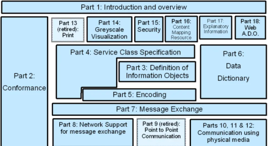
The DICOM standard is organized as a multi-part document, made of 18 independent parts (Figure 1) [1].
Figure 1. The different Parts of the DICOM standard (numbered 1 to 18)
The most important ones are Parts 3, 4, 16, 5 and 2. Part 3 “Information object definitions” provides the specification of the information objects to be exchanged (more than 1000 pages of text), as well as the definition of the semantics of each data element. The main reason why this part is so long and complex is related to the many existing imaging modalities (such as computed tomography, ultrasound, magnetic resonance, positron emission
tomography etc.), that require many technical parameters. Part 4 “Service Class specifications” defines the services for exchanging information, either images or information that is useful to manage images. Part 16 “Content Mapping Resource” addresses the question of terminology, i.e. on the one hand, it defines how existing terminological resources can be used in DICOM (e.g. SNOMED, LOINC, UCUM), and on the other hand, how content items can be grouped together and re-used in DICOM Structured Reporting documents (notion of Templates). Part 5 “Data structure and encoding” specifies how the information objects specified in Part 3 can be organized into a linear bit stream, in order to be sent over a network connection or stored in a file. All aspects related to image compression are addressed in Part 5. Finally, Part 2 “Conformance” specifies how a manufacturer can claim conformance to the DICOM standard for a particular product or implementation, by writing a document called a “conformance statement”. Part 2 explains in detail how this document must be written and the information it must contain.
3.2
Extensions and maintenance
The standard is being continuously updated, either to address new fields of biomedical imaging (for example, surgery applications), or to revisit existing fields (e.g. Magnetic resonance or PET images), in order to face new needs resulting from the evolution of imaging techniques.
Such extensions are made through the publication of “Supplements” to the standard. Several such Supplements are issued every year. Each Supplement includes text modifying the existing parts, e.g. to add a new information object in Part 3, or a new service class in Part 4, or new codes in Part 16, or new data elements in Part 6 “Dictionary” etc. All those changes are put together once a year to deliver a new issue of the standard, e.g. “DICOM 2008”. However, this yearly issue should not be seen as a new “version” of the standard. There is only one single version of the standard, since all changes that are made are supposed to be “backward compatible”: it means that new specifications bring new functionalities, but they never change the way previous services work. The only exception to this rule is the case of retirement, i.e. when service classes are retired from the standard (this happens only to services that are almost never used). The general motivation for this kind of approach is that imaging equipment can be in operation during quite long periods of time (6, 7, up to 10 years) and that it would be non-acceptable to change the standard in ways that would “break”
existing implementations, and cause troubles to installed and working equipment.
A second way of updating the standard exists, through the mechanism of Correction Proposals (CPs). CPs provide the means to raise and fix minor issues and imprecision that cause (or may cause) interoperability problems. CPs are submitted, accepted, worked out and balloted as any other parts of the standard.
4.
BASIC CONCEPTS
4.1
Data Syntax
The syntax used in the DICOM standard is a binary one, based on the transfer of (Tag-Length-Value) triplets. It means that any elementary Data Element is given a binary Tag, e.g. Data element (0028,0010) Rows represents the number of rows in an image. This Tag, composed of 4 bytes is sent first, followed by the corresponding data type, represented in this particular case by ASCII characters ‘U’ and ‘S’ (for Unsigned Short), followed by the length of the value field, in this case “2” because the Data element (0028,0010) Rows is represented by an Unsigned Short, represented by 2 bytes, and finally comes the value itself, e.g. “256”, represented in binary (‘00’ in the least significant byte, and ‘01’ in the most significant byte). So the binary bit stream corresponding to this Data element would be in this example:
… ‘28’ ‘00’ ‘10’ ‘00’ ‘55’ ‘53’ ‘02’ ‘00’ ‘00’ ‘01’ …
As can be seen from this example the values are binary ones, which explains that images in DICOM cannot be edited like, e.g. XML documents. One must use a parser that decodes the TLV triplets in order to put the information in human-readable form.
4.2
Information objects
The specification of an information object (called in DICOM an Information Object Definition or IOD) consists in defining the set of Data elements that shall or may be transmitted. They are organized in groups called ‘modules’. For example, the CT image IOD is depicted by a number
of modules focusing on: (1) the general context of image acquisition (essential information on the patient, the study, the series), (2) the acquisition procedures (particularly the physical acquisition methods, the reconstruction algorithm etc.), (3) the image’s characteristics (size, resolution etc.), and (4) the pixel data themselves. The ‘module’ concept, by gathering together data elements relating to one same information entity (for example ‘Patient Module’, ‘General Study Module’, ‘General Image Module’, ‘Image Plane Module’, ‘Image Pixel Module’), facilitates their reuse in different IODs. These information entities are defined using information models, following the “entity-relationship” formalism. The basic DICOM model is a hierarchy in which a Patient can have one or more Studies, each containing one or more Series, each containing one or more Composite Objects (such as Images, Presentation States, etc.)
A distinction is introduced between Composite objects and Normalized objects. Composite objects gather data elements describing several entities of the real world, whereas Normalized objects focus on one single real world entity.
All sorts of images, whatever the modality (Computed Tomography, Magnetic Resonance, Ultrasound, XRay angio, etc.) are represented by Composite objects, as well as Presentation States, Fiducials, Registration objects, Structured Reports etc.
At the beginning of the standard, image objects were essentially 2D images. Multi-frame objects were introduced to represent in a single Dataset several related images, e.g. in ultrasound (Multi-Frame Ultrasound IOD) and nuclear medicine (Nuclear Medicine IOD). However, the new image objects developed recently for CT, MR, PET etc. (Enhanced CT, Enhanced MR, Enhanced PET, respectively) make use of a new extended multi-frame mechanism. This leads to a better and more efficient organization of common attributes (using so-called functional groups), as well as to faster transmission times.
4.3
Services
Two sets of services exist. A first set of services (called Composite services), applies to Composite objects, whereas a second set (called Normalized services) concerns Normalized objects. The first provide facilities to ‘push’ an object (service C-STORE), or to query a set of objects based on some search criteria (services C-FIND, C-GET and C-MOVE). A last service exist (C-ECHO) to simply test connectivity between two peer application entities.
Normalized services provide facilities to act upon a single object managed by a remote application entity, to create an instance (N-CREATE), delete it DELETE), get information about it GET), modify it (N-SET), launch an action (N-ACTION), report an event (N-EVENT-REPORT).
4.4
Service-Object Pair
Objects, i.e. IODs, can be associated with services to form what is called a ‘Service Object Pair’. For example, a CT image IOD is associated with the Storage service to form the CT Image Storage SOP Class. This abstraction defines a service that can be provided by an application entity (this entity is called the SCP, for Service Class Provider), or used by another application entity (this entity is called the SCU, for Service Class User). These notions of SOP, SCU and SCP are extensively used in DICOM Part 2 to claim the conformance to DICOM of an implementation. A SOP Instance corresponds to the instantiation of the use of a service, e.g. for a particular image. SOP Classes and SOP instances have unique identifiers based upon the OSI Object Identification (numeric form) as defined by the ISO 8824 standard. The SOP instance UIDs provide identifiers for persistent objects such as images.
5.
DICOM MAJOR ACHIEVEMENTS
5.1
Storage and query / retrieve of Composites objects
Storage and Query & Retrieve are the services classes used to communicate any sorts of Composite objects. In fact the exchange and query mechanisms are totally generic with respect to the objects to be exchanged. Requests are based on the use of unique identifiers for Studies, Series and Composite objects (using the SOP Class and SOP instance UIDs).
Composites objects may be exchanged in two ways, either in connected mode, or using removable media. In the first case, composite objects are transmitted as one message per Dataset (a Dataset corresponds to a Composite object instance), after a preliminary stage called Association establishment, during which the two peer application entities negotiate the SOP Classes and Transfer Syntaxes they want to use in the exchange. In the
second case, the composite objects are copied on removable media, together with a file called DICOMDIR, providing a structured description of the Patient / Study / Series / Composite Objects relationships, with links to corresponding files on the media. DICOM does not specify any particular convention for the naming of files. Many kinds of media are supported like Cederoms, DVD, Flash memory etc.
5.2
Image management services
After providing the basic services for exchanging images, DICOM developed services for image management, in close collaboration with CEN TC251/WG4 “Medical Imaging”, a standardization body that was quite active at that time in the area of image management [2]. Such services were acutely needed, both in the US and in Europe, for the development of multi-vendor PACS systems.
Modality Worklist was one of those services whose impact was prominent for successful PACS deployment. Modality Worklist provides the means for a modality to query an information system (e.g. a RIS) about the tasks that have been scheduled for this particular piece of equipment for a certain period of time. More precisely, the modality queries the Modality Scheduled Procedure Steps that were registered in the information system, and this mechanism provides the modality with essential identifying information about the Patient and Study concerned. This is really essential because it is the key for having consistent identifiers in the RIS and the PACS.
A second important service was developed at the same time, it is called “Storage Commitment”. The basic idea is for the modality (or any kind of equipment producing images) to get an explicit confirmation that all the images that have been sent to the image manager have been well received and stored in a safe place, so that the modality can delete those images and free the corresponding storage spaces.
A third image management service is “Performed Procedure Step”, allowing to “close the loop” with the information system. This service allows the information system to be notified about events such as the beginning and completion of the examination and the creation and updating of Performed Procedure Step objects, documenting what was precisely done during a procedure step (which could eventually differ from what was scheduled).
Additional services were added, such as “General Purpose Worklist”, to support the organization of any sort of task (e.g., quality control, image
processing, Computer Assisted Detection or CAD, reporting, transcription), based on a local definition of these tasks.
All these management services have been extensively used in the IHE Integration Profiles specified for Radiology applications, especially those addressing the issue of workflow (for example Scheduled Workflow, Post-Processing Workflow, Reporting Workflow).
5.3
Structured reporting
DICOM’s major focus is on images. However, it became obvious that standards were needed to support the exchange of observations made on images, whatever the origin of such observations, i.e. the analysis of images by a human observer, or through the use of an image analysis algorithm.
DICOM tried (and succeeded !) in addressing such issues in a generic way. The solution was provided by the Structured Reporting concept (SR), introduced in DICOM with Supplement 23, published in 1999.
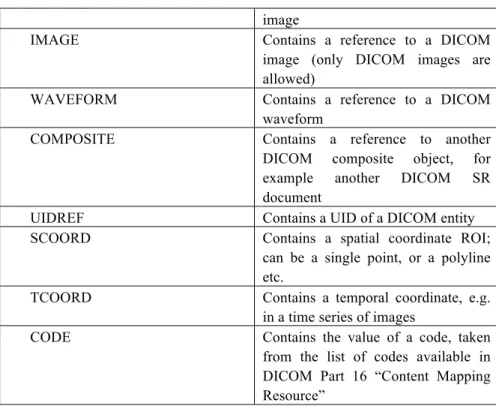
The basic idea is to represent the observations related to a Structured Reporting document as a tree, composed of content nodes (called content items) connected by relationships [3]. Such observations may represent measurements made on images, refer to regions of interest in specific images, or document the context of observation. DICOM introduced 14 content item types, listed in Table 2. What is important to note is that each node has a concept name, that is necessarily a coded entity, and a value, which may be coded or not, depending on the kind of node.
Type of Content Item Comment
CONTAINER A such node has no proper content: it contains the nodes that are connected to it using the CONTAINS relationship
TEXT The content is free text, coded in ASCII, without any presentation features
PNAME Contains a Person Name ; often used to define the observation context DATETIME Contains a Date and Time, also often used to define the observation context TIME Contains a Time, also often used to
define the observation context DATE Contains a Date, also used to define
the observation context
image
IMAGE Contains a reference to a DICOM image (only DICOM images are allowed)
WAVEFORM Contains a reference to a DICOM waveform
COMPOSITE Contains a reference to another DICOM composite object, for example another DICOM SR document
UIDREF Contains a UID of a DICOM entity SCOORD Contains a spatial coordinate ROI;
can be a single point, or a polyline etc.
TCOORD Contains a temporal coordinate, e.g. in a time series of images
CODE Contains the value of a code, taken from the list of codes available in DICOM Part 16 “Content Mapping Resource”
Table 2. The various types of content items of SR trees
Similarly, DICOM introduced a limited number of relationships, with relatively precise semantics such as ‘CONTAINS’, ‘HAS OBSERVATION CONTEXT’, ‘HAS ACQUISITION CONTEXT’, ‘HAS PROPERTIES’, ‘INFERRED FROM’, ‘SELECTED FROM’ and ‘HAS CONCEPT MODIFIER’.
In terms of new DICOM IODs and SOP Classes, Supplement 23 introduced three new classes, addressing various levels of complexity in the use of available content items. The first, called Basic Text SR IOD, addresses the needs of applications exchanging very simple documents, limited to text referring to images. The second, called Enhanced SR IOD, provides the same features plus the possibility to represent measurements and to refer to spatial and temporal coordinates. Finally, the third, called Comprehensive SR IOD may involve any kind of content nodes and authorizes the use of ‘by reference’ relationships (ability to refer to a node anywhere in the SR tree).
Another important feature of DICOM SR was the introduction of Templates. A Template is the specification of a sub-tree that can be instantiated in an SR document. It groups and specifies content item nodes in roughly the same way as modules do, but in a way that is more formalized and dedicated to tree-like structures. Basically, a Template is a table of all content item nodes of a sub-tree, specifying things like requirement type (mandatory or optional), multiplicity, code values allowed for the Concept name attached to each content item node, and eventually, code values allowed for the value of content item node (when represented by a coded entry).
Interested readers can refer to the book written by David Clunie [4] for (far) more details about DICOM SR.
Since the publication of Supplement 23, a wide range of SR documents templates have been standardized to address specific needs in the domains of CAD (e.g. Chest CAD SR, Mammo CAD SR), ultrasound procedure reports (e.g. Ultrasound OB-GYN Procedure report, Vascular Ultrasound Procedure Report, Echocardiographic Procedure Report, Intravascular Ultrasound Procedure Report) and CT/MR reports in cardiology (CT/MR Cardiovascular Analysis Report), among others.
5.4
Web Access to DICOM Objects
Web access to DICOM persistent objects (WADO) was introduced in the standard to facilitate the referencing and retrieval – via the internet protocols http and https – of DICOM persistent objects (images, structured reports, etc.), using URL/URI (Uniform Resource Locator/Identifier). This extension was made in 2003 with Supplement 85, in the form of Part 18 of the standard. It was also recognised by the ISO TC 215 (ISO 17432) [5].
6.
CONCLUSION
The development of the DICOM standard allowed to turn the page of proprietary standards for representing biomedical images and associated metadata. Initial efforts concerned primarily the exchange and management of images acquired on imaging equipment. Image processing is now being considered as well, with extensions of the standard dedicated to radiation therapy, CAD structured reports, spatial registration and fiducials,
segmentation objects and even more recently segmentation surfaces such as meshes.
Besides, DICOM is open to the web technology to communicate and refer to images (or any other composite objects), as demonstrated with the WADO extension. However, DICOM remains based on its original binary syntax, in spite of growing pressure to move to some sort of XML encoding. For example efforts are being devoted in Working Group 20 to facilitate the translation of DICOM SR documents into HL7 Clinical Document Architecture documents, based on XML.
Finally, DICOM has started studying how processing tools, such as plugins or web services, could be more easily shared in the future, based on standard Application Programming Interfaces (API). This initiative, led by Working Group 23 “Application Hosting” received a strong support from the CaBIG initiative (Cancer Biomedical Informatics Grid) and the NCI, because of the perspectives it opens for in vivo imaging in cancer research, especially in molecular imaging, and CAD.
7.
ACKNOWLEDGEMENTS
Many thanks to Joël Chabriais, main representative of the French Society of Radiology in the DICOM Standards Committee, and currently radiologist in the hospital of Aurillac (France), for his continuous and enthusiastic participation to the DICOM standard’s evolution and promotion.
References
[1] Digital Imaging and Communications in Medicine (DICOM) – National Electrical Manufacturers Association, Parts 1 to 18, 2008.
[2] GIBAUD B, GARFAGNI H, AUBRY F, TODD POKROPEK A, CHAMEROY V, BIZAIS Y, DI PAOLA R, « Standardisation in the field of medical image management : the contribution of the MIMOSA model », IEEE Transactions on Medical Imaging, vol. 17, n°1, p. 62-73, 1998.
[3] BIDGOOD WD, Jr. Clinical importance of the DICOM structured reporting standard. Int J Card Imaging 1998; 14:307-315.
[4] CLUNIE D, DICOM Structured Reporting. Bangor: PixelMed Publishing, 2000.
[5] ISO 17432:2004 – « Health Informatics – Messages and communication – Web Access to DICOM persistent objects », TC 215, 18 pages, 2004.