C A S E R E P O R T UDC: 615.46::616.31-08]::616.379-008.64 DOI: 10.2298/VSP150606137B
Apical root-end filling with tricalcium silicate-based cement in a
patient with diabetes mellitus: A case report
Punjenje kanala korena cementom na bazi trikalcijum-silikata kod bolesnika sa
dijabetesom melitusom
Vladimir Biočanin*, Marija Milić†, Milan Vučetić†, Miljana Baćević†,Dina Vasović†, Milka Živadinović†, Dejan Ćetković‡, Dejan Ćalasan†, Božidar Brković† *Department of Dentistry, Faculty of Pharmacy and Health, University of Travnik, Travnik, Bosnia and Herzegovina, and Private Practice, Belgrade, Serbia; †Clinic of Oral Surgery, ‡Anatomy Institute, Faculty of Dental Medicine, University of Belgrade,
Belgrade, Serbia
Abstract
Introduction. The material used for root-end filling has to be biocompatible with adjacent periapical tissue and to stimulate its regenerative processes. Tricalcium silicate cement (TSC), as a new dental material, shows good seal-ing properties with dentin, high compression strengths and better marginal adaptation than commonly used root-end filling materials. Although optimal postoperative heal-ing of periapical tissues is mainly influenced by character-istics of end-root material used, it could sometimes be af-fected by the influence of systemic diseases, such as diabe-tes mellitus (DM). Case report. We presented apical heal-ing of the upper central incisor, retrofilled with TSC, in a diabetic patient (type 2 DM) with peripheral neuropathy. Standard root-end resection of upper central incisor was accompanied by retropreparation using ultrasonic retrotips to the depth of 3 mm and retrofilling with TSC. Post-operatively, the surgical wound healed uneventfully. How-ever, the patient reported undefined dull pain in the oper-ated area that could possibly be attributed to undiagnosed intraoral diabetic peripheral neuropathy, what was evalu-ated clinically. Conclusion. Although TSC presents a suit-able material for apical root-end filling in the treatment of chronic periradicular lesions a possible presence of sys-temic diseases, like type 2 DM, has to be considered in the treatment outcome estimation.
Key words:
periapical diseases; oral surgical procedures; dental cements; silicates; diabetes mellitus, type 2; comorbidity; diabetic neuropathies.
Apstrakt
Uvod. Materijal koji se koristi za retrogradnu opturaciju ka-nala korena trebalo bi da bude biokompatibilan sa okolnim periapeksnim tkivom i da stimuliše procese njegove regene-racije. Trikalcijum silikatni cement (TSC), kao novi dentalni materijal, pokazuje dobro zaptivanje, visoku kompresivnu snagu i bolju ivičnu adaptaciju u odnosu na standardno ko-rišćene materijale za retroopturaciju. Iako postoperativno zarastanje periapikalnog tkiva najviše zavisi od karakteristika materijala za retroopturaciju, ponekad na uspeh zarastanja može uticati i prisustvo neke sistemske bolesti kao što je di-jabetes melitus (DM). Prikaz bolesnika. Prikazali smo api-kalno zarastanje u predelu gornjeg centralnog sekutića, na-kon retroopturacije sa TSC, kod bolesnika sa DM tipa 2 i prisutnom perifernom neuropatijom. Standardna resekcija korena gornjeg centralnog sekutića bila je urađena retropparacijom ultrazvučnim nastavcima do dubine od 3 mm i re-trogradnom opturacijom sa TSC. Zarastanje postoperativne regije bilo je u fiziološkim granicama. Bolesnik se, međutim, žalio na nedefinisan, tup bol u predelu operisane regije koji je verovatno bio povezan sa nedijagnostikovanom intraoral-nom dijabetičintraoral-nom periferintraoral-nom neuropatijom, što je potvr-đeno kliničkim nalazom. Zaključak. Iako TSC predstavlja pogodan materijal za retrogradnu opturaciju kanala korena zuba u lečenju hroničnih periradikularnih lezija, u proceni ishoda lečenja treba imati u vidu i moguće prisustvo perifer-nih manfestacija sistemskih bolesti kao što je DM tipa 2. Ključne reči:
periapeksne bolesti; hirurgija, oralna, procedure; zub, cement; silikati; dijabetes melitus, insulin-nezavisni; komorbiditet; dijabetesne neuropatije.
Fig. 1 – Incorrect root canal 11 filling and slight apical radiolucency.
Fig. 2 – Retropreparation with ultrasonic retrotip. Introduction
The primary goal of periradicular surgery is to seal the apex of the root canal hermetically, preventing the passage of microorganisms or their products into adjacent periapical tis-sues. Traditionally, root-end filling is obtained by amalgam or different types of cement 1. However, modern apical
surgery is still seeking for the material with superb long-time mechanical properties and excellent apical obturation toget-her with biostimulation of regenerative processes of apical tissues 2. Beside the mentioned properties of the material
used, it has to be biocompatible to the neighbouring periapi-cal tissues 3. On the other hand, there are still some possible
complications of periradicular surgery related to disadvanta-ges of materials used for root-end filling 4, 1.
Tricalcium silicate cement (TSC), as a new dental mate-rial, shows mechanical and safety profile which could im-prove the quality of apical obturation 5, 6. It was also shown
that TSC possesses good sealing properties with dentin and high compression strengths 7, 6. TSC provides better marginal
adaptation than commonly used root-end filling materials 8.
Although optimal postoperative healing of periapical tissues is mainly influenced by the characteristics of root-end material used, it could sometimes be affected by the periph-eral appearance of systemic diseases 4. Diabetes mellitus
(DM) results in delayed wound healing and associated com-plications in dental treatments 9, 10. It was already shown that
DM decreased osteoblasts function in the rat model 11. In
ad-dition, microvascular changes found in DM may decrease the reparatory processes of soft and hard tissue and, gradually, could lead to postoperative complications, such as diabetic neuropathy 12–15.
The aim of this report was to present apical healing of the upper central incisor, retrofilled with TSC, in a diabetic patient with possible peripheral neuropathy, as a complica-tion associated with type 2 DM.
Case report
A 53-year-old man, suffering from type 2 DM, with pe-ripheral neuropathy and cardiovascular complications (ASA
III), was referred by his general dental practitioner to the Clinic of Oral Surgery, Faculty of Dental Medicine, University of Belgrade, for root apical surgery of the right central incisor. Clinical examination showed the presence of a fistula in the region of the root apex of the tooth. There were no signs and symptoms of acute dental infection, altho-ugh the patient indicated unpleasant discomfort and unmar-ked chronic pain of the alveolar ridge on the right side. In addition, retroalveolar radiogram was done and short canal filling with well demarcated slight periapical radiolucency were seen around the root apex of tooth (Figure 1). It was decided to perform root-end surgery, implying resection of the root-end and retrofilling with tricalcium silicate cement (TSC) (Biodentine®, Septodont, Saint des Fausses, France)
under high magnifying glass.
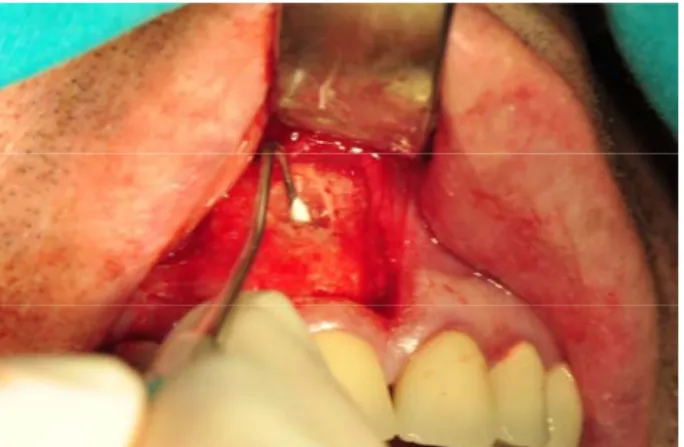
Standard root resection included sectioning the root-end with fissure bar for approximately 2 mm; retro-preparation was done using ultrasonic retro-tips, to the depth of 3 mm (Figure 2). TSC was placed in the 3 mm deep retrograde cavity of the root-end (Figure 3). After the setting time was finished, the wound was debrided and closed primarily.
One month after the operation, the patient complained to constant, undefined, dull pain and discomfort in the operated region, which lasted for the next 3 months. Clinical examination and control retroalveolar radiogram did not show any signs of pathological lesion (Figure 4). There was no fistu-la in the region of oral mucosa or attached gingiva. Also, there were no periodontal pockets around the tooth 11. Regarding that, it was necessary to distinguish the possible presence of vertical root fracture, which usually cannot be diagnosed radiographically. For that reason the re-entry was done. When a full-thickness trapezoid mucoperiostal flap was elevated, al-most complete bone healing in the operated area was present (Figure 5). There were no signs of vertical root fracture. At the end, the operated area was copiously irrigated with saline and interrupted sutures were placed. Follow-up was done 3 and 6 months and 2 years after re-entry and there were no changes both clinically and radiographicaly. During these observation periods the mentioned disturbances at the operated region were recorded occasionally, with different intensity and usually las-ted for several weeks.
Fig. 3 – Root end filling with tricalcium silicate cement (TSC).
Fig. 4 – Control retroalveolar radiogram 3 months after the surgery.
Fig. 5 – Complete bone healing after re-entry. Discussion
Different materials have been used for filling root-ends.
In vitro and in vivo studies have shown that mineral trioxide
aggregate (MTA), as a gold standard, has considerable seal-ing ability and better marginal adaptation to dentin 16–18,
compared to amalgam, super-EBA (ethoxy-benzoic acid) and IRM (intermediate restorative material) cement 19. However, many drawbacks of MTA, such as difficulties with handling, long setting time and high cost, restricts its use as a root-end filling material. In addition, it was found that in higher con-centrations, MTA was toxic to cementoblasts 20.
Tricalcium silicate-based cement – Biodentine®, was
in-troduced as a bioactive material, with the idea of overcoming disadvantages of MTA. Its bioactivity was shown on pulp cells by stimulation biomineralisation 21.Likewise, new
cal-cium silicate-based cement induced odontoblast stimulation and the production of tertiary dentin in the rat pulp injury model 22. Applied directly onto human pulp, TSC induced
stimulation, biomineralization and odontoblast differentiati-on 23, 21. It was also shown that TSC induced osteoblast
diffe-rentiation in mesenchimal stem cells 24. When used as
root-end filling material, TSC showed the least microleakage compared to other cements 25. TSC-based cement produced
more prominent Ca2+ and Si2+ uptake into the root canal
den-tine than MTA when used as a root canal obturation material in bovine incisors 2. Butt et al. 26 suggested that Biodentine
possesses better sealing ability, higher compressive strength and better handling consistency than MTA. The most recent
study of Bhavana et al. 27 revealed that TSC had higher
anti-bacterial and antifungal activity than MTA.
Having in mind all the mentioned advantages and direct biological effect of TSC on bone healing, it was expected to have a successful surgical result after being used for retrofil-ling in the presented patient, showing clinical and radiograp-hic evidence of complete healing. However, the patient re-ported undefined dull pain in the operated area a month after surgery, which lasted for next 3 months. Pain in the operated area could possibly be attributed to undiagnosed intraoral di-abetic peripheral neuropathy, concerning the fact that the pa-tient suffered from type 2 DM for more than 10 years. It was al-so documented that signs of intraoral peripheral neuropathy, such as the loss of intraoral sensation, hyperesthesia, dysesthesia and temporomandibular dysfunction could be related with clinically evident peripheral diabetic neuropathy 28. The
possib-le detrimental influence of diabetic neuropathy in progressi-on of chrprogressi-onic orofacial pain could be also corroborated with unpleasant burning mouth syndrome and nonspecific sore-ness that affect intraoral structures 29. Furthermore, it was
proposed that prolonged effect of local anaesthetic solution could provoke pain in the operating area. Namely, it was already shown that the incidence of diabetic neuropathy inc-reased after spinal and neuraxial block anesthesia 30, 31. Dull
pain that the patient described could possibly be explained by microangiopathy of peripheral dental nerves associated with DM and adjunct prolong ischemic effect of vasocons-trictors from local anaesthetic that was administered in the close proximity to peripheral nerves. Atypical facial pain
(AFP) could be considered in differential diagnosis, having in mind its chronic character. Idiopatic or AFP could be de-scribed as deep or superficial, poorly localised, and someti-mes bilateral pain, predominantly in middle-aged and older women 32, ,33. On the other hand, dull character of pain
locali-sed only in the operated area, could possibly be attributed to peripheral diabetic neuropathy, distinguishing it clinically from AFP.
Conclusion
Tricalcium silicate cement presents a suitable material for apical root-end filling with good mechanical and
biologi-cal properties. However, there are still little data concerning long-term results of using TSC as a root-end filling material in clinical trials, especially in the risk group of patients such as patients with diabetes mellitus, with changed peripheral healing capacity. Further long-term clinical studies are nee-ded to precisely determine clinical and biological behaviour of TSC as a root-end filling material and to confirm the di-rect evidence of regeneration of periapical tissues in humans.
Acknowledgements
This study was supported by the Grant No 175021 from the Ministry of Science of Republic of Serbia.
R E F E R E N C E S
1. Chong BS, Pitt FT. Root-end filling materials: rationale and tis-sue response. Endodontic Topics 2005; 11(1): 114−30. 2. Han L, Okiji T. Uptake of calcium and silicon released from
calcium silicate-based endodontic materials into root canal dentine. Int Endod J 2011; 44(12): 1081−7.
3. Wälivaara D, Abrahamsson P, Isaksson S, Salata LA, Sennerby L,
Dahlin C. Periapical tissue response after use of intermediate
restorative material, gutta-percha, reinforced zinc oxide ce-ment, and mineral trioxide aggregate as retrograde root-end filling materials: A histologic study in dogs. J Oral Maxillofac Surg 2012; 70(9): 2041−7.
4. Lima SM, Grisi DC, Kogawa EM, Franco OL, Peixoto VC,
Gon-çalves-Júnior JF, et al. Diabetes mellitus and inflammatory pulpal
and periapical disease: A review. Int Endod J 2013; 46(8): 700−9.
5. Koubi G, Colon P, Franquin J, Hartman A, Richard G, Faure MO, et al. Clinical evaluation of the performance and safety of a new dentine substitute, Bio dentine, in the restoration of pos-terior teeth-prospective study. Clin Oral Invest 2013;17:243-249.
6. Grech L, Mallia B, Camilleri J. Characterization of set Intermedi-ate Restorative MIntermedi-aterial, Biodentine, BioaggregIntermedi-ate and a proto-type calcium silicate cement for use as root-end filling materi-als. Int Endod J 2013; 46(7): 632−41.
7. Atmeh AR, Chong EZ, Richard G, Festy F, Watson TF. Dentin-cement interfacial interaction: Calcium silicates and polyal-kenoates. J Dent Res 2012; 91(5): 454−9.
8. Ravichandra PV, Vemisetty H, Deepthi K, Reddy JS, Ramkiran D,
Krishna JN, et al. Comparative evaluation of marginal
adapta-tion of Biodentine(TM) and other commonly used root end filling materials: An invitro study. J Clin Diagn Res 2014; 8(3): 243−5.
9. Chaudhary SB, Liporace FA, Gandhi A, Donley BG, Pinuz MS, Lin
SS. Complications of ankle fracture in patients with diabetes. J
Am Academy Orthop Surg 2008; 16(3): 159−70.
10. Kotsovilis S, Karoussis IK, Fourmousis I. A comprehensive and critical review of dental implant placement in diabetic animals and patients. Clin Oral Implants Res 2006; 17(5): 587−99. 11. Verhaeghe J, Herck E, Visser WJ, Suiker AM, Thomasset M,
Ein-horn TA, et al. Bone and mineral metabolism in BB rats with
long-term diabetes: Decreased bone turnover and osteoporo-sis. Diabetes 1990; 39(4): 477−82.
12. Devlin H, Garland H, Sloan P. Healing of tooth extraction sock-ets in experimental diabetes mellitus. Journal of oral and maxil-lofacial surgery 1996; 54(9): 1087−91.
13. Tooke JE. Microvascular function in human diabetes: A physio-logical perspective. Diabetes 1995; 44(7): 721−6.
14. Malik RA, Veves A, Masson EA, Sharma AK, Ah-See AK, Schady
W, et al. Endoneurial capillary abnormalities in mild human
diabetic neuropathy. J Neurol Neurosurg Psychiatr 1992; 55(7): 55−61.
15. Lin JH, Duffy JL, Roginsky MS. Microcirculation in diabetes mellitus: A study of gingival biopsies. Hum Pathol 1975; 6(1): 77−96.
16. Torabinejad M, Chivian N. Clinical applications of mineral triox-ide aggregate. J Endod 1999; 25(3): 197−205.
17. Torabinejad M, Rastegar AF, Kettering JD, Pitt FT. Bacterial leak-age of mineral trioxide aggregate as a root-end filling material. J Endod 1995; 21(3): 109−12.
18. Torabinejad M, Watson TF, Pitt FT. Sealing ability of a mineral trioxide aggregate when used as a root end filling material. J Endod 1993; 19(12): 591−5.
19. Torabinejad M, Smith PW, Kettering JD, Pitt FT. Comparative in-vestigation of marginal adaptation of mineral trioxide aggre-gate and other commonly used root-end filling materials. J Endod 1995; 21(6): 295−9.
20. Hakki SS, Bozkurt BS, Hakki EE, Belli S. Effects of mineral trioxide aggregate on cell survival, gene expression associated with mineralized tissues, and biomineralization of cemento-blasts. J Endod 2009; 35(4): 513−9.
21. Zanini M, Sautier JM, Berdal A, Simon S. Biodentine induces immortalized murine pulp cell differentiation into odontoblast-like cells and stimulates biomineralization. J Endod 2012; 38(9): 1220−6.
22. Tran XV, Gorin C, Willig C, Baroukh B, Pellat B, Decup F, et al. Effect of a calcium-silicate-based restorative cement on pulp repair. J Dent Res 2012;1(12): 1166−71.
23. Laurent P, Camps J, About I. Biodentine(TM) induces TGF-β1 release from human pulp cells and early dental pulp minerali-zation. Int Endod J 2012; 45(5): 439−48.
24. Lee B, Lee K, Koh J, Min K, Chang H, Hwang I, et al. Effects of 3 endodontic bioactive cements on osteogenic differentiation in mesenchymal stem cells. J Endod 2014; 40(8): 1217−22. 25. Pawar AM, Kokate SR, Shah RA. Management of a large
peri-apical lesion using Biodentine(™) as retrograde restoration with eighteen months evident follow up. J Conserv Dent 2013; 16(6): 573−5.
26. Butt N, Talwar S, Chaudhry S, Nawal RR, Yadav S, Bali A. Com-parison of physical and mechanical properties of mineral triox-ide aggregate and Biodentine. Indian J Dent Res 2014; 25(6): 692−7.
27. Bhavana V, Chaitanya KP, Gandi P, Patil J, Dola B, Reddy RB. Evaluation of antibacterial and antifungal activity of new cal-cium-based cement (Biodentine) compared to MTA and glass ionomer cement. J Conserv Dent 2015; 18(1): 44−6.
28. Collin HL, Niskanen L, Uusitupa M, Toyry J, Collin P, Koivisto
AM, et al. Oral symptoms and signs in elderly patients with
type 2 diabetes mellitus. Oral Surg Oral Med Oral Pathol Oral Radiol Endod 2000; 90(3): 299−305.
29. Moore PA, Guggenheimer J, Orchard T. Burning mouth syndrome and peripheral neuropathy in patients with type 1 diabetes mel-litus. J Diabetes Complicat 2007; 21(6): 397−402.
30. Brull R, McCartney CJ, Chan VW, Liguori GA, Hargett MJ, Xu D, et al. Disclosure of risks associated with regional anesthesia: A survey of academic regional nesthesiologists. Reg Anesth Pain Med 2007; 32(1): 7−11.
31. Hebl JR, Kopp SL, Schroeder DR, Horlocker TT. Neurologic com-plications after neuraxial anesthesia or analgesia in patients
with preexisting peripheral sensorimotor neuropathy or dia-betic polyneuropathy. Anesth Analg 2006; 103(5): 1294−9. 32. Pfaffenrath V, Rath M, Pöllmann W, Keeser W. Atypical facial
pain: application of the IHS criteria in a clinical sample. Cephalalgia 1993; 13(Suppl 12): 84−8.
33. Zebenholzer K, Wöber C, Vigl M, Wessely P, Wöber-Bingöl C. Facial pain and the second edition of the International Classification of Headache Disorders. Headache 2006; 46(2): 259−63.
Received on June 6, 2015. Accepted on July 2, 2015. Online First May 2016.