© Birkha¨user Verlag, Basel, 1999
Research Article
Two new aminopeptidases from Ochrobactrum anthropi
active on
D
-alanyl-p-nitroanilide
L. Fanuela, I. Thamma, V. Kostanjeveckib, B. Samynb, B. Jorisa, C. Goffina, J. Branniganc, J. Van Beeumenband
J. M. Fre`rea,*
aLaboratoire d’Enzymologie et Centre d’Inge´nierie des Prote´ines, Universite´ de Lie`ge, Institut de Chimie, B6, B-4000 Sart Tilman (Belgium)
bVakgroep Biochemie, Fysiologie en Microbiologie, Laboratorium voor Eiwitbiochemie en Eiwitengineering, Rijksuniversiteit-Gent, K. L. Ledeganckstraat, 35, B-9000 Gent (Belgium), Fax +32 4 366 33 64,
e-mail: [email protected]
cStructural Biology Laboratory, University of York, Heslington, York YO1 5DD (UK)
Received 7 December 1998; received after revision 15 March 1999; accepted 22 March 1999
Abstract. Two new enzymes which hydrolyseD-alanyl- on the tripeptideL-Ala-Gly-Gly but it was not possible
p-nitroanilide have been detected in Ochrobactrum an- to be certain that the same protein was responsible for thropi LMG7991 extracts. The first enzyme, DmpB, was both p-nitroanilide and peptide hydrolysing activities. purified to homogeneity and found to be homologous to The gene encoding the DmpA protein was cloned and sequenced. The deduced protein sequence exhibits vary-the Dap protein produced by O. anthropi SCRC C1-38
(ATCC49237). The second enzyme, DmpA, exhibits a ing degrees of similarity with those corresponding to similar substrate profile when tested on p-nitroanilide several open reading frames found in the genomes of other prokaryotic organisms, including Mycobacteria. derivatives of glycine andL/D-alanine, but the amounts
produced by the Ochrobactrum strain were not sufficient None of these gene products has been isolated or charac-terised, but a tentative relationship can be proposed with to allow complete purification. Interestingly, the DmpA
preparation also exhibited anL-aminopeptidase activity the NylC amidase from Fla!obacterium sp. K172.
Key words. Peptidase; stereospecificity; amidohydrolase.
Aminopeptidases release the amino-terminal residue from peptide substrates. Most are L-aminopeptidases
and exhibit a wide variety of molecular masses, quater-nary structures, catalytic residues and specificity profiles [1–10]. So far, the only aminopeptidase active on pep-tides containing N-terminal D-residues is that from
Ochrobactrum anthropi SCRC C1-38. It has been char-acterized and named D-aminopeptidase (Dap) (E.C
3.4.11.19) [10, 11]. Its sequence exhibits 25% identity with that of the Streptomyces R61DD-carboxypeptidase
and the residues most important for the catalytic mech-anism of !-lactamases and DD-carboxypeptidases [11–
15] appeared to be conserved. Here we report the isolation and purification of a similarD-aminopeptidase
from O. anthropi LMG7991. Moreover, the same strain produces a second enzyme which hydrolysesD
-alanyl-p-nitroanilide but not its N-acetylated derivative, and thus represents a new potential member of the D
-aminopeptidase family. The study ofD-aminopeptidases
will contribute general information on this poorly char-acterized enzyme group and will help to elucidate the possible evolutionary relationship betweenD -aminopep-* Corresponding author.
tidases andDD-carboxypeptidases. The discovery ofD
-stereospecific enzymes would also offer new tools for enzymatic stereoselective synthesis in organic chemistry [16, 17].
Materials and methods
Enzymes, chemicals and antibodies. Molecular biology and sequencing kits, oligonucleotides, plasmids, purifi-cation gels and columns and Ampholine PAG plates were supplied by Pharmacia Biotech (Uppsala, Swe-den). The restriction enzymes were purchased from Life Technologies (Merelbeke, Belgium) and from Eurogen-tec (Lie`ge, Belgium), and T4 DNA ligase from Boehringer Mannheim (Germany). Substrates and pep-tides were from Bachem (Bubendorf, Switzerland) and Sigma (Bornem, Belgium). Rabbit anti-DAP antibodies were obtained from Gamma (Lie`ge, Belgium).
Selection of D-aminopeptidase-producing strains. Four
strains isolated from soil were obtained from the LMG Culture Collection (Gent, Belgium). Two were O. an-thropi (LMG3306 and LMG7991) [18] and the others were uncertainly classified as related Achromobacter species (LMG1293 which corresponds to ATCC25297 and LMG3499). Cells were grown at 28 °C in nutrient agar medium (1 g beef extract, 2 g yeast extract, 5 g peptone, 5 g NaCl, optionally 15 g agar, 1 l H2O, adjusted to pH 7.4). Cells were stored at −70 °C in 20% glycerol (v/v).
Crude cell extracts obtained by sonication were added to 10 mM D-alanyl-p-nitroanilide (D-Ala-p-Na).
Sam-ples heated at 100 °C for 5 min were also tested to verify the absence of non-enzymatic cleavage of the substrate. Extracts from O. anthropi strains LMG7991 and 3306 exhibited D-aminopeptidase activity.
Achro-mobacter extracts and heated O. anthropi samples did not significantly hydrolyse D-Ala-p-Na.
Production and partial purification of the aminopeptidase activities produced in O. anthropi LMG7991 cells. The cells stored at −70 °C were plated on nutrient medium and grown at 28 °C. Three colonies were resuspended directly in 250 ml of BPY medium and grown for 16 h at 28 °C. This preculture was used to inoculate 15 l of BPY medium in a 20-l fermentor and the culture was grown for 20 h at 28 °C. Cells were collected, resus-pended in 750 ml of Tris-HCl buffer, pH 8.0, containing 0.1 mM EDTA buffer and disrupted with a Constant System Basic Disintegrator (Cell°D, France). The D
-Ala-p-Na-hydrolysing activity was found in the super-natant. The total extract was incubated for 16 h with benzonase in the presence of 1 mM MgCl2 on ice and the insoluble material eliminated by centrifugation and filtration. The D-aminopeptidase activity was
subse-quently recovered in the fraction precipitating between 30 and 95% (NH4)2SO4saturation. After centrifugation, the pellet was redissolved in Tris-EDTA buffer and dialysed against the same buffer. The solution was then diluted fivefold in 10 mM Tris pH 8.0 and the proteins adsorbed on DEAE cellulose. The ion exchanger was filtered, washed with 10 mM Tris-HCl pH 8.0 contain-ing 0.1 M KCl and eluted batchwise with 10 mM Tris-HCl pH 8.0 containing 0.3 M KCl. After filtration, concentration and dialysis against 10 mM potassium phosphate pH 6.0, proteins were loaded on a QSFF column equilibrated with 50 mM potassium phosphate pH 6.0. The exchanger was washed with 0.1 M NaCl in the same buffer and eluted with a linear 0.1–0.3 M NaCl gradient. Two separate peaks exhibiting activity towards D-Ala-p-Na were eluted at NaCl
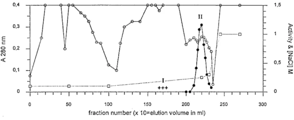
concentra-tions of 0.15 M and 0.25 M (fig. 1).
Figure 1. Elution of the DmpA and DmpB proteins from the QSFF column at pH 6.0. Activity is the v0 value (!A/s) measured for
25-"l aliquots added to 500 "l of 80 "M D-Ala-p-Na in 100 mM Tris-HCl pH 8.0, at 30 °C [!, A 280;!, measured activity (!A×103 s− 1); + + +, detected activity (this activity was too low to be accurately quantified); ", NaCl concentration]. I, 0.15 M NaCl peak
(1) The active fractions eluted at 0.15 M NaCl were pooled, dialysed against 10 mM Tris-HCl pH 8.0 containing 0.2 M KCl and concentrated. This sam-ple was filtered through a Superdex 75 molecular sieve column equilibrated in the same buffer, and the active fractions were analysed by electrophoresis on denaturing and non-denaturing polyacrylamide gels, followed by Coomassie blue staining. Zy-mograms were obtained with the gels run under non-denaturing conditions, by contact with a piece of filter paper previously soaked in a 10 mM solu-tion of D-Ala-p-Na. This enzyme was named
DmpA.
(2) The enzyme eluted at 0.25 M NaCl was purified to 90% by successive chromatographies on QSFF, Phenyl Sepharose Fast-Flow and Sephadex G75 with the help of an A˚kta Explorer apparatus (Phar-macia Biotech, Sweden). Active fractions were loaded on a non-denaturing 8% polyacrylamide gel and the activity detected by a D-Ala-p-Na
zy-mogram. The protein was eluted from the piece of gel containing the activity, concentrated and analysed by SDS-PAGE. This enzyme was named DmpB.
Preparation of genomic DNA. O. anthropi cells were grown in the nutrient medium to a final absorbance at 600 nm (A600) of 1.5. Genomic DNA was prepared as described for the preparation of Streptomyces chromo-somal DNA [19, 20], and was dissolved in water by gentle overnight stirring at 20 °C.
O. anthropi LMG7991 DNA library construction in
pUC18. An incomplete digestion of O. anthropi LMG7991 DNA by EcoRI was performed by incubat-ing the genomic DNA for 30 min at 37 °C with EcoRI (0.5 U EcoRI and 0.1!g DNA/!l) yielding fragments ranging from 1 to 10 kb. This digested genomic DNA (2 !g) was purified by phenol-chloroform extractions, and precipitated. A total of 500 ng of EcoRI-precut and dephosphorylated pUC18 plasmid, T4 DNA ligase and ligation buffer were directly added to the pellet and mixed. After overnight incubation at 16 °C, the ligation mixture was heated at 65 °C for 10 min and used to transform Escherichia coli DH5! competent cells [21]. Transformed cells were spread on LB+ agar plates containing 50!g/ml ampicillin, 20 !g/ml X-gal and 100 !M isopropyl thiogalactoside. Untransformed DH5! cells did not hydrolyse D-Ala-p-Na. After replication
onto nitrocellulose filters (Hybond C), theD
-aminopep-tidase activity was detected as described by Asano et al. [11]. Among the 4000 analyzed colonies, one yellow clone (resulting from the hydrolysis ofD-Ala-p-Na into
p-nitroaniline) was found. After transformation with the plasmid isolated from this clone, the recombinant DH5! cells exhibited high hydrolytic activity onD
-Ala-p-Na.
Plasmid analysis, subcloning and sequencing. The plas-mid (pDML1100) extracted from the positive colony contained a 4.0-kb insert. The insert size was succes-sively reduced to 2.1 kb (pDML1101) and 1.6 kb (pDML1102). Further details concerning these con-structs are available from the authors. The colonies harbouring pDML1102 produced the desired activity and it was concluded that the corresponding gene was located in this fragment. Inserts of reduced sizes were obtained with the help of the Double-Stranded Nested Deletion Kit and were directly sequenced with Reverse (RP) and universal (UP) primers. Sequencing reactions were carried out directly on the two strands with the AutoRead Sequencing Kit and the Thermo Sequenase Labelled Primer Cycle Sequencing Kit for PCR se-quencing. Sequences were read on an Automated Laser Fluorescent DNA Sequencer (Pharmacia). Some DNA compression areas were sequenced with35S-dATP using the T7-Sequencing Kit and Deaza G/A Sequencing Mixes. Analysis of the sequences was performed with the help of the GCG GelAssemble program.
Chemical procedures. Protein N-terminal sequences were determined as described elsewhere [22] on an Ap-plied Biosystems (Perkin Elmer, Foster City, CA) pulsed liquid sequenator. Mrvalues were estimated by SDS-PAGE (10% polyacrylamide). Isoelectric points were measured by isoelectrofocusing (IEF) on Ampho-line PAG plates pH 3.5–9.5, detection of the active bands with theD-Ala-p-Na substrate and measurement
of the pH at the position of the active protein. D
-aminopeptidase activity could be similarly detected af-ter electrophoresis on non-denaturing 8% polyacryl-amide gels by the same zymogram technique [23]. Protein concentrations were estimated on the basis of the absorbance at 280 nm or with the help of the BCA Protein Assay kit (Pierce, Rockford, IL).
D-Aminopeptidase assays and kinetic measurements. The enzyme activity on D-Ala-, L-Ala- and
Gly-p-ni-troanilides was measured in 100 mM Tris-HCl pH 8.0, at 30 °C by monitoring the variation of absorbance at 405 nm (""=11,500 M− 1s− 1) [24]. Estimated errors on v0 values were !5%. The degradation of non-mogenic peptides was followed by thin layer chro-matography analysis (TLC) on silica gel and the amine group of the products visualised by reaction with ninhydrin.
Substrate acetylation. D-Ala-p-Na was dissolved in 50
mM potassium phosphate pH 8.0 buffer. The pH was then adjusted to 9.5 with 1 M KOH, and the solution was chilled on ice. One equivalent of acetic anhydride was added dropwise to the substrate solution under continuous stirring. The reaction mixture was then in-cubated on ice under vigorous stirring until no ninhy-drin-positive compound could be detected. The pH of the solution was adjusted to 8.0 with 1 M KOH. The
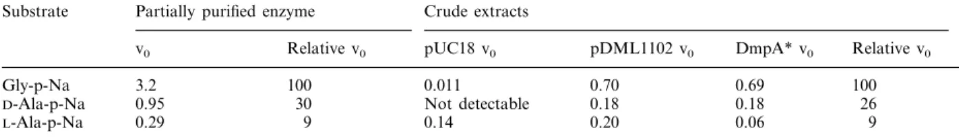
Table 1. Activity of the DmpA protein on p-nitroanilide substrates. The O. anthropi partially purified enzyme (4–40 !g) or crude extracts (corresponding to 50–500 !l of culture) of E. coli XL1-blue cells harbouring the pUC18 or pDML1102 plasmids were incubated with 1 mM substrate in a total volume of 400 !l of 50 mM potassium phosphate buffer pH 8.0 containing 10% dimethylsulphoxide. v0values are in nmol/min and SD values !5%
Crude extracts Substrate Partially purified enzyme
Relative v0 pUC18 v0 pDML1102 v0 DmpA* v0 Relative v0
v0 100 0.011 Gly-p-Na 3.2 0.70 0.69 100 30 Not detectable 0.18 0.95 0.18 26 D-Ala-p-Na 0.29 L-Ala-p-Na 9 0.14 0.20 0.06 9
* The DmpA activity was obtained by subtracting the activity of cells harbouring pUC18 from that of cells harbouring pDML1102.
acetylated substrate solution thus contained 1 equiva-lent of potassium acetate. It was verified that this salt did not influence enzyme activity.
Results
Presence of two distinct D-Ala-p-Na-hydrolysing
activi-ties in O. anthropi LMG7991. Elution of the QSFF column at pH 6.0 yielded two distinct active fractions, centred at 0.15 and 0.25 M NaCl respectively, (fig. 1). The active protein in the latter (DmpB) was purified to 90% homogeneity and its N-terminal sequence, MKFDLSALETFVRTIPQHYKTPXRAVAV(L)KD-X(K), exhibited nearly 60% identical residues with the Dap protein described by Asano et al. [11]. Moreover, its size (55 kDa as determined by SDS-PAGE) and specific activity againstD-Ala-p-Na were also similar to
those of Dap, although its isoelectric pH value was slightly higher (4.9 vs 4.6 measured for the two enzymes under the same conditions). DmpB cross-reacted with rabbit anti-Dap antibodies.
The DmpA protein. After the Superdex 75 molecular sieve chromatography, the DmpA protein was still quite impure as shown by both non-denaturing and SDS-gel electrophoreses. In fact, the activity revealed by the zymograms obtained after non-denaturing elec-trophoresis did not even correspond to one of the major bands revealed by Coomassie blue staining but this activity migrated to a position clearly distinct from those of DmpB and Dap (not shown). Since the amount of DmpA in the preparation was not sufficient, no further attempt was made to purify the protein. A zymogram obtained after gel electrofocusing indicated a pI value of about 5.0.
Preliminary characterisation of the catalytic properties of the enzyme was, however, performed and showed the following relative activities with 1 mM substrates: Gly-p-Na"D-Ala-p-Na"L-Ala-p-Na (table 1).
N-Acetyl-D-Ala-p-Na was not significantly hydrolysed, thus
indicating a clear aminopeptidase specificity. This was confirmed by the fact that hydrolysis of the tripeptide
Ala-Gly-Gly yielded alanine and Gly-Gly as shown by TLC analysis. However, significant hydrolysis was only observed with L-Ala-Gly-Gly and not with D
-Ala-Gly-Gly, suggesting that the stereospecificity was modified compared to that prevailing with the p-nitroanilide derivatives.
Cloning and sequencing of the dmpA gene. To obtain larger amounts of the DmpA protein, the correspond-ing gene was cloned. After growth at 37 °C, the E. coli cells harbouring the pDML1102 plasmid contained in-clusion bodies but also a high amount of solubleD
-Ala-p-Na-hydrolysing activity. Crude extracts exhibited an activity pattern similar to that observed with the origi-nal DmpA protein produced by O. anthropi (table 1). When the E. coli cells harboured pUC18, the crude extracts only hydrolysedL-Ala-p-Na significantly. With
pDML1102, the hydrolysis of Gly-p-Na and D
-Ala-p-Na could easily be measured and the relative reaction rates corresponded well with those observed with the partially purified enzyme. Neither of the crude extracts hydrolysedD-Ala-Gly-Gly. The 2.1-kb EcoRI fragment
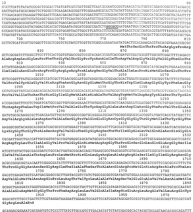
of pDML1101 was completely sequenced on both strands (fig. 2). The sequence has been deposited in the EMBL, Genbank and DDBJ Nucleotide Sequence Databases under the accession number X97669. Since production of the desired activity occurred in only one orientation of the insert in pDML1102, it was con-cluded that the open reading frame (ORF) encoding the enzyme was under the control of the lacZ promoter of the plasmid and was thus on the DNA strand down-stream of this promoter. This strand was translated in the three reading frames and the codons in each frame were compared with a codon frequency table deduced from the sole presently sequenced gene of O. anthropi (the dap D-aminopeptidase gene of O. anthropi SCRC
C1-38). This analysis revealed one ORF encoding a 375-residue polypeptide in the 1.6-kb fragment (fig. 2), downstream of the lacZ promotor, strongly suggesting that this ORF corresponded to the dmpA structural gene and that the 280 bp preceding the initiation codon did not contain the native promoter. The 375-residue
polypeptide did not exhibit any similarity with the se-quence of the Dap protein determined by Asano et al. [11]. Search for Dap (DmpB) proteins in various O. anthropi strains. Western blot analysis of cell extracts of O.
anthropi LMG3306, LMG7991 and SCRC C1-38 strains with anti-Dap antibodies showed that all strains contain a Dap-like protein exhibiting an Mrvalue similar to that of Dap (DmpB in the LMG7991 strain). No such protein
Figure 2. Sequenced O. anthropi LMG7991 genomic DNA fragment containing the dmpA gene, and the derived DmpA protein sequence. Base numbering starts at the first base of the complete sequence. Start codon ATG: 778–780; stop codon TGA: 1903–1905;
dmpA open reading frame: 778–1903; putative Shine-Dalgarno sequence: 769–773. GGCGCC (bold type and underlined) at positions
501–506: NarI site used for the construction of pDML1102. When inserted in pUC18, the 1.6-kb fragment starting at the NarI site is sufficient to result in overproduction of the activity in E. coli cells.
was found in the Achromobacter extracts, in agreement with the absence ofD-Ala-p-Na-hydrolysing activity in
these strains.
Discussion
A new enzyme which hydrolyses Gly-p-Na,D-Ala-p-Na
and L-Ala-p-Na has been purified from O. anthropi
LMG7991 extracts. Its N-terminal sequence, Mr value and catalytic properties indicate that it is similar to the Dap protein, previously described by Asano et al. [11]. Crystallization attempts are now underway. The crystal structures of these proteins should shed further light on their relationship to the DD-peptidase of Streptomyces
R61.
However, the LMG7991 strain produces a second protein which also hydrolyses the same glycine and alanine p-nitroanilide derivatives. In the latter case, preliminary experiments indicated a clear specificity for theD-isomer and acetylation of the free amino group of D-Ala-p-Na decreased the activity to non-detectable
levels. Surprisingly, when assayed on simple tripeptides, the preparation only liberated the N-terminal L-Ala
residue of L-Ala-Gly-Gly. It is possible that the two
activities are due to different proteins, since the partially purified sample was far from homogeneous but the fact that the cellular extracts of E. coli harbouring the pDML1102 plasmid exhibited a similar specificity pat-tern strongly suggests that the same protein was respon-sible for the hydrolysis of the p-nitroanilides and the peptide. If this is confirmed, an interesting reversal of the stereospecificity occurs upon modification of the substrate structure. However, the small amounts avail-able precluded purification of the p-nitroanilide-hy-drolysing enzyme to protein homogeneity.
Cloning experiments yielded a 1.6-kb fragment whose insertion in pUC18 yielded high production ofD
-Ala-p-nitroanilide-hydrolysing activity only when the insert was oriented so that an ORF encoding a novel 375-residue polypeptide was under the control of the lacZ promoter of pUC18, indicating that this ORF corre-sponded to the dmpA gene. A search of the data banks revealed that the genomes of Pseudomonas fluorescens, Pyrococcus horikoshii, Bordetella pertussis and Pseu-domonas aeruginosa contained ORFs encoding putative proteins exhibiting a very high degree of similarity with DmpA. Pairwise comparisons of these putative proteins with DmpA performed with the Bestfit program showed 40–47% identity with scores ranging from 37 to 45 standard deviation units (sdu). Since scores higher than 15 sdu are considered as highly indicative of significant isology, these proteins probably possess enzymatic ac-tivities similar to that of DmpA which would then not be specific to O. anthropi. Thus, other presently
unchar-acterized DmpA-like proteins may well be present in several other bacteria some of which are well-known pathogens.
Moreover, the genomes of M. tuberculosis and M. lep-rae contain ORFs which also encode unknown proteins similar to DmpA, although with somewhat lower scores (10 and 13 sdu, and 20 and 30% identity, respectively). Surprisingly, these two putative mycobacterial proteins exhibit a reliable similarity (about 58% identity with a score of 15 sdu) with the well-characterized NylC ami-dase from Fla!obacterium sp. K172 [25]. This latter enzyme hydrolyses nylon oligomers by a progressive removal of 6-aminohexanoic acid units from the amino terminus. Although this amidase does not hydrolyse other linear amides, dipeptides, tripeptides or casein, it is considered as a linear amidase. It can therefore be hypothesised that DmpA, NylC and the other proteins mentioned above are members of a new amidase superfamily.
Acknowledgments. This work was supported, in part, by the
Bel-gian Program of Interuniversity Poles of Attraction (PAI no. P4/03), by a grant of the FNRS (Brussels) which allowed the purchase of the A˚kta-Explorer apparatus, an Action Concerte´e with the Ministe`re de l’Education, de la Recherche et de la Formation (ARC 93/98-170) and a Geconcerteerde Onderzoeks-actie of the Flemish Government (120.522.93). L.F., B.J. and C.G. are respectively Aspirant and Chercheurs qualifie´s, respectively, of the Fonds National de la Recherche Scientifique (FNRS, Brussels, Belgium). We thank C. Duez for her help and suggestions in molecular genetic methods.
1 Smith E. L. (1955) Aminopeptidases. Methods Enzymol. 2: 83–93
2 Lazdunski A., Murgier M. and Lazdunski C. (1975) Evidence for an aminopeptidase localised near the cell surface of
Es-cherichia coli. Eur. J. Biochem. 60: 349–355
3 Lazdunski C., Busutill J. and Lazdunski A. (1975) Purifica-tion and properties of a periplasmic aminoendopeptidase from Escherichia coli. Eur. J. Biochem. 60: 363–369 4 Khilji M. A., Akrawi A. F. and Bailey G. S. (1979)
Purifica-tion and partial characterisaPurifica-tion of a bovine kidney aminopeptidase (capable of cleaving prolyl-glycylglycine). Mol. Cell. Biochem. 23: 45–52
5 Merkel J. R., Lee C. C. and Freund T. S. (1981) A dimeric, extracellular, heat-stable aminopeptidase produced by a marine pseudomonad. Biochem. Biophys. Acta 661: 32–38 6 Lee C. C. and Merkel J. R. (1981) Selective release and
purification of two periplasmic Alteromonas B-207 aminopep-tidases. Biochem. Biophys. Acta 661: 39–44
7 Burley S. K., David P. R., Sweet R. M., Taylor A. and Lipscomb W. N. (1992) Structure determination and refine-ment of bovine lens leucine aminopeptidase and its complex with bestatin. J. Mol. Biol. 224: 113–140
8 Taylor A. (1993) Aminopeptidases: towards a mechanism of action. Trends Biochem. Sci. 18: 167–172
9 Chapot-Chartier M. P., Rul F., Nardi M. A. and Gripon J. C. (1994) Gene cloning and characterization of PepC, a cysteine aminopeptidase from Streptococcus thermophilus, with se-quence similarity to the eucaryotic bleomycin hydrolase. Eur. J. Biochem. 224: 497–506
10 Asano Y., Nakazawa A., Kato Y. and Kondo K. (1989) Properties of a novel D-stereospecific aminopeptidase from
Ochrobactrum anthropi. J. Biol. Chem. 264: 14233–14239
11 Asano Y., Kato Y., Yamada A. and Kondo K. (1992) Structural similarity ofD-aminopeptidase to carboxypeptidase DD and !-lactamases. Biochemistry 31: 2316–2328 12 Joris B., Ghuysen J. M., Dive G., Renard A., Dideberg O.,
Charlier P. et al. (1988) The active-site-serine penicillin-recog-nizing enzymes as members of the Streptomyces R61 DD-pep-tidase family. Biochem. J. 250: 313–324
13 Joris B., Ledent P., Dideberg O., Fonze´ E., Lamotte-Brasseur J., Kelly J. A. et al. (1991) Comparison of the sequences of class-A !-lactamases and of the secondary structure elements of penicillin-recognizing proteins. Antimicrob. Agents Chemother. 35: 2294–2301
14 Rawlings N. D. and Barrett A. J. (1993) Evolutionary families of peptidases. Biochem. J. 290: 205–218
15 Kurganov B. I. (1993) Can homologous proteins evolve dif-ferent enzymatic activities? Trends Biochem. Sci. 18: 403–406 16 Kato Y., Asano Y., Nakazawa A. and Kondo K. (1989) First stereoselective synthesis of D-amino acid N-alkyl amide catalysed byD-aminopeptidase. Tetrahedron 45: 5743–5754 17 Wong C. H. and Whitesides G. M. (1994) Enzymes in
Syn-thetic Organic Chemistry. Elsevier, Oxford
18 Holmes B., Popoff M., Kiredjian M. and Kersters K. (1988)
Ochrobactrum anthropi gen. nov., sp. nov. from human
clini-cal specimens and previously known as group Vd. Int. J. Syst. Bacteriol. 38: 406–416
19 Hopwood D. A., Bibb M. J., Chater K. F. and Kieser T. (1987) Isolation of chromosomal DNA from Streptomyces
coelicolor A3(2). Methods Enzymol. 153: 123–125
20 Rao R. N., Richardson M. A. and Kuhstoss S. (1987) Isola-tion of high-molecular-weight donor DNA. Methods Enzy-mol. 153: 179–181
21 Sambrook J., Fritsch E. F. and Maniatis T. (1989) Molecular Cloning: A Laboratory Manual, 2nd edn., Cold Spring Har-bor LaHar-boratory Press, Cold Spring Harbour, NY
22 Ledent P., Duez C., Vanhove M., Lejeune A., Fonze´ E., Charlier P. et al. (1997) Unexpected influence of a C-terminal-fused His-tag on the processing of an enzyme and on the kinetic and folding parameters. FEBS Lett. 413: 194–196 23 Gabriel O. (1971) Locating enzymes on gels. Methods
Enzy-mol. 22: 578–604
24 Shibata K. and Watanabe T. (1987) Purification and charac-terization of an aminopeptidase from Mycoplasma sali!arium. J. Bacteriol. 169: 3409–3413
25 Kato K., Ohtsuki K., Koda Y., Maekawa T., Yomo T., Negoro S. et al. (1995) A plasmid encoding enzyme for nylon oligomer degradation: nucleotide sequence and analysis of pOAD2. Microbiology 141: 2585–2590