HAL Id: tel-01316536
https://tel.archives-ouvertes.fr/tel-01316536
Submitted on 9 Jun 2016HAL is a multi-disciplinary open access archive for the deposit and dissemination of sci-entific research documents, whether they are pub-lished or not. The documents may come from teaching and research institutions in France or abroad, or from public or private research centers.
L’archive ouverte pluridisciplinaire HAL, est destinée au dépôt et à la diffusion de documents scientifiques de niveau recherche, publiés ou non, émanant des établissements d’enseignement et de recherche français ou étrangers, des laboratoires publics ou privés.
Regulation of secondary compounds synthesis by
photosynthetic organisms under stress
Parisa Heydarizadeh
To cite this version:
Parisa Heydarizadeh. Regulation of secondary compounds synthesis by photosynthetic organisms under stress. Biochemistry, Molecular Biology. Université du Maine, 2015. English. �NNT : 2015LEMA1019�. �tel-01316536�
Parisa HEYDARIZADEH
Mémoire présenté en vue de l’obtention dugrade de Docteur de l’Université du Maine sous le label de L’Université Nantes Angers Le Mans
École doctorale: Végétal, Environnement, Nutrition, Alimentation, Mer
Discipline: Biologie des Organismes
Spécialité: Biologie, Biochimie Moléculaire et Cellulaire
Unité de recherche: Mer Molécules Santé
Soutenue le 15 décembre 2015
Thèse N: (10)
Regulation of secondary compounds synthesis
by photosynthetic organisms under stress
JURY
Rapporteurs: Fabrice FRANCK, Professeur, Université de Liège
Jean-Paul CADORET, Managing Directeur, Greensea Biotechnologies
Examinateurs: Marc-André SELOSSE, Professeur, Muséum National d’Histoire Naturelle, Paris (Président du jury)
Soulaiman SAKR, Professeur, Agrocampus Ouest, Institut National d’Horticulture et de Paysage, Angers
Directeur de Thèse: Benoît SCHOEFS, Professeur, Université du Maine, Le Mans
Co-encadrantes: Justine MARCHAND, Maître de Conférences, Université du Maine, Le Mans
1
Acknowledgments
I would like to thank my supervisor Professor Benoît Schoefs for his patient guidance, encouragement, support and expertise throughout my graduate experience. At many stages in the course of this research project I benefited from his advice, particularly so when exploring new ideas. His positive outlook and confidence in my research inspired me and gave me confidence. His careful editing contributed enormously to the production of this thesis.
I would also wish to sincere gratitude to my co-supervisor Dr. Justine Marchand for her constant support, guidance, motivation, advice and confidence. I would like to thank my second co-supervisor Dr. Véronique Martin-Jézéquel for her valuable helps and supports. It would never have been possible for me to take this work to completion without their support and encouragement.
I wish to appreciate the member of committee Dr. Jean-Paul Cadoret, Professor Fabrice Franck, Professor Soulaiman Sakr and Professor Marc-André Selosse for their careful editing and endurance. I thank again Dr. Jean-Paul Cadoret and Professor Fabrice Franck to accept to review my thesis.
During my Ph.D. work, I benefited greatly from many fruitful discussions with Brigitte Moreau. I cannot forget the valuable helps of her. I would like to thank Wafâa Boureba and Bing Huang for their help in the lab.
I have a special thank to Dr. Martine Bertrand and Professor Annick Morant-Manceau, who not only helped me in science, but because of the confidante and also good moments they offered to me. I also wish to thank Fanny Laude-Molina that without her helps my stay in France would not be possible.
I wish to say thank to the other people who worked in the lab to progress this thesis, Dr. Ewa Lukomska, Dr. Gaël Bougaran, Dr. Gaëlle Wielgosz and Dr. Aurélie Couzinet.
From Iran's side, I would like to thank my supervisor Dr. Morteza Zahedi for his patient, guidance and support. I would like to extend my sincerest thanks and appreciation to my two co-supervisors Dr. Hosein Zeinali and Dr. Farhad Rejali for their helps and encouragements.
I thank Dr. Mohammad reza Sabzalian and Dr. Ehsan Ataii for their help and supports. Furthermore, I appreciate the support of Isfahan University of Technology (IUT).
I wish to thank all of those people in both universities, Doctoral school VENAM, Région “Pays de la Loire”, Pres LUNAM, Foreign Office of the Republic of France in which this thesis work could not have been done without their generous assistance and helps. Finally, I wish to express my heartfelt gratitude and speciall thanks to my family specially my father and my mother, my life supporters.
2
Abbreviations... 7
1 General introduction ... 9
1.1 The photosynthetic process in land plants and diatoms ... 15
1.2 The reorientation of the carbon metabolism toward the production of secondary compounds... 23
1.3 Influence of light on growth and reorientation of the carbon metabolism ... 26
1.4 Reorientation of the carbon metabolism by biotic factors: the case of arbuscular mycorrhizal ... 28
1.5 Objectives ... 31
1.6 References ... 32
I Regulation of secondary metabolites production in Phaeodactylum tricornutum under different light intensities ... 43
2 Functional investigations in diatoms need more than a transcriptomic approach ... 45
2.1 Abstract ... 45
2.2 Introduction ... 46
2.3 Genome sequencing: towards the system’s part list ... 47
2.4 From the genome to the biochemical model, or who is connected with whom? ... 49
2.5 Physiology requires an integrated model taking into account the cellular compartmentalization of the biochemical activities ... 50
2.6 Combining several ‘omics’ to generate cell models ... 55
2.7 Towards the understanding of metabolic control ... 60
2.8 Conclusions and future perspectives ... 61
2.9 Acknowledgment ... 62
2.10 References ... 62
3 Phaeodactylum metabolism converges to pyruvate formation during growth under different light conditions ... 71
3.1 Abstract ... 71
3.2 Introduction ... 72
3.3 Material and methods ... 73
3.3.1 Phaeodactylum tricornutum ... 73
3.3.2 Experiment strategy and sampling ... 74
3.3.3 Growth determination of microalgae... 75
3.3.4 Pigment extraction ... 75
3.3.5 Photosynthetic and respiratory activity, PI-curve ... 75
3.3.6 Chlorophyll fluorescence yield measurement ... 76
3.3.7 Quantification of intracellular carbon and nitrogen, cellular carbon and nitrogen quotas, C and N uptake rate ... 76
3.3.8 Determination of protein content ... 77
3.3.9 Determination of lipid content ... 77
3
3.3.11 Primer design ... 78
3.3.12 mRNA sampling and extraction ... 78
3.3.13 Real-time quantitative PCR and analyses ... 78
3.4 Results ... 79
3.4.1 Effect of light intensity on the growth of Phaeodactylum tricornutum ... 79
3.4.2 N and C fluxes to lipid, carbohydrate and protein ... 80
3.4.3 Pigment content ... 83
3.4.4 Photosynthetic and respiratory activities ... 84
3.4.5 Photochemical and non-photochemical quenching analysis ... 86
3.4.6 In silico reconstruction of Phaeodactylum tricornutum central carbon metabolism .... 88
3.4.6.1. Central metabolism ... 88
3.4.6.2 CO2 supply ... 91
3.4.6.3 The fate of photosynthetically fixed CO2 ... 93
3.4.7 Changes in selected gene expression during growth ... 93
3.4.7.1 Light intensity influenced expression of genes in different growth phases... 96
3.5 Discussion ... 98
3.5.1 Growth ... 98
3.5.2 The stationary phase: adaptation to carbon deficiency conditions... 102
3.5.3 Adapting to low light condition ... 106
3.5.4 High light adaptation ... 107
3.5.5 Influence of light intensity on the regulation of different pathways ... 108
3.6 Acknowledgments ... 110
3.7 References ... 110
3.8 Supplemental data ... 119
Supplemental data 3.1: Phaeodactylum tricornutum growth curve ... 119
Supplemental data 3.2: Cellular pigment quota ... 120
Supplemental data 3.3: Net photosynthesis and respiratory activities ... 121
Supplemental data 3.4: Kinetic of Chl a fluorescence Management of the incoming light energy ... 122
Supplemental data 3.5: PhotosynthesisIrradiance curve ... 125
Supplemental data 3.6: List of enzymes and related genes ... 127
Supplemental data 3.7: An integrated model for central carbon metabolism in Phaeodactylum tricornutum ... 130
Supplemental data 3.8: Heatmap of up or down-regulated genes under different light intensities ... 131
4
Conclusion of part 1 ... 132
II The impact of light and mycorrhizal endosymbiosis in secondary compound regulation of land plant (Mentha sp.) ... 135
4 Isoprenoid biosynthesis in higher plants and green algae under normal and light stress conditions ... 137 4.1 Abstract ... 137 4.2 Abbreviations ... 138 4.3 Introduction ... 139 4.4 Biosynthesis of isoprenoids ... 140 4.4.1 Toward GPP Biosynthesis ... 140
4.4.2 From GPP to menthol biosynthesis ... 144
4.4.3 Carotenoids ... 147
4.4.4 From GPP to secondary carotenoids ... 147
4.5 Isoprenoid protection against diverse stresses ... 150
4.5.1 Light stress ... 151
4.5.1.1 Light stress and menthol biosynthesis ... 151
4.5.1.2 Light stress and carotenogenesis ... 157
4.6 Genetic engineering ... 158
4.7 Conclusion ... 159
4.8 Acknowledgments ... 159
4.9 References ... 160
5 Mycorrhizal infection, essential oil content and morpho-phenological characteristics variability in three mint species ... 171
5.1 Abstract ... 171
5.2 Introduction ... 172
5.3 Material and methods ... 173
5.3.1 Plant material ... 173
5.3.2 Morpho-phenological characteristics analysis ... 174
5.3.3 Mycorrhizal colonization assessments ... 174
5.3.4 Essential oil isolation ... 175
5.3.5 Statistical analysis ... 175
5.4 Results ... 176
5.4.1 Mycorrhizal colonization assessment ... 176
5.4.2 Morpho-phenological characteristics assessment of mint species ... 176
5.4.3 Essential oil content ... 178
5.4.4 Cluster analysis ... 182
5.5 Discussion and conclusions ... 182
5
5.5.2 Conclusion ... 184
5.6 Acknowledgment ... 185
5.7 References ... 185
6 Photosynthesis under artificial light: the shift in primary and secondary metabolism ... 187
6.1 Abstract ... 187
6.2 Introduction ... 188
6.3 Artificial light sources for photosynthesis ... 189
6.4. Changing light intensity and quality ... 189
6.4.1 Light-emitting diode light(s) can sustain normal plant growth ... 189
6.4.2 Chloroplast differentiation and de-differentiation ... 190
6.4.3 High fluence light-emitting diode triggers production of secondary compounds ... 191
6.4.4 Modification of the metabolism through supplemental monochromatic lighting ... 193
6.5 Photosynthesis in the light of future advances ... 194
6.6 Acknowledgments ... 196
6.7 References ... 196
6.8 Supplemental data ... 199
Supplemental data 6.1. Artificial light sources used in plant cultivation ... 199
7 High performance of vegetables, flowers, and medicinal plants in a red-blue LED incubator for indoor plant production ... 201
7.1 Abstract ... 201
7.2 Introduction ... 202
7.3 Material and methods ... 204
7.3.1 Growth chamber construction ... 204
7.3.2 Light control system... 204
7.3.3 Mint growth evaluation ... 206
7.3.4 Green and potted flower cultivation ... 206
7.3.5 Statistical analysis ... 206
7.4 Results and discussion ... 207
7.4.1 LED light effects on plant growth ... 207
7.4.2 LED light effects on mint essential oil ... 212
7.5 Conclusion ... 212
7.6 Acknowledgments ... 213
7.7 References ... 213
8 The effects of light and mycorrhizal symbiosis on growth parameters and essential oil of three mint species ... 216
8.1 Abstract ... 216
8.2 Introduction ... 216
6
8.3.1 Mycorrhizal inoculation ... 217
8.3.2 Growth chamber condition ... 218
8.3.3 Statistical analysis ... 219
8.4 Results ... 219
8.4.1 Development of Mentha species and AMF under different light conditions ... 219
8.4.2 Essential oil content under different lights ... 221
8.5. Discussion ... 221
8.6 Acknowledgment ... 227
8.7 References ... 227
Conclusion of part 2 ... 230
7
Abbreviation
AbbreviationspH pH gradient across the thylakoid membrane
AM Arbuscular Mycorrhiza
AMF Arbuscular Mycorrhizal Fungi
ATP Adenosine TriPhosphate
CA Carbonic Anhydrase
cDNA Complementary DNA
CF Coupling factor
Chl Chlorophyll
CoA CoEnzyme A
Cx molecules containing x Carbon atoms
Cytbf Cytochromeb6/f
DHA DocosaHexaenoic Acid
DMAPP DiMethylAllyl diPhosPhate
EO Essential Oil
EPA EicosaPentaenoic Acid
Fo basic chlorophyll Fluorescence level
Fm Maximal chlorophyll Fluorescence level
G6P Glucose 6Phosphate
GAP GlycerAldehyde3P
GES GEraniol Synthase
GGPP GeranylGeranyl diPhosPhate
GPP Geranyl diPhosPhate
GPT G6P Phosphate Translocator
IPP Isopentenyl diPhosPhate
LED Light emitting Diodes
LHC LightHarvesting Complex
MEP MethylErythritol 4Phosphate
mRNA Messenger RNA
NPQ Non-photochemical Quenching
NADPH Nicotinamide Adenine Dinucleotide Phosphate
Nudix family of enzymes catalyzes the hydrolysis of aNUcleoside
Diphosphate lInked to another moiety X
OEC Oxygen Evolving Complex
PAR Photosynthetically Active Radiation
PEPC1 PhosphoEnolPyruvate Carboxylase
PEP PhosphoEnolPyruvate
PGT Peltate Glandular Trichomes
PSI PhotoSystem I
PSII PhotoSystem II
PUFA PolyUnsaturated Fatty Acids
PSY Phytoene SYnthase
8
QB Quinone B or plastoquinone
RC Reaction Center
RhNUDX Rosa x hybrida NUDiX hydrolase
9
1General introduction
Since the neolithic revolution, some 15000 years ago, the importance of plants for the human societies never cessed to grow. Although they are used not only as sources for textiles, medicine and energy, but also they are mostly used as a source of food. Their success as source of food resides on the fact that they perform oxygenic photosynthesis, a process allowing them to build organic molecules using water, carbon dioxide and light as a source of energy. A waste of the process is molecular oxygen.
After a complex serie of biochemical reactions, the carbon atoms are combined to produce building block molecules i.e. fatty acids, amino acids and simple sugars such as sucrose that are in turn used as a source of energy or to synthesis more complex compounds such as lipids and proteins. The complex set of reactions involved in these syntheses is referred as the primary metabolism. It provides basic processes like photosynthesis, respiration, growth, development with building blocks, mostly carbohydrates, lipids and proteins. This metabolism is of great importance because it provides the living biomass from which the whole biosphere relies for growth and development.
Plants being mostly nonmotile organisms have to face with the modifications of their biotic and/or abiotic environmental factors. Good examples of these factors are appearance of pathogens, modifications of the growth temperature and/or light intensity. These modifications of the environmental constraints may have different timespan and therefore are susceptible to affect differently plant functioning depending on their duration. Along evolution, plants have acquired several defense mechanisms that can be activated to cope as a response to environmental changes. Among these mechanisms, the reorientation of the metabolism (Figure 1.1) toward the production of secondary metabolites such as phenolic compounds (e.g., anthocyanins, tannins, lignin), nitrogenerated compounds (e.g., alkaloids) and terpenes (e.g. essential oils, terpenoids,
etc.) is very common (Ramawat et al. 2009).
Chapter
10
Figure 1.1. Scheme presenting the effects of a stress on the metabolism orientation in a cell (car: carotenoids).
By definition, secondary metabolites are compounds that are not necessary for cell but play important role in the interactions of the organisms with their environment (Gandhi et al. 2015). As primary metabolites, secondary metabolites have a considerable interest for human societies, serving as odoriferous, spices, colorants for food and textile and medicine (e.g., Gandhi et al. 2015; Adolfsson et al. 2015). Healing with medicinal plants is as old as mankind itself. The oldest written evidence of medicinal plants usage for preparation of drugs has been found on a Sumerian clay slab from Nagpur (India), approximately 5000 years old (Kelly, 2009). Since this pioneer record, the pharmacopea greatly enriched and today, mint is one of the most important medicinal and aromatic plants that has been widely used in food, flavoring, traditional medicine, in cosmetics and pharmaceutical industries (Park et al. 2002; Bhat et al. 2002; Lange et al. 2011).
The clear separation between primary and secondary metabolism is vanishing when internal factors such as the concentration in phytohormones are considered and when secondary metabolites are produced by a restricted number of cells within a tissue producing only primary compounds. A good example is the peltate glandular trichomes
Non stressful condition s Non stressful conditions
11
(PGT) of aromatic plants such as mint, the model plant used for my work, that produce essential oil (EO) by the direction of primary metabolites from mesophyll cells toward PGT. This is why in this manuscript I have chosen the term ‘reorientation of the metabolism’ instead of ‘secondary production’.
Beside land plants, algae1 are another type of organisms performing oxygenic
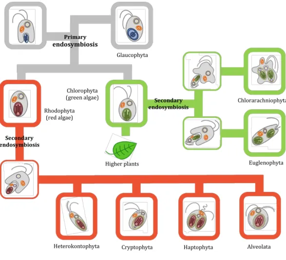
photosynthesis. Plants and algae have originated through a primary endosymbiosis, a process where a non-photosynthetic eukaryote engulfed a cyanobacterium, thereby acquiring a photosynthetic apparatus that became housed within an organelle surrounded by two membranes and it conveniently explains the monophyletic origins of all plastids within eukaryotic cells (Facchinelli & Weber, 2011; Prihoda et al. 2012). This initial endosymbiotic event diverged in the green and red algal lineages, as well as to the Glaucophytes (Figure 1.2). Land plants arose following the evolution of multicellularity within the green algal lineage. In contrast with the evolution of land plants and green algae, the evolutionary history of diatoms is believed to have followed a rather different path. Generally, they have originated from a second non-photosynthetic eukaryote that engulfed a green or a red microalga through a secondary endosymbiosis, resulting a plastid surrounded by four membranes (Gould et al. 2008; Solymosi, 2012). It is believed that a single event is at the origin of the whole Chromalveolata super group, which comprises Heterokonts (also known as Stramenopiles, and to which the diatoms belong), Alveolates (Ciliates, Apicomplexans, and Dinoflagellates), Haptophytes, Cryptophytes, and perhaps also Rhizaria (Facchinelli & Weber, 2011; Prihoda et al. 2012; Kroth 2015). However the question about the number of secondary endosymbioses is still under debate (Keeling, 2013; Kroth, 2015). Regardless to this aspect of the evolution history diatoms, plants and green algae share many similarities at the biochemistry and cell physiology levels. For instance, land plants and diatoms are able to reoriente their metabolism toward the production of secondary metabolites of the terpenoid family, although of different natures. Plants from the Lamiaceae’s botanical family produce essential oil composed by C10 monoterpenoids whereas
diatoms accumulate C18-C22 polyunsaturated fatty acids (PUFAs) (Figure 1.3) (reviewed in Mimouni et al. 2012; see also chapter 4). Nevertheless, because land plants and diatoms evolutionary diverged rapidly, diatoms exhibit unique properties. Convincing evidences are the presence of several carbon concentration mechanisms, full urea cycle, unique pigments and/or lipid compositions and the presence of silica frustules.
Despite a long history of usage, the first written traces about the use of microalgae dates back 2000 years to the Chinese who used Nostoc to survive during famine. The role of microalgae in the biosphere as well as their potential remain rather confidential
1
In this text, the term ‘microalga’ has been concidered to its broad sense, i.e. unicellular photosynthetic micro-organisms and therefore, includes cyanobacteria.
12
until recently (Borowitzka, 1999). Microalgae, especially diatoms (Bacillariophyceae), the second model used in this study, constitute strong oxygen emitter and are responsible for a large part (up to 41%–50%) of the CO2 fixed in oceans (Field et al.
1998; Williams & Laurens, 2010).
Figure 1.2. Schematic view of plastid evolution in the history of photosynthetic eukaryotes. The uptake of a cyanobacterium resulted in a photosynthetic plantae ancestor which subsequently gave rise to the three lineages containing primary plastids: the Chlorophytes (including green algae and the land plants), the Rhodophytes, and the Glaucophytes. The subsequent secondary endosymbioses of green and red algae engulfed by different hosts resulted in the Euglenophyta and Chlorarachniophyta (greens) and in the possibly monophyletic Chromalveolates (reds) that are devided into four major subgroup, i.e. Heterokontophyta/stramenopiles, Cryptophyta, Haptophyta and Alveolata (Facchinelli & Weber, 2011; Kroth, 2015).
In the past, microalgae played a crucial role in the formation of crude oil deposits in ocean floors, which are a rich natural source of fossil fuel (Shukla & Mohan, 2012). This contribution continues today because algae are responsible for a large fraction of the organic carbon being buried on continental margins (Smetacek, 1999).
Primary endosymbiosis Secondary endosymbiosis Secondary endosymbiosis Higher plants Chlorophyta (green algae) Rhodophyta (red algae)
Heterokontophyta Cryptophyta Haptophyta Alveolata
Chlorarachniophyta
Euglenophyta Glaucophyta
13 Figure 1.3. Chemical structure of eicosapentaenoic acid (EPA), geraniol and menthol.
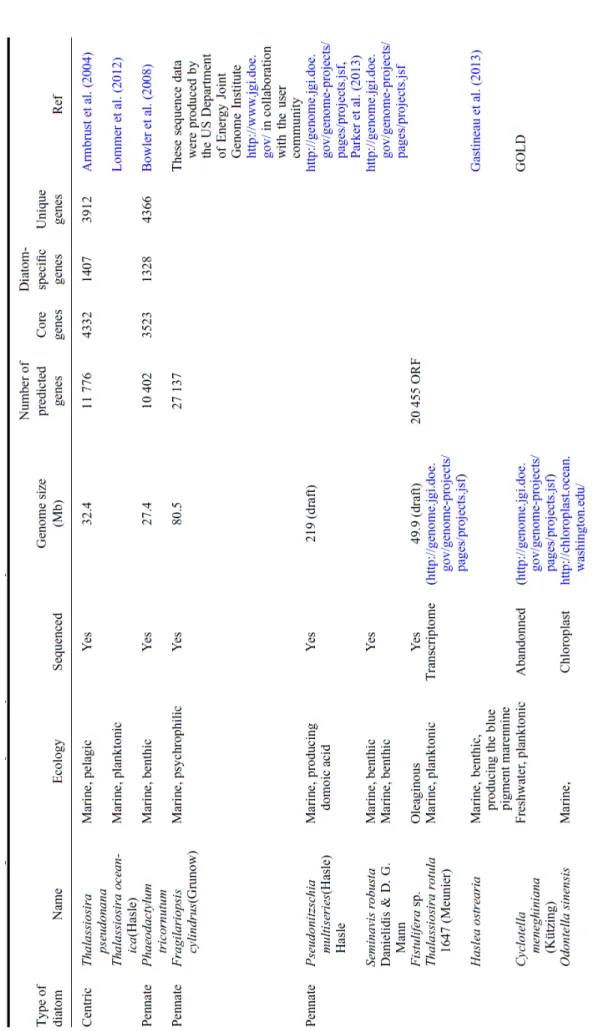
Actually, the human population is up to 7.2 billion people alive today and is expected to coast upward to 9.6 billion by 2050 and 10.9 billion by 2100 (Sullivan, 2014). Several reasons including contribution of human to the acceleration of the depletion in natural resources (Alexandratos & Bruinsma, 2012), increasing the prices of raw materials based on these resources in markets and potentially change the Earth’s climate have increased dramatically the interest for microalgae. This make them as many as major alternative sources of compounds such as polysaccharides, lipids, polyunsaturated fatty acids and pigments (Table 1.1) (land plants: Schoefs, 2002, 2005; microalgae: Cadoret et al. 2012; Mimouni et al. 2012; Hudek et al. 2014) and energy sources, including via genetic engineering and nanotechnology (Monica & Cremonini, 2009; Gordon & Seckbach, 2012; Mimouni et al. 2012; Carrier et al. 2014; Ge et al. 2014). Also their important applications for food, health, cosmetic, waste treatment, energy or pharmaceutical industries are considered (Schoefs, 2003; Gordon & Seckbach, 2012; Mimouni et al. 2012; Gandhi et al. 2015; Gateau et al. 2015).
The production of many of these compounds results from the reorientation of the carbon metabolism toward the production of secondary metabolites (Vinayak et al. 2015). As reflected by the dramatic increase of publications and patents presented in figure 1.4, the field of microalga has become very attractive and this development is strongly impacted the microalgal biotechnology as a topic that began to develop in the middle of the last century (Borowitzka, 1999). Commercial large-scale culture started in the early 1960’s in Japan and the first aquaculture fields appeared in the 1970’s (Muller Feuga, 1996; Pulz & Scheibenbogen, 1998; Borowitzka, 1999; Iwamoto, 2004).
14 Table 1.1 Main storage compounds and oil percentage of some microalgae (Vinayak et al. 2015). High Value Molecules Oil Content (% d.w.) Taxonomy Class Phylum Carotenoids, chlorophyll, tocopherol, lipids 15–32 Tetraselmis suecica Chlorodendrophyceae Chlorophyta Mycosporinelike amino acids, polysaccharides 28–40 Ankistrodesmus sp. Chlorophyceae Chlorophyta Carotenoid, β-carotene, mycosporinelike amino acids, sporopollenin 10 Dunaliella salina Chlorophyceae Chlorophyta Carotenoid, β-carotene, mycosporinelike amino acids 36–42 Dunaliella tertiolecta Chlorophyceae Chlorophyta
Fatty acids, starch 35–65 Neochloris oleoabundans Chlorophyceae Chlorophyta Isobotryococcene, botryococcene, triterpenes 29–75 Botryococcus braunii Trebouxiophyceae Chlorophyta Neutral lipids 58 Chlorella vulgaris Trebouxiophyceae Chlorophyta Neutral lipids 34 Chlorella emersonii Trebouxiophyceae Chlorophyta
EPA, ascorbic acid 15–55 Chlorella protothecoides Trebouxiophyceae Chlorophyta C16 and C18lipids 57 Chlorella minutissima Trebouxiophyceae Chlorophyta EPA 28–69 Nitzschia laevi Bacillariophyceae Heterokontophyta Glycosylglycerides, neutral lipids, TAG 21–31 Thalassiosira pseudonana Coscinodiscophyceae Heterokontophyta Docosahexaenoic acid (DHA) 50–77 Schizochytrium limacinum Labrynthulomycetes Heterokontophyta DHA, Starch 20 Crypthecodinium cohnii Peridinea Myzozoa Neutral lipids 42 Cyclotella sp. Coscinodiscophyceae Ochrophyta EPA, TAG, ω3 LCPUFA 46–68 Nannochloropsis sp. Eustigmatophyceae Ochrophyta
Thus, in a short period of about 30 years, the microalgal biotechnology industry has grown and diversified significantly. Nowadays, the microalgal biomass market produces about 5000 T of dry matter/year but remains ‘confidential’ regarding to that of land plants in terms of production (106 times less than land plants) (Prof. Dussap, personal
communication). Interestingly, the actual market generates a turnover of approximately US$ 125 million in 2004, US$ 271 million in 2010 and US$ 1.6 billion in 2015 (Pulz & Gross, 2004; http://www.biodieselmagazine.com/articles/8604/reportalgaebiofuels technologiesmarketat16bin2015). On the other hand, the global market for botanical and plantderived drugs has been increased from US$ 19.5 billion in 2008 to US$ 32.9 billion in 2013, with an annual growth rate of 11.0% (Trakranrungsie, 2011).
15 Figure 1.4. The time course of the occurrence of the term ʹmicroalga*ʹ in the title of publications (A) and patent families (B) (Source: web of science, core database, last consultation, 29.10.2015; https://www.webofknowledge.com.
1.1 The photosynthetic process in land plants and diatoms
Oxygenic photosynthesis is a biophysicochemical process that converts carbon dioxide into organic compounds using sunlight as a source of energy. In land plants, as in diatoms, photosynthesis takes place in the chloroplasts, and uses water as a source of electrons, releasing oxygen as a waste product (for a recent review, see Hohmann-Marriott & Blankenship, 2011). As mentioned above, the chloroplast in algae and plants has evolved from a cyanobacterial ancestor via endosymbiosis with a primitive eukaryotic host. The chloroplast is a highly compartmentalized organelle, with three membrane systems (outer envelope, inner envelope, and thylakoids) and three soluble spaces (intermembrane space, stroma, and thylakoid lumen) (Figure 1.5.A). A major difference between land plant and diatom chloroplasts resides in the presence of additional surrounding membranes outside the double envelope. They are generally termed periplastid membrane(s) or periplastid/chloroplast endoplasmic reticulum (Solymosi, 2012). The origin of these outer plastid enveloping membranes is still under debate (reviewed in Cavalier-Smith, 2003, 2007; Keeling, 2004; Solymosi, 2012).
Publication year N um ber of p ubl ic ati ons A B
16 Figure 1.5 (A) Structure of chloroplast in land plants, (B) Comparison of the organization of the photosynthetic complexes within thylakoid membranes (Pfeil et al. 2014).
Thylakoid membranes in diatoms (shown in brown) are arranged in groups of three and contain fucoxanthin–chlorophyllprotein complexes for harvesting light (Bertrand, 2010). Note the fourlayer envelopes surrounding the chloroplast as compared to the types found in land plants. Thylakoid membranes in land plants (shown in green) are located inside the chloroplast. They are organized in grana stacks (5–20 vesicles) interconnected by stromaexposed lamellae, and contain chlorophyll–protein complexes for harvesting light (Mustárdy & Garab, 2003).
The photosynthetic apparatus is composed of four multisubunit complexes, namely the wateroxidizing photosystem II (PSII), cytochromeb6/f (cytbf), photosystem I (PSI), and the H+
-translocating ATP synthase (CF0F1) (Nelson & Ben Shem, 2004). These complexes are laterally distributed in land plants, whereas in diatoms, they display a more uniform distribution.
It is mentioned here because some important enzymes involved in the carbon metabolism, namely carbonic anhydrase (CA) and phosphoenolpyruvate carboxylase (PEPC1), have been predicted to be localized in this unique compartment (Solymosi, 2012; Matsuda & Kroth, 2014; see chapter 3). The organization of the thylakoids also
A
17
differs in the two models. In land plants, these membranes are organized in highly stacked (appressed) regions, the so called grana. Grana are interconnected by stromaexposed (nonappressed) regions (Figure 1.5.B). In diatoms, the photosynthetic membranes display groups of 3 weakly stacked thylakoids (for details see Solymosi, 2012 and Figure 1.5).
The photosynthetic apparatus is composed of four macrocomplexes, namely the wateroxidizing PSII, cytbf, PSI, and the H+-translocating ATP synthase (CF0F1) (Nelson
& Ben Shem, 2004). They supply ATP and NADPH for the synthesis of many essential compounds, such as carbohydrates, for autotrophic growth. A PS is composed of a reaction center (RC) and a light harvesting complex (LHC). Two families of pigments are found in LHCs, namely tetrapyrroles and carotenoids. In all photosynthetic organisms, except for most cyanobacteria and red algae, Chlorophyll (Chl) a is aided in its task of harvesting light by accessory pigments, namely other types of Chl (Chl c in diatoms and Chl b in land plants) and carotenoids (Table 1.2). In diatoms, the major carotenoid is fucoxanthin, an allenic ketocarotenoid (Table 1.2). Other important carotenoids in diatoms are the diadinoxanthin and diatoxanthin due to their involvement in the xanthophyll cycle (Bertrand, 2010, Moulin et al. 2010) (Table 1.2). The xanthophyll cycle is a reaction of de-epoxidation triggered by the acidification of the thylakoid lumen.
In land plants, the major carotenoids are lutein and violaxanthin. Other important carotenoids are β-carotene, neoxanthin and zeaxanthin. This last one is involved with violaxanthin in the xanthophyll cycle. The purpose of accessory pigments is to enlarge the range of wavelengths collected by LHCs (Hohmann-Marriott & Blankenship, 2011). In addition to their role in harvesting light, carotenoids play crucial roles in thylakoid organization (Inwooda et al. 2008), in photoprotection of Chl molecules and dissipation of excess energy, for instance, through operation of the xanthophyll cycle (Bertrand, 2010; Goss & Jakob, 2010; Moulin et al. 2011).
Despite the distinct carotenoid composition of brown algae, diatoms and land plants (Table 1.2) they all share a role in photoprotection that includes the xanthophyll cycle (Moulin et al. 2010). Due to lack of space, a detailed comparison of the arrangement of pigments within PSI, PSII and associated LHCs will not be described here, but the interested reader will find relevant information in several recent reviews (Neilson & Durnford, 2010; Busch & Hippler, 2011; Hohmann-Marriott & Blankenship, 2011; Sozer et al. 2011).
It was held for a long time that the different macro-complexes comprising the photosynthetic apparatus were organized linearly along the thylakoid membranes. If this view is still valid for diatoms, (Grouneva et al. 2011) (Figure 1.6), it is no longer accepted for land plants since it has been established that these complexes are laterally
18 Table 1.2. Main chlorophyll and carotenoid types in the various taxons of photosynthetic organisms (Pfeil et al. 2014, Heydarizadeh et al. 2013).
distributed, i.e. localized exclusively in the appressed membranes (PSII), exclusively in the stromaexposed thylakoids (PSI and ATP synthase) or in both types of membranes (cytbf; Anderson, 2002) (Figure1.6).
The differences in thylakoid membrane organization among diatoms and land plants may have implications for biogenesis and turnover of photosynthetic complexes. Because these aspects were not considered in this work, they are not detailed here and the interested reader is requested to consult specialized publications on that topic (Daum & Kühlbrandt, 2011; Austin & Staehelin, 2011).
From the functional point of view, the photons that are harvested by the LHC are directed to the RC of PSII. There, they trigger the release of one electron from one of the two Chl a molecules of the RC.
19 Figure 1.6. The ATP and NADPH that are generated along the photosynthetic apparatus are for CO2 fixation and transformation into organic compounds (Roháček et al. 2008).
This electron is first transferred to the primary acceptor QA (one electron acceptor)
and then to the second electron acceptor QB (two electron acceptor). The changes in the
redox state of QA are reflected by the intensity of the fluorescence emitted by the Chl
molecules of the LHC (Duysens & Sweers, 1963): when QA is oxidized, the level of Chl
fluorescence is minimum. This level is denoted F0. When QA is reduced the Chl
fluorescence is maximum. This level is denoted FM. Using these values, the maximum
quantum yield of PSII photochemistry can be calculated (for details, see chapter 3). The recording of these chlorophyll fluorescence levels in a sample containing billions of PSII require that all the QA are oxidized or reduced simultaneously (Roháček et al. 2008).
This can be obtained by placing the samples in complete darkness for 15 min or by illuminating it with a saturating light, respectively. The electron gap at the RC is filled using an electron coming from the oxidation of a water molecules by the oxygen evolving complex (OEC) that releases oxygen molecules and protons in the thylakoid lumen (Figure 1.6). The rate of oxygen evolving thus reflects the photosynthesis activity. Once QB2 has accumulated two electrons, it leaves PSII to deliver the electrons to the
cytbf. Because charged molecules are unable to cross hydrophobic media such a membrane, QB2 binds 2 protons from the stroma and it is actually QBH2 that it is crossing
the thylakoids membranes until QBH2 pocket of cytbf, located at the other side of the
membrane. While the electrons are delivered, the cotransported protons are released to the lumen of the thylakoids. The electrons are then transferred to the PSI where they
20
are used for production of NADPH. The protons that are delivered in the lumen are transported back to the stroma through the activity of the ATP synthase (Figure 1.6). The biochemical reactions leading to these compounds are common or similar in land plants and diatoms and are described in details in chapter 3. Consequently, they will not be described here.
Under stressless condition, the rate of proton delivery into the lumen is in equilibrium with the rate of proton movement from the lumen to the stroma. In case of stress, for instance during an intense irradiation, the equilibrium is broken and protons accumulate in the lumen, creating a trans-thylakoidal pH gradient ( pH). Lumen acidification triggers the xanthophyll cycle, a molecular device aiming to dissipate the excess of energy. It consists in the reversible de-epoxidation of epoxy-xanthophylls (violaxanthin in land plants and diadinoxanthin in diatoms (Moulin et al. 2010)). To summarize, a proportion of the energy captured by the LHC is used to extract electron from the Chl molecules located in the RC. This proportion is called the photochemical quenching. The rest of the captured energy is dissipated into other mechanisms that are collectively referred as non-photochemical quenching (NPQ). After a continuous illumination, the NPQ relaxes. The analysis of the relaxation kinetic using a nonlinear regression procedure (Roháček, 2010; Roháček et al. 2014) revealed three main components with different shapes, constant rate and underlying mechanisms (Table 1.3). In both models the xanthophyll cycle is the main energy dissipation pathway under short term stress conditions (Roháček, 2010; Roháček et al. 2014).
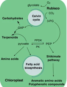
The ATP and NADPH that are generated along the photosynthetic apparatus are used for CO2 fixation and its transformation into building blocks molecules (Figure 1.7). In
brief, storage carbohydrates (chrysolaminarin (polymer of β(1→3) and β(1→6) linked
Table 1.3. Characteristics of the three components composing the NPQ and revelead by the nonlinear regression analyses of the NPQ relaxation. From Roháček, 2010 and Roháček et al. 2014.
Component Fast Medium Slow
Land plants Diatoms Land plants Diatoms Land plants Diatoms Constant rate Of the order of s Of the order of s Of the order of min Of the order of min Of the order of several tens of min Of the order of h
Shape Exponential Exponential Exponential Sigmoid Exponential Exponential Mechanism pH and xanthophyll cycle Fast conformational changes in the membranes State transitions (does not exist in diatoms) pH and xanthophyll cycle Photoinhibition Photoinhibition and partial dissipation of the pH
21
Carbohydrates Calvin cycle
CO2
3-PG
Terpenoids
Aromatic amino acids Polyphenolic compounds O2 glycolate Rubisco p PEP pyruvate PPDK PK Shikimate pathway GAP Fatty acid biosynthesis Amino acids Chloroplast
glucose units) in diatoms and starch (polymer of (1 4) and (1 6) linked glucose units) in land plants are one of the main sinks for carbon fixed during light periods, and it is also incorporated into glucans through gluconeogenesis (Myklestad & Granum, 2009). Another fraction of the fixed carbon is incorporated via pyruvate into fatty acids or via phosphoenolpyruvate into aromatic compounds (Figure 1.7).
The complexity of studying carbon metabolism appears when the pathways are developed and replaced in their cellular context (Figure 1.8). Carbon metabolism is composed by a myriad of enzymatic reactions distributed between the different compartments of the cell i.e. chloroplast, cytoplasm, mitochondria and peroxisome. The complexity is essentially linked to the pathway duplication. A good example is the set of reactions catalyzing the transformation of glyceraldehyde 3-P (GAP) to phosphorenol pyruvate that is present in the cytosol and chloroplast in land plants and also in the mitochondria in diatoms (Figure 1.8). The complexity is even increased by the fact that each reaction is enhanced by an enzyme that is encoded by several isogenes, which expression is differentially regulated by environmental factors. For these reasons, the study of carbon metabolism is very difficult at the biochemical level and inferences from modifications of the gene expression are usually preferred, with all the reservations that imply such reasoning (see chapter 3).
Figure 1.7. General scheme of fixed carbon transformation into building blocks molecules and final products.
22 Figure 1.8. General scheme of central carbon metabolism in land plant (A) and diatom (B) (Martin-Jézéquel et al. 2012). Circles show second part of glycolysis pathway from glyceraldehyde 3-phosphate to pyruvate that exist in plastid and cytosol in land plants and also in mitochondria in diatom. Cytosol gluconate-6-P 6-P-gluconolactone glyceraldehyde-3-P dihydroxyacetone-P fructose-1-6-bisP fructose-6-P 1,3-bisP-glycerate 3-P-glycerate ribulose-5-P TPI FBA PGDH PGL PGK GAPDH GPDH 2-phosphoglycerate pyruvate PFK PGAM PK PPH PEP glucose-6-P glucose-1-P UDP-D-glucose Sucrose-6-P Sucrose Peroxisome glycolate glyoxylate Oxidation Acides gras malate GOX MLS Cycle glyoxylate Acetyl CoA isocitrate succinate ICL 02 H2O2 hydroxypyruvate glycerate HPR SGT GGT NH3 NH3 Peroxisome glycolate glyoxylate Fatty acid oxidation malate GOX MLS Glyoxylate cycle Acetyl CoA isocitrate succinate ICL 02 H2O2 hydroxypyruvate glycerate HPR SGT GGT NH3 NH3 D-fructose D-glucose GPI PGM FBP UT SPS SP FF FF SS A 3-phosphoglycerate ribulose-1-5-bisP ribulose-5-P CO2 PRK 2-phosphoglycolate glycolate O2 1-3-bisP-glycerate glyceraldehyde-3-P glycerate GK fructose-1-6-bisP fructose-6-P dihydroxyacetone-P sedoheptulose-7-P xylulose-5-P ribose-5-P PGK TPI FBA FBP TKL GAPDH Rubisco Chloroplast PGP sedoheptulose1-7-bisP RPE TKL TKL SBP Erythrose-4-P
glucose-6-P glucose-1-P ADP-D-glucose
(1,4-D-glucosyl)(n+1) (1,4-D-glucosyl)(n) starch phosphoenolpyruvate pyruvate Lipides shikimate RPI GPI PGM GPAT ADP-SS AGBE PFK PK PEPS 2-phosphoglycerate PPH PGAM TCA pyruvate serine glycine GDC -SHMT Mitochondria malate OAA succinate citrate MDH PYC1 HCO3 CO2 NH 3 Peroxisome glycolate glyoxylate Oxidation Acides gras malate GDH1 MLS Cycle glyoxylate Acetyl CoA isocitrate succinate ICL Cytosol xylulose-5-P gluconate-6-P 6-P-gluconolactone D-glucose-6-P fructose-6-P erythrose-4-P glyceraldehyde-3-P sedoheptulose-7-P sedoheptulose-1-7-bisP dihydroxyacetone-P fructose-1-6-bisP fructose-6-P 1,3-bisP-glycerate 3-P-glycerate ribulose-5-P TKL TAL TPI FBA RPE PGDH PGL GPI FBP PGK GAPDH GPDH AL SBP 2-phosphoglycerate pyruvate glucose-6-PPGMglucose-1-P GPI PFK PGAM? PK enolase? PEP TCA pyruvate PEP 2-phosphoglycerate 3-phosphoglycerate 1,3-biphosphoglycerate glyceraldehyde 3-phosphate glycerate hydroxypyruvate serine glycine glyoxylate glycolate GDH2 SPT-AGT GDC-SHMT HPR GAPDH PGK PGAM Enolase PK Mitochondrie malate OAA succinate citrate PEPCK PEPC2 CO2 ME1 CO2 MDH PYC1 HCO3 SPT-AGT CO2NH3 3-phosphoglycerate ribulose-1-5-bisP ribulose-5-P CO2 PRK 2-phosphoglycolate glycolate O2 1-3-bisP-glycerate glyceraldehyde-3-P glycerate GK ???? fructose-1-6-bisP fructose-6-P dihydroxyacetone-P erythrose-4-P Sedoheptulose-7-P xylulose-5-P ribose-5-P pyruvate PEP OAA 2-phosphoglycerate glucose-6-P
glucose-1-P Lipides Shikimate PPDK PK PYC2 HCO3 PGK TAL TPI FBA FBP TKL enolase PGM GPI PGAM GAPDH Rubisco Chloroplaste PGP RPE RPI TKL TKL PFK
PEP+ HCO3 OAA
PEPC1
A
23
1.2 The reorientation of the carbon metabolism toward the production of secondary compounds
When photosynthetic organisms are growing in a non-stressful environment, the photosynthetically fixed carbon is mostly oriented toward the synthesis of carbohydrates. The metabolic reorientation consists in reducing the production of carbohydrates and concomitantly inject carbon in (an)other pathway(s) to produce the socalled secondary compounds. Chemically, secondary metabolites can be devided into three groups based on their chemical structure: terpenes, phenolic compounds and alkaloids (Wink, 1988)
Phenolics are a class of chemical compounds consisting of a hydroxyl group (—OH)
bonded directly to an aromatic hydrocarbon group. Based on the number of phenol units in the molecule they are classified as simple phenols, e.g. phenolic acids or polyphenols such as flavonoids. Generally, their primary function is as protection against ultraviolet radiation and pathogens (Manach et al. 2004; Machu et al. 2015).
Alkaloids are a diverse group of low molecular weight, nitrogen containing
compounds derived mostly from aromatic amino acids (made via shikimate pathway) including phenylalanine, tyrosine and tryptophan. These compounds are purported to associate with stress responses including herbivores and pathogens.
Owing to their potent biological activity, many of the approximately 12,000 known alkaloids have a wide range of pharmacological activities including antimalarial (e.g. quinine), anti-asthma (e.g. ephedrine), anti-cancer (e.g. homoharringtonine), analgesic (e.g. morphine), etc. (Wink, 2003; Sinatra et al. 2010; Kittakoop et al. 2014; Pedone-Bonfim et al. 2015).
Terpenoids are the largest class of plant secondary metabolites (Kawoosa et al. 2010;
Akula & Ravishankar, 2011) and they derive from the C5 alkene isoprene. They contain
multiples of 5, 10, 15, 20 or more carbon atoms (Table 1.4) (Breitmaier, 2006; also see chapter 4). The terpenes are generally insoluble in water and synthesized by acetyl-CoA or glycolysis (Pedone-Bonfim et al. 2015). In plants, geranyl diphosphate (GPP), precursor of monoterpenes, is synthesized in plastids from dimethylallyl diphosphate (DMAPP) and isopentenyl diphosphate (IPP) supplied by the methylerythritol 4-phosphate pathway (MEP) (Rodr gue -Concepción & Boronat, 2002).
The monoterpenes in EO are produced through the activity of various monoterpene synthases (Chen et al. 2011). For example, geraniol synthase (GES) converts GPP into geraniol in basil (Iijima et al. 2004). Recently, Magnard et al. (2015) reported the presence of a cytosolic enzyme (RhNUDX1) taking action in the monoterpene alcohols pathway in roses. This discovery makes the basis for monoterpene biosynthesis even more obscure. Very interestingly, monoterpenes similar of those found in mint EO have been detected in the diatom Thalassiosira (Meskhidze et al. 2015).
24 Table 1.4. Diversity of terpenes in photosynthetic organisms.
The other branch in terpenoid biosynthesis is diterpene synthesis that requires geranylgeranyl diphosphate (GGPP) (see chapter 4). The first committed step in carotenoid biosynthesis is head to head condensation of the two C20 molecules of GGPP
by phytoene synthase (PSY) to form phytoene. GGPP is also the precursor for several other groups of metabolites, including Chls. In diatom brown colour is due to the presence of high amounts of the xanthophylls fucoxanthin that masks the other carotenoids (e.g., β-carotene, violaxanthin, diadinoxanthin and diatoxanthin and the chlorophyllous pigments). The absolute and/or relative amounts of individual pigments may differ according to the taxon and its ecology (Bertrand, 2010).
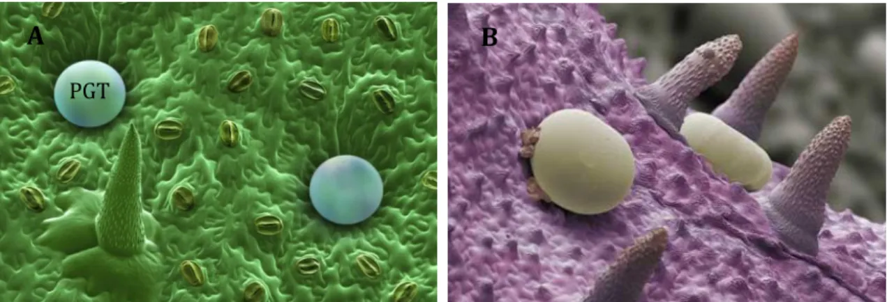
In unicellular organisms such as microalgae the metabolic reorientation per se concerns the whole organisms. In diatoms, modifications in the environmental factors such as nutrient depletion (Reitan et al. 1994; Breteler et al. 2005) or excess light energy (Norici et al. 2011) may increase cellular lipid content in diatoms. In multicellular organisms such as land plants, the metabolic reorientation is often restricted to a part of the organisms. This part could even be restricted to few cells as it is the case for EO production in mint (Bhat et al. 2002; Lange et al. 2011). EO is fabricated and stored in specialized anatomical structures, termed PGT, on leaf surfaces (Lange et al. 2011; Jin et al. 2014) (Figure 1.9. A.B).
The secretory cells of the oil glands responsible for EO biosynthesis can be isolated in high yield from leaves (Fig. 1.9.C) (Gershenzon et al. 1992). The isolated secretory cells from glandular trichomes are capable of de novo biosynthesis of monoterpenes from sucrose as primary carbohydrate precursor (Mc Caskill et al. 1992) and they have been shown to be highly enriched in the enzymes of monoterpene biosynthesis (Lange et al. 2000). Thus, in a first approximation, diatoms and secretory cells could be considered as single cells. However, the two models greatly differ when the carbon and energy sources are considered. Actually diatoms are autotrophs i.e; the photosynthetically fixed CO2 is
Terpene type Number of isoprene units
Number of
carbon atoms Examples
Monoterpene 2 10 Volatile compouds such as menthol,
camphor, limonene, pinene, thujone, nerol (Meskhidze et al. 2015)
Sesquiterpene 3 15 Farnesol, carophyllene, αbergamotene
(Ferriols et al. 2015)
Diterpene 4 20 Chlphytol
Sesterpene 5 25 haslenes ( Rowland et al. 2001)
Triterpene 6 30 lanosterol, achilleol, rhizene (Belt et al. 2003)
25
http://blog.wellcome.ac.uk/2014/07/11/imageoftheweekmint /
Figure 1.9. (A) and (B): Scanning electron microscope pictures of the surface of Mentha piperita leaf (B: Svoboda et al. 2001). PTG are oil glands responsible in which EO is synthesized and stored. It gives to mint its characteristic aroma. The spikes are small hairs (non glandular hairy trichome) on the leaf. The pictures have been colored using false colors. (C): Scheme of a transversal section of PGT of Mentha piperita (Mc Caskill & Croteau 1999). A B C PGT Secretory cells Essential oil storage cavity Stalk cell basal cell
used for further biosynthesis, including terpenoids, and the energy for biosynthesis is mostly arising from photosynthesis. In contrast, PGT are heterotrophic cells i.e. the energy needed for these syntheses does not come from photosynthesis because PGT of aromatic plants generally have non-green plastids while secreting terpenoids (Lange et al. 2013; Jin et al. 2014). Therefore, these cells rely on exogenous supply of sucrose from underlying leaf tissues, i.e. mesophyll, to use as carbon source for monoterpene production (Jin et al. 2014). Sturm & Tang (1999) found higher expression of genes encoding enzymes for sucrose catabolism in secretory cells, such as sucrose synthase, neutral and alkaline invertases that are important to transfer carbon from sucrose in non-photosynthetic tissues. These enzymes convert sucrose to hexose phosphates. In most plants glucose 6-phosphate (G6P), which is synthesized from sucrose in the cytosol, seems to be the preferred hexose phosphate taken up by non-green plastids. The transporter proteins responsible for this import of carbon into plastids are known as G6P-phosphate translocator (GPT) and secretory cells are enriched in GPT (about 30 times more than green leaf cells) (Jin et al. 2014). Two genes coding for GPT, GPT_1 and GPT_2, have been described in the Arabidopsis genome, while GPT_2 is non-essential and generally expressed at lower levels than GPT_1 (Niewiadomski et al. 2005).
26
Regardless the model under consideration, the available data have revealed that triggering the metabolic reorientation involves a complex signaling network, the description of which is out of the scope of this general introduction. Among these factors light appears to be one of the most important and fundamental environmental factor affecting the orientation of the carbon metabolism (Bowler et al. 2010; Lemoine & Schoefs, 2010; Meitao Dong et al. 2012; Lan et al. 2013). Actually, light can affect the regulation of central carbon metabolism through its two characteristics i.e. wavelength and intensity (Fan et al. 2013; Li et al. 2013). The reorientation of the carbon metabolism toward the production of secondary metabolites is rather well understood when the final pathways are considered (e.g., Lemoine & Schoefs, 2010) but little is known about the pathway used to carbon dispatch between the different pathways, especially under light stress in diatom and land plants (Rech et al. 2008; Vidoudez & Pohnert, 2012). In addition, it has become evident that the production of certain metabolites is highly dependent on the development of the cells and several ecological interactions are mediated by these strongly regulated metabolites (Barofsky et al. 2010; Barofsky et al. 2009, 2010; Vidoudez & Pohnert, 2008).
1.3 Influence of light on growth and reorientation of the carbon metabolism
Land plants and microalgae being photosynthetic organisms, the presence of light is mandatory for their growth and development. As mentioned above photons are harvested by pigments and the energy associated to the photons is used to drive photosynthesis (Chen & Blankenship, 2011). In nature, including outdoor cultures, sunlight is the continuous source of photons. Per se, indoor culture requires artificial lighting, that are typically outfitted with fluorescent and/or incandescent bulbs providing a general spectrum that is accommodating to the human eyes but not necessarily supportive to plant development (Folta et al. 2005; Li et al. 2013) (see chapters 6 and 7). The incandescent or fluorescent bulbs contain filaments that must be periodically replaced, consume a lot of electrical power and also generate heat, making impossible their use close to the canopy (Tennessen et al. 1994; Singh et al. 2015). Incandescent and fluorescent bulbs differ by their operational lifetime: around of 20,000 h for fluorescent bulbs whereas only 1000 h for incandescent bulbs (Barta et al. 1992; Tennessen et al. 1994). Beside these traditional lighting systems, another lighting source, the Light emitting diodes (LED), rapidly developed (Girón González, 2012). This lighting system has been used less than two decades to test plant responses to narrow wavelength irradiations and has shown to be a promising lighting technology for future because LED usage eliminated limitations of traditional lighting systems environments (Olvera-Gonzalez et al. 2013; Li et al. 2013).
LEDs have an extraordinary lifetime (about 100,000 h), require little maintenance, and they can be placed close to plants and can be configured to emit high light fluxes
27
even at high light intensities (Barta et al. 1992; Tennessen et al. 1994; Singh et al. 2015). The emergence of energy efficient LEDs has opened up a new methodology for conversion of photonic wavelengths to organic compounds in photosynthetic macro and microorganisms (Tan Nhut et al. 2005; Lan et al. 2013; Fan et al. 2013; Olvera-Gonzalez et al. 2013).
Because the photosynthetic activity is a light dependent process, it is usually thought that increasing the irradiance level is proportionally increasing the photosynthetic activity and thus growth. This is true until a certain level of irradiance (Terry et al. 1983) above which the photosynthetic activity is saturated (Nguyen-Deroche et al. 2012). When the irradiance levels exceed the electron transport capacity, the photosynthetic organisms are stressed and the photosynthetic machinery can be damaged. These damaged can lead to an arrest of photosynthesis (Ritchie, 2010). To avoid such a situation, defense mechanisms such as the xanthophyll cycle, state transition, chloroplast cycling and photoinhibition are activated to dissipate the excess of absorbed energy (Moulin et al. 2010; Allorent et al. 2013; de Marchin et al. 2014; Spetea et al. 2014). It is interesting to note that some of these mechanisms might not exist in some taxons. For instance state transition, which consists in changing the relative antenna size of PSII and PSI does not exist in diatoms. The energy invested in these defense mechanisms being not available for growth, the relation between growth rate and irradiance level reaches a stationary phase or even presents a decreasing phase (Geider et al. 1985). How the carbon metabolism is impacted by the growth light intensity in diatoms remain largely unknown. This is mostly due to the fact that the completion of the genome sequences of diatoms is very recent (Phaeodactylum
tricornutum: Bowler et al., 2008; Thalassiosira pseudonana: Armburst et al. 2004) and
the reconstitution of the cellular metabolism including prediction of enzyme localization even more fresh and still under discussion (Kroth et al. 2008; Fernie et al. 2012; Kroth, 2015). The transcriptome of P. tricornutum has been studied in a few contexts, such as silicon metabolism (Sapriel et al. 2009), short term light acclimation (Nymark et al. 2009), carbon fixation, storage and utilization (Chauton et al. 2013) and nitrogen stress (Levitan et al. 2015) but growth related modifications in gene expression induced by different light intensities in P. tricornutum are not yet described.
A similar study could not be started in mint because of a general lack of information at the genomic level. When this thesis was initiated, only one paper was reporting mRNA from isolated M. piperita and M. spicata secretory cells that were used to generate a cDNA library (Lange et al. 2000). Recently, next generation sequencing methods have been applied to mint and revealed a more complex metabolism than expected (Jin et al. 2014). Altogether, the knowledge about the development of PGT, terpene production and its regulation is very limited making it difficult a study on carbon reorientation toward the production of EO (Glas et al. 2012; Tissier, 2012).
28
In addition to be an energy shuttle for photosynthetic organisms, light provides information from the environment to the organisms. For instance, it has been shown that the presence of light has a positive impact on shoot branching (Djennane et al. 2014) and bud burst (Henry et al. 2011) are mechanisms regulated by light. These information are decoded by photoreceptors such as phytochrome (red/farred, blue light, UVB photoreceptors) (Lin, 2000; Rockwell et al. 2006; Heijde & Ulm, 2012). If the knowledge about photoreceptors is really advanced in land plants, it remains in its infancy in microalgae and especially in diatoms (Hegemann, 2008; Fortunato et al. 2015) and will not be treated here. Many studies over the last several decades have clearly shown that variation in light quality can affect growth and control developmental transitions of diatoms and land plants, including mint (diatoms: Mouget et al. 2004; Huysman et al. 2013 land plants Urbonavic iu te et al. 2008; Li et al. 2012; Tarakanov et al. 2012; Colquhoun et al. 2013; Bula et al. 1991; Duong et al. 2002, Duong & Nguyen, 2010; Kurilcik et al. 2008; Barisic et al. 2006; mint: Nishioka et al. 2008; Malayeri et al. 2011). Indeed, several results showed that LED light is more suitable for land plant growth than fluorescent lamps (Bula et al. 1991; Folta et al. 2005; Li et al. 2010; Olvera-Gonzalez et al. 2013). To the best of our knowledge, there is no report about the effect of different quality of LED light on plant growth and essential oil production in Mentha taxon, except the study performed in this Ph.D thesis (see chapter 7).
1.4 Reorientation of the carbon metabolism by biotic factors: the case of arbuscular mycorrhizal
As mentioned earlier, land plants and diatoms have emerged from first and secondary endosymbiosis, respectively. When Earth colonization by plants started, plants had only rudimentary xylem for conducting mineral sap (Gerrienne et al. 2011), probably making difficult mineral nutrition (Selosse & Le Tacon, 1998). Plants being rooted they were immobile and unable to move to capture nutrients from their immediate environment. To circumvent this difficulty, plants established a symbiotic associations with Glomalean fungi from the Glomeromycota, ancestors of modern arbuscular mycorrhizal fungi (AMF), about 460 million years ago (Simon et al. 1993; Taylor et al. 1995; Redecker et al. 2000). This is estimated to be some 300–400 million years before the appearance of root nodule symbioses with nitrogen fixing bacteria (Finlay, 2008). In this respect, symbiosis with AMF is the most ancient and widespread form of fungal symbiosis with plants. Indeed today, more than 74% of plant species and more than 90% of the cultivated species still acquire nutrients from soil using AMF, reflecting the evolutionary success of this mutualistic symbiosis (Wang & Qiu, 2006; Smith & Read, 2008; Heijden et al. 2015). These interactions between organisms pervade all ecosystems and strongly influence the structure of natural populations and
29
communities (Cairney, 2000). It is interesting to note that only 150–200 species of AMF have so far been distinguished on the basis of morphology, but DNAbased studies suggest the true diversity of these symbionts may be very much higher (Fitter, 2005; Soka & Ritchie, 2015).
From the functional point of view, the symbiosis is primarily seen as a trade contract between the two partners: the fungus provides the plants with water and mineral, especially phosphate. AMF being heterotrophs, they require an external source of carbon for energy and cellular synthesis that the plant is ‘offering’. Thus, photosynthetic products under the form of glucose and fructose to the fungus, are converted to trehalose and lipids (Pfeffer et al. 1999; Doidy et al. 2012). Lipids translocate to the extraradical mycelium for further metabolism, as they are the major forms of carbon storage in AM fungal spores, hyphae, and vesicles (Cox et al. 1975). The trading of compounds occurs through the unique highly branched fungal structures, the so called arbuscule, which grow intracellularly without penetrating the host plasmalemma (Finlay, 2008) (Figure 1.10)
Depending on the AMF species, it is assumed that the total carbohydrate cost of the arbuscular mycorrhiza (AM) symbiosis can be up to 20% of the host plant photosynthetic production (Harrison, 1999; Kaschuk et al. 2009; Lendenmann et al. 2011; van der Heijden et al. 2015). Clearly, AMF constitutes additional carbon sinks that sources, the above ground part of plants, should feed.
Figure 1.10. A) light microscopic image of Mentha spicata root with arbuscules inside root cells. The black circle represent an arbusculecontainig root cell that is explained by detailes in partB (picture B from Recorbet et al. 2008)
Consequently, the photosynthetic activity of mycorrhized plants is expected to be enhanced. Actually, many reports describe stimulatory effects of photosynthesis in
plant cell wall periarbuscular membrane
intraradical hyphae apoplastic compartment A B plant plasma membrane
30
mycorrhizal plants (Louche-Tessandier et al. 1999; Valentine et al. 2001). Boldt et al. (2011) reported a decrease, and Parádi et al. (2003) and Adolfsson et al. (2015) found no effect of mycorrhization on Chl content and fluorescence. In contrast, Adolfsson et al. (2015) demonstrated that photosynthesis in Medicago truncatula was enhanced through branching enhancement. The reason for this discrepancy remains unclear as well as the mechanism by which AM could influence these photosynthetic parameters in plant leaves. Arbuscular mycorrhization also induces carbon reorientation. For instance, in the root, mycorrhization induces the synthesis of secondary carotenoids that eventually are used for the production of C13/C14 apocarotenoids regulating the timelife
of the arbuscules (Walter et al. 2015).
The activation of the carotenoid metabolism has likely additional function relevant for symbiosis due to plastid role in the biosynthesis of gibberellin, abscisic acid and strigolactone (Seddas et al. 2010; Walter et al. 2015; Takeda et al. 2015). In addition, the increased amounts of glutamate and aspartate in roots of AM plants (Lohse et al. 2005; Schliemann et al. 2008; Rivero et al. 2015) suggest a stimulation of amino acid biosynthetis and N assimilation in plastids upon mycorrhization. A mycorrhiza related activation of plastidic metabolism was also inferred from the increased abundance of some fatty acids (palmitic and oleic acids) in M. truncatula roots (Lohse et al. 2005; Schliemann et al. 2008), presumably reflecting the extension and de novo synthesis of the plasma membrane around the arbuscule, the so-called periarbuscular membrane (Figure 1.10) (Gaude et al. 2012a; b). Gutjahr et al. (2011) also showed that although starch is dispensable for mycorrhization, it can, when present, be used as a second energy source for AM symbiosis. Likewise, the comparison of the membrane proteome between mycorhizal and non-mycorrhizal roots of M. truncatula indicated a reduced abundance of plastidic proteins having role in carbon import to plastids, ammonium assimilation and glycolysis, thereby suggesting that part of carbon skeletons devoted to plastid metabolism might be highjacked to sustain the fungal development within host roots (Abdallah et al. 2014).
For the reasons explained earlier, much less is known about carbon reorientation in the photosynthetic cells of AM plants. This reorientation should occur at least in aromatic plants because a higher production of secondary metabolic is often recorded in AM plants (del Rosario Cappellari et al. 2015; Copetta et al. 2006; Khaosaad et al. 2006; Zeng et al. 2013; Kumar et al. 2014 a; b; Ratti et al. 2015). Last but not least, it is worth to mention that mycorrhization enhance plant fitness by increasing resistance or tolerance to biotic and abiotic stresses (Newsham et al. 1995; Auge, 2001; Aloui et al. 2011). How light intensity and light quality influence the carbon reorientation mechanisms in the photosynthetic tissue remains unknown, as to the best of our knowledge, there is no report showing the interaction between mycorrhizal symbiosis and different LED light quality on plant productivity.