i
Université de Montréal
Development and plasticity of locomotor circuits in the zebrafish
spinal cord
par
LAURA D. KNOGLER
Département de pathologie et biologie cellulaire Faculté de médecine
Thèse présentée à la Faculté de médecine en vue de l’obtention du grade de doctorat (Ph.D.)
en pathologie et biologie cellulaire
Novembre, 2014
ii
Université de Montréal
Faculté des études supérieures et postdoctorales
Cette thèse de doctorat intitulée :
« Development and plasticity of locomotor circuits in the zebrafish
spinal cord »
Présentée par: LAURA D. KNOGLER
a été évaluée par un jury composé des personnes suivantes:
Guy Doucet, Ph.D. Président-rapporteur Pierre Drapeau, Ph.D. Directeur de recherche Roberto Araya, Ph.D. Membre du jury Paul De Koninck, Ph.D. Examinateur externe Louis-Eric Trudeau, Ph.D. Représentant du doyen de la FES
iii RÉSUMÉ
Un objectif important en neurobiologie est de comprendre le développement et l'organisation des circuits neuronaux qui entrainent les comportements. Chez l'embryon, la première activité motrice est une lente contraction spontanée qui est entrainée par l'activité intrinsèque des circuits spinaux. Ensuite, les embryons deviennent sensibles aux stimulations sensorielles et ils peuvent éventuellement nager, comportements qui sont façonnées par l'intégration de l'activité intrinsèque et le rétrocontrôle sensoriel. Pour cette thèse, j'ai utilisé un modèle vertébré simple, le poisson zèbre, afin d'étudier en trois temps comment les réseaux spinaux se développent et contrôlent les comportements locomoteurs embryonnaires.
Pour la première partie de cette thèse j'ai caractérisé la transition rapide de la moelle épinière d'un circuit entièrement électrique à un réseau hybride qui utilise à la fois des synapses chimiques et électriques. Nos expériences ont révélé un comportement
embryonnaire transitoire qui précède la natation et qu'on appelle « double coiling ». J'ai démontré que les motoneurones spinaux présentaient une activité dépendante du glutamate corrélée avec le « double coiling » comme l'a fait une population d'interneurones
glutamatergiques ipsilatéraux qui innervent les motoneurones à cet âge. Ce travail (Knogler et al., Journal of Neuroscience, 2014) suggère que le « double coiling » est une étape distincte dans la transition du réseau moteur à partir d'un circuit électrique très simple à un réseau spinal entrainé par la neurotransmission chimique pour générer des comportements plus complexes.
Pour la seconde partie de ma thèse, j'ai étudié comment les réseaux spinaux filtrent l'information sensorielle de mouvements auto-générés. Chez l'embryon, les neurones sensoriels mécanosensibles sont activés par un léger toucher et ils excitent en aval des interneurones sensoriels pour produire une réponse de flexion. Par contre, les contractions spontanées ne déclenchent pas ce réflexe même si les neurones sensoriels sont toujours activés. J'ai démontré que les interneurones sensoriels reçoivent des entrées glycinergiques pendant les contractions spontanées fictives qui les empêchaient de générer des potentiels d'action. L'inhibition glycinergique de ces interneurones, mais pas des autres neurones
iv
le courant inhibiteur. Ce travail (Knogler & Drapeau, Frontiers in Neural Circuits, 2014) suggère que la signalisation glycinergique chez les interneurones sensoriels agit comme un signal de décharge corolaire pour l'inhibition des réflexes pendant les mouvements auto-générés.
Dans la dernière partie de ma thèse, je décris le travail commencé à la maîtrise et terminé au doctorat qui montre comment la plasticité homéostatique est exprimée in vivo aux synapses centrales à la suite des changements chroniques de l'activité du réseau. J'ai démontré que l'efficacité synaptique excitatrice de neurones moteurs spinaux est augmentée à la suite d’une diminution de l'activité du réseau, en accord avec des études in vitro précédentes. Par contre, au niveau du réseau j'ai démontré que la plasticité homéostatique n'était pas nécessaire pour maintenir la rythmicité des circuits spinaux qui entrainent les comportements
embryonnaires. Cette étude (Knogler et al., Journal of Neuroscience, 2010) a révélé pour la première fois que l'organisation du circuit est moins plastique que l'efficacité synaptique au cours du développement chez l'embryon.
En conclusion, les résultats présentés dans cette thèse contribuent à notre compréhension des circuits neuronaux de la moelle épinière qui sous-tendent les comportements moteurs simples de l'embryon.
Mots clés: les circuits de la moelle épinière, générateur central de motifs, décharge corollaire, plasticité homéostatique, comportement locomoteur, poisson zèbre embryonnaire,
v SUMMARY
A fundamental goal in neurobiology is to understand the development and organization of neural circuits that drive behavior. In the embryonic spinal cord, the first motor activity is a slow coiling of the trunk that is sensory-independent and therefore appears to be centrally driven. Embryos later become responsive to sensory stimuli and eventually locomote, behaviors that are shaped by the integration of central patterns and sensory
feedback. In this thesis I used a simple vertebrate model, the zebrafish, to investigate in three manners how developing spinal networks control these earliest locomotor behaviors.
For the first part of this thesis, I characterized the rapid transition of the spinal cord from a purely electrical circuit to a hybrid network that relies on both chemical and electrical synapses. Using genetics, lesions and pharmacology we identified a transient embryonic behavior preceding swimming, termed double coiling. I used electrophysiology to reveal that spinal motoneurons had glutamate-dependent activity patterns that correlated with double coiling as did a population of descending ipsilateral glutamatergic interneurons that also innervated motoneurons at this time. This work (Knogler et al., Journal of Neuroscience, 2014) suggests that double coiling is a discrete step in the transition of the motor network from an electrically coupled circuit that can only produce simple coils to a spinal network driven by descending chemical neurotransmission that can generate more complex behaviors.
In the second part of my thesis, I studied how spinal networks filter sensory information during self-generated movement. In the zebrafish embryo, mechanosensitive sensory neurons fire in response to light touch and excite downstream commissural
glutamatergic interneurons to produce a flexion response, but spontaneous coiling does not trigger this reflex. I performed electrophysiological recordings to show that these interneurons received glycinergic inputs during spontaneous fictive coiling that prevented them from firing action potentials. Glycinergic inhibition specifically of these interneurons and not other spinal neurons was due to the expression of a unique glycine receptor subtype that enhanced the inhibitory current. This work (Knogler & Drapeau, Frontiers in Neural Circuits, 2014) suggests that glycinergic signaling onto sensory interneurons acts as a corollary discharge signal for reflex inhibition during movement.
vi
In the final part of my thesis I describe work begun during my masters and completed during my doctoral degree studying how homeostatic plasticity is expressed in vivo at central synapses following chronic changes in network activity. I performed whole-cell recordings from spinal motoneurons to show that excitatory synaptic strength scaled up in response to decreased network activity, in accordance with previous in vitro studies. At the network level, I showed that homeostatic plasticity mechanisms were not necessary to maintain the timing of spinal circuits driving behavior, which appeared to be hardwired in the developing zebrafish. This study (Knogler et al., Journal of Neuroscience, 2010) provided for the first time
important in vivo results showing that synaptic patterning is less plastic than synaptic strength during development in the intact animal.
In conclusion, the findings presented in this thesis contribute widely to our understanding of the neural circuits underlying simple motor behaviors in the vertebrate spinal cord.
Keywords: spinal cord circuitry, central pattern generator, corollary discharge, homeostatic plasticity, locomotor behavior, embryonic zebrafish, developmental neurobiology
vii
LAY SUMMARY
The spinal cord is an important part of our nervous system and drives motor movements. If the normal function of the spinal cord is disrupted, for example due to
developmental defects or to injury or disease, then motor movements are compromised or lost entirely (paralysis). Although we know a great deal about the different kinds of neurons and connections present in the spinal cord there still remains much to understand about how the circuit functions as a whole. My research uses a simple vertebrate, the zebrafish, to study the neural circuits within the spinal cord that contribute to spontaneous and sensory-evoked behaviors early in development. By using techniques that allow me to record the activity of individual neurons in combination with microscopy and behavioral studies I can examine the development, function and plasticity of connections between different types of neurons and how this activity relates to motor movements. My research has revealed three key findings about spinal cord circuits.
First, my colleagues and I identified a novel developmental behavior that precedes mature swimming in the zebrafish. This behavior occurs spontaneously and is driven by a small group of neurons in the spinal cord that initially send only electrical signals to each other, unlike the mature nervous system that uses chemical neurotransmitters. We showed that a population of excitatory spinal interneurons is active during this behavior and that these neurons first begin to develop chemical connections using the neurotransmitter glutamate at this developmental stage. These findings help explain how the earliest development proceeds in the zebrafish spinal cord and will help us understand these processes in terrestrial
vertebrates, including mammals.
Second, I described a simple mechanism by which the zebrafish avoids being startled by somatosensation (touch) resulting from its own movement. During motor movements, the spinal cord sends a signal to inhibit a sensory interneuron that is usually responsible for conveying information about light touch. As a result, a touch stimulus resulting from the fish’s own movement does not cause a reflexive startle response whereas any touch stimulus coming from the external environment (e.g. a predator) efficiently evokes a response. These findings may be important for understanding how animals filter out self-generated sensory
viii
stimuli and may have implications for human diseases such as schizophrenia where filtering may be abnormal or absent.
Finally, in the last study I describe how the spinal circuits that are responsible for producing swimming behavior can be adjusted in response to changes in the environment. By experimentally reducing the activity of neurons throughout the fish, I found that neurons could use homeostatic mechanisms to change their synapse properties in order to maintain normal activity levels. This work also showed that despite these experimental treatments to globally reduce the activity of neurons, swimming behavior was mostly unaffected. These results help us understand how genetics combine with environmental factors (nature versus nurture) to generate a functional spinal cord circuit that produces swimming.
ix TABLE OF CONTENTS
RÉSUMÉ ... III SUMMARY ... V LAY SUMMARY ... VII TABLE OF CONTENTS...IX LIST OF FIGURES AND TABLES ... XV LIST OF ABBREVIATIONS ... XVII ACKNOWLEDGEMENTS ... XIX
CHAPTER 1 ... 1
I. GENERAL INTRODUCTION ... 1
I.1.THEDEVELOPMENTOFMOTORBEHAVIORS ... 2
I.1.1. Historical studies of embryonic behavior ... 2
I.1.2. Embryonic motor behaviors in lower vertebrates ... 4
I.1.3. Embryonic motor behaviors in mammals ... 5
I.1.4. Summary of behavioral studies ... 7
I.2.THENEURALCONTROLOFLOCOMOTION ... 7
I.2.1. Central pattern generators driving locomotion ... 8
I.2.2. Descending control and sensory feedback modulate locomotion ... 11
I.2.3. Activity-dependent plasticity in spinal circuits ... 13
I.3.THEEMBRYONICZEBRAFISHASASIMPLEVERTEBRATEMODEL ... 15
I.3.1. Timeline of locomotor behaviors in the developing zebrafish ... 17
I.3.2. Classification of spinal sensory and CPG neurons in the early embryo ... 19
I.3.3. Glycinergic spinal interneurons ... 21
I.3.4. Glutamatergic spinal interneurons ... 24
I.3.5. Experimental methods to study locomotor circuits ... 25
x
CHAPTER 2 ... 28
II. ARTICLE: "A HYBRID ELECTRICAL AND CHEMICAL CIRCUIT IN THE SPINAL CORD GENERATES A TRANSIENT NOVEL EMBRYONIC MOTOR BEHAVIOR" ... 28
LINKERSTATEMENT ... 29
II.1.ABSTRACT ... 31
II.2.INTRODUCTION ... 32
II.3.MATERIALSANDMETHODS ... 33
II.3.1. Fish maintenance ... 33
II.3.2. Drug applications ... 33
II.3.3. Lesions ... 34
II.3.4. Behavioral analysis ... 34
II.3.5. Electrophysiology ... 34
II.3.6. DNA microinjection and confocal microscopy ... 35
II.3.7. Statistical analyses ... 36
II.4.RESULTS ... 36
II.4.1. Transitional appearance of double coiling ... 36
II.4.2. Double coiling does not depend on mechanosensory transduction ... 38
II.4.3. Glutamatergic neurotransmission is required for double coiling ... 38
II.4.4. Normal frequency of occurrence of double coiling requires a descending excitatory drive from the hindbrain ... 39
II.4.5. Blockade of glycinergic neurotransmission promotes multiple coils ... 40
II.4.6. Double coiling-related activity recorded in primary motoneurons ... 42
II.4.7. Blockade of glutamatergic neurotransmission abolishes mixed events in motoneurons ... 44
II.4.8. Candidate glutamatergic interneurons in the early motor network ... 44
II.4.9. Glutamatergic CiD interneurons are active during spontaneous behaviors ... 45
II.4.10. CiD neurons contact ipsilateral MNs and other CiDs with putative glutamatergic synapses ... 47
xi
II.5.1. Double coiling: A novel intermediate behavior ... 48
II.5.2. The integration of chemical neurotransmission into an existing electrical circuit 49 II.5.3. Neural activity patterns generating double coiling ... 50
II.6.REFERENCES ... 52
II.7.FIGURES ... 56
CHAPTER 3 ... 68
III. ARTICLE: "SENSORY GATING OF AN EMBRYONIC ZEBRAFISH INTERNEURON DURING SPONTANEOUS MOTOR BEHAVIORS" ... 68
LINKERSTATEMENT ... 69
III.1.ABSTRACT ... 71
III.2.INTRODUCTION ... 72
III.3.MATERIALSANDMETHODS ... 73
III.3.1. Zebrafish maintenance ... 73
III.3.2. Electrophysiology and pharmacology ... 73
III.3.3. Statistical analyses ... 75
III.4.RESULTS ... 75
III.4.1. Intrinsic properties of embryonic CoPA interneurons are similar to other spinal neurons ... 75
III.4.2. Embryonic CoPAs show spontaneous long-lasting glycine-mediated depolarizations that shunts excitation ... 77
III.4.3. Embryonic CoPAs have slow glycine-mediated synaptic currents ... 79
III.4.4. CoPA interneurons express PhTX-insensitive AMPA receptors that mediate rectifying glutamatergic mEPSCs ... 81
III.4.5. Embryonic CoPAs are not part of the network driving ipsilateral coils and are shunted by glycinergic inhibition during contralateral coils ... 83
III.4.6. Embryonic CoPAs receive brief glutamatergic excitation followed by long lasting, shunting glycinergic inputs in response to touch ... 85
III.4.7. Other classes of embryonic spinal neurons show different activity patterns during spontaneous and touch-evoked activity ... 86
xii
III.4.8. CoPAs receive rhythmic inhibition in phase with excitation to ipsilateral MNs
during burst swimming episodes ... 88
III.5.DISCUSSION ... 89
III.5.1. Shunting of embryonic CoPA interneurons prevents inappropriate activation of the touch reflex during spontaneous or ongoing evoked behaviors ... 90
III.5.2. CoPAs and other neurons in the sensory pathway are excluded from the electrically coupled network ... 93
III.5.3. Strong AMPAergic excitation allows brief activation of CoPA interneurons in response to external touch stimuli prior to the onset of shunting glycinergic inhibition . 94 III.5.4. Sensory gating in other spinal networks ... 95
III.6.REFERENCES ... 96
III.7.FIGURES ... 102
CHAPTER 4 ... 112
IV. ARTICLE: "SYNAPTIC SCALING AND THE DEVELOPMENT OF A MOTOR NETWORK" ... 112
LINKERSTATEMENT ... 113
IV.1.ABSTRACT ... 115
IV.2.INTRODUCTION ... 116
IV.3.MATERIALSANDMETHODS ... 117
IV.3.1. Zebrafish maintenance ... 117
IV.3.2. Pharmacological injections ... 117
IV.3.3. Neural recordings ... 118
IV.3.4. Intrinsic excitability ... 119
IV.3.5. NMDA-induced slow oscillations ... 119
IV.3.6. Muscle recordings ... 119
IV.3.7. Acute pharmacological treatment ... 120
IV.3.8. Analysis ... 120
IV.3.9. Tnfα expression ... 120
xiii
IV.3.11. Behavioral recordings ... 121
IV.4.RESULTS ... 123
IV.4.1. Scaling up occurs at glutamatergic synapses upon activity blockade in the zebrafish embryo ... 123
IV.4.2. Absence of synaptic scaling at glycinergic synapses ... 125
IV.4.3. TNFα induces synaptic scaling down ... 126
IV.4.4. No changes in intrinsic cellular excitability ... 127
IV.4.5. Homeostatic plasticity of network activity and motor behavior ... 128
IV.4.6. Comparable acute changes in synaptic amplitudes do not disrupt network activity patterns ... 130
IV.5.DISCUSSION ... 131
IV.5.1. Neuronal activity ... 132
IV.5.2. Behavioral consequences ... 133
IV.6.REFERENCES ... 134
IV.7.FIGURES ... 138
CHAPTER 5 ... 148
V. GENERAL DISCUSSION ... 148
V.1.GENERALSIGNIFICANCE ... 149
V.2.SPONTANEOUSACTIVITYINDEVELOPINGLOCOMOTORNETWORKS .... 149
V.2.1. Transient, spontaneous motor behaviors appear as the locomotor network matures ... 149
V.2.2. CPG circuits driving spontaneous double coiling in the zebrafish ... 150
V.2.3. The contribution of gap junctions to spontaneous coiling and swimming ... 151
V.2.4. Double coiling as an indicator of locomotor CPG function ... 153
V.2.5. Future work ... 154
V.3.SENSORIMOTORCIRCUITSINTHEEMBRYONICSPINALCORD ... 155
V.3.1. Sensory interneurons gate activation of the locomotor CPG during fictive embryonic motor behaviors ... 156
xiv
V.3.3. Developmental reorganization of sensorimotor circuits ... 159
V.3.4. Future work ... 161
V.4.STABILITYANDPLASTICITYOFSPINALLOCOMOTORCIRCUITS ... 162
V.4.1. Synaptic scaling ... 162
V.4.2. Intrinsic plasticity ... 164
V.4.3. Developmental role of spontaneous activity in locomotor circuits ... 165
V.4.4. Future work ... 166
V.5.OUTLOOKSFORTHESTUDYOFLOCOMOTORCIRCUITS ... 167
V.5.1. Genetic tools for visualizing and manipulating network activity ... 168
V.5.2. Anatomical studies of the "connectome" ... 169
V.5.3. Computer modeling and bioengineering of integrated circuits ... 170
V.5.4. Final thoughts ... 171
REFERENCES ... 173
APPENDIX A ... 185
xv
LIST OF FIGURES AND TABLES
CHAPTER 1
Figure 1. The locomotor central pattern generator (CPG) in swimming vertebrates ... 9
Figure 2. Organization of the locomotor system in vertebrates ... 12
Figure 3. Functional neuron classes in the vertebrate spinal cord ... 16
Figure 4. Overview of embryonic zebrafish behaviors ... 18
Figure 5. Neurons of the embryonic zebrafish spinal cord ... 20
CHAPTER 2 Figure 1. Double coiling appears during embryonic development and depresses network activity ... 56
Figure 2. Glutamatergic neurotransmission is required for double coiling ... 58
Figure 3. Double coils require a descending excitatory drive from the hindbrain ... 59
Figure 4. Decreased glycinergic signaling results in an increase in multiple coiling ... 60
Figure 5. Motoneurons show mixed electrical and synaptic events corresponding to glutamate-dependent fictive double coils ... 61
Figure 6. Blockade of glutamatergic neurotransmission abolishes mixed events in motoneurons but not SBs or PICs ... 64
Figure 7. Glutamatergic CiD interneurons are highly active at early embryonic stages and in contrast to CoPAs fire bursts of action potentials during spontaneous behaviors ... 65
Figure 8. Embryonic spinal CiD neurons have putative synapses that contact caudal primary MNs and other CiDs ... 67
CHAPTER 3 Table 1. Properties of glycinergic mPSCs in spinal neurons from 26-29 hpf embryos ... 81
Table 2. Properties of AMPAergic mPSCs in CoPA interneurons from 26-29 hpf embryos ... 82
Figure 1. Intrinsic properties of embryonic CoPA interneurons are similar to other spinal neurons ... 102
xvi
Figure 2. Embryonic CoPAs show spontaneous activity in the form of a long-lasting
depolarization that has low strychnine sensitivity and shunts excitation ... 103 Figure 3. Embryonic CoPAs have slow glycinergic mPSCs ... 105 Figure 4. Embryonic CoPAs are inactive during fictive ipsilateral coils and are depolarized by glycinergic inputs during fictive contralateral coils ... 107 Figure 5. Embryonic CoPAs receive brief glutamatergic excitation then long lasting, shunting glycinergic inputs in response to touch ... 109 Figure 6. CoPAs receive rhythmic inhibition in phase with excitation to ipsilateral MNs
during burst swimming episodes ... 111
CHAPTER 4
Figure 1. A significant increase in glutamatergic mPSC amplitude was only seen following
chronic blockade of network activity with TTX from 1 or 2dpf to 4dpf ... 138 Figure 2. Chronic TTX or CNQX treatment results in a scaling up of glutamatergic mEPSC amplitudes at 4dpf ... 139 Figure 3. Chronic TTX, CNQX, or AP-5 treatment has no effect on glycinergic mIPSC
amplitudes at 4dpf ... 140 Figure 4. Chronic TNFα treatment results in a scaling down of glutamatergic mEPSC
amplitudes at 3dpf ... 141 Figure 5. Chronic CNQX or TNFα treatment does not alter cellular excitability ... 142 Figure 6. Slow oscillation frequency underlying fictive swimming in motoneurons is
unchanged following chronic CNQX or TNFα treatment ... 143 Figure 7. Chronic TNFα or CNQX treatment does not significantly alter the patterning of
motor input to muscle cells at 3dpf or 4dpf, respectively ... 144 Figure 8. Larval swimming behavior is not altered following chronic TNFα or CNQX
treatment ... 145 Figure 9. Acute CNQX or CTZ treatment significantly altered maximum synaptic amplitude (and contraction frequency of motor input - only in the second case) to muscle cells at 3dpf or 4dpf, respectively ... 146
xvii
LIST OF ABBREVIATIONS
α-Btx alpha-bungarotoxin
AMPA α-Amino-3-hydroxy-5-methyl-4-isoxazolepropionic acid APV (2R)-amino-5-phosphonovaleric acid
dlc dorsolateral commissural CiA circumferential ascending CiD circumferential descending
CNQX 6-cyano-7-nitroquinoxaline-2,3-dione CoBL commissural bifurcating longitudinal CoLo commissural local
CoPA commissural primary ascending CoSA commissural secondary ascending CPG central pattern generator
CTZ cyclothiazide
DLR diencephalic locomotor region DNA deoxyribonucleic acid
dpf days post-fertilization DRG dorsal root ganglion
EPSC excitatory post-synaptic current ER estrogen receptor
GABA gamma-aminobutyric acid
GCaMP genetically encoded calcium indicator GFP green fluorescent protein
hpf hours post-fertilization IC ipsilateral caudal IN interneuron KCl potassium chloride LTD long-term depression LTP long-term potentiation
xviii MLR mesencephalic locomotor region
MN motoneuron
mPSC miniature post-synaptic current mRNA messenger ribonucleic acid NMDA N-Methyl-D-aspartate PhTX philanthotoxin
PIC periodic inward current
RB Rohon-Beard
SB synaptic burst
SBEM serial blockface scanning electron microscope
syp synaptophysin
TNFα tumor necrosis factor-alpha TTX tetrodotoxin
UAS upstream activating sequence VeLD ventral lateral descending
xix
ACKNOWLEDGEMENTS
First and foremost I would like to thank my supervisor Dr. Pierre Drapeau for his mentorship during my graduate studies. He has been an endless source of optimism and information not to mention hilarious anecdotes throughout my time in the lab. I appreciate how supportive Pierre has been of my research ideas. He has encouraged me to participate in numerous courses and conferences which have allowed me to thrive independently. I am very grateful for his invaluable guidance during my studies and in my future research plans.
I would also like to acknowledge the numerous contributions of Dr. Louis Saint-Amant to my graduate studies. Particularly in the early stages of my project, Louis generously offered his time and expertise for experimental troubleshooting. He also encouraged me to think more critically about my work and offered many valuable insights.
I appreciate the many useful and entertaining discussions with zebrafish colleagues past and present including Dr. Gary Armstrong, Dr. Kessen Patten, Dr. Sean Low, Sébastien Côte and Alexandra Lissouba. I am grateful to our laboratory technician Meijiang Liao for her molecular biology expertise as well as her cheerful help in general lab matters. Thanks as well to Guy Laliberté and Marina Drits for zebrafish care. Many thanks to my other friends and fellow graduate students in the department especially Dr. Ariel Wilson who was a major source of inspiration and moral support throughout my studies.
Many thanks to my thesis jury members Dr. Guy Doucet, Dr. Paul De Koninck and Dr. Roberto Araya for their important contributions at this final stage of my doctorate as well as to my doctoral committee members Dr. Graziella di Cristo and Dr. Karl Fernandez for their support and feedback over the course of my studies. I would also like to thank Dr. Kerry Delaney at UVic for his scientific mentorship at the earliest stage of my research career.
I was fortunate to have participated in the excellent Neurobiology course at the Marine Biological Laboratory run by Dr. Grae Davis and Dr. Tim Ryan. This was an extremely inspiring and rewarding scientific experience unlike anything else. I am thankful to have had this opportunity and I hope to return to Woods Hole many times over the course of my career.
Last but not least, I am tremendously grateful to my partner Joel Ryan for his unwavering support during my PhD. I could not have done this without him. Vielen Dank!
1 CHAPTER 1
2
I.1. THE DEVELOPMENT OF MOTOR BEHAVIORS
In order to survive, animals of all species must quickly develop motor behaviors that allow them to perform essential tasks such as prey capture and predator evasion. Motor movements first appear during embryonic stages as neural circuits of the spinal cord begin to develop. In general, the earliest movements are driven by the spontaneous activation of spinal circuits but are later triggered by external stimuli in the environment or modified by sensory feedback as additional neurons are born and integrated into the network. As a result, a series of transient motor behaviors is produced during development that reflects the ongoing maturation of the underlying circuitry. These behaviors exhibit remarkable similarity across species, from which we can identify key aspects of vertebrate development. Understanding their cellular bases remains an important challenge for modern research.
I.1.1. Historical studies of embryonic behavior
Embryology has fascinated biologists for centuries as a means to understand how animals, including humans, develop in the natural world. The morphology of the embryo initially attracted scientists interested in comparative anatomy, however interest rapidly shifted towards the physiology of the embryo and particularly the behaviors that were produced during development. Studies dating back to the 17th century report the earliest movements of mussels and oysters (Leeuwenhoek, 1695) as well as in ovo movements of the chick embryo (Harvey, 1651). However, it was due to the work of two scientists centuries later, the physiologist W. Preyer and the anatomist G. E. Coghill, that the development of animal behavior truly established itself as a field of study. Their work stimulated a great deal of interest in researching developmental physiology that continues to this day.
In his 1885 book "Specielle Physiologie des Embryo," Preyer described his detailed observations of embryonic movement in a variety of vertebrates including the frog and chick embryo. He concluded from his novel findings and previous research that the earliest
movements occurred at a time when embryos were not yet responsive to sensory stimuli, which was in clear contrast to the widely held belief that these behaviors were purely reflexive. Coghill's approach was to correlate the stages of behavioral development with cellular anatomy in order to begin the process of unravelling the underlying causes of
3
behavior. He focused on the salamander amblystoma, for which he characterized discrete, sequential spontaneous and sensory-evoked embryonic behaviors that corresponded to the appearance of different classes of neurons and the connections between them. Coghill also performed lesion experiments to observe the effects of severing these connections on behavior. In his book "Anatomy and the Problem of Behavior," published in 1929, he summarized his findings and encouraged thinking about the development of behaviors in parallel with corresponding changes in the nervous system. Despite the organized, scientific approach that Preyer and Coghill brought to the study of embryonic behavior, the scientific community at the time was mainly composed of "reflexologists" who believed that local reflexes responding to sensory stimulation were the "primary units of embryonic behavior" (Hamburger, 1989) and became linked together to create more complex movements. The reflexologists therefore did not accept the idea that early embryonic behaviors could arise spontaneously (e.g. without sensory stimulation) and these opposing ideas created a controversy in the field that remained deadlocked with little advance in research.
A turning point in the field came decades later with the research of V. Hamburger, in particular his classic 1963 paper titled "Some aspects of the embryology of behavior." This article summarized key observations and experiments relating to motor behaviors in the vertebrate embryo across species including fish, amphibians, birds and mammals and included several important new findings from his own work in the chick embryo. From this survey of fundamental studies he presented a cohesive argument for "spontaneous and reflexogenic motility...as two independent basic constituents of embryonic behavior. The former is considered to be the primary component...". This work prompted an explosion of studies in the field and an increasing interest in comparative physiology that led to our current
understanding of embryonic vertebrate behavior.
In the following sections I will provide a brief overview of the research from lower vertebrates and mammals that have significantly contributed to our progress in understanding the development of motor behaviors.
4
I.1.2. Embryonic motor behaviors in lower vertebrates
As described above, Coghill observed early amphibian behavior in amblystoma, a simple vertebrate. He described five stages of behavior, beginning with the non-motile stage and followed by progressively more complex movements as the embryo began to move spontaneously and in response to touch with stereotyped flexures, coils, "S-bends," and swimming (Coghill, 1929). From these observations, he made many astute predictions about the role of individual neurons in these behaviors. For example, he proposed that some neurons in the motor system might have intrinsic rhythmic activity, that commissural connections were important for the left-right alternations of swimming and that the trunk of the animal might contain all of the necessary elements for producing locomotion. His work provided new insight into how animal behaviors could be studied in parallel with cellular anatomy in order to decipher how the nervous system produces behavior. Although spontaneous and sensory-evoked behaviors seem to arise simultaneously in amblystoma, this work nonetheless
profoundly changed the way biologists regarded behavioral physiology and certainly laid the foundation for future studies of the development of behavior and the nervous system in other vertebrates.
Around this same time, many researchers were also investigating motor behaviors in fish with a similar aim to understand how the rhythmic locomotor activity of swimming is produced. Amongst cartilaginous and bony (teleost) fish, spontaneous movements are observed during development that progress from single to alternating flexions as movements mature into swimming behaviors guided by sensory feedback (reviewed by Hooker, 1952). In one teleost fish, the toadfish, embryos begin moving spontaneously at 9 days post-fertilization (dpf) while still in the egg and do not begin to respond to touch until 27 dpf, only after
hatching has occurred (22 dpf) and swimming activity has already begun (25 dpf) (Tracy, 1926). The particularly long latency between the onset of spontaneous and sensory-evoked behaviors in the toadfish provided strong evidence that the basic circuits for locomotion can develop prior to sensory feedback and that they can be activated spontaneously. Furthermore, these results lent support to the idea that intrinsic rhythmic activity could drive behaviors during early development.
5
Finally, the work of Hamburger in the chick embryo greatly advanced our
understanding of behavioral development. In the chick embryo, which hatches at 20 dpf, spontaneous flexions of the head and undulating contractions of the trunk ("S-waves") first appear at 3.5 dpf but a response to tactile stimulation cannot be elicited until 7 dpf
(Hamburger & Hamilton, 1951). From 7 dpf onward, embryonic movements are uncoordinated and no longer resemble the spontaneous motor patterns of amblystoma,
suggesting that embryonic behaviors might share a generalized vertebrate pattern from which certain specializations arise. A careful quantification of early movements showed that the frequency of spontaneous activity continually increased with age up to a peak at 13 dpf then decreased sharply by 19 dpf (Hamburger et al., 1951). During the stage of decreasing frequency of spontaneous movements the duration of the motor activity became longer, hinting at an important transition towards sustained locomotor movements. Another important experiment around this time demonstrated that the spontaneous motor activity in the chick embryo was as the result of neurogenic and not myogenic activity and used anatomical investigations to show that the onset of motility correlated with innervation of the muscle fibres by neurons (Visintini and Levi-Montalcini, 1939). Hamburger and Balaban (1963) performed lesion experiments in the spinal cord that resulted in the autonomous production of rhythmic activity in the embryo above and below the lesions. Together, these findings
suggested that spontaneous activity was generated by intrinsic rhythmic activity from spinal neurons and so it was declared that "spontaneous, self-generated motility is the major issue in the development of behavior" (Hamburger, 1963).
I.1.3. Embryonic motor behaviors in mammals
Compared to lower vertebrates, mammals begin to show spontaneous movements at later stages of body development, particularly in the human fetus where the earliest
movements begin at approximately five weeks of age (Felt et al., 2011). Well over a century of research has been devoted to understanding the development of human fetal movements (reviewed by Hooker, 1952; Lacquaniti et al., 2012) but here I will focus on key observations and experiments from model organisms.
6
From early studies in several species including rats, guinea pigs, cats and sheep, it appeared that spontaneous and reflexive movements develop nearly simultaneously in mammals (reviewed by Hamburger, 1963). In the embryonic cat, spontaneous movements of the neck begin at 24 dpf and responses to touch appear just days later, at 28 dpf (Windle & Griffin, 1931). The authors noted that these early movements strongly resembled the flexions seen in amblystoma and appeared "fish-like," hinting at a highly conserved sequence of motor development across vertebrates. However, the majority of early studies in mammalian
embryos were focused on reflex development and failed to describe spontaneous activity in adequate detail.
In an effort to relate the results of his earlier studies in the chick to mammalian development, Hamburger and colleagues began a thorough examination of spontaneous and sensory-evoked embryonic motor development in the rat (Narayanan et al., 1971). They found that periodic spontaneous motor activity in the rat embryo began at 16 dpf then underwent a similar progression where activity rates peaked early then decreased in frequency in much the same pattern as in the chick (Hamburger et al., 1951) and cat (Windle & Griffin, 1931;
Windle et al., 1933). These spontaneous movements in the rat embryo were described as undulations of the trunk that strongly resembled previous observations in the chick and also persisted in the isolated trunk. However, they saw that rat embryos already responded to tactile stimuli at 16 dpf, showing that relative to the chick and other lower vertebrates, spontaneous activity starts relatively late in the rat and tactile inputs are sensed early (Narayanan et al., 1971). Furthermore, they observed that movements were not jerky and disorganized as seen in later stages of spontaneous motor activity in the chick but remained smooth and integrated throughout maturation. This may be related to the delayed onset of spontaneous motor activity in mammals that appears late in limb development after joints are formed (Narayanan et al., 1971).
The reason that the time course of development of spontaneous and sensory-evoked movement in the spinal cord is different in mammals may arise from the fact that they have long gestation periods where the embryo is protected to a great extent from the external environment. From these and subsequent studies in mammals it became clear that although
7
the timeline of the development of motor behaviors may be slightly different from lower vertebrates it nonetheless follows a highly conserved sequence of developmental stages.
I.1.4. Summary of behavioral studies
Comparative studies across vertebrate species reveal that spontaneous motor activity is a conserved feature of embryonic development. These periodic movements do not require sensory input, suggesting that at theses early stages rhythmicity must be provided by the nervous system itself. While spontaneous, centrally generated motor activity can account for the earliest embryonic behaviors, sensory feedback and reflexes also contribute and indeed these behaviors strongly resemble one another. As the nervous system continues to mature, spontaneous and touch-evoked movements develop from slow, single flexions to repeated, alternating fast contractions in a stereotyped sequence. These common features of embryonic behavior suggest that the underlying organization of motor circuits is very similar across vertebrates. As tools became available to study developmental physiology at the cellular level, researchers were eager to investigate how the activity of individual cells of the nervous
system contributed to these well-characterized motor behaviors.
I.2. THE NEURAL CONTROL OF LOCOMOTION
As far back as the 18th and 19th centuries, experiments on spinalized animals revealed that circuits residing in the spinal cord were sufficient to produce a basic pattern of
locomotion (Clarac 2008; Guertin, 2009). The general interpretation of these results was that reflexes activated by local, peripheral feedback acted on central spinal circuits to produce patterned locomotor behaviors (Sherrington, 1910). It was not until the early 20th century that T.G. Brown showed that activity must originate from central patterns in the spinal cord because it persisted under doses of anaesthesia that abolished peripheral reflexes in cats, rabbits and guinea pigs (Brown, 1911, 1914). Subsequent studies of the isolated nervous system of invertebrates including the crayfish (Hughes & Wiersma, 1960) and the locust (Wilson, 1961) showed that rhythmic motor patterns could be produced in the isolated nerve cord that resembled motor activity in the intact animal. This work led to the idea that "… central patterning is the necessary and often the sufficient condition for determining the main
8
characteristic features of almost all actions" (Bullock, 1961) and thus the term "central pattern generator" (CPG) was established to refer to the basic spinal circuits that drive patterned motor output without sensory feedback. This work was happening at the same time as the behavioral observations of Hamburger and colleagues and thus the connection was quickly made between the concept of central pattern generators and the centrally generated nature of spontaneous motor behavior in the chick embryo. Additional pathways carrying sensory information could then be integrated into this existing CPG circuit during development in order to initiate and control motor patterns. Here, I will summarize the essential components of the CPG and sensory feedback circuits in order to describe our current understanding of how the developing nervous system controls motor behaviors.
I.2.1. Central pattern generators driving locomotion
The CPG has been a subject of extensive research since the 1980s and several important principles have emerged regarding the components and organization of the spinal CPG of vertebrates. The spinal cord is a bilaterally symmetrical network of neurons whose output controls movements on each side of the body independently. The spinal CPG consists of core groups of neurons on each side of the spinal cord that activate muscles as well as connections between the two sides to coordinate out of phase activity (Goulding, 2009). In terrestrial mammals such as mice, each limb is controlled by a CPG network at the
corresponding level of the spinal cord consisting of several interconnected modules that control individual joints (Kiehn, 2006; Goulding, 2009), while in swimming vertebrates such as lamprey or zebrafish, the components of the CPG network are more evenly distributed along the length of the spinal cord to control trunk musculature (Grillner, 2006; Wiggin et al., 2012).
Decades of experimental evidence from the lamprey (Grillner, 2003, 2006) and
xenopus tadpole (Roberts et al., 1998) have produced a basic wiring diagram for the
vertebrate swim CPG consisting of four functional classes of neurons (Figure 1). First, each segment contains a group of motoneurons, known as a motor pool, that each activates a single adjacent muscle. The recruitment and activation of these motoneurons depends on the
9
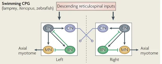
Figure 1. The locomotor central pattern generator (CPG) in swimming vertebrates.
The swimming CPG is composed of four functional classes of spinal neurons that are
connected in such a way that they generate a rhythmic patterned locomotor output. Excitatory glutamatergic interneurons (EINs, grey cells) are rhythmically active and excite all three other CPG neuron cell types during swimming. Commissural glycinergic interneurons (CINs, blue cells) project across the spinal cord to provide the mid-cycle inhibition needed for left-right coordination. Ipsilateral glycinergic interneurons (IINs, green cells) provide inhibition to the CPG that may limit motorneuron (MN, yellow cell) activation in order to increase the frequency of swimming. MNs are the output neuron of the spinal cord and excite adjacent muscle fibers to produce movement. Source: adapted from Goulding, Nature Reviews Neuroscience, 2009.
10
Excitatory CPG interneurons are rhythmically active and release glutamate to drive on-cycle rhythmic firing in downstream motorneurons (as well as other spinal interneurons) during swimming (Buchanan and Grillner, 1987; Buchanan et al., 1989; Li et al., 2006). Excitation of motoneurons is driven by the activation of both N-Methyl-D-aspartate (NMDA) and α-Amino-3-hydroxy-5-methyl-4-isoxazolepropionic acid (AMPA) glutamate receptors that produce slow and fast excitatory post-synaptic potentials (EPSPs) (Dale & Grillner, 1986; Li et al., 2003) and blocking these receptors prevents slow and fast swimming, respectively (Grillner et al., 1991). Excitatory premotor interneurons have descending axons so that the initial activation of rostral CPGs is conveyed along the spinal cord and locomotion proceeds in a rostral to caudal direction (Figure 1).
Additional neurons contribute to the coordination of the left and right halves of the spinal cord so that activity alternates between the two sides of the body to produce forward locomotion such as swimming or walking (Figure 1). A group of commissural inhibitory interneurons use glycine to provide mid-cycle inhibition to the contralateral spinal cord to ensure that bilateral muscles are contracting out of phase with each other (Buchanan, 1982; Dale, 1995). Finally, ipsilateral glycinergic interneurons provide local inhibition within the CPG including direct synapses onto motoneurons (Rovainen, 1974, 1982; Buchanan, 1982; Buchanan & Grillner, 1987). The organization of the CPG for both walking and swimming thus ensures two important features of locomotion: excitatory interneurons drive rhythm generation in the network via motoneurons while commissural inhibitory interneurons pattern left-right coordination.
Rhythm generation in the embryonic spinal CPG in some species is driven not by glutamate but instead by passive depolarization that spreads between cells via gap junctions. Gap junctions are channels composed of connexin proteins that allow the bidirectional passage of electrical currents as well as small signaling molecules such as cyclic AMP (reviewed by Pereda, 2014). Gap junctions appear to be prevalent in embyronic vertebrate motor circuits between many types of neurons including motorneurons and the premotor interneurons that are crucial for generating rhythmicity in the CPG (reviewed by Kiehn & Tresch, 2002). Furthermore, there is accumulating evidence to suggest that some CPG neurons, including motoneurons, may have instrinsic pacemaker properties that contribute to
11
the generation of rhythmic activity (Li, 2011). These findings suggest that rhythm generation can be produced by intrinsic oscillations and electrical coupling of neurons in the network prior to the development of chemical synapses and is supported by biologically realistic CPG modeling studies (Kepler et al, 1990; Loewenstein et al., 2001; Horn et al., 2012). It is thought that as chemical synaptogenesis proceeds the overall contribution of gap junctions decreases but they nonetheless continue to play a role in some aspects of mature nervous system function such as lateral inhibition or activity synchronization (Kiehn & Tresch, 2002).
I.2.2. Descending control and sensory feedback modulate locomotion
The locomotor CPG can be driven at the earliest stages by intrinsic pacemaker activity or by the application of pharmacological agents such as NMDA (Cohen & Wallen, 1980; Brodin et al., 1985) but once the network is more developed the CPG is activated to produce locomotion mainly by descending inputs from higher brain regions with additional
modulation from sensory feedback in the spinal cord (Figure 2). In the mature vertebrate nervous system, output neurons in subcortical pathways are linked via the basal ganglia to the tectum (superior colliculus), the mesencephalic (MLR) and diencephalic (DLR) locomotor command regions, or other brainstem motor centres where signals then activate reticulospinal neurons that will project to the spinal cord (Grillner et al., 2013). Studies from a number of species have identified descending excitatory inputs from the brainstem that activate certain CPG modules and thus produce a specific motor behavior (Buchanan & Grillner, 1987; Deliagina et al., 2002; Jordan, 1998; Kimura et al., 2013; Severi et al., 2014). For example, hindbrain reticulospinal Mauthner cells present in fish and amphibians send descending projections into the spinal cord that trigger fast escape behaviors (Korn & Faber, 2005).
Sensory pathways may also activate and modulate the spinal CPG directly through local sensory neurons and interneurons as shown by the simple touch reflex in xenopus (Li et al., 2003). The sense of light touch is mediated by sensory neurons located all over the body, therefore our skin can in fact be considered as the largest sense organ (Gallace & Spence, 2010). Sensory feedback from local pathways plays an important role to ensure continuing adaptation of ongoing locomotor behaviors in a changing environment (Rossignol et al., 2006).
12
Figure 2. Organization of the locomotor system in vertebrates.
Motor pathways in aquatic and terrestrial vertebrates share a similar neuroanatomical structure. Local control of muscle movements is regulated by pools of motor neurons in the spinal cord that are part of a dispersed locomotor central pattern generator (CPG) network. Spinal motor centres are modulated by proprioceptive sensory feedback through sensory afferents. Descending reticulospinal (RtS), rubrospinal (RbS) and vestibulospinal (VS) pathways control the locomotor network in the spinal cord, although the reticulospinal pathway is the primary pathway for initiating locomotion. The reticulospinal pathway can be activated by the mesencephalic locomotor region (MLR), which has inputs from the basal ganglia and the thalamus. The cerebellum coordinates motor behaviors by mediating sensory and internal feedback and optimizing the motor pattern to the task at hand. It also coordinates spinal motor actions via supraspinal motor pathways. Connections from the motor cortex refine and initiate motor actions (dotted arrow). The black arrows indicate direct command pathways, the grey arrows indicate feedback pathways. Source: adapted from Goulding, Nature Reviews Neuroscience, 2009.
13
Neuromodulatory inputs are also known to modify the function of CPG networks (Harris-Warrick, 2011). Evidence from both invertebrates and vertebrates suggest that
neuromodulators such as serotonin or dopamine may be essential for normal CPG function by either being connected within the circuit itself or by "priming" or modulating the network in a functional state, for example by regulating the excitability of CPG components (Katz, 1995).
Finally, in the developing embryo spinal or hindbrain neurons with intrinsic pacemaker properties may activate CPG networks prior to the maturation of sensory and neuromodulatory pathways (Broch et al., 2002; Tong & McDearmid, 2012) and there is evidence for continuing pacemaker activity in adults as well (Li, 2011; Smith et al., 2000). A complex combination of intrinsic and/or extrinsic neuromodulatory and synaptic interactions in addition to the pacemaker properties of neurons must therefore be considered when studying the generation of locomotion by spinal CPG networks.
I.2.3. Activity-dependent plasticity in spinal circuits
The function and dynamics of the network can also be modified as a result of plasticity mechanisms that alter the intrinsic properties of neurons or their synaptic connections. The best-studied type of plasticity is Hebbian plasticity, which consists of positive-feedback mechanisms that either strengthen strong synapses (via long-term potentiation, LTP) or weaken weak synapses (via long-term depression, LTD). LTP is thought to be a ubiquitous plasticity mechanism at excitatory synapses and particularly important in the experience-dependent refinement of neural circuits (reviewed by Malenka & Bear, 2004). Given the high degree of spontaneous activity in the developing zebrafish locomotor network, it is highly possible that Hebbian plasticity plays a role in fine-tuning spinal synapses during
development. In counterbalance to Hebbian plasticity, which operates over short timescales (minutes) and small spatial distances (< 10 µm), homeostatic plasticity is thought to integrate neuronal activity over hours and days to rebalance network activity via negative-feedback mechanisms that seek to maintain stability in the network (Turrigiano & Nelson, 2004). Homeostatic plasticity may include extrinsic mechanisms such as the multiplicative scaling up or down of synapse strengths (synaptic scaling) or the addition or removal of synapses and
14
intrinsic mechanisms such as the redistribution of ion channels within a neuron (reviewed by Turrigiano, 2012).
Homeostatic plasticity mechanisms are thought to be crucial for compensating for changes in network activity such as during development or disease (Turrigiano, 2008). These mechanisms could be particular important in sensory and motor circuits, which are important for survival behaviors. Decades ago, researchers had observed that the amplitude of EPSPs onto motoneurons scaled up following experimentally-induced decreases in activity in cats and rats with lesions or pharmacological disruptions of activity (Gallego et al., 1979; Manabe et al., 1989; Webb & Cope, 1992), but it was not clear how to interpret these findings in light of their apparent contradiction to the known principles of Hebbian plasticity. Since this time, the field of homeostatic plasticity has exploded and many other in vivo studies have
confirmed that these compensatory mechanisms are engaged at the cellular level following a reduction of activity in sensory (Deeg & Aizenman, 2011; Desai et al., 2002; Kaneko et al., 2008; Whiting et al., 2009) or motor pathways (Gonzales-Islas & Wenner, 2006; Wilhelm et al., 2009).
In order to show that these compensatory mechanisms are truly homeostatic it is crucial to also examine the consequences of this type of plasticity on the function of neural networks and the rhythmic behaviors they produce such as walking or swimming. For example, the escape swimming CPG network of the mollusc functionally recovers within a day following the lesion of commissural projections through the reversal of a key synaptic connection from inhibitory to excitatory (Sakurai & Katz, 2009). Furthermore, spontaneous activity in the chick embryo recovers just hours after chronic exposure to glutamatergic and cholinergic receptor antagonists (Chub & O'Donovan, 1998). The mechanism for this
recovery is thought to involve an upregulation of glycine/GABA signaling that could recover initial activity rates because these neurotransmitters are depolarizing in the embryo. Beyond these studies however, little work has been done to study the homeostatic regulation of locomotor circuits controlling behavior despite an abundance of pharmacological and genetic tools available with which to manipulate and measure network activity in an intact animal.
15
I.3. THE EMBRYONIC ZEBRAFISH AS A SIMPLE VERTEBRATE MODEL In order to understand the fundamental organization of the nervous system it is advantageous to study neural circuits in a model organism with relatively few neurons and thus a reduced complexity compared to higher vertebrates such as mammals. Over the past decades, neurobiology has benefited greatly from the use of simple vertebrate models such as
xenopus and lamprey. The recent availability of genetic tools in the zebrafish has made it a
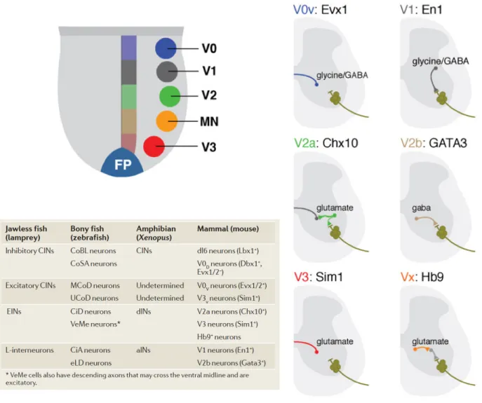
powerful and increasingly popular model organism for studying questions in modern neuroscience including the neural control of locomotion. Despite the obvious differences in movement between the swimming of aquatic vertebrates such as fish and the walking of terrestrial vertebrates such as humans, the primary functional classes of spinal neurons and thus the organization of the CPG and associated sensory and neuromodulatory circuits, appear to be highly conserved (Jessell, 2000; Goulding, 2009; Figure 3).
Studying the embryonic zebrafish offers several experimental advantages. The embryonic spinal cord consists of only eight classes of interneurons (INs) with only a few types of each in any somite (trunk segment) and a known, limited number of sensory and motoneurons (MNs) that assemble into simple circuits, simplifying the identification and description of individual neurons. The intrinsic and synaptic properties of spinal neurons and muscle fibres such as morphology, resting membrane potential and mEPSC amplitude and frequency are well-documented for comparative purposes (Nguyen et al., 1999, Ali et al., 2000a). In spite of their simplicity, embryonic spinal circuits are already functional as
evidenced by the presence of robust, stereotyped embryonic behaviors in zebrafish that begin within the first 24 hours of development (see below). We can therefore investigate the
function and plasticity of developing locomotor circuits in the zebrafish from synapse to behavior at stages in development that would be very difficult to study in prenatal mammals.
16
Figure 3. Functional neuron classes in the vertebrate spinal cord.
Upper left, distinct dorsoventral domains in the developing spinal cord give rise to motoneurons (MNs) and four cardinal interneuron populations (V0 - V3). Right, many of these interneuron subtypes can be identified based on their expression of postmitotic transcription factors, axonal morphology and neurotransmitter identity. Source: Grillner & Jessell, Current Opinion in Neurobiology, 2009. Lower table, the proposed homology of spinal neurons from different vertebrate species including zebrafish. Source: Goulding, Nature Reviews Neuroscience, 2009.
17
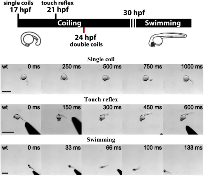
I.3.1. Timeline of locomotor behaviors in the developing zebrafish
The development of the zebrafish spinal cord CPG is rapid following fertilization (Figure 4). Beginning at 17 hours post-fertilization (hpf), the zebrafish embryo begins to produce slow, large amplitude 1 Hz spontaneous contractions of the trunk (Saint-Amant & Drapeau, 1998). This behavior, known as coiling, peaks in frequency at 19 hpf then rapidly declines to below 0.1 Hz by 26 hpf (Saint-Amant & Drapeau, 1998). By 21 hpf embryos develop a glutamate-dependent response to touch consisting of one to three alternating contractions of the tail (Saint-Amant & Drapeau, 1998; Downes and Granato, 2006; Pietri et al., 2009). Neither the response to touch nor spontaneous coiling depends on supraspinal inputs (Saint-Amant & Drapeau, 1998; Pietri et al., 2009).
From 29 hpf onward, embryonic zebrafish no longer produce coils but instead swim in response to touch or, to a lesser degree, spontaneously (Saint-Amant, 2006). Swimming behavior is driven by chemical glutamatergic and glycinergic neurotransmission and initially appears as a long burst of low-amplitude 10 Hz contractions (Kimmel et al., 1995) that develops following hatching into mature, 40 Hz “beat and glide” swimming by 4 dpf (Buss and Drapeau, 2001). Swimming activity recovers in the isolated spinal cord (Downes and Granato, 2006; Pietri et al., 2009) and can develop in the absence of descending inputs in the presence of NMDA (Chong and Drapeau, 2007), demonstrating that the neural circuitry within the spinal cord is sufficient to produce all of these earliest motor behaviors.
18
Single coil
Touch reflex
Swimming
Figure 4. Overview of embryonic zebrafish behaviors.
Upper figure, developmental timeline in hours post-fertilization (hpf) of embryonic zebrafish motor behaviors. Single coils are spontaneous contractions that appear at 17 hpf. The touch reflex appears at 21 hpf in response to somatosensory stimuli. Double coils are spontaneous, alternating contractions that appear at 24 hpf. Around 30 hpf, both spontaneous and touch-evoked coiling transitions to swimming behavior. Lower panels, time-lapse images of a spontaneous single coil (20 hpf embryo; scale bar, 1 mm), the touch reflex (26 hpf embryo; scale bar 500 µm) and swimming (48 hpf; scale bar, 1 mm). Source: lower panels adapted from Low et al., Journal of Neurophysiology, 2012.
19
I.3.2. Classification of spinal sensory and CPG neurons in the early embryo
A great deal of knowledge of the embryonic zebrafish spinal cord is built on work from previous anatomical and behavioral studies with the xenopus tadpole and lamprey (Figure 5). In particular, several key studies describing the morphology and order of appearance of the earliest zebrafish spinal neurons and their projections was invaluable for comparative studies of the organization of spinal circuits (Mendelson, 1986; Myers et al, 1986; Bernhardt et al., 1990; Hale et al., 2001; Downes et al., 2002).
Proposed functional classes were initially assigned based on the anatomical features of neurons including the dorsoventral position of their soma, dendritic morphology and axonal projection, as these features would determine to a large extent the synaptic inputs and outputs of the neurons. Electrophysiological recordings of cellular activity further refined these categories by providing the intrinsic and synaptic properties of different neuronal classes as well as the contribution of different cell types to particular behaviors (Liao & Fetcho, 2008). The embryonic spinal cord is a particularly tractable circuit not only because there are fewer neurons present at this time but also because at these early stages it is thought that neuron classes have a primitive function that has yet to undergo further diversification and specialization (Goulding et al., 2002; Goulding & Pfaff, 2005; Kimura et al., 2006).
Neurotransmitter identity is another key feature of neurons that defines their role within the network, with glutamate and glycine being the major chemical neurotransmitters driving behavior in the zebrafish, as in other lower vertebrates (Grillner et al., 1991). A thorough classification of the neurotransmitter properties of spinal neurons in zebrafish embryos and larvae has been an important tool for predicting the function of neuronal classes (Higashijima et al., 2004b). Finally, with the increasing availability of genetic tools many neuronal classes are now defined by a combinatorial “code” of transcription factors that is conserved across vertebrates and provides a means to target different neuron types for experimental manipulations (Jessell, 2000).
20
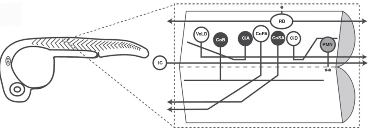
Figure 5. Neurons of the embryonic zebrafish spinal cord.
In the 26 hpf, fewer than twenty neurons with axons are present per segment of the spinal cord and they can be grouped in nine distinct cell types based on morphology. One spinal segment is shown in an open book configuration as if cut along the midline in a sagittal section. Glycinergic neurons are shown in black, cholinergic motoneurons in gray and glutamatergic neurons in white with the exception of the VeLD (which are GABAergic) and the IC cell (whose neurotransmitter identity is unknown). The asterisks denote points of exit from the spinal cord, * to the skin, ** to the ventral root. Please see the text for detailed descriptions of the cell types. Source: Saint-Amant, Progress in Brain Research, 2010.
21
As described above, the vertebrate locomotor CPG consists of four key components: motoneurons, descending excitatory interneurons and commissural and ascending inhibitory interneurons. In zebrafish embryos, three primary motoneurons are present per spinal
hemisegment that innervate the adjacent muscles of either the dorsal, medial or ventral trunk (Myers et al., 1986; Figure 5). In addition to their distinctive axonal morphology, primary motoneurons are identified by their large, ventral soma, cholinergic identity and the expression of transcription factors from the Islet and Mnx family (Seredick et al., 2012). Secondary motoneurons do not begin growing out their axons until 26 hpf and likely contribute to the subsequent maturation of swimming behaviors (Drapeau et al., 2002). Motoneurons and their innervation of muscle fibres at the neuromuscular junction has been a well-studied field. Thus the focus of my work in this thesis is on the sensory and CPG circuits that generate motor patterns rather than the output of the motoneurons themselves.
It should be noted that the earliest activation of the zebrafish spinal cord (coiling) precedes central chemical neurotransmission and is driven entirely by electrical activity (Saint-Amant & Drapeau, 2001). A rostral pacemaker neuron, the ipsilateral caudal (IC) cell, projects a descending axon that periodically excites large groups of ipsilateral neurons
through gap junctions, leading to the earliest motor behavior (Tong & McDearmid, 2012; Fig. 5). Just hours later, chemical neurotransmission has developed extensively and appears to shape the majority of activity in the spinal cord (Buss & Drapeau, 2001), although at some synapses electrical coupling persists in the larva and adult (Fetcho, 1992; Kimura et al., 2006). The presence of the purely electrical circuit is very transient and likely contributes to chemical synaptogenesis and the development of a mature locomotor network. In the
following sections, I will outline the major classes of glycinergic and glutamatergic interneurons that have been shown to play a role in the earliest embryonic behaviors.
I.3.3. Glycinergic spinal interneurons
The earliest chemical synapses to form in the zebrafish spinal cord are glycinergic, starting at 20 hpf, followed a few hours later by glutamatergic synapses (Saint-Amant & Drapeau, 2000). The role of glycinergic interneurons in embryonic locomotor circuits is complicated by the fact that both GABA and glycine are depolarizing in the vertebrate
22
nervous system at early stages (Ben-Ari, 2002). Glycinergic inputs are depolarizing at all developmental stages of zebrafish discussed in this thesis (< 5 dpf) and the exact timing of the transition to a mature, hyperpolarizing response is not known. Depolarization occurs because the intracellular chloride concentration is high in immature neurons therefore the opening of GABA and glycine receptors causes chloride ions to flow out and depolarize the cell.
However, depolarizing GABA and glycine can nonetheless be either excitatory or inhibitory depending on the resting membrane potential and threshold for action potential firing (Jean-Xavier et al., 2007). It has been postulated that depolarizing glycinergic signaling in early development provides an advantage in that it provides low levels of excitation in an immature circuit that activate voltage-gated calcium channels to promote maturation without causing overexcitation (Ben-Ari, 2002).
The population of neurons responsible for the earliest glycinergic activity in the zebrafish spinal cord is currently unknown but a recent study has identified glycine
immunoreactive neurons in the hindbrain and rostral somite of the spinal cord at 20 hpf whose appearance coincides with the arrival of glycinergic inputs to spinal neurons (Moly et al., 2014). Indeed, lesion experiments suggest the presence of descending glycinergic inputs from the rostral spinal cord that synapse onto motoneurons and other spinal neurons at these early stages (Saint-Amant & Drapeau, 2000; Pietri et al., 2009). Much more is known about the identity of glycinergic spinal neurons in the later, larval zebrafish and their activity during locomotor behaviors.
The main classes of commissural glycinergic interneurons in the embryonic spinal cord are commissural secondary ascending (CoSA) and commissural bifurcating longitudinal (CoBL) interneurons (Bernhardt et al., 1990; Fig. 5). These neurons have extended axons by 24 hpf and express the transcription factor evx1 (Suster et al., 2009), although the CoSAs in fact appear to be a heterogeneous class of neurons that may include both glycinergic and glutamatergic neurons (Higashijima et al., 2004b). There is evidence to suggest that
glutamatergic CoSAs appear first, followed by glycinergic CoSAs at later stages (> 36 hpf) (Satou et al., 2012), therefore interpreting the role of this class of neurons is aided by specific markers of neurotransmitter identity. Studies of the activity of glycinergic spinal zebrafish neurons during larval behaviors using the expression of GFP driven by the glycine transporter