OATAO is an open access repository that collects the work of Toulouse
researchers and makes it freely available over the web where possible
Any correspondence concerning this service should be sent
to the repository administrator: tech-oatao@listes-diff.inp-toulouse.fr
This is an author’s version published in:
http://oatao.univ-toulouse.fr/21138
To cite this version:
Khebizi, Noura and Boudjella, Hadjira and Bijani, Christian and Bouras, Noureddine
and Klenk, Hans-Peter and Pont, Frédéric and Mathieu, Florence
and Sabaou,
Nasserdine Oligomycins A and E, major bioactive secondary metabolites produced by
Streptomyces sp. strain HG29 isolated from a Saharan soil. (2018) Journal de
Mycologie Médicale / Journal of Medical Mycology, 28 (1). 150-160. ISSN 1156-5233
Official URL:
https://doi.org/10.1016/j.mycmed.2017.10.007
Oligomycins A and E, major bioactive
secondary metabolites produced by
Streptomyces sp. strain HG29 isolated from
a Saharan soil
N. Khebizi
a, H. Boudjella
a
,
*
, C. Bijani
b
, N. Bouras
a
,
c,
H.-P. Klenk
d, F. Pont
e, F. Mathieu
t, N. Sabaou
aa Laboratoire de biologie des systèmes microbiens (LBSM), école normale supérieure de Kouba, Alger, Algeria b Laboratoire de chimie de coordination (LCC), CNRS, université de Toulouse, UPS, /NPT, LCC, 205, route de Narbonne, 31077 Toulouse, France
c
Département de biologie, faculté des sciences de la nature et de la vie et sciences de la terre, université de Ghardaïa, BP 455, 47000 Ghardaïa, Algeria
d School of biology, Newcastle university, Ridley building, NE1 7RU Newcastle upon Tyne, United Kingdom e Proteomics group, lnserm UMR1037, centre de recherches en cancérologie de Toulouse (CRCT), Toulouse, France
t Laboratoire de génie chimique, CNRS, INPT, UPS, université de Toulouse, Toulouse, France
KEYWORDS
Streptomyces;Taxonomy;
Antifungal activity;
Oligomycins;
Minimal inhibitory
concentrations
Summary
An actinobacterial strain, HG29, with potent activity against pathogenic, toxigenic
and phytopathogenic fungi was isolated from a Saharan soil sample of Algeria. On the basis of
morphological and chemotaxonomic characteristics, the strain was classified in the genus
Streptomyces.
Analysis of the
16S rRNAgene sequence showed a similarity level of 99.3% with
Streptomyces gancidicusNBRC 15412
T. The comparison of its cultural and physiological cha
racteristics with this species revealed significant differences. Moreover, the phylogenetic tree
showed that strain HG29 forms a distinct phyletic line within the genus
Streptomyces.Production
of antifungal activity was investigated by following kinetics in shake broth. The highest
antifungal activity was obtained after five days of fermentation, and in the dichloromethane
extract. Two active compounds, NK1 and NK2, were purified by HPLC using a C18 column. Their
chemical structures were identified through nuclear magnetic resonance experiments and mass
spectrometry as oligomycins E and A, respectively, which have not been reported to be produced
• Corresponding author.
E-mail address:
h-boudjella@live.fr
(H. Boudjella).
by S. gancidicus. The two bioactive compounds exhibited significant antifungal activity in vitro
showing minimal inhibitory concentrations (MICs) values between 2 and 75 µg/mL.
Introduction
Filamentous fungi cause a wide range of infectious diseases in humans, animais and plants [1,2]. ln addition, their ability to produce mycotoxins such as aflatoxins, ochratoxins and patulin can be harmful to humans and animais at certain concentrations after ingestion of contaminated food and feed [3]. Current antifungal compounds are limited in their ability to treat infections due to the increase of the resis tance in fungi and the toxicity of a wide range of antifungal molecules in use such as polyenes [4,5]. This phenomenon motivates the search for new bioactive compounds with strong activity against pathogenic fungi. ln order to find new solutions, several approaches were developed, includ ing the search for new antifungal compounds from micro organisms [6]. The actinobacteria are known as the most attractive source of several types of bioactive metabolites, especially members of the genus Streptomyces, which pro duce over two-thirds (70%) of the clinically useful antibiotics of natural origin [7]. The Algerian Saharan soils are rich and diversified in actinobacteria with interesting antimicrobial properties [8-13].
ln this paper, we describe the taxonomy of an actinobac terial strain isolated from a Saharan soil sample, during a primary screening program to search for antimicrobial compounds. The strain, designated HG29, showed an inter esting antifungal producing potential active against several pathogenic and toxigenic species of filamentous fungi. Two active compounds were purified, and their structures and activities were determined.
Materials and methods
Strain isolation and maintenance
The actinobacterial strain HG29 was isolated from a Saharan soil sample collected from Hoggar, Tamanrasset (Southern Algeria, 22°491N, 5°25'E), by serial dilution agar plating
method using chitin-vitamin B agar [14], supplemented with actidione (80 µg/ml). Pure culture of the strain was main tained at 4 °C on ISP4 medium slants.
Taxonomie studies of HG29 strain
The morphological characteristics were investigated using the media of the International Streptomyces Project (ISP2, ISP3 and ISP4) [15], Bennett medium and nutrient agar [16] after 7, 14 and 21 days of incubation at 30 °C. The mycelia organiza
tion and sporulation were observed by light microscopy. For the chemotaxonomic analysis, biomass was obtained from cultures grown in shake flasks (250 rpm) using ISP2 medium. After 5 days of incubation at 30 °c, cells of strain
HG29 were harvested by centrifugation, washed twice with
distilled water and hydrolyzed for diaminopimelic acid iso mers [ 17], whole-cell sugar pattern [ 1 8] and phosphol i pids analyses [19].
For the physiological characteristics, 79 tests were per formed according to the methods of Locci [20]. They concer ned the production of melanoid pigments on ISP6 and ISP7 media and nitrate reductase, the assimilation of 26 carbohy drates and derivatives as sole carbon source, the utilization of 9 amino acids as sole nitrogen sources, the degradation of 7 organic acids on ISP9 medium, hydrolysis of 11 organic compounds (adenine, guanine, xanthine, casein, Tween 80, gelatin, starch, cellulose, esculin, arbutin and urea), the sensitivity to 5 different inhibitory compounds including violet crystal (0.05%), sodium azide (0.02%), potassium tellurite (0.01%), phenol (0.1%) and lysozyme (0.005%), the growth in the presence of different concentrations of NaCl (0 2 3 5 7, 9 and 10% w/v), at different temperatures (25, 30'. 35,
'..m'.
45 and 50 °C) and various pH values (3, 5, 7, 9 and 10). Ail thetests were performed at 30 °C (except for temperature tests)
and observations were recorded at 7, 14 and 21 days. For molecular analysis, the DNA of the strain HG29 was extracted with a DNA extraction kit (MasterPure ™ Gram Positive DNA Purification Kit, Epicentre® Biotechnologies,
Madison, WI). The 16S rRNA gene was amplified by PCR using two primers, 10-30F (5'-GAGTTTGATC-CTGGCTCA-3') and 1500R (5'-AGAAAGGAGGTGATCCAGCC-3'), as described by Rainey et al. [21].
Amplification was carried out in a 50 µL reaction volume containing 1.5 U of AmpliTaq Gold Taq polymerase (Applied Biosystems, Foster City, CA), dNTPs (0.25 mM each), 1 µM of each primer, and 100 ng of genomic DNA. Reaction condi tions were: 97 °C for 4 min, followed by 35 cycles of 97 °c for
45 s, 52 °C for 45 s, and 72 °C for 45 s, with a final elongation
step at 72 °C for 10 min. The amplified products were visua lized on a 0.8% (w/v) agarose gel stained with ethidium bromide. PCR products were purified with a purification kit (Qiagen, Hilden, Germany). The primers used for sequencing are listed in Coenye et al. [22].
For the 16S rRNA gene phylogenetic analyses, the sequence obtained was compared for similarity with sequen ces present in the EzTaxon-e server (http://www.eztaxon-e. ezbiocloud.net/; [23]). The sequences were aligned with reference sequences of representatives of the genus Strep
tomyces retrieved from the GenBank database. Phylogenetic
analyses were conducted using Molecular Evolution Genetics Analysis (MEGA) software version 5.0 [24]. Phylogenetic trees were constructed using the neighbor-joining method of Saitou and Nei [25]. The resulting topologies of trees were evaluated by bootstrap analyses [26] on 1000 replicates.
Primary screening for antimicrobial activity
Antimicrobial activity of the strain HG29 against fungi and bacteria was detected using the agar cross streak method.
The actinobacterium was streaked as a dense straight line on ISP2 medium plates and incubated at 30 °C for 10 days. After good growth, target strains were streaked at a right angle to HG29 strain, and then incubated at 30 °C for 48 h. The antimicrobial activity was evaluated by measuring the dis tance of inhibition between the target-microorganisms and the HG29 strain colony margin. Tests were conducted in triplicate.
The target-fungi tested were mostly pathogenic or toxigenic for humans, or phytopathogenic strains. They included twenty filamentous fungi (Aspergillus carbonarius M333, A. niger OT304, A. parasiticus CBS 100926,
A. westerdijkiae NRRL 3174, A. nidulans KE202, A. terreus
CT290, A. fumigatus CF140, A. flavus NRRL 3251, Fusarium
culmorum, F. equiseti, F. proliferatum, F. graminearum
Fg3, F. sporotrichioides, F. moniliforme, F. oxysporum f. sp. lini, Fusarium oxysporum f. sp. albedinis, Penicillium
expansum, P. glabrum, Botrytis cinerea and Umbelopsis ramanniana NRRL 1829) and five strains of Candida albicans
(M1, M2, M3, IPA200 and IPA988). ln addition, four strains of bacteria (Bacillus subtilis ATCC 6633, Staphylococcus aureus ATCC 25923, Klebsiella pneumoniae E40 and Pseudomonas
aeruginosa ATCC 27853) were tested. The strains without
accession number resulted from our laboratory collection.
Time course of antifungal activity production
and growth
Fermentations were carried out in liquid medium containing glucose 1%, peptone 0.5% and MgCI2 0.1% [27]. The seed
culture was prepared using a spore suspension in sterile distilled water with a final optical density (OD600) of 0.1, and from a fresh culture of HG29 strain grown on ISP4 medium at 30 °C for 10 days. One milliliter of the spore suspension was inoculated into Erlenmeyer flasks (250 ml) containing 50 ml of the seed medium consisting of (g/L): 4 beef extract; 1 yeast extract; 4 peptone; 2.5 NaCI and 10 glucose, pH 7.2 [27]. The Erlenmeyer flasks were prepared and incubated on a shaker (250 rpm) at 30 °C for 48 h. Aliquots (5%, v/v) of this seed culture were transferred into 100 ml of the fermentation medium in 500-mL Erlenmeyer flasks. The cultures were incubated on a rotary shaker (250 rpm) at 30 °C for 10 days. The antifungal activity pro duction, the pH and the dry cell weight (DCW) were measu red every 24 h.
The antifungal activity was evaluated by agar well diffu sion method against Aspergillus carbonari us M333. The spore suspension was prepared in sterile distilled water and adjus ted as inoculum to a final concentration of 1 to 2 x 10⁶ CFU/
ml. An amount of 20 ml of Sabouraud agar (10 g/L agar) was inoculated with 15 µL of spore suspension and then poured into a Petri dish. The plates were kept at room temperature for 30 min to allow the medium to solidify. Four wells of 10 mm diameter were aseptically bored into the culture medium, and 200 µL of each culture supernatant were loa ded in each well. The plates were incubated at 30 °C for 48 h after a diffusion process of 2 h at 4 °C. Diameters of the inhibition zones around the wells were measured. The results were expressed as mean
±
standard deviation (SD) from triplicate experiments. Growth was studied by determining the DCW using the method of Bou ras et al. [28].Extraction and purification of antifungal
compounds
For the production of antibiotics, repeated fermentations were carried out to obtain a total of 4.0 L of culture broth. Extraction of antifungal compounds from the culture filtrate was carried out on the day of optimal antifungal activity production. The culture broth was centrifuged at 5000 g for 15 min. The culture filtrate obtained was extracted with dichloromethane solvent (1 :1 v/v). The organic phase was concentrated with a rotary evaporator at 40 °C. The dry crude extract was dissolved in methanol and bioassayed against A. carbonarius M333 by the paper disk diffusion method. Each paper disk (6 mm diameter) was impregnated with 40 µL of the crude extract. Ali paper disks prepared were then air dried, sterilized under UV and then placed on the surface of Sabouraud agar, previously inoculated with
A. carbonarius M333 as the target-fungus. Then, the plates
were kept at 4 °C for 2 h to allow the diffusion of antifungal substances in the medium. Afterwards, the plates were incubated at 30 °C for 48 h. The antifungal activity was determined by measuring the diameters of inhibition zones around each paper disk.
The purification and the analysis of the crude extract were carried out by HPLC (Agilent Technologies). The equip ment consisted of a variable wavelength detector (UV-Vis G1315D detector array), a pump system G1311C, and an auto-sampler G1329B. Data were acquired using Agilent ChemStation (Rev. B 04.03) software. The stationary phase consisted of a C18 reverse-phase column (Zorbax SB, 5 µm, 9.6 x 250 mm). The samples were analyzed by continuous linear gradient solvent system from 40 to 100% methanol in water during 25 min, using a flow rate of 1 ml/min and ultra violet detection at 220 nm. Fractions were collected sepa rately, concentrated and bioassayed against A. carbonarius M333 by the paper disk diffusion method.
Spectroscopie and spectrometric analyses
The UV-visible of the active compounds was determined in methanol solution with a Perkin-Elmer Lambda 20 UV /Vis spectrophotometer. The mass spectrum was recorded with a LCQ ion-trap mass spectrometer (Finnigan MAT, San Jose, California, USA) equipped with a nanospray ion electro-spray ionization ESI source (positive and negative ion mode). 1H
and 13C NMR spectroscopy were used for the characterization
of the active molecules. NMR sample was prepared by dis solving 2 mg of each purified compound in 600 µL of MeOD. Ali spectra were recorded on a Bruker Avance 500 spectro meter equipped with a 5 mm triple resonance inverse z gradient probe (TBI 1H, 31P, BB). Ali chemical shifts for 1H
and
ne
are relative to TMS using 1 H (residual) orne
chemicalshifts of the solvent as a secondary standard. The tempera ture was set at 298 K. Ali the 1 H and 13C signais were assigned
on the basis of chemical shifts, spin-spin coupling constants, splitting patterns and signal intensities, and by using 1H-1H
COSY45, 1H-13C HSQC, 1H-13C HMBC and nc_1H experi
ments. Gradient-enhanced 1H COSY45 was realized included
24 scans per increment. 1H-13C correlation spectra using a
gradient-enhanced HSQC sequence (delay was optimized for
Gradient-enhanced HMBC experiment was performed allow ing 62. 5 ms for long-range coupling evolution (340 scans were accumulated). Typically, 2048 t2 data points were collected for 320 t1 increments.
Minimal inhibitory concentrations
The minimal inhibitory concentrations (MICs) were determi ned by the conventional agar dilution method [29]. The target-microorganisms included fifteen filamentous fungi (Aspergillus carbonarius M333, A. niger OT304,
A. parasiticus CBS 100926, A. westerdijkiae NRRL 3174, A. nidulans KE202, A. terreus CT290, A. fumigatus CF140, A. flavus NRRL 3251, Fusarium culmorum, F. equiseti, F. proliferatum, F. oxysporum f. sp. lini, Penicillium expan sum, P. glabrum and Umbelopsis ramanniana NRRL 1829),
one yeast (Candida albicans C200) and four bacteria (Bacillus
subtilis ATCC 6633, Staphylococcus aureus ATCC 25923, Klebsiella pneumoniae E40 and Pseudomonas aeruginosa
ATCC 27853). Suspensions of target-microorganisms were prepared in sterile distilled water with an inoculum size of 1 x 10⁶ to 3 x 10⁶ CFU/ml for fungi and 108 CFU/ml for
bacteria. The stock solutions were prepared by dissolving 3 mg of each antifungal powder in 3 ml of methanol. The agar media (Mueller Hinton for bacteria and Sabouraud for fungi) were supplemented with different concentrations of the antifungal compound (0.5, 1, 2, 3, 4, 5, 6, 7, 8, 10, 20, 30, 40, 50, 75 and 100 µg/ml) and then poured into Petri dishes. Two standard antifungal drugs, amphotericin B (AMB) and itraconazole (ITR) were used as the positive controls. A negative control was included in the test by inoculating the target-microorganisms onto the media without the anti fungal compounds. The agar surface of the plates containing a concentration of antifungal agent and the control plates were spot inoculated with 1 µL of the suspension of each target-microorganism. The plates were incubated for 24- 48 h at 37 °C for bacteria and 48-72 h at 28 °C for fungi. Tests were performed in duplicate. The MICs were determi ned as the lowest concentration of the antifungal compound
that inhibited the growth of the target-microorganism, as determined by the lack of visual growth compared to the negative control.
Results and discussion
Taxonomy
Good growth of strain HG29 was observed on ISP2, ISP4 and Bennett media. The growth was moderate on ISP3 and weak on nutrient agar medium. At maturity, the aerial mycelium was dark greenish blue and produced spirals chains with 3-8 coils, each chain borne by a short sporophore and containing 10-50 spores (ESM Fig. 1 ). The substrate mycelium was pale beige and non-fragmented, producing a brownish soluble pigment on ISP2 and Bennett media.
Strain HG29 contained LL-diaminopimelic acid isomer and glycine. Whole-cell hydrolysates contained galactose, glu cose, ribose and traces of mannose, which are typical of cell wall type IC [18]. Diagnostic phospholipid detected was phosphatidylethanolamine corresponding to type PII phos pholipids [30]. Based on the chemotaxonomic and morpho logical properties, strain HG29 was identified as a member of the genus Streptomyces [31].
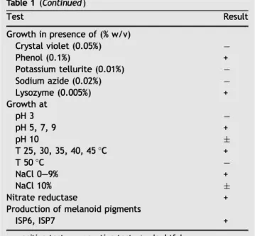
The physiological characteristics of strain HG29 are given in Table 1. The strain produced melanoid pigments on the ISP6 and ISP7 media and nitrate reductase. Growth occurred at temperatures between 25 and 45 °C, at pH 5, 7 and 9 and at a sait concentration of 9% NaCl (w/v). lt can utilize ail major organic compounds tested except dulcitol, erythritol, melezitose, sorbose, trehalose, glycine, histidine, trypto phane, valine, adenine, cellulose, guanine, benzoate and tartrate. lt was unable to grow in the presence of crystal violet (0.05%), potassium tellurite (0.01%) and sodium azide (0.02%).
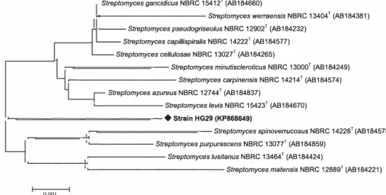
The phylogenetic analysis using the 16S rRNA gene sequence (1406 nucleotides) confirmed that HG29 strain belonged to the genus Streptomyces. The sequence was deposited in GenBank under the accession number
Streptomyces gancidicus NBRC 15412 T (AB 184660)
.+---Streptomyces werraensis NBRC 13404T (AB184381)
Streptomyces pseudogriseolus NBRC 12902T (AB184232)
Streptomyces capil/ispira/is NBRC 14222T (AB184577)
Streptomyces cellulosae NBRC 13027T (AB184265)
�---Streptomyces minutiscleroticus NBRC 13000T (AB184249)
�---Streptomyces carpinensis NBRC 14214T (AB184574)
Streptomyces azureus NBRC 12744T (AB184837)
'---Streptomyces levis NBRC 15423T (AB184670) L....---
♦
Strain HG29 (KP868649)�---Streptomyces spinoverrucosus NBRC 14228T (AB184578) �---< '---Streptomyces purpurascens NBRC 13077T (AB184859) �---Streptomyces lusitanus NBRC 13464T (AB184424) �---<
'---Streptomyces matensis NBRC 12889T (AB184221) 0.001
Figure 1 Phylogenetic tree derived from nearly complete 16S rRNA gene sequences showing relationships between the strain HG29 and the related type species of the genus Streptomyces. The tree was constructed using the neighbor-joining method. Bootstrap values are indicated at nodes (2: 50%). Bar 0.001 substitutions per nucleotide position.
Table 1 Physiological characteristics of strain HG29. Test Hydrolysis of Adenine Arbutin Casein Cellulose Esculin Guanine Starch Tween 80 Gelatin Urea Xanthine
Carbone source utilization (1% w/v) Adonitol Arabinose Cellobiose Dextrin Dulcitol Erythritol Fructose Fucose Galactose Glucose Glycerol lnositol Lactose Maltose Mannitol Mannose Melezitose Melibiose Raffinose Rhamnose Ribose Sucrose Sorbitol Sorbose Trehalose Xylose
Nitrogen source utilization (0.1% w/v) L-Alanine L-Asparagine Glycine L-Histidine L-Proline L-Serine L-Threonine L-Tryptophane L-Valine
Decarboxylation of sodium salts Acetate Benzoate Citrate Oxalate Propionate Pyruvate Tartrate Result
±
+ + + + + + + + + + + + + + + + + + + + + + + + + + + + + + + + + + + + + + Table 1 (Continued) TestGrowth in presence of(% w/v) Crystal violet (0.05%) Phenol (0.1%) Potassium tellurite (0.01%) Sodium azide (0.02%) Lysozyme (0.005%) Growth at pH 3 pH 5, 7, 9 pH 10 T 25, 30, 35, 40, 45 °C T 50 °C NaCl 0-9% NaCl 10% Nitrate reductase
Production of melanoid pigments ISP6, ISP7
+: positive test; -: negative test; ±: doubtful.
Result + + +
±
+ +±
+ +Table 2 Antimicrobial activity of Streptomyces sp. HG29 by the agar cross streak method.
Target-fungi Aspergillus carbonarius M333 A. niger OT304 A. parasiticus CBS 100926 A. westerdijkiae NRRL 3174 A. nidulans KE202 A. terreus CT290 A. fumigatus CF140 A. flavus NRRL 3251 Fusarium equiseti F. moniliforme F. sporotrichioides F. cu/morum FC200
Fusarium oxysporum f. sp. a/bedinis
F. oxysporum f. sp. lini F. graminearum Fg3 F. proliferatum Penicillium glabrum P. expansum Botrytis cinerea Umbelopsis ramanniana NRRL 1829
Candida a/bicans IPA200
C. albicans IPA988 C. a/bicans M1 C. a/bicans M2 C. albicans M3
Bacil/us subtilis ATCC 6633
Staphy/ococcus aureus ATCC 25923
Klebsiel/a pneumoniae E40
Pseudomonas aeruginosa ATCC 27853
Distance of inhibition (mm) 39.0 ± 1.0 37.3 ±0.6 22.5 ± 0.5 18.3 ± 0.6 5.7
±
1.1 4.5 ± 0.5 3.3±
0.6 2.1±
0.8 34.0 ± 1.0 32.6 ±0.6 27.7 ± 0.6 26.0 ± 1.0 26.8 ±0.8 25.0 ± 1.0 20.0 ± 1.0 20.3 ±0.6 27.6 ±0.6 27.0±1.0 27.0±1.0 4.8±
1.2 3.8 ± 0.8 3.0 ± 1.0 2.8 ± 0.8 5.2±
0.8 6.2 ± 1.2 20.0 ± 1.0 13.5±
1.3 0.0 0.0 Values are mean ± SD of three independent experiments.KP868649. The highest similarity Level was 99.3% with Strep·
tomyces gancidicus NBRC 15412T. The phylogenetic tree
illustrated in Fig. 1 showed that the HG29 strain forms a distinct phylogenetic position with closely related species of the genus Streptomyces. However, our strain could be dis· tinguished from S. gancidicus by some phenotypic properties such as the color of aerial mycelium (gray for S. gancidicus), the production of melanoid pigments on ISP6 and ISP7 media, the utilization of cellobiose, raffinose, sucrose and trypto· phane, the decarboxylation of sodium citrate and growth in presence of lysozyme [32].
Primary screening of antimicrobial activity
The results of the screening for antimicrobial activity of strain HG29 are displayed in Table 2. Streptomyces sp. HG29 showed an antifungal activity against ail the fungi tested. The antifungal activity was significantly strong (> 30 mm) against Aspergillus carbonarius M333, A. niger OT304, Fusarium equiseti and F. moniliforme; strong to moderate (21-30 mm) against F. sporotrichioides, Penicil·
lium glabrum, P. expansum, Botrytis cinerea, A. parasiticus
CBS 100926, F. culmorum FC200, F. oxysporum f. sp.
albedinis and F. oxysporum f. sp. lini, and moderate
(10-20 mm) against A. westerdijkiae, F. proliferatum and
F. graminearum. However, a weak antifungal activity was
detected (< 10 mm) against A. nidulans KE202, A. terreus CT290, A. fumigatus CF140, A. flavus NRRL 3251, Umbelopsis
ramanniana NRRL 1829 and all tested yeasts.
The antibacterial activity was moderate against Bacillus
subtilis ATCC 6633 and Staphylococcus aureus ATCC 25923,
but no activity was found against f. coli ATCC 10536 and
Klebsiella pneumoniae E40.
Consequently, A. carbonarius M333, the most sensitive strain, was selected as the indicator fungus for determining the antifungal activity in shake culture media.
Time course of antifungal activity production
and growth
The kinetics of antibiotic production, growth and pH were investigated in shake medium, as shown on Fig. 2. The antifungal activities were detected on the first day of fer• mentation against A. carbonarius M333, reaching a maximum the 5th day. The antifungal activity production was found to be correlated with the cell growth. The pH decreased to an acid state until the 3rd day and then increased gradually and reached its maximum (pH 8.2) on the 10th day of fermentation.
Isolation and purification of antifungal
compounds
Streptomyces sp. HG29 was grown for five days. The anti·
fungal substances were extracted with dichloromethane from the culture filtrate. The active dichloromethane extract was subjected to HPLC analysis to determine the antifungal metabolite profiles. The HPLC profile showed two major peaks active against A. carbonarius M333, and were designed as NK1 (retention time (RT) = 14.99 min) and NK2 (RT = 15. 77 min). These two peaks were collected and re· injected into the HPLC system until total purification was achieved (ESM Fig. 2). The pure bioactive compounds had a light yellowish color.
:z: 8
i
...
•····:J'••···�···••j "- ···-l'···E.. 'I. _ ..-···•!
7 ···�···�···r···� Ê E 20 18 ; 16,;
·t
], 14 C: C: < 12 +---'----'---'----'---'----'---'---'--...l----1-5,0 4,5 4,0 3,5 3,0 2,5 � � 2,0g
1,5 1,0 0,5 +---r---ir---r---...,...--..----r--...,...--�-�---1-0,0 0 2 3 4 5 6 7 8 9 10 Time (days)Figure 2 Time course of pH, growth (dashed curve) and antifungal activity production (solid curve) against Aspergillus carbonarius M333. Values include the diameter of the well (10 mm). Each measure represents average ± standard deviation from three replicates per treatment.
Spectroscopie studies
The UV-visible spectra of the pure products NK1 and NK2 exhibited Âmax at 240 nm (NK1 ), and 232 nm (NK2) in MeOH.
The mass spectra were obtained in positive and negative mode. The molecular weights of the active compounds are M
=
820 (NK1) and M=
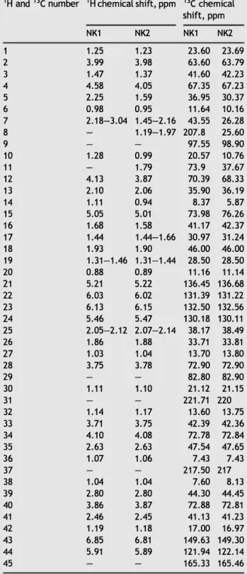
790 (NK2).Table 3 1H and ne NMR data assignments of NK1 and NK2
compounds in CD3CN at 298 K (See Figs. 3 and 4 for numbering
of hydrogen and carbon atoms).
1 H and ne number 1 H chemical shift, ppm ne chemical
shift, ppm 1 2 3 4 5 6 7 8 9 10 11 12 13 14 15 16 17 18 19 20 21 22 23 24 25 26 27 28 29 30 31 32 33 34 35 36 37 38 39 40 41 42 43 44 45 NK1 1.25 3.99 1.47 4.58 2.25 0.98 2.18-3.04 1.28 4.13 2.10 1.11 5.05 1.68 1.44 1.93 1.31-1.46 0.88 5.21 6.03 6.13 5.46 2.05-2.12 1.86 1.03 3.75 1.11 1.14 3.71 4.10 2.63 1.07 1.04 2.80 3.86 2.46 1.19 6.85 5.91 NK2 1.23 3.98 1.37 4.05 1.59 0.95 1.45-2.16 1.19-1.97 0.99 1.79 3.87 2.06 0.94 5.01 1.58 1.44-1.66 1.90 1.31-1.44 0.89 5.22 6.02 6.15 5.47 2.07-2.14 1.88 1.04 3.78 1.10 1.17 3.75 4.08 2.63 1.06 1.04 2.80 3.87 2.45 1.18 6.81 5.89 NK1 23.60 63.60 41.60 67.35 36.95 11.64 43.55 207.8 97.55 20.57 73.9 70.39 35.90 8.37 73.98 41.17 30.97 46.00 28.50 11.16 136.45 131.39 132.50 130.18 38.17 33.71 13.70 72.90 82.80 21.12 221.71 13.60 42.39 72.78 47.54 7.43 217.50 7.60 44.30 72.88 41.13 17.00 149.63 121.94 165.33 NK2 23.69 63.79 42.23 67.23 30.37 10.16 26.28 25.60 98.90 10.76 37.67 68.33 36.19 5.87 76.26 42.37 31.24 46.00 28.50 11.14 136.68 131.22 132.56 130.11 38.49 33.81 13.80 72.90 82.90 21.15 220 13.75 42.36 72.84 47.65 7.43 217 8.13 44.45 72.81 41.23 16.97 149.30 122.14 165.46
The 1 H and ne chemical shifts of NK1 and NK2 compounds
are given in Table 3 and structures in Figs. 3 and 4. The 13e, HSQe and HMBe spectra show 45 carbon signais for NK1 and NK2 molecules. lt was possible to discern 3 ketone groups (8c
207.8 to 221.7), 6 hydroxyl group (8c 63.6 to 82.8), 1 ester
function (8c 165.33), 6 sp2-hybridized carbons (8c from 121. 9
to 146.63) and 25 sp3-hybridized carbons (8
c 7.4 to 47.5) for
molecule NK1, 2 ketone groups (8c 217 .0 and 220.0), 1 ester
function (8c 165.46) and 5 hydroxyl group (8c 63.79 to 82.90),
6 sp2-hybridized carbons (8
c from 122.1 to 149.3) and 27 sp3-
hybridized carbons (8c 7.4 to 47.7) for compound NK2. The
hydrogens of the hydroxyl group are not observed due to rapid exchange with MeOD. The 2D 1 H-1 H permitted the
establishment of connectivity between the groups of the NK1 and NK2 molecules.
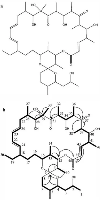
a
b
20 6 - 1H COSY correlation--IH-13C HMBC con-clation
Figure 3 Structure of bioactive compound NK1 (a) and HMBe and COSY correlations (b).
42
20
6
Figure 4 Structure of bioactive compound NK2 (a) and HMBC and COSY correlations (b).
The structures of the NK1 and NK2 compounds were determined by NMR and mass spectrometry to be oligomycin E [33] and oligomycin A [34], respectively.
Oligomycins are a subfamily of the macrolides class. The first oligomycin was described as a new antifungal antibiotic produced by Streptomyces diastatochromogenes [35]. Later, nine isomers named A, B, C, D, E, F, G, H and I were identified. They refer to a closely related molecular species whose basic structure consists of a macrolide ring of ketide units [36]. The oligomycins A and E differ in two side sub stituents [33,34] linked to the carbon in positions 8 and 11, as shown on Figs. 3 and 4. Oligomycin A is a highly specific inhibitor of mitochondrial ATP synthase that induces apop tosis in various cell types [37]. The oligomycin antibiotics are also produced by other species of Streptomyces including
S. bottropensis [38], S. ostreogriseus [39], S. libani [40],
S. griseolus [41], S. avermitilis [42] and S. diastaticus [43]. However, their isolation has so far not been reported from
S. gancidicus. This species produced the gancidin (molecular
weight 210), a non-ribosomal peptide with antitumoral, antifungal and antibacterial properties [44,45]. The search for a possible phylogenetic relationship between the strain HG29 and the strains of oligomycins-producing Streptomyces was realized by the analysis and comparison of 16S rRNA gene sequences. The constructed phylogenetic tree (ESM Fig. 3) indicated that the strain HG29 constituted a distinct phyletic line with similarity levels ranging from 98 to 95%.
ln addition to their antifungal activity, the oligomycins were reported to have other biological activities, including insecticidal and nematicidal activities [46] and immunosup pressive properties [39]. Oligomycins have not found clinicat applications as antifungal agents because of their high toxi city to eukaryotic cells. The animal toxicity of oligomycins was reported since 1954 by Smith et al. [35]. The toxic levels by intravenous or intraperitoneal administration lied bet ween 1.25 and 2.5 mg per kg of body weight. As a conse quence of these toxic effects, eukaryotic cells may develop serious syndromes (including neurological disorders) that may degenerate into life-threatening diseases [47]. Conse quently, oligomycins are not used therapeutically. They are used only in the research for studying mitochondrial function and dysfunction, and to modulate ATP synthesis in studies of cell or organ functions. They are also used for understanding the pathological mechanisms of some diseases such as Par kinsonian syndrome or immunodeficiency diseases, where mutations in genes encoding subunits of the ATP synthase are known to be responsible for human diseases [48,49]. ln recent years, several methods were developed by chemical modification of the oligomycin A (at the side chain), which resulted in compounds with Lower toxic properties than oligomycin A [50,51].
Determination of the minimum inhibitory
concentrations
The obtained results are illustrated in Table 4. The two antifungal compounds showed almost the same biological activities against the majority of filamentous fungi. The anti
Aspergillus effects of NK1 and NK2 (MIC, 2-10 µg/ml) deter
mined with A. carbonarius M333, A. niger OT304,
A. parasiticus CBS 100926 and A. westerdijkiae NRRL 3174
were comparable or better than those of amphotericin B (AMB) and itraconazole (ITR). ln contrast, their anti
Fusarium effects (MIC, 2-30 µg/ml) determined with F. culmorum, F. equiseti, F. proliferatum and F. oxysporum f. sp. lini were comparable to AMB, but much
better than ITR. The anti-Penicillium effect (MIC, 3 µg/ml) determined with P. expansum and P. glabrum was similar to AMB and better than that of ITR. However, the MIC values of the two compounds against
C.
albicans C200 and ail testedbacteria were superior to 100 µg/ml.
Regarding the MIC values determined with Aspergillus species (MIC, 10 µg/ml), Fusarium species (MIC, 2-30 µg/ml) and Penicillium species (MIC, 3 µg/ml), the effects of NK1 and NK2 are very important against these molds which are pathogenic and/or toxigenic for humans or phytopathogenic. The obtained values are comparable with AMB and better than those of ITR. These standard drugs used
Table 4 Minimum inhibitory concentrations (MIC) of the two antifungal compounds NK1 and NK2 produced by the strain HG29 and standard antifungal compounds against tar-get-microorganisms. Target-microorganisms MIC (µg/mL)a NK1 NK2 AMB ITR Aspergillus carbonarius 2 2 10 M333 A. westerdijkiae NRRL 8 10 > 100 7 3174 A. flavus NRRL 3251 > 100 > 100 5 A. parasiticus CBS 100926 4 3
0.5
A. nidulans KE20275
75
2 3 A. niger OT304 4 3 6 A. terreus CT29075
75
> 100 5 A. fumigatus CF140 100 100 1 10 Penicillium expansum 3 3 3 5 P. gtabrum 3 3 1 5 Botrytis cinerea 3 3 3 5 Fusarium cutmorum 4 2 2 > 100 F. equiseti 2 2 1 > 100 F. moniliforme 2 2 2 > 100 F. proliferatum 20 30 > 100 > 100 F. oxysporum f. sp. lini 8 10 3 > 100 Umbetopsis ramanniana > 100 > 100 0.2 > 100 NRRL 1829 Candida atbicans C200 > 100 > 100 > 100Bacillus subtilis ATCC > 100 > 100 > 100 > 100
6633 Staphy/ococcus aureus > 100 > 100 > 100 > 100 ATCC 25923 Klebsietta pneumoniae > 100 > 100 > 100 > 100
E40
Pseudomonas aeruginosa > 100 > 100 > 100 > 100 ATCC 27853AMB: amphotericin B; ITR: itraconazole.
a MIC values represent the average of two repetitions. as positive controls are potent antifungal compounds [29]. The lowest MIC value found was 2 µg/ml against
A. carbonarius M333 and F. equiseti which were included
in the tests due to their ability to produce mycotoxins [52,53]. The antimicrobial spectrum of oligomycins discove red by Smith et al. [35] was similar to our obtained results showing a strong activity against several fungal species. Moreover, at 100 µg/ml, no activity was observed against Gram-positive and Gram-negative bacteria or yeasts. They are also similar in solubility properties (soluble in organic solvents, but insoluble in water). ln our study, oligomycin A (NK2) showed an ultraviolet absorption maxima at 232 nm, similar to finding of Smith et al. [35].
Comparison between the distance of inhibition in the primary screening (Table 2) and MIC values of oligomycins A and E showed a direct correlation, whenever there is a strong distance of inhibition, the MIC value is Lower and vice versa. Except for the antibacterial activity against
8.
subtilisATCC 6633, S. aureus ATCC 25923, detected by the streak method with moderate distances of inhibition, while the MICs were > 100 µg/ml. This result could be explained by
the presence of another antibiotic or more, showing anti-bacterial activity in the fermentation broth of strain HG29.
Conclusion
The results of the present study showed that the strain HG29 produced oligomycins A and E and shared 99.3% similarity with Streptomyces gancidicus. However, these two antibio-tics are not produced by this nearest species. DNA-DNA hybridization experiments between strain HG29 and the closely related species S. gancidicus should determine the originality of strain HG29 or its assignment to the
S. gancidicus species.
Despite the toxicity of oligomycins against eukaryotic cells, their extremely high potency against mycotoxigenic and phytopathogenic fungi, as well as their antitumor acti-vity make these antibiotics an attractive scaffold for rational design of new chemotherapeutic agents. Further develop-ment of oligomycin derivatives (chemical modification or semi-synthesis) could be potentially useful for reducing the toxic effect and improvement of their therapeutic potential. On these bases, the possible use of oligomycins to treat infectious and non-infectious diseases would be an interest-ing perspective. Therefore, availability of oligomycins in substantial quantities by fermentation could be useful for such investigations. Based on the results presented in this paper, we propose a potent producer strain of oligomycins A and E.
Disclosure of interest
The authors declare that they have no competing interest.
Acknowledgements
This work was gratefully supported by the Comité mixte Franco-Algérien de l' Accord Programme TASSILI (PHC No. 15 MDU 932; Campus France: 33000WC).
Appendix A. Supplementary data
Supplementary data associated with this article can be found, in the online version, at http:/ /dx.doi.org/10.1016/j. mycmed.2017.10.007.
References
[1) Benedict K, Thompson Ill GR, Deresinski S, Chiller T. Mycotic infections acquired outside areas of known endemicity, United States. Emerg Infect Dis 2015;21 :1935-41.
[2] Vallabhaneni S, Mody RK, WalkerT, ChillerT. The global burden of fungal diseases. Infect Dis Clin North Am 2016;30:1-11. [3] Riba A, Bouras N, Mokrane S, Mathieu F, Lebrihi A, Sabaou N.
Aspergillus section Flavi and aflatoxins in Algerian wheat and derived products. Food Chem Toxicol 2010;48:2772-7. [4] Arendrup MC. Update on antifungal resistance in Aspergillus
and Candida. Clin Microbiol Infect 2014;20:42-8.
[5] Delarze E, Sanglard D. Defining the frontiers between antifungal resistance, tolerance and the concept of persistence. Drug Resist Updat 2015;23:12-9.
[6] Baltz RH. Renaissance in antibacterial discovery from actino• mycetes. Curr Opin Pharmacol 2008;8:557-63.
[7] Demain AL. From natural products discovery to commercial• ization: a success story. J lnd Microbiol Biotechnol 2006;33:486-95.
[8] Aouiche A, Bijani C, Zitouni A, Mathieu F, Sabaou N. Antimicro bial activity of saquayamycins produced by Streptomyces
sp. PAL114 isolated from a Saharan soit. J Mycol Med 2014;24:e17-23.
[9] Badji B, Riba A, Mathieu F, Lebrihi A, Sabaou N. Activité anti· fongique d'une souche d' Actinomadura d'origine saharienne sur divers champignons pathogènes et toxinogènes. J Mycol Med 2005;15:211-9.
[10] Belghit S, Driche EH, Bijani C, Zitouni A, Sabaou N, Badji B, et al. Activity of 2, 4-Di-tert-butylphenol produced by a strain of Streptomyces mutabilis isolated from a Saharan soil against
Candida albicans and other pathogenic fungi. J Mycol Med 2016;26:160-9.
[11] Boubetra D, Sabaou N, Zitouni A, Bijani C, Lebrihi A, Mathieu F. Taxonomy and chemical characterization of new antibiotics produced by Saccharothrix SA 198 isolated from a Saharan soil. Microbiol Res 2013;168:223-30.
[12] Boudjella H, Zitouni A, Coppel Y, Mathieu F, Monje M, Sabaou N, et al. Antibiotic R2, a new angucyclinone compound from Streptosporangium sp. Sg3. J Antibiot (Tokyo) 2010;63:709-11.
[13] Toumatia 0, Yekkour A, Goudjal Y, Riba A, Coppel Y, Mathieu F, et al. Antifungal properties of an actinomycin D-producing strain, Streptomyces sp. IA1, isolated from a Saharan soil. J Basic Microbiol 2015;55:221-8.
[14] Hayakawa M, Nonomura H. Humic acid-vitamin agar, a new medium for the selective isolation of soil actinomycetes. J Ferment Technol 1987;65:501-9.
[15] Shirling EB, Gottlieb D. Methods for characterization of Strep tomyces species. lnt J Syst Bacteriol 1966;16:313-40.
[16] Waksman SA. The actinomycetes classification identification and descriptions of genera and species, 2. Baltimore: Williams and Wilkins; 1961.
[17] Becker B, Lechevalier MP, Gordon RE, Lechevalier H. Rapid differentiation between Nocardia and Streptomyces by paper chromatography of whole-cell hydrolysates. Appl Microbiol 1964;12:421-3.
[18] Lechevalier MP, Lechevalier H. Chemical composition as a criterion in the classification of aerobic actinomycetes. lnt J Syst Bacteriol 1970;20:435-43.
[19] Minnikin D, Patel P, Alshamaony L, Goodfellow M. Polar lipid composition in the classification of Nocardia and related bac teria. lnt J Syst Bacteriol 1977;27:104-17.
[20] Locci R. Streptomyces and related genera. ln: Williams ST, Sharpe ME, Holt JG, editors. Bergey's manual of systematic bacteriology, 4. Baltimore: Williams and Wilkins; 1989.
[21] Rainey FA, Ward-Rainey N, Kroppenstedt RM, Stackebrandt E. The genus Nocardiopsis represents a phylogenetically coherent taxon and a distinct actinomycete lineage: proposai of Nocardiopsaceae fam. nov. lnt J Syst Bacteriol 1996;46: 1088-92.
[22] Coenye T, Falsen E, Vancanneyt M, Hoste B, Govan JR, Kersters K, et al. Classification of Alcaligenes faecalis-like isolates from the environment and human clinicat samples as Ralstonia gilardii sp. nov. lnt J Syst Evol Microbiol 1999;49:405-13.
[23] Kim OS, Cho YJ, Lee K, Yoon SH, Kim M, Na H, et al. lntroducing EzTaxon-e: a prokaryotic 16S rRNA gene sequence database with phylotypes that represent uncultured species. lnt J Syst Evol Microbiol 2012;62:716-21.
[24] Tamura K, Peterson D, Peterson N, Stecher G, Nei M, Kumar S. MEGAS: molecular evolutionary genetics analysis using maxi mum likelihood, evolutionary distance, and maximum parsimo• ny methods. Mol Biol Evol 2011 ;28:2731-9.
[25] Saitou N, Nei M. The neighbor-joining method: a new method for reconstructing phylogenetic trees. Mol Biol Evol 1987;4:406-25.
[26] Felsenstein J. Confidence limits on phylogenies: an approach using the bootstrap. Evolution (N Y) 1985;39:783-91.
[27] Mander P, Choi YH, Seong JH, Na BH, Cho SS, Lee HJ, et al. Statistical optimization of a multivariate fermentation process for enhancing antibiotic activity of Streptomyces sp. CS392. Arch Pharm Res 2013;36:973-80.
[28] Bouras N, Mathieu F, Sabaou N, Lebrihi A. Nutritional require ments for the production of dithiolopyrrolone antibiotics by
Saccharothrix algeriensis NRRL B-24137. Enzyme Microb Tech nol 2006;39:1423-9.
[29] Lass-Fior C, Mayr A, Perkhofer S, Hinterberger G, Hausdorfer J, Speth C, et al. Activities of antifungal agents against yeasts and filamentous fungi: assessment according to the methodology of the European Committee on Antimicrobial Susceptibility Test· ing. Antimicrob Agents Chemother 2008;52:3637-41.
[30) Lechevalier MP, De Bievre C, Lechevalier H. Chemotaxonomy of aerobic actinomycetes: phospholipid composition. Biochem Syst Ecol 1977;5:249-60.
[31] Holt J, Krieg N, Sneath P, Staley J, Williams S. Bergey's manual of determinative bacteriology, . 9th ed., Baltimore: Williams and Wilkins; 1994.
[32) Kampfer P, Glaeser SP, Parkes L, van Keulen G, Dyson P. The family Streptomycetaceae. ln: Rosenberg E, DeLong E, Lory S, Stackebrandt E, Thompson F, editors. The prokaryotes. Hei delberg, Germany: Springer Verlag; 2014.
[33) Kobayashi K, Nishino C, Ohya J, Sato S, Mikawa T, Shiobara Y, et al. Oligomycin E, a new antitumor antibiotic produced by
Streptomyces sp. MCl-2225. J Antibiot (Tokyo) 1987;40:1053-7.
[34) Carter GT. Structure determination of oligomycins A and C. J Org Chem 1986;51:4264-71.
[35) Smith RM, Peterson WH, McCoy E. Oligomycin, a new antifungal antibiotic. Antibiot Chemother 1954;4:962-70.
[36) Sato S, lwata F, Yamada S, Katayama M. Neomaclafungins A-1: oligomycin-class macrolides from a marine-derived actinomy cete. J Nat Prod 2012;75:1974-82.
[37) Palmer RA, Potter BS. X-Ray structures and absolute configu rations of the antibiotics oligomycins A, B and C: inhibitors of ATP synthase. J Chem Crystallogr 2008;38:243-53.
[38) Yamazaki M, Yamashita T, Harada T, Nishikiori T, Saito S, Shimada N, et al. 44-Homooligomycins A and B, new antitumor antibiotics from Streptomyces bottropensis.
Producing organism, fermentation, isolation, structure eluci• dation and biological properties. J Antibiot (Tokyo) 1992;45:171-9.
[39) Kim HS, Han SB, Kim HM, Kim YH, Lee JJ. 41-Demethylhomoo ligomycin B, a new immunosuppresant antibiotic from Strepto• myces ostreogriseus. J Antibiot (Tokyo) 1996;49:1275-7.
[40) Kim BS, Moon SS, Hwang BK. Isolation, identification, and antifungal activity of a macrolide antibiotic, oligomycin A, produced by Streptomyces libani. Can J Bot 1999;77:850-8.
[41) Grammatikova N, Bibikova M, Spiridonova 1, Kabanov A, Kat linskiy A. Streptomyces griseolus no. 182 - a novel organism producing oligomycin antibiotics. Taxonomy, fermentation, and isolation. Antibiot Chemoterapy 2002;48:11-5.
[42) Lin X, Wen Y, Li M, Chen Z, Guo J, Song Y, et al. A new strain of
Streptomyces avermitilis produces high yield of oligomycin A with potent anti-tumor activity on human cancer cell lines in vitro. Appl Microbiol Biotechnol 2009;81 :839-45.
[43) Yang PW, Li MG, Zhao JY, Zhu MZ, Shang H, Li JR, et al. Oligomycins A and C, major secondary metabolites isolated from the newly isolated strain Streptomyces diastaticus. Folia Microbiol (Praha) 2010;55:10-6.
[44) Aiso K, Arai T, Suzuki M, Takamizawa Y. Gancidin, an antitumor substance derived from Streptomyces sp. J Antibiot Ser A 1956;9:97-101.
[45) de Carvalho MP, Abraham WR. Antimicrobial and biofilm inhi biting diketopiperazines. Curr Med Chem 2012;19:3564-77.
[46) Enomoto Y, Shiomi K, Matsumoto A, Takahashi Y, lwai Y, Harder A, et al. Isolation of a new antibiotic oligomycin G produced by Streptomyces sp. WK-6150. J Antibiot 2001;54:308-13.
[47) Symersky J, Osowski D, Walters DE, Mueller DM. Oligomycin frames a common drug-binding site in the ATP synthase. Proc Natl Acad Sei 2012;109:13961-5.
[48) Hêiglinger GU, Carrard G, Michel PP, Medja F, Lombès A, Ruberg M, et al. Dysfunction of mitochondrial complex I and the proteasome: interactions between two biochemical deficits in a cellular model of Parkinson's disease. J Neurochem 2003;86:1297-307.
[ 49) Xu T, Pagadala V, Mue lier D. Understanding structure, f unction, and mutations in the mitochondrial ATP synthase. Microb Cell 2015;2:105-25.
[50) Lysenkova LN, Saveljev OY, Grammatikova NE, Tsvetkov VB, Bekker OB, Danilenko VN, et al. Verification of oligomycin A structure: synthesis and biological evaluation of 33-dehydroo ligomycin A. J Antibiot (Tokyo) 2017;70:871-7.
[51) Lysenkova LN, Turchin KF, Korolev AM, Dezhenkova LG, Bekker OB, Shtil A, et al. Bioorganic & medicinal chemistry synthesis and cytotoxicity of oligomycin A derivatives modified in the side chain. Bioorg Med Chem 2013;21 :2918-24.
[52) Battilani P, Camarde Leggieri M. OTA-grapes: a mechanistic model ta predict ochratoxin A risk in grapes, a step beyond the systems approach. Toxins 2015;7:3012-29.
[53) Goswami RS, Dong Y, Punja ZK. Host range and mycotoxin production by Fusarium equiseti isolates originating from gin seng fields. Can J Plant Pathol 2008;30:155-60.