Development of a highly specific sandwich ELISA for
the detection of Listeria monocytogenes, an important
foodborne pathogen
Julie V. Coutu
1, Céline Morissette
1, Sabato D’Auria
2and Monique Lacroix
1*
1
INRS-Institut Armand-Frappier, Research Laboratories in Sciences Applied to Food, Canadian Irradiation Centre, Laval, Québec, Canada.
2
Laboratory for Molecular Sensing, IBP-CNR, Naples, Italy. Accepted 10 November, 2014
ABSTRACT
In this study, a sandwich ELISA was developed to address both rapidity and specificity in order to detect the p60 protein secreted by Listeria monocytogenes, a harmful foodborne bacterial pathogen. By sequentially combining the use of a monoclonal antibody against a L. monocytogenes specific 11-amino acids peptide sequence on the protein p60 and polyclonal antibodies against the whole p60 protein, 103 CFU/ml of L.
monocytogenes were specifically detected without cross reaction with Listeria innocua, Escherichia coli, Salmonella Typhimurium and S. aureus. The detection was preceded by an incubation period of 18 h with
minimal experimental manipulations. Among the sample preparation procedures tested, samples directly from L. monocytogenes bacterial culture allowed better detection as compared to cell-free supernatant samples. This sandwich ELISA is an experimental design that could be easily adapted for use in food processing industries for routine monitoring of washing and sanitizing procedures.
Keywords: Listeria monocytogenes detection, sandwich ELISA, p60 protein.
*Corresponding author. E-mail: monique.lacroix@iaf.inrs.ca. Tel: 450-687-5010 #4489. Fax: 450-686-5501.
INTRODUCTION
Listeria monocytogenes is among the most important
pathogenic bacteria that are harmful for human health as well as have detrimental impact on food industry. One of the two most recent Canadian outbreaks of L.
monocytogenes infections that occurred in 2008 was
caused by contaminated delicatessen. Among the whole genus Listeria, which are gram-positive bacteria,
monocytogenes specie is the only one highly pathogenic
for human (Liu, 2006). It is an opportunistic pathogen mainly affecting children, elder, pregnant women and other immunosuppressed persons. Listeriosis can be transmitted by direct or indirect contact from one individual to another as well as from the mother to the foetus (Posfay-Barbe and Wald, 2009). The entrance of the bacterium in the organism is mainly by the digestive system with infected food, containing from 103 to 106
bacteria per gram of food ingested (Posfay-Barbe and Wald, 2009; Tompkin, 2002).
Because L. monocytogenes is facultative intracellular, it infects liver and spleen cells and then disseminate in the blood system. If the immune system is too weak, the infection spread to the central nervous system or uterus in the case of pregnant women. L. monocytogenes can be found in about 5% of healthy people stool (Posfay-Barbe and Wald, 2009). L. monocytogenes
characteristics make it hard to control in food premises. This bacterium is psychotropic, which means that L.
monocytogenes can grow in food storage conditions at
4°C. Indeed, Listeria spp. can grow at temperature from 0 to 45°C. Being facultative anaerobes, the microorganism can grow with or without the presence of oxygen. It tolerates and resists to extreme conditions such as pH
really low or high (from 5.1 to 9.6) (Carpentier and Cerf, 2011; Chasseignaux et al., 2002; Lungu et al., 2009). Monitoring L. monocytogenes in food premises is crucial, since it indicates the efficiency of cleaning and sanitizing methods. According to the Canadian Food Inspection Agency (CFIA), non-food contact surfaces also need to be monitored as prevention for any post-production contamination. Loads of samples can be tested from the environment of a food premise and CFIA requires re-samples subsequently to a positive control result of L.
monocytogenes in the next 5 days after a first sample
analysis (Farber et al., 2011). The large amount of samples and the need for quick results are forcing scientists to develop rapid, inexpensive, sensitive and specific methods to detect L. monocytogenes.
Conventional detection methods of bacterial pathogens in food require enrichment, isolation and identification and are time consuming. It can take up to 96 h before obtaining a result that can eliminate a negative sample (Mandal et al., 2011). Thus, results are often available only after the product is released on the market (Kawasaki et al., 2011). Therefore, additional methods have been developed to increase the rapidity of analysis. Real-time PCR is now widely used to detect all kinds of bacteria. This method is accurate but has some disadvantages. Specific and expensive equipment is needed to run those tests, where bacteria are detected through their DNA or RNA (Mandal et al., 2011; Fan et al., 2011; Jasson et al., 2010). Also, specialized personnel are needed to ascertain accurate results. Another inconvenience of the PCR-related method is the sensitivity to the presence of inhibitors. Components found in selective enrichment broth or in environmental samples such as nucleases and phenolic molecules can inhibit reagents used in the PCR method (Mandal et al., 2011; Jadhav et al., 2012).
Alternative methods such as immunological assays largely used in microbial detection are based on the highly specific reaction between an antibody and its antigen. The enzyme linked immunoassays (ELISA) commonly employed are accurate, cost-effective, simple, easy to interpret and results are obtained notably faster than conventional culture methods (Jadhav et al., 2012; Jain et al., 2012). Immunoassay developed using whole bacteria as antigen can detect 103 to 105 CFU/ml while immunoassays using protein as antigen can detect only a few ng/ml of protein (Mandal et al., 2011). Therefore, the selection of a specific protein discriminating for L.
monocytogenes such as p60 is needed to obtain high
resolution in the immunoassay. This p60 protein is largely found in the culture supernatant of L. monocytogenes (Bubert et al., 1994; Yu et al., 2004). Indeed, only 25% of p60 protein produced by L. monocytogenes is cell-associated which means that 75% of the protein is found free in the supernatant (Ruhland et al., 1993). However, detection of L. monocytogenes cannot be carried out using an antibody against the whole p60 in reason of the cross-reactivity with p60-like proteins from other species
of Listeria (Bubert et al., 1994; Yu et al., 2004; Bubert et al., 1992). Consequently, the selectivity of detection can be achieved using monoclonal antibodies produced against a small and highly conserved 11-amino acids peptide which is specific to p60 protein secreted by L.
monocytogenes species only (Bubert et al., 1992).
In this paper, i) polyclonal antibodies against the whole p60 protein and ii) monoclonal antibodies against the specific 11-amino acids peptide were combined to design a sandwich ELISA. This method was used to specifically detect six different strains of L. monocytogenes among other Listeria species. This sandwich ELISA was used as a part of an experimental design which will involve the use of biopolymers membrane as a support suitable for the detection of L. monocytogenes directly in the enrichment broth reducing hence the required duration of analysis.
MATERIALS AND METHODS
Antibodies and recombinant p60 protein production
Monoclonal antibody and recombinant p60 production was carried out by Genescript USA Inc., (NJ, USA) as described previously in a recent paper (Beauchamp et al., 2012). Briefly, specific antigenic peptide (QQQTAPKAPTE) derived from the p60 protein of L.
monocytogenes was synthesized, purified and coupled to a carrier
protein which was used as the antigen to immunize five Balb/c mice. Cell fusion, sub-cloning of positive parental clones and expansion of positive sub-clones were screened using ELISA. Monoclonal antibodies were purified from the supernatant of hybridoma cells and analyzed by ELISA and Western Blot. For the recombinant p60, competent E. coli BL21 was transformed with pUC57 DNA plasmid and p60 was purified using a nickel column via poly-histidine-tag. The resulting recombinant p60 was used for the production of polyclonal antibodies, which was also carried out by Genscript USA Inc. Four rabbits were inoculated with 1.25 to 1.5 mg of recombinant p60 protein emulsified in the T-MaxTM adjuvant. Each rabbit was immunized three times in a period of 33 to 36 days. Upon final bleed, antibodies from the serum of each rabbit were affinity-purified against recombinant p60.
Bacterial strains and growth conditions
A total of six (6) strains of L. monocytogenes were used: HPB 2812, HPB 1043, HPB 2569 (serotypes 1/2a), HPB 2558, HPB 2739 and HPB 2371 (serotypes 1/2b) as well as one strain of Listeria innocua (LSPQ 3285), all purchased from Laboratoire de Santé Publique du Québec (Ste-Anne-de-Bellevue, QC, Canada). For cross-reactivity monitoring, Escherichia coli, Salmonella Typhimurium and
Staphylococcus aureus were used. Serial dilutions of 24 hours
culture of the strain were sub-cultured in a total of 10 ml of Tryptic Soy Broth (TSB, Alpha Biosciences, Inc., Baltimore, MD, USA) for 18 h at 37°C. Freshly inoculated suspensions were serially diluted in 10 ml of saline (0.8% NaCl w/v) prior to spreading 100 µl onto Tryptic Soy Agar (TSA, Alpha Biosciences, Inc., Baltimore, MD, USA) plates, in triplicate (n = 3). Inoculated TSA plates were incubated at 37°C for 48 h. Culture supernatants from TSB were collected after centrifugation at 5,500 rpm for 10 min followed by filtration using a 0.2 mm syringe filter (Sarstedt, Montreal, QC, Canada) in order to obtain cell-free supernatants. Colonies growing on TSA plates were counted to determine the initial concentration of the culture.
Western blot experiments
Cell-free supernatant obtained from filtration were concentrated by a factor of 40 using a 30 kDa Amicon Ultra centrifugal filter unit (Millipore, ON, Canada) and diluted 1:1 in Laemmli sample buffer (Bio-Rad Laboratories, ON, Canada). Each sample was heated at 100°C for 5 min in order to denature the protein prior loading onto a 10% polyacrylamide gel for SDS-Page. Electrophoresis was achieved for 3 h at 120 V. Wet transfer of the proteins on nitrocellulose membrane was achieved at 100 V for 1 h under cold conditions. Nitrocellulose membranes were blocked for 1 h at room temperature using phosphate buffer saline (PBS) containing 5% skimmed milk. After one washing step for 5 min with PBS with 0.05% Tween 20, anti-p60 polyclonal antibodies were added (0.1 µg/ml) and incubated overnight at 4°C under stirring. After 3 washes, the nitrocellulose membrane was incubated with 0.1 µg/ml horseradish peroxidase (HRP) labelled secondary monoclonal anti-rabbit antibodies (ProSci Inc., Poway, CA, USA) for one h at 20 to 22°C. After 3 additional washes, the reaction was developed with the Western blotting detection reagent (Thermo Scientific, Ottawa, ON, Canada) before exposing to a photographic film (GE Healthcare Life Sciences, QC, Canada). All strains of Listeria were compared and recombinant p60 was used as a positive control.
Indirect ELISA experiments
Indirect ELISA was used to confirm both antibody reactivity with recombinant p60 and p60 protein from cell-free supernatant of different strains of Listeria. First, 100 µl of each sample of p60 protein was loaded onto the respective well and incubated at 4°C overnight. The next morning, the wells were washed three times with 200 µl of Washing Buffer (PBS + 0.05% Tween 20) for 10 min. Then, 100 µl of Blocking Buffer (PBS + 1% BSA) was added to each well and the plate was incubated at 25°C for 1 h. After another washing step, 100 µl of the rabbit polyclonal antibodies (5 µg/ml) diluted in the Diluting Buffer (PBS + 0.05% Tween 20 containing 0.25% BSA) was added to each well and the plate was incubated at 37°C for 1 h. Following another washing step, 100 µl of the secondary goat anti-rabbit antibodies coupled with HRP, diluted in dilution buffer was added to each well and incubated at 37oC for 1 h. Following the incubation, the wells were washed 3 times and 100 µl of 3,3′,5,5′-tetramethylbenzidine (TMB) was added to each well for a 4-min colour development. The colour reaction was stopped by the addition of 50 µl of 2M H2SO4 and the optical density was read at 450 nm.
Sandwich ELISA experiments
A 96-well polystyrene microplate was used for sandwich ELISA experiments. A volume of 100 µl of mouse monoclonal anti-p60 antibodies (8 µg/ml) was added into each well for an overnight incubation at 4°C. Then, the wells were washed three times with 200 µl of washing buffer for 10 min each. The non-specific binding sites were blocked using 1% BSA in PBS for 1 h at 20 to 22°C. After washing, 100 µl of samples were added to the wells and incubated for 1 h at 37°C. Following washing steps, rabbit polyclonal anti-p60 antibodies were added (8 µg/ml in Diluting Buffer) and incubated for another hour at 37°C. The HRP-coupled mouse monoclonal anti-rabbit antibodies (ProSci Inc., Poway, CA, USA), diluted in Diluting Buffer was added for another hour at 37°C. After the incubation period of time, the wells were washed 3 times and 100 µl TMB was added to each well for a 4-min colour development. To stop the colour reaction, 50 µl of 2M H2SO4 was added and the optical density was read at 450 nm. Sandwich ELISA experiments on bacterial culture and unfiltered supernatant were achieved under sterile environment.
RESULTS
Western blot and indirect ELISA analysis with polyclonal anti-p60 antibodies
Biochemical analysis and immunoreactivity using anti-p60 monoclonal antibodies and native anti-p60 proteins from
Listeria species have been reported previously (Beauchamp et al., 2012). Therefore, in this study, analysis using rabbit polyclonal antibodies was carried out to confirm the reactivity between the antibody and the native p60 protein from Listeria since the recombinant form of the p60 was used for the immunization. Concentrated cell-free supernatant of six strains of L.
monocytogenes and one strain of L. innocua were tested.
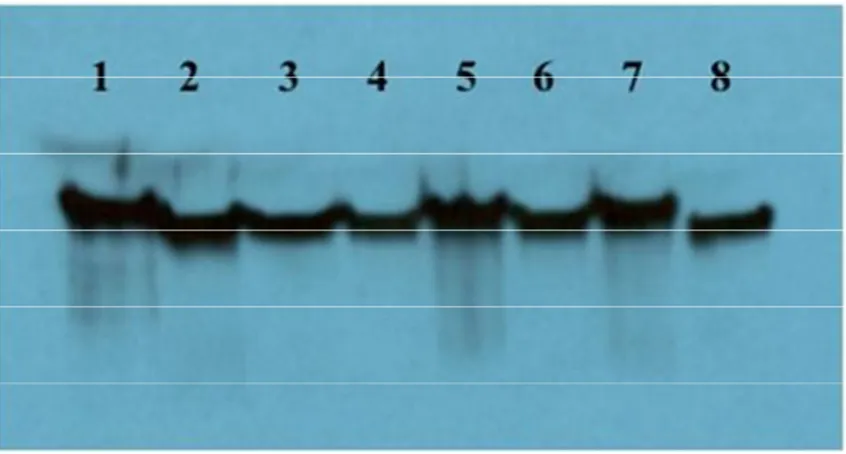
Western blot and indirect ELISA analysis obtained using polyclonal antibodies are showed respectively in Figure 1 and Table 1.
Sandwich ELISA for the detection of L.
monocytogenes
Mouse monoclonal and rabbit polyclonal antibodies that both recognize p60 protein from all strains of L.
monocytogenes were selected as reagents to implement
a sandwich ELISA. The monoclonal antibody against the specific 11-amino acids peptide sequence in the p60 protein was utilized as the capture antibody to ensure the specificity of the test while the polyclonal antibody allows for detecting the captured antigen. After the specific epitope on L. monocytogenes p60 protein is captured by the monoclonal antibody, different epitopes on the whole protein are then recognized by the polyclonal antibodies, allowing consequently the detection of the captured protein. Besides, the use of different species of antibodies (mouse and rabbit) allows for the amplification of the resulting signal. Prior to the analyses of bacterial supernatants, technical ELISA parameters were optimized using the recombinant p60 as the antigen. Hence, the concentration of each antibody showing an optimal signal to background ratio was selected. All strains of L. monocytogenes as well as four other different bacterial strains were used to evaluate cross reactivity. Results of bacterial detection are presented in Table 2.
Limit of detection
The limit of detection of the optimized sandwich ELISA was established with different dilutions of one strain of L.
monocytogenes (HPB 2812) which was incubated for 18
h of incubation at 37°C before performing the immunoassay detection. Results obtained with HPB 2812 were compared with results obtained for L. innocua used as a negative control. Bacterial count was determined by plate count on TSA as described above. Dilutions of
Figure 1. Western blot analysis of concentrated cell-free supernatants from 18-h cultures of various Listeria species using a polyclonal anti-p60 antibody. Lanes: 1: recombinant p60 protein; 2: L. monocytogenes HPB 1043; 3: L. monocytogenes HPB 2371; 4: L. monocytogenes HPB 2558; 5: L. monocytogenes HPB 2569; 6: L. monocytogenes HPB 2739; 7: L. monocytogenes HPB 2812; 8: L. innocua.
Table 1. Optical density obtained by Indirect ELISA using polyclonal anti-p60 antibodies and cell-free supernatant samples from 18-h culture of different Listeria species and strains.
Organisms Optical density (450 nm)
Listeria innocua 2.06 ± 0.28 Listeria monocytogenes HPB 2812 2.18 ± 0.12 HPB 1043 2.33 ± 0.14 HPB 2739 2.16 ± 0.17 HPB 2371 1.98 ± 0.12 HPB 2569 2.15 ± 0.12 HPB 2558 2.18 ± 0.15 Negative control (TSB) 0.09 ± 0.01 Results are represented as mean ± standard deviation.
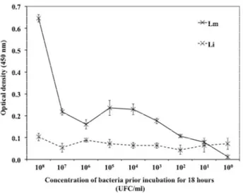
culture supernatant obtained after 18 h of incubation were filtered to collect the free supernatant. The cell-free supernatant samples were tested using the sandwich ELISA. Results are presented in Figure 2 as optical density of sandwich ELISA in regard to the number of colony-forming units. The absorbance of 0.1 is associated to a bacterial count of < 102 CFU/ml. Between 103 and 107 CFU/ml, a signal of 0.2 on average was observed while 108 CFU/ml was associated with an optical density of 0.8.
Bacterial detection using culture broth, culture supernatant and cell-free supernatant
In the purpose of reducing the experimental labour prior to detect the p60 protein in the samples, three methods
Table 2. Optical density obtained by optimized sandwich ELISA using monoclonal anti-p60 antibodies as capture antibodies (8 µg/ml) and polyclonal anti-p60 antibodies (8 µg/ml) as detection antibodies using cell-free supernatant samples obtained from different strains of bacteria incubated for 18 h at 37°C.
Organisms Optical density (450 nm)
L. monocytogenes HPB 2812 0.83 ± 0.16 HPB 1043 1.15 ± 0.01 HPB 2739 0.60 ± 0.12 HPB 2371 0.51 ± 0.03 HPB 2569 0.75 ± 0.10 HPB 2558 0.45 ± 0.04 L. innocua 0.10 ± 0.02 E. coli 0.02 ± 0.01 S. Typhimurium 0.01 ± 0.01 S. aureus 0.09 ± 0.01
Results are represented as mean ± standard deviation.
of sample preparation were compared. Optimized sandwich ELISA was used to detect the antigenic targeted in bacterial broth, in whole supernatant and in cell-free supernatant. All samples were harvested following an 18-h incubation period at 37°C of the bacterial suspension freshly sub-cultured. To avoid contamination that may occur, all experiments were done under sterile conditions whereas buffers and antibodies solutions into to the microplate were aspirated using vacuum. Also, the period of time for the colour development of TMB was diminished to 1 min instead of 4 min in order to avoid maximal optical density. To confirm that none of the sample preparation methods
Figure 2. Optical density from our sandwich ELISA using monoclonal anti-p60 antibodies as capture antibodies (8 µg/ml) and polyclonal anti-p60 antibodies (8 µg/ml) as detection antibodies using cell-free supernatant samples from L. monocytogenes HPB 2812 (Lm) and L. innocua (Li). Concentration represents the CFU/ml of the initial dilution before the incubation period of 18 h at 37°C. Error bars represent the standard deviation of the mean.
cause cross-reactivity or undesired background, L.
monocytogenes HPB 2812 and L. innocua were tested in
parallel. Optical densities obtained are presented in Figure 3.
DISCUSSION
Polyclonal anti-p60 antibodies reactivity and specificity
No discrimination of detection between L. monocytogenes and L. innocua was expected since it
has been demonstrated that amino acid sequences of p60 protein from different Listeria species have conserved identical regions (Bubert et al., 1992). Also, previous works on polyclonal antibodies against p60 from
L. monocytogenes showed cross-reactivity with proteins
from other Listeria species (Yu et al., 2004; Bubert et al., 1992; Wieckowska-Szakiel et al., 2002). However, since the capture monoclonal antibody ascertains the specificity, the polyclonal rabbit antibody allows for detecting the reaction between the monoclonal antibody and the antigen. Given that polyclonal antibodies can recognize a variety of epitopes on the antigen, even if the specific epitope on p60 protein recognized by the monoclonal antibody is hidden, polyclonal antibodies will allow reaction on other sites on the antigenic molecule. Both results show a good recognition between the antibodies and the supernatant from all strains of Listeria. Figure 1 shows that polyclonal antibodies are specific to p60 protein and that no cross-reactivity occurred with
Figure 3. Comparison of sample preparation methods prior to antigen detection of L. monocytogenes HPB 2812 (Lm) and L. innocua (Li) using optimized sandwich ELISA. Error bars represent the standard deviation of the mean.
other proteins found in culture supernatant. However, proteins transferred during Western blot analysis are denatured due to the action of SDS which could alter recognition between native protein antigen and antibody. On the contrary, ELISA maintains protein conformation. Results showed in Table 1 demonstrate that anti-p60 polyclonal antibodies can detect the protein in its native structure.
Sandwich ELISA for the detection of L.
monocytogenes
As expected, all L. monocytogenes strains were detected at different extent while no reactivity was obtained with the other strains including L. innocua. The absence of cross-reactivity with other bacterial species and the
non-monocytogenes strains of Listeria demonstrated the
specificity of the detection. Several immunoassays reported for the detection of L. monocytogenes usually present results for one strains and do not monitor cross-reactivity while other assays target the whole cell, which does not ensure specificity (Kawasaki et al., 2011; Jadhav et al., 2012; Cho and Irudayaraj, 2013; Heo et al., 2014; Yang et al., 2006). Our results demonstrate that the combination of both monoclonal and polyclonal anti-p60 antibodies is suitable for a sandwich immunoassay that could be easily adapted to other formats. Sandwich ELISA is very versatile and can be innovate using many kinds of supports such as functionalized microplate, silicon, polycarbonate, polystyrene, gold nanoparticles and chitosan (Jain et al., 2012; Yang et al., 2006; Albarghouthi et al., 2000; Darain et al., 2009; Dixit et al.,
2011; Dixit et al., 2010; Gan et al., 2011). Therefore, this sandwich ELISA in 96-well plate could be easily adapted using a variety of support in order to optimize the efficiency of the method.
Limit of detection
As presented in Figure 2, the optical density varies around 0.1 independently to the number of L. innocua in the culture. Optical density signal observed with 103 CFU/ml of L. monocytogenes was higher than the one monitored for 108 CFU/mL of L. innocua (p ≤ 0.05), which demonstrates the possibility of detection with a limit as low as 103 CFU/ml of L. monocytogenes only after 18 h of incubation. In comparison, other studies showed rapid methods for detection of L. monocytogenes within a limit of 102 and 105 CFU/mlfor an analysis period for up to 30 to 50 h (Jasson et al., 2010; Jadhav et al., 2012; Jasson et al., 2009; Cruz et al., 2012). As an example, a commercial test detected from 105 to 106 CFU/ml of L.
monocytogenes in food samples after 12 h of incubation
(Jadhav et al., 2012). In this study, the designed sandwich ELISA is advantageously comparable due to the limit of detection that was lower (103 CFU/ml instead of 105 to 106 CFU/ml) with only 6 h more of incubation. In the goal of improving the limit of detection, steps of culture incubation and antigen capture by monoclonal antibodies could be combined. Then, as the bacteria grows and secretes the p60 protein in its supernatant, immobilized monoclonal anti-p60 antibodies may bind the p60 protein immediately. Following this combined incubation period, only steps needed to detect the captured antigen (detection with polyclonal antibodies and use of secondary antibodies for signal amplification purposes) would be necessary to obtain the optical density results.
Bacterial detection using culture broth, culture supernatant and cell-free supernatant
Results presented in Figure 3 demonstrated that using cell-free supernatant leads to the lower optical density signal while bacterial culture gives the higher signal. Optical density reading from unfiltered supernatant is slightly higher than that of cell-free supernatant, but the whole bacterial culture allows for significantly higher signals (p ≤ 0.05). These results demonstrate that the sample preparation may cause the loss of the molecule of interest. Moreover, the absence of detection of L.
innocua in bacterial culture samples indicates that using
unprocessed sample does not lead to increased background due to higher content of non-specific components as compared to the cell-free supernatant. These experiments demonstrated that this sandwich ELISA does not require any preparation prior analysis which is highly advantageous as compared to PCR
method which requires DNA purification and amplification or magnetic immuno-separation to remove matrix interference (Cho and Irudayaraj, 2013; Heo et al., 2014; Churchill et al., 2006; Shriver-Lake et al., 2013). Immunoassays are less sensitive to non-related components in matrix interference, however some L.
monocytogenes detection immunoassays require heat
killing and formalin fixation processes or flagellum and protein extraction (Churchill et al., 2006). These kinds of sample preparation greatly increase experimental labour required to obtain results. Using this sandwich ELISA, it was demonstrated that a specific assay with a low limit of detection directly in the bacterial culture suspension can be performed and could be highly useful for detection of surfaces or food samples in industries.
CONCLUSION
In conclusion, a version of sandwich immunoassay was developed using a combination of specific anti-p60 monoclonal antibody and a polyclonal antibody against p60 protein from L. monocytogenes in order to improve the sensitivity of the detection method. It was demonstrated that the polyclonal antibody can specifically detect the protein p60 among all proteins contained in the cell-free supernatant without discriminating L. monocytogenes and L. innocua. However, the sequential
use of monoclonal and polyclonal anti-p60 antibodies in the sandwich ELISA in 96-well microplate allowed for specific results in less than 24 h, without observation of cross reactivity with other strains of bacteria. Moreover, this method permitted a detection limit as low as 103 CFU/ml, compared to other methods that detect 104 or 105 CFU/ml. Furthermore, it was demonstrated that sample preparation such as supernatant harvesting and filtration are not necessary to ascertain specificity and sensibility of the detection. It is believed that sandwich ELISA designed in this study could be adapted in another immunoassay version with versatile bioactive polymer supports for a quicker detection of L. monocytogenes.
ACKNOWLEDGEMENTS
This research was supported by the Ministère de l'Économie, de l'Innovation et des Exportations of Quebec, International Program (MEIE) as well as by a grant from S.M. Group International Inc at the Armand-Frappier Foundation University of INRS. This research was also in the frame CNR Commessa “Development of biochips for food safety and health” (SD).
REFERENCES
Albarghouthi M, Fara DA, Saleem M, El-Thaher T, Matalka K , Badwan A, 2000. Immobilization of antibodies on alginate-chitosan beads. Int J Pharm, 206(1-2):23-34.
Beauchamp S, D'Auria S, Pennacchio A, Lacroix M, 2012. A new competitive fluorescence immunoassay for detection of Listeria monocytogenes. Anal Meth, 4(12):4187-4192.
Bubert A, Kuhn M, Goebel W, Kohler S, 1992. Structural and functional properties of the p60 proteins from different Listeria species. J Bacteriol, 174(24): 8166-8171.
Bubert A, Schubert P, Kohler S, Frank R , Goebel W, 1994. Synthetic peptides derived from the Listeria-monocytogenes P60-protein as antigens for the generation of polyclonal antibodies specific for secreted cell-free L-monocytogenes P60-proteins. Appl Environ Microbiol, 60(9):3120-3127.
Carpentier B, Cerf O, 2011. Review - Persistence of Listeria monocytogenes in food industry equipment and premises. International journal of food microbiology, 145(1):1-8
Chasseignaux E, Gerault P, Toquin MT, Salvat G, Colin P , Ermel G, 2002. Ecology of Listeria monocytogenes in the environment of raw poultry meat and raw pork meat processing plants. FEMS Microbiol Lett, 210(2):271-275
Cho IH , Irudayaraj J, 2013. Lateral-flow enzyme immunoconcentration for rapid detection of Listeria monocytogenes. Analyt Bioanalyt Chem, 405(10):3313-3319.
Churchill RL, Lee H , Hall JC, 2006. Detection of Listeria monocytogenes and the toxin listeriolysin O in food. J Microbiol Methods, 64(2):141-170.
Cruz CD, Win JK, Chantarachoti J, Mutukumira AN , Fletcher GC, 2012. Comparing rapid methods for detecting Listeria in seafood and environmental samples using the most probably number (MPN) technique. Int J Food Microbiol, 153(3):483-487.
Darain F, Gan KL, Tjin SC, 2009. Antibody immobilization on to polystyrene substrate--on-chip immunoassay for horse IgG based on fluorescence. Biomed Microdevice, 11(3):653-661.
Dixit CK, Vashist SK, MacCraith BD, O'Kennedy R, 2011. Multisubstrate-compatible ELISA procedures for rapid and high-sensitivity immunoassays. Nat Protocols, 6(4):439-445.
Dixit CK, Vashist SK, O'Neill FT, O'Reilly B, MacCraith BD , O'Kennedy R, 2010. Development of a high sensitivity rapid sandwich ELISA procedure and its comparison with the conventional approach. Analyt Chem, 82(16):7049-7052
Fan C, Hu Z, Mustapha A, Lin M, 2011. Rapid detection of food- and waterborne bacteria using surface-enhanced Raman spectroscopy coupled with silver nanosubstrates. Appl Microbiol Biotechnol, 92(5):1053-1061.
Farber JM, Kozak GK, Duquette S, 2011. Changing regulation: Canada's new thinking on Listeria. Food Control, 22(9):1506-1509 Gan N, Jia L, Zheng L, 2011. A novel sandwich electrochemical
immunosensor based on the DNA-derived magnetic nanochain probes for alpha-fetoprotein. J Automat Method Manag Chem, 2011:957805.
Heo EJ, Song BR, Park HJ, Kim YJ, Moon JS, Wee SH, Kim JS, Yoon Y, 2014. Rapid detection of Listeria monocytogenes by real-time PCR in processed meat and dairy products. J Food Prot, 77(3):453-458 Jadhav S, Bhave M, Palombo EA, 2012. Methods used for the
detection and subtyping of Listeria monocytogenes. J Microbiol Meth, 88(3):327-341
Jain S, Chattopadhyay S, Jackeray R, Abid CK, Kohli GS , Singh H, 2012. Highly sensitive detection of Salmonella typhi using surface aminated polycarbonate membrane enhanced-ELISA. Biosens Bioelect, 31(1):37-43
Jasson V, Jacxsens L, Luning P, Rajkovic A , Uyttendaele M, 2010. Alternative microbial methods: An overview and selection criteria. Food Microbiol, 27(6):710-730
Jasson V, Rajkovic A, Debevere J , Uyttendaele M, 2009. Kinetics of resuscitation and growth of L. monocytogenes as a tool to select appropriate enrichment conditions as a prior step to rapid detection methods. Food Microbiol, 26(1):88-93.
Kawasaki S, Fratamico PM, Kamisaki-Horikoshi N, Okada Y, Takeshita K, Sameshima T , Kawamoto S, 2011. Development of the Multiplex PCR Detection Kit for Salmonella spp., Listeria monocytogenes, and Escherichia coli O157:H7. Jarq-Japan Agric Res Quart, 45(1):77-81. Liu D, 2006. Identification, subtyping and virulence determination of
Listeria monocytogenes, an important foodborne pathogen. J Med Microbiol, 55(6):645-659.
Lungu B, Ricke SC , Johnson MG, 2009. Growth, survival, proliferation and pathogenesis of Listeria monocytogenes under low oxygen or anaerobic conditions: a review. Anaerobe, 15(1-2):7-17
Mandal PK, Biswas AK, Choi K, Pal UK, 2011. Methods for rapid detection of foodborne pathogens: An overview. Am J Food Technol, 6(2):87-102.
Posfay-Barbe KM , Wald ER, 2009. Listeriosis. Seminars in fetal and neonatal medicine, 14(4):228-233
Ruhland GJ, Hellwig M, Wanner G , Fiedler F, 1993. Cell-surface location of Listeria-specific protein p60 - detection of Listeria cells by indirect immunofluoresence. J Gen Microbiol, 139 :609-616.
Shriver-Lake LC, Golden J, Bracaglia L, Ligler FS, 2013. Simultaneous assay for ten bacteria and toxins in spiked clinical samples using a microflow cytometer. Analyt Bioanalyt Chem, 405(16):5611-5614. Tompkin RB, 2002. Control of Listeria monocytogenes in the
food-processing environment. J Food Prot, 65(4):709-725
Wieckowska-Szakiel M, Bubert A, Rozalski M, Krajewska U, Rudnicka W, Rozalska B, 2002. Colony-blot assay with anti-p60 antibodies as a method for quick identification of Listeria in food. Int J Food Microbiol, 72(1-2):63-71.
Yang L, Banada PP, Chatni MR, Seop Lim K, Bhunia AK, Ladisch M , Bashir R, 2006. A multifunctional micro-fluidic system for dielectrophoretic concentration coupled with immuno-capture of low numbers of Listeria monocytogenes. Lab Chip, 6(7):896-905. Yu KY, Noh Y, Chung M, Park HJ, Lee N, Youn M, Jung BY, Youn BS,
2004. Use of monoclonal antibodies that recognize p60 for identification of Listeria monocytogenes. Clin Diagnostic Lab Immunol, 11(3):446-451.
Citation: Coutu JV, Morissette C, D’Auria S, Lacroix M, 2014.
Development of a highly specific sandwich ELISA for the detection of Listeria monocytogenes, an important foodborne pathogen. Microbiol Res Int, 2(4): 46-52.

