REFERENCE POPULATIONS FOR SHOULDER STUDIES SHOULD BE SELECTED CAREFULLY 1 Cédric Schwartz, 1,2 Vincent Denoël, 1,3 Olivier Brüls, 1,4 Jean-Louis Croisier, 1,4 Bénédicte Forthomme
1Laboratory of Human Motion Analysis - LAMH, University of Liège, Belgium
2
Department of Architecture, Geology, Environment and Constructions, University of Liège, Belgium
3Department of Aerospace and Mechanical Engineering, University of Liège, Belgium
4Department of Physical Medicine and Rehabilitation, University of Liège, Belgium
Email: [email protected], website: www.lamh.ulg.ac.be
SUMMARY
To assess various shoulder pathologies / treatments, non pathological populations are often used as references. However, some factors may influence significantly the scapular kinematics within a healthy population and consequently alter the final kinematic evaluation. Results of 3D shoulder assessment found in this study show that small (≈5°) but significant differences exist between gender and between the dominant and non-dominant arms. Therefore the populations used for referential data should be selected carefully.
INTRODUCTION
Knowledge of the normal scapula kinematic is of great importance in order to correctly evaluate and/or detect clinical pathologies, sport adaptations, and treatment or rehabilitation program effects. Several factors such as age, gender, dominant side have been shown to affect the shoulder motion [1, 2, 3]. Within these factors, the dominance effect is still controversial [3, 4] and gender studies [2,5] are limited to the humeral range of motion. As differences induced by the composition of the reference population could introduce bias and inaccurate results of the studied populations, the influence of these factors on the shoulder motion should be quantified. The present study aims at evaluating dominance and gender effects on the scapula-thoracic kinematic during shoulder abduction.
METHODS
The study included 11 men (age: 22.4 ±2.5 years; Body Mass Index or BMI: 22.6±2.2 kg.m-2) and 11 women (age: 22.2 ±1.8 years; BMI: 21.0±1.5 kg.m-2). None of the subjects have ever practiced more than 2 hours per week a sport involving the upper limbs. They had no inequality of the lower limbs, they never suffered from kyphosis, scoliosis, and never undergone upper limbs or thoracic lesions and/or surgery. Additional clinical tests confirmed that the volunteers do not suffer from any sub-coraco-acromial conflict and/or tendinous.
The 3D scapula kinematic was tracked at a sampling rate of 100 Hz using four Codamotion CX1 units (Charnwood Dynamics, Rothley, UK). Six markers were placed on each scapula following Bourne [6] recommendations, 4 others on
the middle of each humerus in order to avoid the deltoid area and finally 4 on the thorax following ISB recommendations [7]. Scapular and humerus orientations were expressed relatively to the thorax using Cardan decomposition proposed by the ISB [7] and Senk [8] (in order to avoid gimbal lock at 0°) respectively. Finally, the scapula kinematic was reported wrt the humerus elevation. After getting familiar with the exercise, the subjects realized 5 maximal active humeral elevations in the frontal plane (abduction) with arm rotated externally for their dominant and non-dominant arms. The reported results are based on the average of the trial repetitions. To cancel the influence of the initial posture, the scapular angles were shifted to 0° at the starting position. Descriptive statistics (mean and standard deviation) of the scapular motion were calculated for each step of 1°. Paired and unpaired two-sample t-tests were used to evaluate the dominance and gender effects respectively. The level of significance was set at p < 0.05.
RESULTS AND DISCUSSION
Significant asymmetries were observed between the dominant and non-dominant arms during abduction in the male (Figure 1) but not in the female population (Figure 2).
Figure 1: Effect of dominance on the male population
scapular kinematic - scapular rotations (mean and standard deviation) wrt humeral elevation relatively to the thorax.
However these asymmetries remain low in terms of amplitude (inferior to 5°). In both male and female populations, the scapula had a larger upward rotation on the dominant side even if the difference is statistically significant only in the male population. For men, a larger posterior tilt was also observed on the dominant side. The results obtained in this paper confirm some previous results [3] of the literature stating that some kinematic asymmetries may be present for healthy populations. The observed differences in the kinematic have probably not a unique cause but are the consequence of various parameters. The larger physical activity of men may partly explain the increased asymmetries observed in the male population.
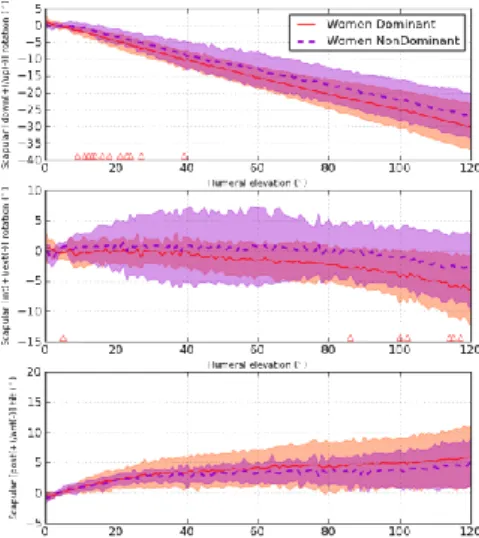
Figure 2: Effect of dominance on the female population
scapular kinematic - scapular rotations (mean and standard deviation) wrt humeral elevation relatively to the thorax. Gender comparison shows that women had larger external rotation and smaller posterior tilt than men (Figure 3) during abduction. The differences reached approximately 5° at 120° of humeral elevation. Upward rotations were similar for men and women. Differences observed in the scapular pattern between men and women have not been reported before. However several studies have observed morphological [9] and motor control strategies differences [10] between genders that could explain the reported differences.
The gender and dominance effect observed in this study remains relatively small in terms of amplitude at least up to 120° of humeral elevation. These differences may be difficult to observe visually by the physicians and therefore emphasized the interest of systems, which provide
quantified measurements of the kinematic. Larger
asymmetries may occur for larger humeral elevations but skin marker based systems are sensitive to soft tissue artifacts, which increase significantly for humeral elevation superior to 120°.
Electromyographic (EMG) measurements could give further insights to the activation strategies underlying the scapular kinematic and help to identify the origins of the asymmetries reported in this study. Subject-specific musculo-squeletic modeling would also be an interesting tool to test the origins of the scapular kinematic differences. Indeed, it would be
possible to study independently each parameter of interest without the effect of inter-individual variability.
Figure 3: Effect of gender on the dominant scapular
kinematic - scapular rotations (mean and standard deviation) wrt humeral elevation relatively to the thorax.
The strict recruitment criterions lead to homogenous populations in terms of physical activity, health, age and body mass index in order to limit the influence of external parameters. One should however be cautious if extrapolating these results to other populations.
CONCLUSIONS
Gender and dominance were shown in this study to have small but significant effect on the scapular kinematic. Special care should consequently be given to the gender composition of the studied populations as well as the arm used for the comparisons. Carefully selected reference populations may improve the quality of clinical evaluations.
ACKNOWLEDGEMENTS
The authors thank T. Fedrigo and E. Rigaux for their help in the data collection. This work was partly supported by the Fédération Wallonie-Bruxelles (Belgium).
REFERENCES
1. Dayanidhi S, et al., Clin Biomech (Bristol, Avon).
20(6):600–606, 2005.
2. Barnes CJ, et al., J Shoulder Elbow Surg. 10(3):242– 246, 2001.
3. Matsuki K, et al., J Shoulder Elbow Surg. 20(4):659– 665, 2011.
4. Yoshizaki K, et al., J Shoulder Elbow Surg. 18(5):756-753, 2009.
5. Vairo GL, et al., Sports Med Arthrosc Rehabil Ther
Technol. 4:33, 2012.
6. Wu G, et al., J Biomech. 38(5):981–92, 2005.
7. Bourne DA, et al., Ann Biomed Eng. 39(2):777-785,
2011.
8. Senk M et al., Clin Biomech (Bristol, Avon). 21(Suppl 1):S3–8, 2006.
9. Krobot A, et al., Med Biol Eng Comput. 47(5):497–506, 2009.
10. Anders C, et al., J Electromyogr Kinesiol. 14(6):699– 707, 2004