Regulation of membrane-type 1 matrix metalloproteinase activity by
vacuolar H
+-ATPases
Erik MAQUOI1,2, Karine PEYROLLIER1, Agnès NOËL, Jean-Michel FOIDART and Francis FRANKENNE
Laboratory of Tumor and Development Biology, University of Liège, Tour de Pathologie (B23), Sart Tilman, B-4000 Liège, Belgium
Abstract
Membrane-type 1 matrix metalloproteinase (MT1-MMP) is a key enzyme in normal development and malignant processes. The regulation of MT1-MMP activity on the cell surface is a complex process involving autocatalytic processing, tissue inhibitor of MMPs (TIMP) binding and constitutive internalization. However, the fate of internalized MT1-MMP is not known. Acidification of intracellular vacuolar compartments is essential for membrane trafficking, protein sorting and degradation. This acidification is controlled by vacuolar H+-ATPases, which can be selectively inhibited by bafilomycin-A1. Here, we treated human tumour cell lines expressing
MT1-MMP with bafilomycin-A1, and analysed its effects on MT1-MMP activity, internalization and processing.
We show that the activity of MT1-MMP on the cell surface is constitutively down-regulated through a vacuolar H+-ATPase-dependent degradation process. Blockade of this degradation caused the accumulation of TIMP-free active MT1-MMP molecules on the cell surface, although internalization was not affected. As a consequence of this impaired degradation, pro-MMP-2 activation was strongly enhanced. This study demonstrates that the catalytic activity of MT1-MMP on the cell surface is regulated through a vacuolar H+-ATPase-dependent degradation process.
Keywords: internalization, matrix metalloproteinase 2 (MMP-2), proteinase, proteinase inhibitor, tissue inhibitor of matrix met-alloproteinases 2 (TIMP-2), tumour cell.
Abbreviations used: bafilo, bafilomycin-A1 ; DMEM; Dulbecco's modified Eagle's medium ; mAb, monoclonal
antibody ; MMP, matrix metalloproteinase ; MT-MMP, membrane-type MMP ; TIMP, tissue inhibitor of MMPs ; rhTIMP, recombinant human TIMP ; RT-PCR, reverse transcriptase PCR.
INTRODUCTION
Matrix metalloproteinases (MMPs) form a family of neutral zinc endopeptidases which play pivotal roles in several normal and pathological processes, including embryogenesis, tumour invasion, metastatic dissemination and angiogenesis [1,2]. MMPs are subgrouped into soluble and anchored MMPs, called membrane-type MMPs (MT-MMPs) [3]. MT1-MMP appears more destructive than secreted MMPs [3-5]. It displays broad collagenolytic, glycoproteolytic, gelatinolytic and fibrinolytic activities [3,6,7]. It also activates other MMPs, including pro-MMP-2 [8]. In the MT1-MMP-deficient mouse, collagen turnover is severely compromised and activation of pro-MMP-2 is suppressed [5,9]. Uncontrolled MT1-MMP activity is a key determinant in cancer metastasis and tumour angiogenesis [1,10].
MT1-MMP activity is regulated by several mechanisms. Pro-MT1-MMP is activated by furin [11,12]. Mature MT1-MMP can be inhibited by all the tissue inhibitors of MMPs (TIMPs), excluding TIMP-1. Whereas TIMP-2 is the major inhibitor of MT1-MMP [13], it also participates in the activation of MMP-2 by mediating the binding of pro-MMP-2 to MT1-MMP through the formation of a ternary complex [13,14]. When present in excess relative to active MT1-MMP, TIMP-2 completely blocks this activation process [15,16]. MT1-MMP activity can also be down-regulated through autocatalytic and non-autocatalytic shedding processes [17]. Emerging evidence indicates that the internalization of cell-surface-anchored MT1-MMP represents an additional regulatory mechanism of its activity [18,19]. However, the fate of internalized MT1-MMP remains completely unknown.
It is well established that acidification of intracellular vacuolar compartments (e.g. endosomes and lysosomes) plays an important role in membrane trafficking, protein sorting and degradation [20]. This acidification is caused by vacuolar H+ -ATPases, which can be selectively inhibited by bafilomycin-A1 (bafilo) [21,22]. We have
demonstrated [23] that, in A2058 melanoma cells, MT1-MMP is required to trigger the binding of extracellular TIMP-2 molecules. Membrane-bound TIMP-2 is then internalized rapidly and degraded intracellularly through an MT1-MMP-dependent and pH-sensitive mechanism. Indeed, treatment of these cells with bafilo abolished TIMP-2 degradation and induced its accumulation in an intact form [23]. This observation prompted us to investigate the
1
These authors contributed equally to this study.
2
potential implication of vacuolar H+-ATPases in the regulation of MT1-MMP activity. Our results demonstrate that, in addition to suppressing the intracellular degradation of TIMP-2, inhibition of H+-ATPases also prevents the degradation of MT1-MMP and increases its activity at the cell surface.
MATERIALS AND METHODS Chemicals
A broad-spectrum hydroxamic acid-based synthetic MMP inhibitor, GI129471 [24], and bafilo (Sigma, St. Louis, MO, U.S.A.) were dissolved in DMSO. Pronase E was from Roche Molecular Biochemicals (Penzberg, Germany). BSA (fraction V) was from Sigma.
Preparation of conditioned media and cell extracts
A2058 human melanoma cells transfected with MT1-MMP cDNA (S.I.5 clone) or control vector (C.IV.3 clone), HT1080, and BT549 cells were plated in 96-well plates (6 x 10⁴ cells/well) in serum-free Dulbecco's modified Eagle's medium (DMEM) supplemented with 0.1 % BSA (DMEM-BS A) as described previously [23]. Once adherent, cells were supplemented with 0.1% DMSO or 1-100 nM bafilo (final concentrations). In some experiments, 1 µM GI129471 was also added. Culture supernatants were collected after 24 h (except where otherwise stated) and cell monolayers were extracted in RIPA buffer as described in [23].
Cellular distribution of bound 125I-labelled recombinant human TIMP-2 (125I-rhTIMP-2) rhTIMP-2 was prepared and iodinated as described in [23]. S.I.5 cells were incubated in DMEM-BSA
supplemented with 125I-rhTIMP-2 (0.6 nM) in the presence or absence of bafilo (100 nM) at 37 °C for 24 h. Media were collected, cells were washed in DMEM-BSA, exposed to 0.25 % Pronase E at 4 °C for 30 min to release cell-surface-bound radiolabelled TIMP-2, and the monolayers were dislodged by gentle pipetting. After centrifugation (2000 g, 5 min), supernatants (cell-surface-bound fraction) were collected and pellets (internalized fraction) were resuspended in PBS. The radioactivity of the three fractions was measured in a gamma counter.
125
I-rhTIMP-2-binding assay
S.I.5 cells were incubated with DMSO (0.1 %) or bafilo (100 nM) at 37 °C for 24 h. The monolayers were washed in DMEM-BSA to remove unbound TIMP-2, followed by a 30 min incubation at -20 °C to fix the cells. Binding was performed with 125I-rhTIMP-2 (0.6 nM) in DMEM-BSA in the presence or absence of excess unlabelled rhTIMP-2 (500 nM) for 30 min at 4 °C. Cells were washed with DMEM-BSA, and lysed with NaOH (0.5 M) supplemented with 0.1 % SDS for determination of radioactive counts in a gamma counter. Specific binding was calculated by subtracting the non-specific radioactivity (measured in the presence of unlabelled rhTIMP-2) from cell lysate-associated radioactivity (measured in the absence of unlabelled rhTIMP-2). Non-specific binding never exceeded 0.5 % of total added radioactivity. The number of cells was determined by DNA quantification. Results are expressed as a percentage of the total radioactivity specifically bound per µg of DNA.
Gelatin zymography, Western-blot analysis and TIMP-2 quantification
Gelatinolytic activities in conditioned media were analysed by gelatin zymography as described in [23]. Total cell extracts and pronase-treated cells were analysed by immunoblotting using monoclonal antibodies (mAbs) 1E12 and 2D7 against the N-terminal pro-domain and the haemopexin-like domain of MT1-MMP, respectively, as described in [23]. TIMP-2 concentrations in conditioned media were determined by ELISA [23].
RNA isolation and quantitative reverse transcriptase PCR (RT-PCR)
Total DNA-free RNA was extracted from S.I.5 cells treated for 24 h with DMSO (0.1 %) or bafilo (100 nM) using the High Pure RNA Isolation kit (Roche Molecular Biochemicals). MMP-2,
MT1-MMP, TIMP-2 mRNA and 28 S rRNA were quantified by RT-PCR as described in [25]. All results were corrected for differences in RNA concentration by dividing the number of copies of target mRNAs by the number of copies of 28 S rRNA.
Statistical analysis
Results are expressed as means ± S.E.M. from at least triplicate determinations. Statistical comparisons are based on the two-sample Student's t test or Mann-Whitney U test. Differences were considered to be statistically significant at P < 0.05.
RESULTS AND DISCUSSION
Bafilo induces activation of pro-MMP-2
To assess the potential implication of vacuolar H+ -ATPases in the regulation of MT1-MMP activity, A2058 melanoma cells overexpressing recombinant MT1-MMP (clone S.I.5) were used as a model system. The primary function assigned to MT1-MMP is the processing of pro-MMP-2 to its active form at the cell surface [8]. Therefore, S.I.5 cells and mock-transfected cells (clone C.IV.3) were treated for 24 h with increasing
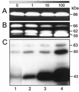
concentrations of bafilo (0-100 nM), a specific inhibitor of vacuolar H+ -ATPases [21,22]. Aliquots of conditioned medium were analysed by gelatin zymography to monitor the activation of MMP-2. We have reported previously that C.IV.3 cells, which express very low levels of endogenous MT1-MMP, were unable to activate pro-MMP-2. In contrast, S.I.5 cells efficiently processed pro-MMP-2 (66 kDa) into the intermediate (62 kDa) and, to a lesser extent, into the fully mature (59 kDa) form through an MT1-MMP-dependent process [23]. In C.IV.3 cells, bafilo did not promote MMP-2 activation, whereas in S.I.5 cells a dose-dependent stimulation of pro-MMP-2 processing to the 62-kDa intermediate and the active 59-kDa species was induced (Figures 1A and 1B). Quantitative RT-PCR analysis indicated that bafilo did not alter the expression level of mRNA for MMP-2 (Figure 2). A similar
stimulation of pro-MMP-2 activation was also observed in bafilo-treated HT1080 cells, a fibrosarcoma cell line expressing high levels of endogenous MT1-MMP (results not shown).
TIMP-2 has been shown to play a dual role in pro-MMP-2 maturation, promoting activation at low concentrations but inhibiting it at higher ones [13,16]. The concentration of secreted TIMP-2, as assessed by ELISA, was elevated in the medium of bafilo-treated S.I.5 cells (47.6 ± 1.2 and 151 ± 24.9 pM for vehicle- and bafilo-treated cells, respectively; P < 0.003). This higher concentration arose most probably from the impaired TIMP-2 degradation [23] since TIMP-2 mRNA level was only slightly increased (1.12-fold) in the presence of bafilo (Figure 2). This observation thus raised the possibility that the higher TIMP-2 concentration detected in bafilo-treated cells might promote MMP-2 processing. To rule out this hypothesis, S.I.5 cells were incubated for 24 h with increasing concentrations of rhTIMP-2 (0.01-100 nM). None of these concentrations was able to increase MMP-2 processing (results not shown), demonstrating that the enhanced MMP-2 activation induced by bafilo did not result from the higher level of secreted TIMP-2.
Figure 1 Influence of bafilo treatment on pro-MMP-2 activation and MT1-MMP processing
C.I.V.3 (A) and S.I.5 (B and C) cells were exposed to increasing concentrations of bafilo (lanes 2-4) or 0.1 % DMSO (lane 1) in serum-free conditions. After 24 h, conditioned media were analysed by gelatin zymography (A and B) and cell lysates by immunoblotting using the mAb 2D7 (C). The molecular masses of pro-, intermediate and mature forms of MMP-2 (66, 62 and 59 kDa, respectively) as well as immunoreactive pro-, mature and inactive MT1-MMP proteins (63, 60 and 43 kDa, respectively) are shown on the right.
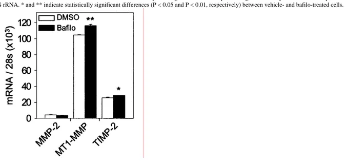
Figure 2 Quantitative RT-PCR analysis showing the influence of bafilo on MMP-2, MT1-MMP and TIMP-2 mRNA levels in S.I.5 cells
S.I.5 cells were treated with DMSO (0.1%) or bafilo (100 nM) for 24 h. Total RNA was extracted and analysed by quantitative RT-PCR. All results were corrected for differences in RNA concentrations by dividing the number of copies of target mRNAs by the number of copies of 28 S rRNA. * and ** indicate statistically significant differences (P < 0.05 and P < 0.01, respectively) between vehicle- and bafilo-treated cells.
Bafilo promotes the accumulation of TIMP-free active 60-kDa MT1-MMP at the cell surface
Activation of pro-MMP-2 is essentially governed by the abundance of active MT1-MMP on the cell surface [12,26]. Therefore, we examined the effect of bafilo on the expression and/or activity of MT1-MMP. Lysates of S.I.5 cells treated or not with bafilo were subjected to immunoblotting using mAb 2D7 directed against the haemopexin-like domain of MT1-MMP (Figure 1C). In vehicle-treated cells (Figure 1C, lane 1), MT1-MMP was represented by protein bands at 63 kDa (proenzyme), 60 kDa (mature enzyme) and 43 kDa (autocatalytically processed and inactive form). Bafilo slightly increased the level of the 63-kDa species. In contrast, a more pronounced increase was observed for the 60- and 43-kDa bands (Figure 1C, lanes 2-4). The level of these latter forms clearly correlated with the enhanced activation of pro-MMP-2 (Figures 1B and 1C). This bafilo-dependent accumulation of MT1-MMP molecules can only partly be attributed to an increased level of the corresponding mRNA (Figure 2), suggesting that bafilo essentially regulates the amount of MT1-MMP at a post-transcriptional level. Similar to the effect of bafilo in blocking the acidification of lysosomes and endosomes, NH4Cl also
promoted the accumulation of MT1-MMP molecules in S.I.5 cells (results not shown). Moreover, the increased level of MT1-MMP induced by bafilo was not restricted to the S.I.5 cells since a similar increase was observed in bafilo-treated HT1080 and BT549 cells (results not shown).
To identify the cellular distribution of the different MT1-MMP species, lysates of S.I.5 cells, treated or not with bafilo, were prepared before (both membrane-anchored and intracellular proteins) or after (intracellular proteins only) treating the cells with pronase. The resulting lysates were then analysed by immunoblotting using mAbs 1E12 and 2D7. In preliminary experiments, surface biotinylation followed by immunoprecipitation of total lysates with mAb 2D7 revealed the complete disappearance of the biotinylated 60- and 43-kDa MT1-MMP forms from the lysate of pronase-treated cells, confirming the efficacy of this digestion (results not shown). As shown in Figure 3, the 63-kDa pro-MT1-MMP was insensitive to the pronase treatment, indicating that its localization was restricted to the intracellular compartment. This distribution pattern is in agreement with the furin-dependent intracellular activation process previously reported for latent MT1-MMP [11,12]. On the other hand, the 60-kDa MT1-MMP, whose level is increased by bafilo, disappeared completely after pronase treatment. This observation demonstrates that mature MMP resides essentially at the cell surface, thus accounting for the higher MT1-MMP activity (as reflected by the increased pro-MT1-MMP-2 processing) detected in bafilo-treated cells. The 43-kDa form was observed in lysates of cells treated or not with pronase; however, it was at a higher level in the latter, suggesting that this species is localized at the cell surface as well as in the intracellular compartment. Although the level of the 43-kDa species was increased by bafilo, its cellular distribution remained unchanged.
Figure 3 Immunoblot showing unchanged distribution of MT1-MMP molecules in bafilo-treated cells
S.I.5 cells were treated for 24 h with DMSO (0.1 %) or bafilo (100 nM) in serum-free conditions and subjected to pronase digestion to remove cell-surface proteins (intracellular MT1-MMP) or left untreated (total MT1-MMP). The corresponding cell lysates were analysed by immunoblotting using the mAbs 1E12 (upper panel) and 2D7 (lower panel).
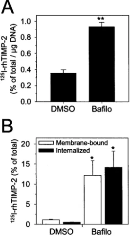
Figure 4 Influence of bafilo on 125I-rhTIMP-2 binding and internalization
S.I.5 cells were treated with DMSO (0.1 %) or bafilo (100 nM) in serum-free conditions for 24 h in the absence (A) or presence (B) of 125 I-rhTIMP-2 (0.6 nM). (A) After 24 h, the cells were washed in serum-free medium and fixed for 30 min at -20 °C to prevent internalization. Fixed cells were incubated with 125I-rhTIMP-2 (0.6 nM) for 30 min at 4 °C and the cell-associated radioactivity was quantified by gamma
counting. Results are expressed as the percentage of the total radioactivity specifically bound per µg of DNA. (B) Membrane bound 125
I-rhTIMP-2 was quantified by subjecting the cells to pronase (0.25%) digestion. The radioactivity remaining in the cells after the pronase treatment was defined as internalized 125I-rhTIMP-2. Results were expressed as percentages of 125I-rhTIMP-2 initially added to the cells. * and ** indicate statistically significant differences (P < 0.05 and P < 0.01, respectively) between vehicle- and bafilo-treated cells.
The ratio between TIMP-2-bound MT1-MMP (which serves as a cell-surface receptor for MMP-2) and TIMP-free active MT1-MMP (which functions as an activator of pro-MMP-2) is critical for MMP-2 processing [16]. As illustrated above, bafilo increased the amount of active 60-kDa MT1-MMP as well as the level of secreted TIMP-2. To further examine the impact of this treatment on the number of TIMP-free active MT1-MMP molecules on the cell surface, the abundance of this species was quantified by measuring the binding of 125I-rhTIMP-2 to the surface of vehicle- or bafilo (100 nM)-treated S.I.5 cells. Different reports have shown that the binding of TIMP-2 was restricted to the active MT1-MMP [26,27]. Furthermore, we have demonstrated previously that the binding of
125
I-rhTIMP-2 to S.I.5 cells was MT1-MMP-dependent [23]. Cell monolayers were fixed briefly at -20 °C before the addition of radiolabelled TIMP-2 to negate the influence of endocytosis on the number of cell-surface receptors. Specific binding of 125I-rhTIMP-2 to S.I.5 cells was increased significantly in the presence of bafilo (Figure 4A), suggesting that despite the oversecretion of TIMP-2, bafilo promoted the accumulation of TIMP-free
active MT1-MMP molecules at the cell surface. According to recent data [18,19], the level of active MT1-MMP at the cell surface is regulated through internalization. Therefore, we considered the possibility that bafilo may
promote the accumulation of active MT1-MMP at the cell surface through the inhibition of its internalization. To test this hypothesis, we monitored MT1-MMP internalization indirectly by following the fate of 125I-rhTIMP-2 (used as a tracer), since Uekita and co-workers [18] have shown that the internalization of exogenous TIMP-2 closely reflects the internalization of MT1-MMP. Therefore, S.I.5 cells were treated for 24 h with vehicle or bafilo (100 nM) in the presence of 125I-rhTIMP-2 and were then subjected to pronase digestion to discriminate between membrane-bound (pronase-released radioactivity) and internalized (pronase-resistant radioactivity) 125I-rhTIMP-2. As shown in Figure 4(B), the total radioactivity associated with untreated cells was partitioned between the plasma membrane and the intracellular compartment. In the presence of bafilo, the total cell-associated radioactivity was increased and was equally distributed between the two cellular compartments (Figure 4B), demonstrating that bafilo does not prevent the internalization of the tracer. These observations are consistent with other studies illustrating the lack of influence of bafilo on the internalization of other plasma-membrane proteins [28,29]. Bafilo inhibits the degradation of MT1-MMP
To further investigate the influence of bafilo on the activity of MMP, we next examined the fate of MMP molecules in S.I.5 cells. Indeed, it was reported previously that, in the absence of TIMP-2, the active MT1-MMP undergoes autocatalytic conversion to the 43-kDa inactive form, which diminishes the surface availability of active MT1-MMP [26,30]. For that purpose, S.I.5 cells were plated in serum-free DMEM-BSA and, once
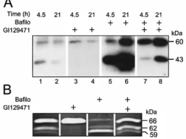
adherent, they were treated immediately with vehicle or bafilo (100 nM), supplemented or not with a broad-spectrum synthetic MMP inhibitor (GI129471; 1 µM) to inhibit the autocatalytic processing of the 60-kDa MT1-MMP. Cells were lysed after 2, 4.5 and 21 h and the lysates were analysed by immunoblotting using mAb 2D7 (Figure 5A). At 2 h, only a faint band corresponding to active MT1-MMP was observed, irrespective of the treatment that was applied (results not shown). In untreated cells, the 60- and 43-kDa MT1-MMP species were detected in lysates after a 4.5 h incubation (Figure 5A, lane 1). After 21 h, the abundance of the 43-kDa species, and to a lesser extent that of the 60-kDa form, decreased noticeably (Figure 5A, lane 2), suggesting the occurrence of an effective turnover of these MT1-MMP species. When the incubation was performed in the presence of GI129471, the processing of the 60- to the 43-kDa MT1-MMP species was completely blocked. Despite this inhibition, GI129471 did not prevent the decreased immunoreactivity of the 60-kDa form observed after 21 h (Figure 5A, lanes 3 and 4), indicating that the formation of the 43-kDa form is not a prerequisite for the
degradation of the 60-kDa MT1-MMP. Previous experiments have shown that GI129471 did not alter MT1-MMP mRNA level in S.I.5 cells, thus ruling out a potential transcriptional regulatory effect of this drug [23]. In contrast, a 4.5 h incubation with bafilo was sufficient to accumulate large amounts of both 60-and 43-kDa MT1-MMP (Figure 5A, lane 5). Furthermore, this treatment prevented the decrease of the 60-kDa species observed after 21 h in vehicle-treated as well as in GI129471-treated cells, leading to a massive accumulation of the 60-and 43-kDa forms (Figure 5A, lane 6). When compared with cells treated with GI129471 alone, the combined treatment with both GI129471 and bafilo strongly increased the intensity of the 60-kDa band (Figure 5A, lanes 3 and 7). This combined treatment also abolished the partial disappearance of the 60-kDa form observed after 21 h in vehicle- and GI129471-treated cells (Figure 5A, lanes 2, 4 and 8). However, when combined with bafilo, GI129471 failed to completely block the formation of the 43-kDa species. This failure to prevent the autoproteolytic processing of the 60-kDa form is most likely a consequence of the altered stoichiometry between the number of active 60-kDa MT1-MMP (which was increased by bafilo) and GI129471 molecules. This assumption is supported by the observation that, under these conditions, GI129471 also failed to prevent the MT1-MMP-dependent activation of pro-MMP-2 (Figure 5B) as well as the binding of TIMP-2 to the plasma membrane (results not shown), two processes that were completely abolished in the absence of bafilo. Alternatively, the processing of 60-kDa MT1-MMP may occur intracellularly where the concentration of the synthetic inhibitor is lower than in the extracellular medium. Altogether, these data reveal that the degradation of the 60- and 43-kDa MT1-MMP species is mediated by a H+ -ATPase-dependent and MMP-independent mechanism.
The precise cellular mechanism through which bafilo promotes the accumulation of active MT1-MMP on the cell surface remains uncertain. Recent studies have demonstrated that MT1-MMP is constitutively internalized in clathrin-coated pits before being routed to early endosomes [18,19]. After delivery to early endosomes, the most common fates of internalized molecules are degradation in the late endosomes and lysosomes or recycling to the cell surface. The control of the internal pH of these vesicles, through the activity of vacuolar H+ -ATPases, is essential for endocytic sorting [30]. Consistent with a role for H+ -ATPases in this process, bafilo has been shown to prevent the delivery of internalized molecules to lysosomes [28,31,32] as well as the acidification of these organelles, thereby interfering with the activity of lysosomal enzymes that have acidic pH optima. The increased level of active MT1-MMP on the surface of bafilo-treated cells suggests that, in conditions where delivery to and/or degradation in the lysosomes are impaired, MT1-MMP recycles back to the plasma membrane, thus accounting for the elevated MT1-MMP activity. Further study is needed to precisely define the intracellular trafficking of MT1-MMP.
Figure 5 Effect of bafilo on the internalization and processing of MT1-MMP
S.I.5 cells were treated with bafilo (100 nM), GI129471 (1 µM) or both in serum-free conditions for up to 24 h. (A) Cell lysates obtained after 4.5 and 21 h were analysed by immunoblotting using the mAb 2D7. The molecular masses of the MT1-MMP-immunoreactive proteins are indicated on the right. (B) Conditioned media collected after 24 h were analysed by gelatin zymography. The molecular masses of pro-, intermediate and mature form of MMP-2 (66, 62 and 59 kDa, respectively) are shown on the right.
In conclusion, we have shown here that MT1-MMP activity on the cell surface is down-regulated through a constitutive vacuolar H+ -ATPase-dependent intracellular degradation process. Hence, blockade of this
degradation by using a specific inhibitor of vacuolar H+ -ATPases promotes the accumulation of TIMP-free active MT1-MMP molecules on the cell surface, thus increasing pro-MMP-2 activation as well as MT1-MMP
autocatalysis to its inactive 43-kDa form. Thus the intracellular degradation of MT1-MMP appears to be a new layer of control in the complex regulation of MT1-MMP-dependent pericellular proteolysis.
ACKNOWLEDGEMENTS
This work was supported by grants from the Communauté Française de Belgique (Actions de Recherches
Concertées), the Commission of European Communities, the Fonds National de la Recherche Scientifique (FNRS, Belgium), the Fédération Belge contre le Cancer, the Fonds spéciaux de la Recherche (University of Liège), the Centre Anticancéreux près l'Université de Liège, the FB Assurances, the Interuniversity Attraction Poles
(I.U.A.P.) from the Federal Office for Scientific, Technical and Cultural Affairs (O.S.T.C, Brussels, Belgium) and the D.G.T.R.E. from the "Région Wallonne" (Belgium). A. N. is a research senior associate from FNRS.
REFERENCES
1 Egeblad, M. and Werb, Z. (2002) New functions for the matrix metalloproteinases in cancer progression. Nat. Rev. Cancer 2, 161-174 2 McCawley, L. J. and Matrisian, L. M. (2000) Matrix metalloproteinases: multifunctional contributors to tumor progression. Mol. Med. Today 6, 149-156
3 Seiki, M. (1999) Membrane-type matrix metalloproteinases. Acta Pathol. Microbiol. Immunol. Scand. 107, 137-143
4 Hotary, K., Allen, E, Punturieri, A., Yana, I. and Weiss, S. J. (2000) Regulation of cell invasion and morphogenesis in a three-dimensional type I collagen matrix by membrane-type matrix metalloproteinases 1, 2, and 3. J. Cell Biol. 149, 1309-1323
5 Holmbeck, K., Bianco, P., Caterina, J., Yamada, S., Kramer, M., Kuznetsov, S. A., Mankani, M., Robey, P. G., Poole, A. R., Pidoux, I. et al. (1999) MT1-MMP-deficient mice develop dwarfism, osteopenia, arthritis, and connective tissue disease due to inadequate collagen turnover. Cell 99, 81-92
6 d'Ortho, M.-P., Stanton, H. Butler, M. Atkinson, S. and Murphy, G. (1998) MT1-MMP on the cell surface causes focal degradation of gelatin films. FEBS Lett. 421, 159-164
7 Hiraoka, N., Allen, E, Apel, I. J., Gyetko, M. R. and Weiss, S. J. (1998) Matrix metalloproteinases regulate neovascularization by acting as pericellular fibrinolysins. Cell 95, 365-377
8 Sato, H., Takino, T., Okada, Y., Cao, J., Shinagawa, A., Yamamoto, E. and Seiki, M. (1994) A matrix metalloproteinase expressed on the surface of invasive tumour cells. Nature (London) 370, 61-65
endochondral ossification and angiogenesis in mice deficient in membrane-type matrix metalloproteinase I. Proc. Natl. Acad. Sci. U.S.A. 97, 4052-4057
10 Sounni, N. E., Devy, L., Hajitou, A., Frankenne, F., Munaut, C., Gilles, C., Deroanne, C., Thompson, E. W., Foidart, J. M. and Noël, A. (2002) MT1-MMP expression promotes tumor growth and angiogenesis through an up-regulation of vascular endothelial growth factor expression. FASEB J. 16, 555-564
11 Pei, D. and Weiss, S. J. (1996) Transmembrane-deletion mutants of the membrane-type matrix metalloproteinase-1 process progelatinase A and express intrinsic matrix-degrading activity. J. Biol. Chem. 271, 9135-9140
12 Maquoi, E., Noël, A., Frankenne, F., Angliker, H., Murphy, G. and Foidart, J.-M. (1998) Inhibition of matrix metalloproteinase 2 maturation and HT1080 invasiveness by a synthetic furin inhibitor. FEBS Lett. 424, 262-266
13 Strongin, A. Y., Collier, I., Bannikov, G., Marmer, B. L., Grant, G. A. and Goldberg, G. I. (1995) Mechanism of cell surface activation of 72-kDa type IV collagenase. Isolation of the activated form of the membrane metalloprotease. J. Biol. Chem. 270, 5331-5338
14 Butler, G. S., Butler, M. J., Atkinson, S. J., Will, H., Tamura, T., Schade van Westrum, S., Crabbe, T., Clements, J., d'Ortho, M.-P. and Murphy, G. (1998) The TIMP2 membrane type 1 metalloproteinase "receptor" regulates the concentration and efficient activation of progelatinase A. A kinetic study. J. Biol. Chem. 273, 871-880
15 Strongin, A. Y., Marmer, B. L., Grant, G. A. and Goldberg, G. I. (1993) Plasma membrane-dependent activation of the 72-kDa type IV collagenase is prevented by complex formation with TIMP-2. J. Biol. Chem. 268, 14033-14039
16 Kinoshita, T., Sato, H., Okada, A., Ohuchi, E., Imai, K., Okada, Y. and Seiki, M. (1998) TIMP-2 promotes activation of progelatinase A by membrane-type 1 matrix metalloproteinase immobilized on agarose beads. J. Biol. Chem. 273, 16098-16103
17 Toth, M., Hernandez-Barrantes, S., Osenkowski, P., Bernardo, M. M., Gervasi, D. C., Shimura, Y., Meroueh, 0., Kotra, L. P., Gálvez, B. G., Arroyo, A. G. et al. (2002) Complex pattern of membrane type 1 matrix metalloproteinase shedding. Regulation by autocatalytic cells surface inactivation of active enzyme. J. Biol. Chem. 277, 26340-26350
18 Uekita, T., Itoh, Y., Yana, I., Ohno, H. and Seiki, M. (2001) Cytoplasmic tail-dependent internalization of membrane-type 1 matrix metalloproteinase is important for its invasion-promoting activity. J. Cell Biol. 155, 1345-1356
19 Jiang, A., Lehti, K., Wang, X., Weiss, S. J., Keski-Oja, J. and Pei, D. (2001) Regulation of membrane-type matrix metalloproteinase 1 activity by dynamin-mediated endocytosis. Proc. Natl. Acad. Sci. U.S.A. 98, 13693-13698
20 Mellman, I., Fuchs, R. and Helenius, A. (1986) Acidification of the endocytic and exocytic pathways. Annu. Rev. Biochem. 55, 663-700 21 Yoshimori, T., Yamamoto, A., Moriyama, Y., Futai, M. and Tashiro, Y. (1991) Bafilomycin A1, a specific inhibitor of vacuolar-type H(+)-ATPase, inhibits acidification and protein degradation in lysosomes of cultured cells. J. Biol. Chem. 266, 17707-17712
22 Bowman, E. J., Siebers, A. and Altendorf, K. (1988) Bafilomycins: a class of inhibitors of membrane ATPases from microorganisms, animal cells, and plant cells. Proc. Natl. Acad. Sci. U.S.A. 85, 7972-7976
23 Maquoi, E., Frankenne, F., Baramova, E., Munaut, C., Sounni, N. E., Remade, A., Noël, A., Murphy, G. and Foidart, J. M. (2000) Membrane type 1 matrix metalloproteinase-associated degradation of tissue inhibitor of metalloproteinase 2 in human tumor cell lines. J. Biol. Chem. 275, 11368-11378
24 Campion, C., Dickens, J. P. and Crimmin, M. J. (1990) PCT Patent WO 90/05719
25 Blanc, J. F., Bisson, C., Frankenne, F;, Noël, A., Munaut, C., Colige, A., Collette, J., Rosenbaum, J. and Foidart, J. M. (2002) Hepatocarcinoma cell lines down-regulate matrix metalloproteinase-2 expression in human hepatic myofibroblasts. Int. J. Oncol. 20, 1129-1136
26 Hernandez-Barrantes, S., Toth, M., Bernardo, M. M., Yurkova, M., Gervasi, D. C., Raz, Y., Sang, Q. A. and Fridman, R. (2000) Binding of active (57 kDa) membrane type 1-matrix metalloproteinase (MMP) to tissue inhibitor of metalloproteinase (TIMP)-2 regulates MT1-MMP processing and pro-MT1-MMP-2 activation. J. Biol. Chem. 275, 12080-12089
27 Zucker, S., Drews, M., Conner, C., Foda, H. D., DeClerck, Y. A., Langley, K. E., Bahou, W. F., Docherty, A. J. and Cao, J. (1998) Tissue inhibitor of metalloproteinase-2 (TIMP-2) binds to the catalytic domain of the cell surface receptor, membrane type 1-matrix metalloproteinase 1 (MT1-MMP). J. Biol. Chem. 273, 1216-1222
28 van Deurs, B., Holm, P. K. and Sandvig, K. (1996) Inhibition of the vacuolar H(+)-ATPase with bafilomycin reduces delivery of internalized molecules from mature multivesicular endosomes to lysosomes in HEp-2 cells. Eur. J. Cell Biol. 69, 343-350
29 Clague, M. J., Urbe, S., Aniento, F. and Gruenberg, J. (1994) Vacuolar ATPase activity is required for endosomal carrier vesicle formation. J. Biol. Chem. 269, 21-24
30 Lehti, K., Lohi, J., Valtanen, H. and Keski-Oja, J. (1998) Proteolytic processing of membrane-type-1 matrix metalloproteinase is associated with gelatinase A activation at the cell surface. Biochem. J. 334, 345-353
31 Mukherjee, S., Ghosh, R. N. and Maxfield, F. R. (1997) Endocytosis. Physiol. Rev. 77, 759-803
32 van Weert, A. W., Dunn, K. W., Gueze, H. J., Maxfield, F. R. and Stoorvogel, W. (1995) Transport from late endosomes to lysosomes, but not sorting of integral membrane proteins in endosomes, depends on the vacuolar proton pump. J. Cell Biol. 130, 821-834