The overexpression of the efflux pump Tpo1 leads to the bleomycin resistance in Saccharomyces cerevisiae.
Par Siham Berra
Programmes de biologie moléculaire Faculté de médecine
Mémoire présenté à la Faculté de médecine en vue de l’obtention du grade de M.Sc.
en biologie moléculaire
Février 2012 © Siham Berra
Faculté de médecine
Ce mémoire intitulé:
The overexpression of the efflux pump Tpo1 leads to the bleomycin resistance in Saccharomyces cerevisiae.
Présentée par: Siham Berra
A été évalué par un jury composé des personnes suivantes: Martine Raymond présidente rapportrice Dindial Ramotar directeur de recherche Richard Bertrand membre du jury
Résumé
La bléomycine est un antibiotique cytotoxique, son potentiel génotoxique est plus important quand elle est utilisée en combinaison avec des agents antinéoplasiques sur le cancer testiculaire, que sur les autres types qui développent souvent une résistance envers la drogue. Notre but consiste alors de mettre en évidence ce mécanisme de résistance en utilisant l’organisme modèle Saccharomyces cerevisiae.
Nous avons démontré au sein de notre laboratoire, que les levures délétées au niveau de leur coactivateur transcriptionnel Imp2, présentent une hypersensibilité à la bléomycine, en raison de son accumulation toxique dans la cellule. Ceci suggère que Imp2 pourrait réguler l’expression d’une ou de plusieurs pompes à efflux, capables d’expulser la bléomycine à l’extérieur de la cellule.
Pour tester notre hypothèse, nous avons recherché des suppresseurs multicopies capables de restaurer la résistance à la bléomycine chez le mutant imp2, et c’est ainsi que nous avons identifié l'activateur transcriptionnel Yap1. Ce dernier se lie à une région spécifique localisée au niveau du promoteur et permet d’activer l'expression d'un sous-ensemble de gènes, codant pour des pompes à efflux, impliquées dans la résistance aux drogues.
Selon la littérature, au moins 27 pompes à efflux ont été identifiées chez la levure Saccharomyces cerevisiae, certaines d’entre elles disposent du site de liaison pour Yap1, tels que Qdr3, Tpo2 et Tpo1. Afin de déterminer si une de ces pompes expulse la bléomycine, nous avons créé des mutations simples et doubles en combinaison avec IMP2, aussi nous avons verifié si les mutants étaient sensibles à la drogue et enfin, nous avons testé si la surexpression de Yap1 pouvait restaurer le phénotype sauvage chez ces mutants, via l’activation de pompes à efflux.
Abstract
Bleomycin is a cytotoxic antibiotic that, when used in combination with antineoplastic agents, has more genotoxic potential on testicular cancer than other types of cancer, which often develop resistance to the drugs. Our goal is to identify the resistance mechanism, using the organism Saccharomyces cerevisiae as a model.
In our laboratory, we have demonstrated that deleted yeast strains on their transcriptional coactivator Imp2 have presented hypersensitivity to bleomycin due to the toxic accumulation inside the cell. This led us to believe that Imp2 might regulate the expression of one or more efflux pumps capable of expelling bleomycin outside the cell.
To test our hypothesis, we sought multi-copy of suppressors capable of restoring bleomycin resistance in the mutant imp2. As a result we identified the transcriptional activator Yap1, which binds to a specific region within the promoter and activates the expression of subset of genes, encoding efflux pumps that are involved in drug resistance.
Based on the literature, at least 27 efflux pumps have been identified in Saccharomyces cerevisiae. Some of these efflux pumps have binging sites for Yap1; such as Qdr3, Tpo2 and Tpo1. To determine whether or not one of these pumps expelled bleomycin, we proceded by single and double mutations in combination with IMP2. We also verified if these single and double mutants were sensitive to the drug, and then we have examined whether the overexpression of Yap1 could restore the wild phenotype in these mutants through the activation of efflux pumps.
Table of contents
Résumé ...iii
Abstract ... iv
Table of contents ... v
List of tables ... vii
List of figures ... viii
List of abbreviations ... x
Acknowledgments ... xv
1
Introduction ... 2
1.1 Bleomycin ... 2
1.1.1 Bleomycin structure ... 3
1.1.2 Mechanism of action of bleomycin ... 4
1.2 Mechanisms of resistance ... 9
1.2.1 DNA repair pathways ... 9
1.2.2 Major mechanisms leading to resistance ... 12
1.3 Transcriptional co-activator Imp2 provides resistance ... 12
1.4 Polyamines ... 13
1.4.1 Polyamine synthesis pathway ... 13
1.4.2 The importance of polyamine in the cell ... 14
1.4.3 Polyamines related to cancer ... 15
1.5 The transcriptional factor Yap1 ... 15
1.5.1 Yap1 structure ... 16
1.5.2 Role and mechanism of action of Yap1 ... 17
1.6 Yap1 regulates the expression of efflux pumps ... 18
1.6.1 Major Classes of efflux pumps ... 18
1.6.2 Polyamines efflux pumps confer multi-drug resistance ... 19
1.6.3 Characterisation of the MDR efflux pump Tpo1 ... 21
1.6.4 The regulation of TPO1 by transcriptions factors ... 23
1.6.5 The orthologue of TPO1 in human cells ... 25
2
Material and methods ... 28
2.1 Yeast strains and growth media ... 28
2.2 Spot test ... 30
2.3 Spermidine excretion assay ... 31
2.4 Fluorescence microscopy ... 31
2.6 Bacterial transformation ... 33
2.6.1 Chemical transformation using CaCl2 ... 33
2.6.1.1Preparation of competent cells ... 33
2.6.1.2 Transformation of competent cells ... 34
2.6.2 The electroporation ... 34
2.6.2.1 Preparation of electrocompetent cells ... 34
2.6.2.2 Electroporation of electrocompetent cells ... 35
2.7. Plasmid extraction from E.Coli ... 35
2.8. Yeast Genomic DNA extraction ... 36
2.8.1 The procedure for extracting genomic DNA ... 36
2.8.2 Purification of extracted genomic DNA ... 37
2.9 RT-PCR ... 38
2.9.1 RNA extraction ... 38
2.9.2 RNA denaturing agarose gel electrophoresis ... 39
2.9.3 The synthesis of cDNA ... 39
2.9.4 PCR reaction ... 40
2.10 Plasmid constructions ... 41
2.10.1 Primers design... 41
2.11 Dissection of tetrads ... 45
3
Results ... 47
3.1 imp2 mutants are hypersensitive to bleomycin and polyamines ... 47
3.2 The multicopy plasmid bearing YAP1 gene restores bleomycin and polyamine resistance to imp2Δ mutant ... 50
3.3 The single mutants tpo2 and qdr3 show parental resistance to polyamines and bleomycin, except tpo1 ... 53
3.4 The activation of TPO1 via the overexpression of Yap1, confers bleomycin and spermidine resistance to imp2Δ mutant ... 55
3.5 The overexpression of Tpo1 confers resistance to imp2Δ mutant toward bleomycin and polyamine ... 60
3.6 Yap1-GFP does not localize to the nucleus of agp2Δ in response to spermidine ... 66
4
Discussion ... 71
5
Conclusion ... 78
List of tables
Table 1. List of strains and their genotype ... 28
Table 2. List of plasmids ... 30
Table 3. List of the components used for transformation ... 32
Table 4. Design of PCR primers for TPO1 and QDR3 genes ... 42
List of figures
Figure 1. Bleomycin A5 structure ... 4
Figure 2. Mechanism of generation activated bleomycin ... 6
Figure 3. Mechanism of generation DNA lesions ... 7
Figure 4. Bleomycin generates double strand breaks ... 8
Figure 5. Base excision repair pathway ... 11
Figure 6. Schematic structure of Yap1 ... 16
Figure 7. Northern blot analysis ... 21
Figure 8. Model of Tpo1 structure ... 22
Figure 9. Position of consensus sequences recognized by transcription factors. ... 24
Figure 10. Design of plasmid carrying QDR3 gene ... 43
Figure 11. Design of plasmid carrying TPO1 gene ... 44
Figure 12. Design of plasmid carrying TPO1 gene tagged with MYC tag. ... 44
Figure 13. imp2 null mutants displayed more sensitivity to bleomycin and spermidine compared to the wild type. ... 49
Figure 14. The excretion assay of 3H spermidine during the time course 0-6 min. .... 50
Figure 15. The complementation with pYAP1 restored resistance to imp2Δ. ... 53
Figure 16. tpo1 exhibited partial resistance to bleomycin compared to tpo2 and qdr3 ... 55
Figure 17. tpo1Δimp2Δ exhibited higher sensitivity toward drugs compared to other double mutants. ... 57
Figure 18. pYAP1 rescues partially tpo1Δimp2Δ but completely tpo2Δimp2Δ and qdr3Δ imp2Δ ... 59
Figure 19. pTPO1 restores resistance to imp2 mutant towards drugs. ... 61
Figure 20. Lithium does not affect the phenotype of the wild type and mutants. ... 63
Figure 22. Overexpression of TPO1 restored full resistance to tpo1Δimp2Δ towards BLM and SPD. ... 64 Figure 23. Cells lacking the YAP1 gene displayed parental resistance towards BLM and SPM. ... 66 Figure 24. Cellular localization of Yap1-GFP in reponse to H2O2 and spermidine in
the wild type, imp2Δ and agp2Δ. ... 68 Figure 25. Expression of endogenous AGP2 gene. ... 69 Figure 26. Predicted Model showing how the resistance to bleomycin can occur ... 80
List of abbreviations
3’ PG 3’phosphoglycolate 4-NQO 4-nitroquinoline 1-oxide 5'-G-Py-3' 5’ Guanine-Pyrimidine 3’ 5'-G-Py-Pu-3 ' 5’ Guanine-Pyrimidine-Purine-3’ 5'-G-Py-Py-3’ 5’ Guanine-Pyrimidine-Pyrimidine-3’ 5’ P 5’ phosphate
ABC ATP binding cassette
ADH Alcohol dehydrogenase promoter AGP2 High affinity polyamine permease AP Apurinic/apyrimidinic site
Apn1 Apurinic/apyrimidinic endonuclease Atr1 Amino triazole resistance
BER Base excision repair BLM Bleomycin
bZIP Basic leucine zipper CaCl2 Calcium cloride
cAMP Cyclic adenosine monophosphate c-CRD C-terminal cysteine-rich region cDNA Complementary DNA
Ci Curie unit of measuring the radioactivity (1 Ci = 37 GBq) Co2+ Cobalt
Crm1 protein β-karyopherin-like nuclear exporter CTT1 Cytosolic catalase 1
Cys Cystein
DAM Decarboxylated S-adenosyl methionine DHA1 Drug: H+ antiporter-1
dH2O Distilled water
DNA Deoxyribose nucleic acid DNase Deoxyribonuclease
dNTP Desoxy adénine tri-phosphate dRpase DNA deoxyribophosphodiesterases DTT Dithiothreitol
EDTA Ethylene diamine tetra acetic acid ENA1 Sodium extrusion 1
Fe II Reduced iron
FLR 1 Fluconazole resistance 1
Flr1p FLuconazole resistance1 protein GAL2 Galactose 2 permease
GFP Green fluorescent protein GLR1 Glutathione reductase Glu Glutamic acid
Gpx3p Glutathione peroxidise like protein 3 GRX1 Glutharedoxin 1
HAL3 Halotolerance H2O2 Hydrogen peroxide
His Histidine IAA Isoamyl alcohol
IMP2 Innositol monophosphate 2 K+ Potassium
KAc Potassium acetate
Kan Kanamycin kV Kilovolts
LB Luria-Bertani media Leu Leucine
LRR C-terminus leucine- rich repeat MALS Maltase permease
MALT Maltose permease
MCPA 2-methyl-4-chlorophenoxyacetic acid MDR Multidrug resistance
MGBG Methylglyoxal bis-guanylhydrazone µFD Microfaraday
M-MLV RT Moloney Murine Leukemia Virus Reverse Transcriptase MMS Methyl methanesulfonate
MOPS Morpholino propanesulfonic acid mRNA Messenger ribonucleic acid
Msn2 Multicopy suppressor of SNF1 mutation 2 Msn4 Multicopy suppressor of SNF1 mutation 4 NaAc Acetic acid
NADPH Nicotinamide adenine dinucleotide phosphate-oxidase NaI Sodium iodide
NaOH Sodium hydroxide
Na/MES Morpholinoethanesulfonic acid sodium salt n-CRD N-terminal cysteine-rich region
NEM N-ethylmaleimide
NES Leucine-rich nuclear export signal NLS Nuclear localization Signal
NSAID Non-steroidal anti-inflammatory drugs O2 Oxygen
OD Optical density
ODC Ornithine decarboxylase Pdr 1-3 Pleiotropic drug resistance1-3 PCR Polymerase chain reaction PEG Polyethylene glycol Qdr2-3 Quinidine resistance 2-3 Rad DNA repair protein RNA Ribonucleic acid Rnase Ribonuclease Rpm Rotation per minute ROS Reactive oxygen species S. cerevisiae Saccharomyces cerevisiae
SNQ2 Sensitivity to 4-NitroQuinoline-N-oxide SAM S-Adenosyl methionine
SAMD Adenosylmethionine decarboxylase enzyme SAMDC S-adenosyl methionine decarboxylase enzyme SDS Sodium Dodecyl Sulfate
Ser Serine
Skn7 Suppressor of Kre Null 7 SOD1-2 Superoxide dismutase1-2 SPD Spermidine
SPM Spermine SS Single stranded
SSAT SPD acethyltransferase enzyme (SSAT) SSAT SPM acethyltransferase enzyme (SSAT) TE/LiAc Tris EDTA / Lithium acetate
TETRAN Tetracycline transporter-like protein Thr Threonine
Tpo1-4 Polyamine transporter 1-4
Tris-HCl 2-Amino-2-hydroxymethyl-1, 3-propanediol hydrochloride TRR1-2 Cytoplasmic thioredoxin reductase1-2
TSA1 Thiol peroxidase 1 URA Uracil
UV Ultraviolet
War1 Weak acid resistance 1 Yap1 Yeast activator protein
Ycf1p Yeast cadmium factor 1 protein YOR1 Yeast oligomycin resistance 1
YPD Yeast extract peptone dextrose (YEPD) YRE Yeast response element
To my family, for their enduring support, Encouragements and love.
Acknowledgments
It is a pleasure to thank those who made this work possible.
First I owe my deepest gratitude to Dr. Dindial Ramotar for his supervision, constant guidance, and his constructive criticisms throughout my Masters program. He provided me unflinching encouragement and valuable support in various ways. His confidence in my abilities, his enthusiasm, dedication to sciences and passion for teaching inspired and enriched my growth not only as a scientist but as a person also.
Besides, I gratefully acknowledge Dr. Mustapha Aouida for his help, expertise and his patience; he always granted me his time kindly, even for answering some of my unintelligent questions. Special thanks go in particular to Xiaoming Yang and Rad Ramotar who devoted all their time for helping us. I am deeply indebted to them for their technical and moral support, encouragements and for their enthusiastic discussions about science, life and religions.
I would like to thank Dr Elliot Drobetsky, for his help, his valuable advices and his important support throughout my studies and research work.
I am fortunate to have worked with so many colleagues who I consider good friends and good scientists. To Jim Daley, Karima El Fadili, Rim Marrakchi, Nathalie Jouvet, Emily Ayoub, Jeremy Poshmann and Chadi Zakaria: thank you all for your helpful advices, efforts, and constructive comments on my presentations and for your friendship. Also, I am thankful to my lab colleagues especially Khalid Talal, Ousmane Male and Salmaan Hasgarally for their help, and their friendship as well.
This thesis would not be completed without the support given specially by my brother who reviewed my master thesis, and provided me some instructive comments. I addressed particular thanks to Dr. Jim Daley who despite being busy, he accepted to review my thesis, To Nathalie Jouvet who helped me in editing my master thesis, and to Rad Ramotar who was all the time available for me, when I needed her.
I wish to convey my sincere gratitude to: Dr. Martine Raymond and Dr. Richard Bertrand for accepting to be part of my jury members in order to review and comment my master thesis.
I sincerely thank my loving parents, brothers, my sister and my brother in law, for their enduring support, care and love. Without my family I would never be here, and I would never be the person I am.
Lastly, I would like to thank all my friends of research center whether or not they contributed to the realization of this work.
1 Introduction
1.1 Bleomycin
Bleomycins are a family of glycopeptide antibiotics that were originally isolated as a fermentation product from Streptomyces verticillis cultures [1, 2] by Umezawa et al. in 1966 [1, 3]. Because of their cytotoxicity effect resulted from their DNA cleavage potency, they are considered as powerful anti-cancer drugs [1, 4, 5].
The clinically used compound is bleomycin sulfate (bleonoxane), which is a combination of 3 analogues of bleomycin – A2 (roughly 60%), B2 (approximately 30%) and minor amount of A5 [6]. Bleomycin has been used clinically since the early 1970’s [7] to treat efficiently a number of malignancies, namely lymphomas, squamous cell carcinoma of the cervix, head, neck, and Hodgkin’s disease [5, 8-11]. In combination therapy with cisplatin and etoposide, bleomycin is also used to treat testicular cancer, in spite of its side effect in inducing lung fibrosis in 10% of patients [12]. It is curative for 80 % patients [13, 14]; however, the 20 % remaining develop resistance towards the drug, limiting its therapeutic efficacity [13, 15].
Though the mechanism(s) describing the development of drug resistance towards bleomycin have not yet been found, a number of mechanisms whereby cells can become resistant to cytotoxic drugs include: 1) reduced permeability or uptake; 2) enhanced efflux; 3) inactivation of the drug by elevated levels of bleomycin hydrolase; 4) enhanced repair of bleomycin induced DNA lesions [16-20].
Depending on the level of the resistance they provide, most of these mechanisms that lead to resistance play a significant role in declining the efficacy of cancer chemotherapy.
1.1.1 Bleomycin structure
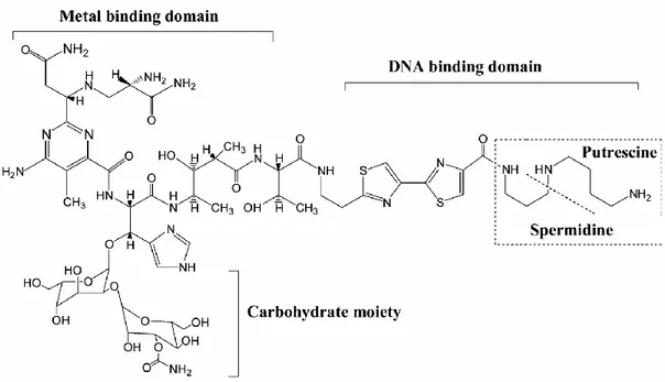
All bleomycin family members have a common structure but differ only in their amino acid side-chain at their C-terminal [21]. An example of bleomycin analogue A5 is illustrated in figure 1. Bleomycin A5 comprises three functional domains: C-terminal domain, N-terminal domain and the carbohydrate moety.
The C-terminal DNA-binding domain comprises a bithiazole moiety and a positively charged polyamine-like region [22-27]. It has been suggested that these two portions interact together with the DNA and might be involved in the sequence selective DNA cleavage.This was supported by previous studies showing that the substitution of bithiazole moity by two coumpounds; thriazole [28], and monothiazole [29] respectively resulted in altering the sequence from GC-3’ to 5’-GT-3’ [28] and loosing the ability of selective DNA cleavage [29].
The N-terminal metal-binding domain consists on β-hydroxyhistidine and pyrimidoblamic acid moieties [30]. It binds to the molecular oxygen as well as both iron and cobalt which are respectively redox-active and non-redox-active metal ions [24, 26, 31-34]. It interacts preferentially with iron, as it enhances the production of DNA lesions [35, 36].
The role of the carbohydrate moiety remains unknown, although some recent studies have shown that when bleomycin was lacking this domain, its DNA cleavage
potency became low [25, 37-39], and the ration of DSB to SSB was then reduced given that the deglycobleomycin generates essencially SSB, which were 300 fold less cytotoxic than the DSB [38].
Figure 1. Bleomycin A5 structure
Bleomycin comprises 3 major domains: N-terminal metal binding domain, C-terminal metal binding domain and the carbohydrate moiety [40].
1.1.2 Mechanism of action of bleomycin
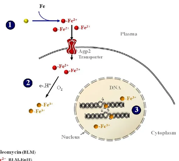
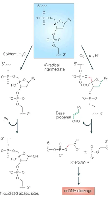
Bleomycin requires molecular oxygen and iron metal as a co-factor [41] to generate a narrow set of DNA lesions [42]. The reduced form of iron (Fe II) binds to the iron metal binding domain of bleomycin and becomes oxidized in the presence of oxygen. The complex (Blm–Fe(II)–O2) is then converted to an activated form [43, 44]
that is able to intercalate with the DNA as an oxidant causing an abstraction of the hydrogen atom from the 4’ carbon of deoxyribose, and finally resulting in the
production of an intermediate radical (Fig. 2).
Depending on the absence or presence of oxygen, this intermediate will be either oxidized, generating oxidized apurinic/apyrimidinic (AP) sites [42, 45, 46], or reacts with O2 to form a 4’peroxy radical that is reduced to a 4’hydroperoxide. The
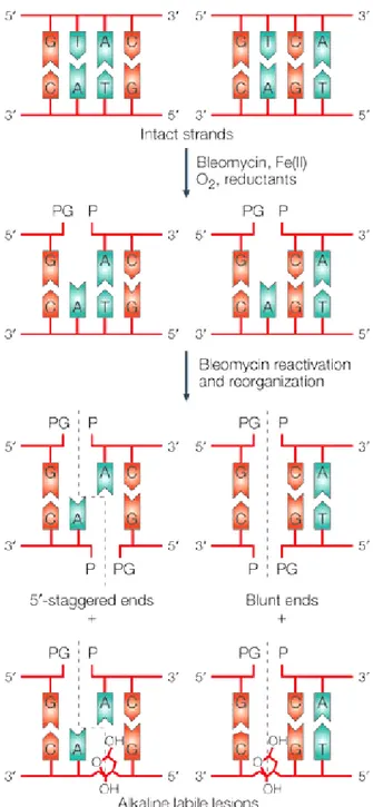
resulting product goes through chemical transformations, eventually inducing a single stranded DNA cleavage with 3’phosphoglycolate/5’-phosphate (3.-PG/5.-P) ends [42, 45, 47, 48], (Fig. 3), at a pyrimidine base (thymine or cytosine) to a guanine (5'-G-Py-3') [49]. Depending on the site of the residue 3’, which is derived from the primary cleavage [50], a secondary cleavage occurs generating either 5'-staggered ends for the sequence 5'-G-Py-Pu-3 ', or blunt ends for 5'-G-Py-Py-3’ (Fig. 4).
Such DNA lesions are considered highly genotoxic, and it is still not clear how cells could develop drug resistance to bleomycin.
Figure 2. Mechanism of generation activated bleomycin
Bleomycin binds to the iron metal by forming a bleomycin-Fe (II) complex (1), this complex becomes activated once Fe (II) is intracellularly oxidized (2), generating free radicals and causing DNA lesions (3).
Figure 3. Mechanism of generation DNA lesions
Once bleomycin is activated, it intercalates with DNA, abstracts a hydrogen atom from the carbon 4’ of desoxyribose and generates a radical intermediate. Depending on the availability of oxygen; the radical intermediate will generate either oxidative AP sites or single and double-strand break. Nature review [6].
Figure 4. Bleomycin generates double strand breaks
The activated bleomycin initiates a single-stranded DNA cleavage, followed by a second cleavage that depends on the residue 3’ created during the first cleavage. Nature review[6].
1.2 Mechanisms of resistance
Up to now, there is not a well accepted theory that is able to explain with certainty why some types of cancer respond better to the bleomycin treatment than those that are resistant. Cancer cells are selectively killed by anticancer drugs, because most of them are defective in their checkpoint responses [51-54], and proliferate rapidely; they would not have sufficient time to repair the DNA damage, and therefore keep dividing despite of unrepaired DNA. Furthermore, some cancer cells are repair-defective [55-62] in the first place, and this leads to their genomic instability [62-64].
Although, normal cells are continuously subjected to DNA damage, as well as cancer cells, they possess several pathways that lead to their resistance [65]. The identification of these pathways could provide us with an insight into the mechanism of increasing the cytotoxicity of bleomycin in specific cancer cells.
1.2.1 DNA repair pathways
Bleomycin is a radiomimetic agent that generates mostly the same types of DNA damages (DSB and AP sites) caused by ionizing radiations (IR) [66]. Its cytotoxicity is particularly related to its potency to induce DSB [67].
Unlike the IR, where the ratio of DSB to SSB is 1/20 [68], each molecule of bleomycin generates in vivo and in vitro 8 to 10 DNA breaks [50], and for 6-10 SSB, 1 DSB is produced [69]. In recent studies on Chinese hamster lung fibroblasts, it has
been demonstrated that bleomycin induced 2-3 times more DSB in G1 or G2/M phases than in S-phase [70].
In yeast, depending on the type of damage generated by bleomycin, whether it is single or double cleavage, cells can select the appropriate pathway to repair its DNA damage. When the double strand breaks (DSB) occur, the DNA repair predominantly proceeds through the homologuous recombination pathway [71] where Rad52 epistasis group proteins (Rad51, Rad52, Rad54, Rad55/57 and Rad59) are involved [72, 73].
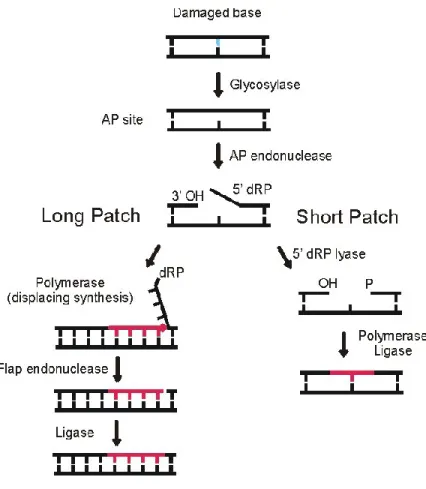
In the case of a single strand break and oxidized AP site, the DNA repair process is carried out by base excision repair (BER) pathway [74], which required the major apurinic/apyrimidinic endonuclease 1(Apn1) [75, 76] homologous to APN1 in human cells.This pathway consists of the following: first of all, the BER pathway is initiated by the glycosylase enzyme necessary for the recognition and the excision of a damaged base that generates an AP site [77, 78], which in turn recruits the endonuclease Apn1. This latter cleaves the 5’ phosphodiester bonds to the AP site yielding free 3′-hydroxyl and 5′-deoxyribose phosphate moieties [79, 80] that will be subsequently removed by DNA deoxyribophosphodiesterase (dRpase). Depending on
the total number of nucleotides that are excised through the repair process, the resulting gap is filled either by one single nucleotide (through a short-patch sub-pathway), or by two or more new synthesized nucleotides. These are incorporated by DNA polymerase through long-patch sub-pathway and sealed with DNA ligase [81], (Fig. 5).
Recent studies demonstrated that the expression level of APN1 determined by immunohistochemical analysis is increased in a number of human cancer cells, such as the prostate and the lung cancers [82-85]. It has also been supported that the increased level of APN1 observed in testicular cancer correlates with bleomycin resistance by providing a 3-fold increase in protection against bleomycin [86].
Figure 5. Base excision repair pathway
During the BER pathway, the glycolase recognizes and excises the DNA damage generating an AP site that is cleaved by Apn1 endonuclease, then a DNA polymerase is recruited to fill the gap by synthesizing one or more nucleotides through the short pach or the long patch. J Biol Chem. [81].
1.2.2 Major mechanisms leading to resistance
Current findings from Ramotar’s lab raised the possibility that the resistance to bleomycin can be related to drug efflux as well as drug uptake. As it has been demonstrated, when the AGP2 gene – a transporter of bleomycin – was mutated, it reduced the drug uptake, and hence induced an (~3000-fold) increase in resistance with high efficiency [87].
A similar effect is induced by the overexpression of an efflux pump that extrudes the drug from the cytoplasm, and thus, reduces the effective intracellular concentration [88, 89]. In agreement, it has been shown recently that the principal mechanism leading to the resistance of stem cells to chemotherapy is related to the expression of multi-functional efflux transporters in human stem cells [90].
1.3 Transcriptional co-activator Imp2 provides resistance
IMP2 is a well known gene that provides resistance against bleomycin among the S.cerevisiae genes. It was characterized as a gene encoding a transcriptional co-activator that turns on the gene expression of maltase permease (MALS), maltose permease (MALT) and galactose permease (GAL2) [91, 92].
It has been demonstrated that the IMP2 gene is required to mediate cellular resistance to oxidative DNA damaging agents that generate free radicals such as H2O2
and bleomycin. When IMP2 is deleted, mutants become 15-fold hypersensitive to bleomycin [93]. Recent findings suggest that the hypersensitivity exhibited by imp2 mutants towards bleomycin, is not due to the expression deficiency of drug efflux
pumps such as Snq2, Yor1, Flr1, Atr1 [94-100] or defect in antioxidant activity [101]. To date, there is no evidence indicating that Imp2 directly regulates genes implicated in oxidative stress response [102]. It could manifest via a specific gene target that regulates by itself the expression of an efflux pump responsible for expelling bleomycin.
1.4 Polyamines
In spite of the fact that bleomycin is a hydrophilic molecule unable to diffuse passively across the cell membrane [6], its spermidine substituent, which is positively charged, might have a role in the cellular uptake. In fact, as bleomycin has an affinity to a specific transporter (Agp2) responsible for its uptake [87], probably through the recognition of the polyamine region, then it must have an affinity to efflux pumps, that are specific to polyamines such as spermine, spermidine and putreschine. These polyamines are natural and small molecules; they are water soluble and have an aliphatic carbon chain that is positively charged [103].
1.4.1 Polyamine synthesis pathway
The polyamine synthesis pathway is initiated by the conversion of arginine – resulting from the urea cycle – into ornithine. The latter is decarboxylated to form putrescine through the action of the ornithine decarboxylase (ODC) enzyme. Meanwhile, S-adenosyl methionine (SAM), known to be involved in methyl group transfers, is converted to a decarboxylated form (DAM) by the adenosylmethionine decarboxylase enzyme (SAMD). The decarboxylated S-Adenosyl methionine donates
its aminopropyl group to putrescine to produce spermidine and spermine, by spermidine and spermine synthetase, respectively [104] .
In the polyamine catabolism pathway, spermine and spermidine are acetylated to N-acetyl SPM and N-acetyl SPD, respectively, via SPM and SPD acethyltransferase enzyme (SSAT). These two acetylated polyamines could be converted back to putrescine through a catalyzation reaction mediated by flavin-dependent polyamine oxidase [104] .
1.4.2 The importance of polyamine in the cell
Since polyamines are known to be positively charged, they bind to DNA, RNA and phospholipid macromolecules [105] that carry a negative charge, then get involved in many cellular processes including cell growth and proliferation, differentiation, chromatin structure, gene expression, transcription, signal transduction, membrane stability, ion transport, and cell signalling [106-108] .
In recent studies, it has been reported that when polyamines bind to chromatin, they alter DNA and RNA synthesis by increasing or decreasing the accessibility of genomic sites for DNA and RNA synthetases. However, when polyamines are depleted, chromatin becomes more accessible to DNA digestion. Other studies supported that polyamines like spermidine and spermine play a role in DNA condensation and segregation, and induce DNA conformational transition [103].
1.4.3 Polyamines related to cancer
Polyamine levels are tightly regulated by their influx and efflux across the plasma membrane, in such a way that their level is never extremely low or high, which would result in either failure in supporting cell growth or toxicity [108-111].
Many studies focused on cancer research, have explored the polyamine biosynthetic pathway for the development of agents that inhibit tumor growth. It has been suggested that molecules such as methylglyoxal bis-guanylhydrazone (MGBG), known as a potent inhibitor of polyamine biosynthesis [112], inhibit the proliferation of 99% of many types of cancer cells in culture, and more than 95 % of transplanted cancers in animals. This occurs by the inhibition of the S-adenosyl methionine decarboxylase enzyme (SAMDC) that is involved in the polyamine pathway [103]. Thereby, the cell is depleted from its natural polyamines, and the toxic analogue of polyamine is then accumulated into the cell resulting in cell death.
1.5 The transcriptional factor Yap1
To search for possible efflux pumps, a study was carried out that consisted of creating a multi-copy plasmid bearing the whole genomic DNA that derived from imp2 null mutant, and introduced into imp2Δ [102]. The main aim was to identify multi-copy suppressor genes that would restore the normal resistance to the imp2Δ mutant in response to bleomycin. This study showed that only 3 clones restored the normal phenotype, and all of them were carrying the entire YAP1 gene (Ramotar D., unpublished data), while no efflux pump gene was reported.
1.5.1 Yap1 structure
Yap1 was isolated based on its ability to recognize and to bind to the recognition element TTAGTCA of virus 40 (Simian vacuolating virus) [113, 114]. Yap1 possesses 3 major domains: a basic leucine zipper DNA binding domain (bZip), a C-terminal cysteine-rich region (c-CRD) and an N-terminal cysteine-rich region (n-CRD) [115] (Fig. 6). The bZip domain contains a leucine zipper domain necessary for dimerization of DNA, and a basic DNA binding domain, that share a homology with members of the mammalian Jun family of transcription factors, AP1.
Because its bZip domain contains a DNA-binding domain, Yap1 was characterized as a member of the Jun family based on transcription factors [116].
NES
NLS bZIPn-CRD c-CRD
60 150 279 313 565 650
Yap1
NLS bZIPNES
n-CRD c-CRD
60 150 279 313 565 650
Yap1
Figure 6. Schematic structure of Yap1
Yap1 comprises three conserved domains: a basic leucine zipper DNA binding domain (bZIP), a C-terminal cysteine-rich region (c-CRD) and an N-terminal cysteine-rich region (n-CRD).The Nuclear Localization Signal (NLS) is located at the N-terminal whereas the leucine-rich Nuclear Export Signal (NES) is located at C-terminal. Adapted from Nature review [115].
1.5.2 Role and mechanism of action of Yap1
In Saccharomyces cerevisiae, the basic leucine-zipper transcription factor Yap1 [116] regulates the gene expression of antioxidant enzymes like the reactive oxygen species (ROS) – removing enzymes such as: SOD1-2 CTT1 and TSA1, and also the components of the cellular thiol-reducing pathways that include the REDOX group TRR1-2 GLR1 and GRX1, necessary for maintaining the cytosol in a reduced state by NADPH [117]. In addition, Yap1 plays a major role in response to H2O2
[118, 119], and many electrophilic thiol-reactive chemicals such as N-ethylmaleimide (NEM), and the lipid peroxidation by-products 4-hydroxynonenal [120] and malondialdehyde [119].
Under non stress conditions, Yap1 is freely imported and exported from the nuclear compartiment [121, 122], whereas upon activation by oxidative stress – mainly caused by H2O2 [123], –Yap1 is rapidely redistributed in the nucleus because
of the inhibition of its nuclear exportation [121, 124].
In response to oxidative stress, the Gpx3p Peroxidase (GPX)-like enzyme initially perceives the hydrogen peroxide signal, then transduces it to Yap1p resulting in the formation of two intramolecular disulfide bonds: one is between theYap1 N-terminal CRD Cys303 and the C-terminal CRD Cys598 [117], and the other one is between the Cys310 and Cys629, it stabilize the activated form of Yap1 [125]. These two conformational changes mediate interactions with a conserved amino-terminal α-helix [115], and therefore, mask the nuclear export signal sequence in the c-CRD (C-terminal rich domain) of Yap1. This then becomes non-accessible to the Crm1
protein (β-karyopherin-like nuclear exporter), known to export Yap1 from the nucleus to the cytoplasm [121].
As a result, the oxidized Yap1 accumulates in the nucleus, where it regulates the expression of up to 71 genes (one half is regulated by Yap1 alone, whereas the second half depends on both Yap1 and the transcription factor Skn7) [123, 126, 127], through its binding to the recognition element YRE: T (T/G) ACTAA [128-130] that is located within the promoter of target genes.
1.6 Yap1 regulates the expression of efflux pumps
It has been found that Yap1 does not regulate only antioxidants, but mediates drug resistance in Saccharomyces cerevisiae by regulating the expression of efflux pumps such as: Flr1p and Ycf1p [94, 131] which are involved in diazaborine resistance.
1.6.1 Major Classes of efflux pumps
Efflux pumps are subdivided into two main classes of pumps depending on their source of energy: ATP binding cassette (ABC) transporters and multifacilitator superfamily MFS. The ABC proteins are energized by ATP hydrolysis, while MFS proteins use the proton-motive force across the plasma membrane to translocate compounds [132-138].
Most of the efflux pumps specific to polyamines are part of the MFS superfamily. They are classified in 2 sub-families: DHA1 (the drug), H+ antiporter
families have 12 putative membrane-spanning helices; whereas the DHA2 family has 14 predicted spanners [139]. Although it has been shown that many efflux pumps confer resistance to a range of structurally dissimilar compounds (including antibiotics and drugs) [140-142], the molecular mechanisms remain not well elucidated [141-146].
1.6.2 Polyamines efflux pumps confer multi-drug resistance
Among the 28 MFS [89] pumps identified to date in the Saccharomyces cerevisiae, 5 have been described: Tpo1, Tpo2, Tpo3, Tpo4 [147-150], Qdr3 [151, 152], and also Qdr2 [150].
The QDR3 gene encodes an MFS efflux pump that belongs to the DHA1 family [140]. It is located in the yeast plasma membrane [151, 152], and provides resistance to a variety of drugs including the polyamines [149], the antimalarial drug quinidine, the cisplatin, the bleomycin and the herbicide barban [152]. Its homologue – QDR2 – produces similar effects. A genome wide screen revealed that QDR3 also provides resistance to the antifungal drug fluconazole [153], selenomethionine [154], the antiarrhythmic drug amiodarone [155], and magnesium dichloride [156]. This screen was carried out by testing the sensitivity of the Qdr3 mutant to the drugs mentioned above. Unlike Qdr3, Qdr2 is involved in potassium uptake and might be capable of coupling K+ ion transport with the export of specific substrates such as quinidine, that have been shown to interfere with K+ uptake [157].
Besides Qdr2 and Qdr3, other efflux pumps have been described. Tpo1, Tpo2 Tpo3 and Tpo4 are also members of the DHA1 family as drug: H+ antiporters.
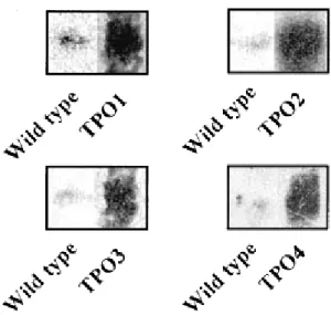
All of them share a similarity in their amino acid sequences [149], and thus have the same specificity to polyamine substrates like spermine, except Tpo1 and Tpo4 that are specific also to putrescine and spermidine, in addition to spermine [158]. Igarashi’group reported that TPO1, TPO2, TPO3 and TPO4 encoded specific polyamine transporters. They overexpressed each one of them, and then examined their role in response to methylglyoxal bis-guanylhydrazone (MGBG) [112], Co2+ – an inorganic bivalent cation – was used as a control since it does not have any effect on polyamine transport.The four overexpressed efflux pumps demonstrated resistance to 1 mM MGBG compared to the wild type, but not to Co2+, indicating that Tpo1, Tpo2, Tpo3 and Tpo4 transport specific polyamines [149].
This was confirmed by a northern blot experiment showing that cells overexpressing TPO genes have an increased level of their TPO mRNA expression, in response to 0.3 mM of spermine, but not as much as TPO1 mRNA, which was the most expressed compared to TPO2 TPO3 and TPO4 mRNA (Fig. 7).
Figure 7. Northern blot analysis
Each TPO gene was overexpressed and transformed into wild type strain. Wild types and transformed strains were cultured in the presence of 0.3mM of spermine. The expression level of TPO1, TPO2, TPO3 and TPO4 mRNA in wild types was compared to those overexpressing TPO genes by northern blot analysis Biochem Journal [149].
1.6.3 Characterisation of the MDR efflux pump Tpo1
Although these proteins have been shown to confer resistance to a range of structurally dissimilar compounds (including antibiotics and drugs) [140-142], besides polyamines, Tpo1 [148], remains the best characterized efflux pump. It is involved in the extrusion of the artesunate [159], the herbicide 2, 4-D [160, 161], and the antimalarial drugs quinidine [162].
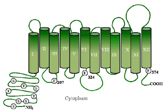
The TPO1 gene is located on chromosome XII and encodes a membrane protein consisting of 586-amino-acid residues [163], specific to putrescine, spermidine, and spermine.
In its structure, Tpo1 possesses (Fig. 8):
- 3 glutamate acids Glu-207, Glu-324 and Glu-574, necessary for polyamine transport [149, 164].
- A long hydrophilic N-terminal region rich in serine and threonine residues, including serine 19, Threonine 52. These have been shown to enhance the activity of Tpo1 when phosphorylated by the protein kinase C [150].
- Ser 342, that is located in the cytoplasmic loop between the VI and VII transmembrane segment, helps releasing Tpo1 from the endoplasmic reticulum to the plasma membrane once it is phospohrylated by cAMP-dependent protein kinases [150].
Figure 8. Model of Tpo1 structure
The DHA1 efflux pump Tpo1 has in its N-terminal glutamic acid, serine and threonine residues, necessary for its transport activity. Adapted from Biochem Journal. [164].
Tpo1 is mainly located on the yeast plasma membrane [147], and it catalyzes the efflux of polyamines at acidic pH, however when it is overexpressed, it is found in both plasma and vacuolar membrane. This has been demonstrated by a study showing that when Tpo1 is expressed from a single copy plasmid, it is detected only on the plasma membrane, whereas when it is overexpressed using a multi copy plasmid, Tpo1 is produced in many copies that will be located on plasma and vacuole membranes [150].
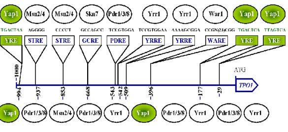
1.6.4 The regulation of TPO1 by transcriptions factors
The TPO1 gene includes within its promoter region many known specific binding sites that are recognized by specific transcription factors such as Pdr1, Pdr3, Skn7, Msn2, Msn4, Wrr1, and War1 (Fig. 9). These are involved in the transcriptional regulation of the gene depending on the type of stress that the cell undergoes [165].
For example, in response to the stress induced by the antimalarial drug artesunate, TPO1 is dependent on the zinc finger Pdr1p for its transcriptional control [159]. It also requires Pdr3 – that shares 36% of its amino acid identity with Pdr1p – and Pdr1p, in case cells are under stress, caused by the chlorophenoxy herbicides 2-methyl-4-chlorophenoxyacetic acid (MCPA) and 2,4-D [159, 160].
Based on the YEASTRACT database (www.yeastract.com), the transcription factor Yap1 is found to activate the transcription of the TPO1 gene by binding to its recognition site YRE [159], when cells are exposed to oxidative stress (H2O2).
It has been shown by Sá-Correia’s group that Yap1 is involved in resistance to polyamine, since yap1 mutant exhibits an extreme sensitivity to spermine, spermidine and putrescine compared to the wild type [166]. Consistent with this finding, another study focused especially on the role of polyamine, and demonstrated that in presence of free iron, polyamines function as pro-oxidants able to induce oxidative stress [167]. This explains why Yap1 confers resistance to polyamine as it does to H2O2.
Conferring resistance to polyamine does not necessarily mean that only one efflux pump is involved in polyamine detoxification. This might result from the transcriptional regulation of a set of genes specific to polyamine substrate such as TPO1 TPO2, TPO3, TPO4 and QDR3 [166].
Figure 9. Position of consensus sequences recognized by transcription factors.
Transcription fators control the regulation of TPO1 by binding to specific motifs. Yap1 recognized and binds to a concensus sequence located in 3 positions on the TPO1 gene: 994 (Forward strand), 29, 177 (Reverse strand). Adapted from Biotechnol.Review [159].
1.6.5 The orthologue of TPO1 in human cells
To date, no efflux pumps specific to bleomycin have been identified in mammalian cells. In the human genome, 48 ABC efflux pumps have been identified [168]. However, none of them have been shown yet to confer resistance to bleomycin, as Tpo1 does in saccharomyces cerevisiae.
Based on blast search, a tetracycline transporter-like protein (TETRAN) has been the first MFS protein characterized in human cells as an orthologue to TPO1 [169]. Since TETRAN shares some similarity with TPO1, it is that which is believed to expel specifically indomethacin (non-steroidal anti-inflammatory drugs (NSAIDs) in yeast. It is supported that TETRAN functions probably as an efllux pump in the same way as Tpo1 and confers resistance to some NSAID when overexpressed [169]. Such a protein could lead us in further studies to check its specificity for other substrates and eventually bleomycin.
Since no efflux pump specific to bleomycin has been identified in mammalian cells, we decided to use the powerful genetic system of saccharomyces cerevisiae, the simplest and most well-known representative of eukaryotic cells, for evaluating toxic effects of bleomycin and characterizing potential efflux pumps that probably have their homologuous in mammalian cells and function similarly as in yeast.
The main goal from this work is first to explore and understand how cells could develop resistance to the drug in yeast, then extrapolate the information from yeast to human. This will provide an insight into how cancer cells treated with bleomycin develop cancer.
In this work, we provide evidence that Yap1 regulates the expression of the multidrug efflux pump TPO1 that in turn provides resistance to imp2mutants toward bleomycin. However, we still do not know if this regulation is direct, or mediated by other proteins such as the transcription factor Skn7p, since we know that half of the 71 proteins induced by oxidative stress depend on both Yap1p and Skn7p [127].
2 Material and methods
2.1 Yeast strains and growth media
The yeast strains used in this work are listed in Table 1. Cells were grown either in YPD medium containing 1% Yeast Extract (MULTICELL) peptone (BIO BASIC INC) and 2% dextrose (BIO BASIC INC), or selective medias contained Yeast Nitrogen Base 10x (DIFCO), 2% dextrose (BIO BASIC INC), supplemented with the amino acids necessary for cells growth depending on the genotype.
Solid media was obtained by the addition of 1.5% Agar (MULTICELL).
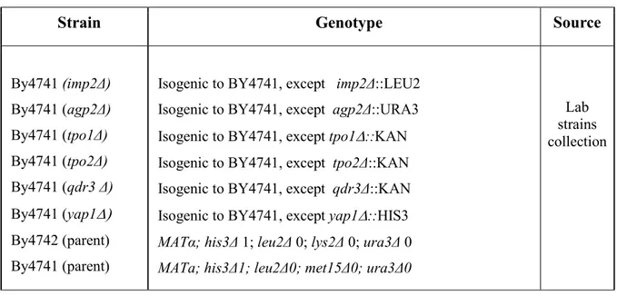
Table 1. List of strains and their genotype
Strains used in the present study:
Strain Genotype Source
By4741 (imp2Δ) By4741 (agp2Δ) By4741 (tpo1Δ) By4741 (tpo2Δ) By4741 (qdr3 Δ) By4741 (yap1 By4742 (parent) By4741 (parent)
Isogenic to BY4741, except imp2Δ::LEU2 Isogenic to BY4741, except agp2Δ::URA3 Isogenic to BY4741, except tpo1::KAN
Isogenic to BY4741, except tpo2Δ::KAN Isogenic to BY4741, except qdr3Δ::KAN Isogenic to BY4741, except yap1::HIS3 MATα; his3Δ 1; leu2Δ 0; lys2Δ 0; ura3Δ 0 MATa; his3Δ1; leu2Δ0; met15Δ0; ura3Δ0
Lab strains collection
Strains made for the present study:
Strains Genotype Source
Wt (parent) / pYCP50 Wt (parent) / pYAP1 imp2Δ / pYCP50 imp2Δ / pYAP1 tpo1Δ / pYCP50 tpo2Δ / pYCP50 qdr3 Δ / pYCP50 tpo1Δ / pYAP1 tpo2Δ / pYAP1 qdr3 Δ / pYAP1 tpo1Δimp2Δ / pYCP50 tpo2Δimp2Δ / pYCP50 qdr3Δimp2Δ / pYCP50 tpo1Δimp2Δ / pYAP1 tpo2Δimp2Δ / pYAP1 qdr3Δimp2Δ / pYAP1 BY4741 / pYCP50 BY4741 / pYAP1
Isogenic to BY4741,except imp2Δ::LEU2 / pYCP50 Isogenic to BY4741,except imp2Δ::LEU2 / pYAP1 Isogenic to BY4741, except tpo1::KAN / pYCP50 Isogenic to BY4741, except tpo2Δ::KAN / pYCP50 Isogenic to BY4741, except qdr3Δ::KAN / pYCP50 Isogenic to BY4741, except tpo1::KAN / pYAP1 Isogenic to BY4741, except tpo2Δ::KAN / pYAP1 Isogenic to BY4741, except qdr3Δ::KAN / pYAP1
Isogenic to BY4741, except tpo1 Δ ::KAN Imp2Δ::LEU2 / pYCP50 Isogenic to BY4741, except tpo2Δ::KAN Imp2Δ::LEU2 / pYCP50 Isogenic to BY4741, except qdr3Δ::KAN Imp2Δ::LEU2 / pYCP50
Isogenic to BY4741, except tpo1 Δ ::KAN Imp2Δ::LEU2 / pYAP1 Isogenic to BY4741, except tpo2Δ::KAN Imp2Δ::LEU2 / pYAP1
Isogenic to BY4741, except qdr3Δ::KAN Imp2Δ::LEU2 / pYAP1
This work Wt (parent) / pQDR3 Wt (parent) / pTPO1 Wt (parent) / pTW438 imp2Δ/ pTW438 imp2Δ/ pTPO1 imp2Δ/ pMYC-TPO1 imp2Δ/ pQDR3 tpo1ΔImp2Δ / pTW438 tpo1ΔImp2Δ / pTPO1 tpo1ΔImp2Δ / pQDR3 By4741 / pQDR3 By4741 / pTPO1 By4741 / pTW438
Isogenic to BY4741, except imp2Δ::LEU2 / pTW438 Isogenic to BY4741, except imp2Δ::LEU2 / pTPO1 Isogenic to BY4741, except imp2Δ::LEU2 / p MYC-TPO1 Isogenic to BY4741, except imp2Δ::LEU2 / pQDR3
Isogenic to BY4742, except tpo1 Δ ::KAN imp2Δ::LEU2 / pTW438 Isogenic to BY4742, except tpo1 Δ ::KAN imp2Δ::LEU2 / pTPO1 Isogenic to BY4742, except tpo1 Δ ::KAN imp2Δ::LEU2 / pQDR3
This work
Wt(parent)/ pYAP1-GFP imp2Δ/ pYAP1-GFP agp2Δ/ pYAP1-GFP
BY4741 / pYAP1-GFP Isogenic to BY4741, except imp2Δ::LEU2 /pYAP1-GFP Isogenic to BY4741, except agp2Δ::URA3 /pYAP1-GFP
This work
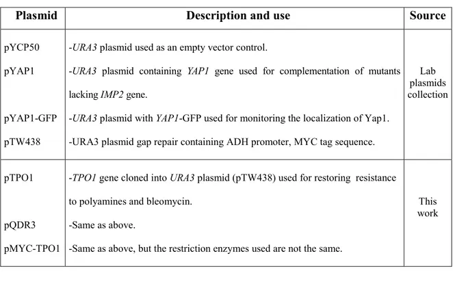
Table 2. List of plasmids
Plasmid Description and use Source
pYCP50 pYAP1
pYAP1-GFP pTW438
-URA3 plasmid used as an empty vector control.
-URA3 plasmid containing YAP1 gene used for complementation of mutants lacking IMP2 gene.
-URA3 plasmid with YAP1-GFP used for monitoring the localization of Yap1. -URA3 plasmid gap repair containing ADH promoter, MYC tag sequence.
Lab plasmids collection pTPO1 pQDR3 pMYC-TPO1
-TPO1 gene cloned into URA3 plasmid (pTW438) used for restoring resistance to polyamines and bleomycin.
-Same as above.
-Same as above, but the restriction enzymes used are not the same.
This work
2.2 Spot test
Yeast strains were inoculated in 1 ml of YPD liquid media and were grown overnight at 30 °C with agitation. The next day, cells were subcultured in 2 ml at 30 °C on the rotary shaker. After 3-4 hours of subculture, cells reached exponential phase (OD: 0.6). Serial dilutions were performed, and from each one 4μl or 5 μl were spotted on agar plates contained or not different concentration of drugs: spermidine, spermine, bleomycin, 4NQO, and H2O2, depending on the experiment. Plates were
incubated for two days at 30°C before being photographed by the ALPHA imager robot.
2.3 Spermidine excretion assay
The strains By4741, imp2Δ were harvested at A600=0.5, then washed 2 times
with a buffer containing 20mM Na/MES, pH 5.0 and 10 mM glucose, and resuspended in the same buffer. The excretion assay was initiated by the addition of
3Hspermidine (16.6 Ci /mmol Perkin Elmer) in 2ml of cells. Suspended cells were
incubated at 30 °C with shaking for 0, 2, 4 and 6 min. After each incubation, cells were washed 3 times with the buffer described above supplemented with 1 mM of cold spermidine and resuspended in 400 µl of water. The radioactivity content was measured by adding 5 ml of scintillation liquid (Amersham Biosciences) to each sample, and using liquid scintillation spectrometry.
2.4 Fluorescence microscopy
We overexpressed the YAP1 gene as a GFP fusion gene into a wild type, imp2 and agp2 strains. Transformed cells were grown in 2 ml of selective liquid media –URA and harvested at OD600 0.5. Next, the cultures were diluted and
incubated in the absence and presence of 0.5mM H2O2, and 18 mM spermidine for 10
min and 30 min respectively. Cells were first fixed in methanol 50%, then stained with DAPI (4’,6-diamidino-2-phenylindoledihydrochloride) (DAPI; 2.5 pg/ml), and viewed using a Leica DMRE microscope.
2.5 Yeast transformation
Depending on the genotype, strains were grown either in 1 ml of YPD liquid or 1 ml of selective media, over night at 30 °C. The following day, cells were sub-cultured in a fresh media, for 3-4 hours. After incubation, they were washed twice with sterilized water followed by a quick spin (30 sec) at 1000 rpm. The pellet was washed with 600 ul of TE/LiAc solution (10 mM Tris - pH 7.5, 1mM EDTA, 100 mM LiAc - pH 7.5), then centrifuged at room temperature and resuspended in 30 ul of the same solution which was sequentially supplemented with a list of components listed in the Table 2, below.
Table 3. List of the components used for transformation
Samples Volume Wild type or mutant strain Controls Cell culture SS carrier DNA Plasmid Empty vector PEG solution* 30 µl 5 µl 1-2 µl - 150 µl 30 µl 5 µl - 1-2 µl 150 µl 30 µl 5 µl - - 150 µl
Cells were incubated at 30°C for one hour, followed by a heat shock in a water bath at 42oC for 15 min. Next, we microcentrifuged them at 3000 rpm for 1 min, removed the supernatant and resuspended their pellet in 100 µl of sterilized water by pipeting up and down. This volume was plated on YPD or appropriate selective agar plates that were then incubated at 30°C. The single colonies detected after 2-3 days, were patched on new agar plates.
2.6 Bacterial transformation
2.6.1 Chemical transformation using CaCl2
2.6.1.1 Preparation of competent cells
A single bacterial DH5colony inoculated in 2 ml of LB liquid, was grown over night at 37°C with moderate shaking. The following day, cells were subcultured in 50 ml LB for 4-5 hours at 37°C, followed by a spin down at 4°C for 10 min at 4000 rpm. The supernatant was discarded while the pellet was washed twice with sterilized water, resuspended in 100 mM of cold CaCl2 and then chilledon ice for 1
hour. After ice incubation, cells were spun down. The supernatant was then spun down and the pellet recovered. At this step, one can either place the tube containing suspension cells on ice for an immediate use or can add 100 mM CaCl2 and 50 %
glycerol, then aliquote 50 µl of cells into 1.5 ml eppendorf tubes and store them at -80 °C.
2.6.1.2 Transformation of competent cells
Competent cells already stored at -80 oC were thawed gently on ice. For each transformation, 25 µl was aliquoted into pre-chilled 1.5 ml eppendorf tubes, and then 1-2 µl of DNA (plasmid of genomic DNA) was added to the aliquot. At this step, 50 µl can also be used instead of 25 µl.
The tube containing the mix of the DNA and cells was incubated on ice for 30 min followed by a heat shock in a water bath at 42 °C for 90 sec.
After that, the tube was left on ice to cool for 1-2 min, then 1 ml of LB was added to the culture and incubated in the shaker at 37 °C for 1-2 hours.
After incubation, cells were spun down at high speed for 5 min and the pellet was recovered only with 200 µl of its supernatant. This was then plated onto LB agar media supplemented with 100 µg/ml Ampicillin. Plates were inverted and incubated at 37 °C over night. After 14-16 hours bacterial colonies appeared.
2.6.2 The electroporation
2.6.2.1 Preparation of electrocompetent cells
A single bacterial DH5colony picked from the LB plate or from the frozen glycerol stock of bacterial cells, was inoculated into 3 ml LB media liquid, and incubated over night at 37°C in the shaker. The next day, cells were subcultured in 30 ml of fresh LB media at 37°C. After 4-5 hours of incubation, cells were washed with MiliQ water 3 times and centrifuged at 4°C for 10 min at 4000 rpm. The pellet was re-suspended in 20 µl of miliQ water for immediate use.
2.6.2.2 Electroporation of electrocompetent cells
0.5 -1 µl of transforming DNA was added to 20 µl of electrocompetent cells, the content was mixed gently, then placed along walls of a 0.1 cm prechilled cuvet on ice for 30 min. After the incubation, cells were electroshocked according to the conditions below:
-The voltage: 2.5 kV -The capacitance: 25 F -The resistance: 200 ohm -Time constant: 2.5-2.6
Once cells are electroporated, 0.5 ml of LB media is added immediately, and the transformed cells are transferred to a 1.5 ml eppendorf tube then incubated at 37 °C for 30-60 min. After the incubation, cells were spread on LB agar media + 100ug/ml Ampicillin, and incubated at 37 °C over night. After 14-16 hours bacterial colonies appeared.
2.7. Plasmid extraction from E.Coli
One single colony picked from a LB/Amp transformation plate, was inoculated into 4 ml LB liquid containing 100ug/ml Ampicillin, and grown over night at 37°C in the shaking incubator. The next day; the sample was centrifuged at high speed (4000 rpm) for 10 min. The supernatant was carefully poured-off and the bacterial pellet was resuspended in 0.1 ml of solution 1 (50mM glucose, 25 mM Tris-HCl - pH 8.0 and 10 M EDTA - pH 8.0).
Next, 0.2 ml of solution 2 (1% SDS, 0.2 N NaOH) was added, and the contents was mixed by inverting the tube gently 5 times, then the tube was left to stand at room temperature. After 5 min of incubation, 0.15 ml of solution III (3 M potassium acetate K+, 5M glacial acetic acid) was added, and then the tube was incubated on ice for 10 min, and centrifuged at 12000 rpm for 5 min.
In order to get rid of cell debris, the supernatant was transferred to a new eppendorf, to which was added 300 µl phenols twice, followed by adding the same volume of chloroform, twice also. The DNA was pelleted by centrifugation at 12000 rpm for 5 min, washed with ethanol 95%, and then span down at the same speed for 3 min.
After this step, the ethanol was poured out and the pellet was dried in the fume hood. Once the ethanol had evaporated, we dissolved the pellet in 50 µl of dH2O, then
stored it at -20 °C.If necessary, before storing, the DNA can be incubated with RNase for 30 min at 37 °C, however this step is not required in case we use the plasmid mini-prep kit.
2.8. Yeast Genomic DNA extraction
2.8.1 The procedure for extracting genomic DNA
Yeast cells were grown in 5 ml of appropriate media over night at 30°C. Next, they were centrifuged in 15 ml conical tubes at 4000 rpm for 1 min at 4 °C and then resuspended in 200 µl of breaking buffer (TE, 100 mM CaCL2 and 0.1 % SDS) by
by 200 µl of acid washed glass beads (425-600 micron). We capped the tubes tightly, and vortexed them for 2 min by placing them inside a mini bead beater. After repeating this step 2 times more, we spun the cells for 5 min at 15000 rpm to separate the lysed cells from the upper phase that we collected in a new tube.
300 µl of 6M NaI and 10 µl glassmilk were added and mixed with 100 µl of supernatant. After 5 min of vortexing at high speed, the sample was pelleted by centrifugation and washed 3 times with 400 µl of new wash buffer. After the last wash, we let the tube drain at room temperature on a clean paper towel until the pellet get dried , then resuspended in 10-100 µl of dH2O.
At this point, the sample could be centrifuged to separate the glassmilk from the supernatant containing dissolved DNA then used immediately, or alternatively stored at -20 °C.
2.8.2 Purification of extracted genomic DNA
In order to purify the extracted genomic DNA, 1/10 volume of 3 M NaAc and 2 volumes of 100% ethanol were added and mixed with the sample by inverting the tube many times, before incubation at -80°C for 30 min.
Next, the sample was spun down at 12000 rpm for 15 min, and the pellet was washed with ethanol (70%) followed by a short spin down (1 min). Finally, the pellet was air-dried and the DNA was resuspended in 20 µl of dH2O and stored at -20 °C.
2.9 RT-PCR
2.9.1 RNA extraction
RNA was extracted using a RiboPure-Yeast extraction kit (Ambion). 3 ml of yeast culture, grown overnight at 30 °C, was centrifuged at high speed. We discarded the supernatant while resuspending the pellet in 480 μl Lysis Buffer followed by 48 μl 10% SDS and 480 μl Phenol: Chloroform: IAA. After adding these lysis reageants, we transferred the cell mixture to the tubes already filled with 750 µl of cold zircona beads. Tubes were capped tightly then vortexed at high speed by using the bead beater. After 10 min of vortexing, cells were spun down at 16,000xg for 6 min to separate the pellet and the beads from the aqueous phase containing RNA that we transferred to 15 conical tubes. 1.9 ml binding buffer, followed by 1.25 ml ethanol (100%) were added and mixed thoroughly with the partially purified RNA. This RNA mixture was drawn through the filter cartridge, collected in new tubes, then centrifuged for 1-2 min at high speed. Once done, we washed the filters with wash solutions (700 µl of wash solution 1, 500 µl of wash solution 2/3, twice). They were then transferred to new tubes in which we eluted the RNA in 25-50 µl of elution solution.
Once the RNA is isolated, it is recommended to treat it with DNase since RNA contains chromosomal DNA. In order to remove contaminating DNA, we incubated 50-100 µl of the RNA sample with 4 μl DNase I (8 U) in 1/10 volume 10X DNase 1 buffer at 37 °C in a water bath. After 30 min of digestion, we inactivated the DNase by adding 0.1 volume DNase inactivation reagent, and then we let the reaction
stand at room temperature for 5 min. The sample was centrifuged for 2-3 min at 10,000 xg. Finally the total RNA was transferred to a new tube and its absorbance was measured at 260 nm.
2.9.2 RNA denaturing agarose gel electrophoresis
10 ml 10X MOPS running buffer (0.3 M MOPS - pH 7.0, 0.1 M sodium acetate,0.01 M EDTA) and 18 ml of 37% formaldehyde were added to 1 g agarose already dissolved in 72 ml water. The gel was poured, then covered by 1X MOPS running buffer. For RNA samples, we added 0.5-3X volumes of formaldehyde load dye to 1-3 µg RNA, before heating the sample to 65-70°C for 5 min to denaturate RNA, and then loading it.
2.9.3 The synthesis of cDNA
For cDNA synthesis, we added 0.2 µl of random primers and 1 µl of 10 mM dNTP to each RNase free tube containing 2 -5 µg of total RNA and we filled it to 12 µl with sterilized distilled water. The mixture was heated to 65 °C for 5 min, and immediatly cooled on ice. We spun down the sample then added 4 µl 5X First-Strand Buffer, 2 µl 0.1 M DTT and 1 µl RNase OUTTM (40 units/µl). We mixed gently the sample that we placed in a water bath at 37 °C for 2 min. After 2 min of incubation, 1 µl (200 units) of M-MLV RT (Reverse transcriptase) was gently mixed with the contents of the tube that we incubated for 10 min at 25 °C, 50 min at 37°C, and 15 min at 70 °C. At this step cDNA is ready to use as a template for PCR reactions.
2.9.4 PCR reaction
We designed the following primers using Gene Runner software, to assess the expression level of TPO1 and QDR3:
-RT-PCR-TPO1-F: 5'- TTATATCGCACAAAGAACTACC-3' -RT-PCR-TPO1-R: 5'- GCATAATGATAGGCCTCGTC-3' -RT-PCR-QDR3-F: 5'- ATGCAAGCCCAAGGTTCAC-3' -RT-PCR-QDR3-R: 5'- TCCTCACTCTTCAACCTAC-3'
We used the following primers already designed by a previous student, to detect the expression of AGP2, by using the ACTIN as a control:
-RT-PCR-AGP2-F 5’-TTGATTGCTATTTCCGGTGTCA-3’ -RT-PCR-AGP2-R 5’GGGCCCTCCACGGTATAAAG-3’
-RT-PCR-ACT1-F1 5'- GTTTTGCCGGTGACGACGCTCCTCGTGCTG-3' -RT-PCR-ACT1-R1 5'-CGGCTTGGATGGAAACGTAGAAGGCTGGAACG-3'
The master mix was prepared by adding these reagents: 1-4 µl cDNA, 5 µl 10 X taq polymerase buffer, 1 µl of forward primer, 1 µl of reverse primer, 1 µl of taq polymerase (5U/µl) and 5 µl of 10mM dNTP. We filled up the tube with sterilised water to 50 µl.
The PCR designed programs are:
-TPO1: 1- 95 ºC for 2 minutes, 2- 94 ºC for 0.5 minute, 3- 58 ºC for 0.5 minute, 4- 72 ºC for 2.20 minutes, 5- 22 times repeat of steps 2-4, 6- 72 ºC for 7 minutes, and 7- 4 ºC.
-QDR3: 1- 95 ºC for 2 minutes, 2- 94 ºC for 0.5 minute, 3- 51 ºC for 0.5 minute, 4- 72 ºC for 2.20 minutes, 22 times repeat of steps 2-4, 6- 72º C for 7 minutes, and 7- 4 ºC.
-AGP2:1- 95 ºC for 2 minutes, 2- 94 ºC for 0.5 minute, 3- 55 ºC for 0.5 minute, 4- 72 ºC for 2.30 minutes, 22-30 times repeat of steps 2-4, 6- 72 ºC for 2:30 minutes, and 7- 4 ºC.
2.10 Plasmid constructions
2.10.1 Primers design
We used the plasmid pTW438 as a substrate in the gap repair assay. It contains a Myc tag, URA cassette and an ADH promoter.
It was digested with HindIII and SmaI. The linear DNA was gel purified. The TPO1 and the QDR3 genes were amplified by PCR using genomic DNA of the strain BY4741 as a template, then purified. The following PCR primers designed for untagged TPO1 and QDR3 are:
Table 4. Design of PCR primers for TPO1 and QDR3 genes
Primers for TPO1: The product size is 1.7 kbp Primers for QDR3: The product size is 2 kbp
Table 5. Design of PCR primers for TPO1 gene tagged with MYC tag Name Sequence TPO1-F TPO1-R 5’-GCACAATATTTCAAGCTATACCAAGCATACAATAAGCTTCTCACC ATGTCGGATCATTCTCCCAT-3’ 5’-TATGTAACGTTATAGATATGAAGGATTTCATTCGTCTGTCGACCCT TAAGCGGCGTAAGCATACT-3’ QDR3-F QDR3-R 5’-GCACAATATTTCAAGCTATACCAAGCATACAATAAGCTTCTCACC ATGCAAGCCCAAGGTTCACA-3’ 5’-TATGTAACGTTATAGATATGAAGGATTTCATTCGTCTGTCGACCC TTAATCAATTTTGTCGTACA-3’ Gene Sequence MYC-TPO1-F MYC-TPO1-R 5'-ACCATGGCGTCCGAGCAAAAGCTCATTTCTGAAGAGGACTTGCGG TCGGATCATTCTCCCATTTC-3’ 5’-TATGTAACGTTATAGATATGAAGGATTTCATTCGTCTGTCGACCC TTAAGCGGCGTAAGCATACT-3’