The effects of administration of a nuclear factor κB inhibitor on pulmonary endothelial dysfunction after cardiopulmonary bypass:
impact on oxygenation and hemodynamics and development of therapeutic and preventive modalities in a porcine model
Par Cristian Rosu M.D.
Programme de Sciences Biomédicales Université de Montréal
Faculté de Médicine
Mémoire présenté à la Faculté des Études Supérieures en vue de l’obtention du grade de
Maîtrise en Sciences Biomédicales January 2013 ©, Cristian Rosu, 2013
Les effets sur la dysfonction endothéliale pulmonaire de l’administration d’un inhibiteur du facteur nucléaire κB: impact sur l’hémodynamie et l’oxygénation et le développement de modalités thérapeutiques et préventives dans un modèle porcin.
Rosu C, Denault AY, Carrier M, Perrault LP
Introduction: La circulation extracorporelle (CEC) peut entraîner une dysfonction endothéliale pulmonaire et l’hypertension pulmonaire. Le SN50 agit au niveau de la signalisation cellulaire pour prévenir ces réactions à la CEC et pourrait renverser la dysfonction endothéliale pulmonaire post-‐CEC sans effets néfastes sur
l’hémodynamie.
Méthodes: Quatre groups de porcs ont reçu un parmi quatre traîtements avant de subir 90 minutes de CEC et 60 minutes de reperfusion: (1) milrinone nébulisé; (2) sildenafil nébulisé; (3) placebo nébulisé; et (4) SN-‐50 intraveineux. Un monitoring hémodynamique invasif a été utilisé. La réactivité vasculaire des artères
pulmonaires de deuxième ordre a été évaluée face à l’acétylcholine et la bradykinine.
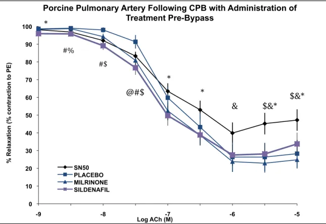
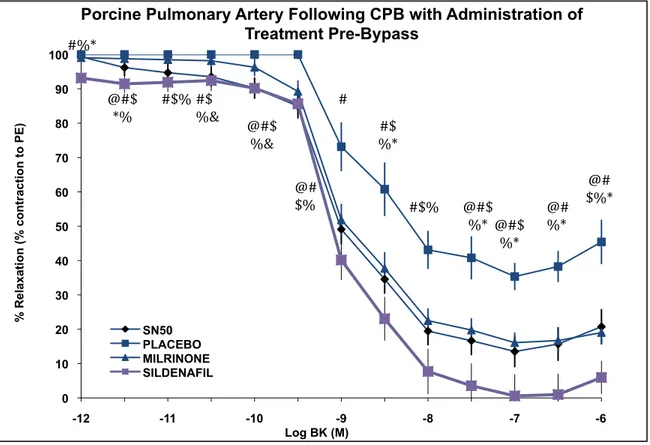
Résultats: Le sildénafil produit une augmentation significative de la pression de l’artère pulmonaire (PAP) moyenne à 60 minutes de reperfusion par rapport au début de la chirurgie. Les relaxations dépendantes de l’endothélium face à la
bradykinine étaient meilleurs dans les groupes milrinone et SN-‐50 et surtout dans le groupe sildénafil par rapport au groupe placébo. Le SN-‐50 produisait de moins bonnes relaxations dépendantes de l’endothélium face à l’acétylcholine que les autres traitements incluant placébo.
Conclusion: Le sildénafil prévient mieux la dysfonction endothéliale pulmonaire que les autres traitements. Les bénéfices du SN-‐50 sont possiblement sous-‐estimés vu que la dose n’a pas pu être ajustée à la durée de CEC. Le sildenafil inhalé mérite une étude plus importante chez l’humain et le SN-‐50 dans un model de CEC animal.
Dysfonction endothéliale – hypertension pulmonaire – circulation extracorporelle– facteur nucléaire κB – inhibiteurs de la phosphodiestérase
The effects of administration of a nuclear factor κB inhibitor on pulmonary endothelial dysfunction after cardiopulmonary bypass: impact on oxygenation and hemodynamics and development of therapeutic and preventive
modalities in a porcine model.
Rosu C, Denault AY, Carrier M, Perrault LP
Background: Cardiopulmonary bypass (CPB) can lead to pulmonary endothelial dysfunction and consequent pulmonary hypertension. The novel agent SN-‐50 acts at the level of the transduction pathway to prevent these responses and may limit or reverse post-‐CPB pulmonary endothelial dysfunction and pulmonary hypertension without the untoward effects on hemodynamics seen with other known therapies. Methods: Four groups of Landrace-‐Yorkshire swine that received one of four treatments before undergoing 90 minutes of normothermic CPB and 60 minutes of reperfusion were compared: (1) Nebulized milrinone; (2) nebulized sildenafil; (3) placebo consisting of nebulized NaCl solution; and (4) intravenous SN-‐50. Invasive hemodynamic monitoring was used throughout all experiments. Vascular reactivity of second-‐degree pulmonary arteries was evaluated in response to acetylcholine and bradykinin.
Results: Sildenafil produced a significant increase in mean pulmonary artery pressure (PAP) at 60 minutes after CPB compared to baseline. Both the sildenafil and milrinone groups had increased mean PAP/MAP ratio at 60 minutes after CPB compared to baseline, however this ratio was not different between the groups. Endothelial-‐dependent relaxations to bradykinin were improved in the SN-‐50 and milrinone groups and especially the sildenafil group as compared to placebo. SN-‐50 produced worse endothelium-‐dependent relaxations in response to acetylcholine compared to the other groups including placebo.
Conclusion: Sildenafil better prevented pulmonary endothelial dysfunction than all other treatments. The improvements seen with SN-‐50 may be suboptimal as dose could not be titrated to length of CPB. Inhaled sildenafil and SN-‐50 both merit further study in human trials and animal models, respectively.
Endothelial dysfunction – pulmonary hypertension – cardiopulmonary bypass – nuclear factor κB – phosphodiesterase inhibitors
TABLE OF CONTENTS RÉSUMÉ……….….ii SUMMARY………...iii TABLE OF CONTENTS………..………iv-v LIST OF FIGURES……….………vi-vii LIST OF ABBREVIATIONS………..……….viii-xiii ACKNOWLEDGEMENTS………..……….…xiv
FIRST CHAPTER : Introduction……….………1
SECOND CHAPTER : The vascular endothelium………..4
Endothelial cell anatomy……….………..5
Normal endothelial cells functions……….………..5
Control of vascular tone……….………6
Control of growth of vascular smooth muscle cells……….………8
Vasorelaxant factors……….……….10
Nitric Oxide……….………..10
Prostacyclin (PGI2) ……….……….13
Endothelium-derived hyperpolarizing factor (EDHF) ……….………..14
Adenosine……….……….16
Bradykinin……….………16
Histamine……….……….17
Vasoconstricting factors (EDCFs) ……….………18
Prostaglandins/thromboxanes……….………18
Endothelin……….………19
The influence of the renin-angiotensin system……….………...21
Angiotensin II……….………...22
Humoral control of circulation……….……….23
Epinephrine and norepinephrine……….………23
Vasopressin……….………..24
Reactive oxygen and nitrogen species……….……….25
Nuclear factor κB……….………...26
THIRD CHAPTER : The physiologic response to cardiopulmonary bypass……….30
Hemostatic system……….……….32
Anticoagulation……….………32
Prothrombotic factors……….………..34
Fibrinolysis and coagulopathy……….……….38
Inflammatory effects……….……….40 Complement system……….……….40 Neutrophils……….………...41 Monocytes……….……….42 Lymphocytes……….……….43 Platelets……….………43 Endothelial cells……….………...44
Pulmonary injury……….………46
FOURTH CHAPTER : Pulmonary hypertension in cardiac surgery……….………..53
FIFTH CHAPTER : Options for treatment of post-cardiopulmonary bypass pulmonary hypertension……….……….…………..59
Non-‐pharmacologic therapies……….………..60
General cardiovascular management……….………...62
Inhaled nitric oxide……….………62
Prostanoids and prostanoid analogs……….………66
Endothelin receptor antagonists……….………..70
Phosphodiesterase inhibitors……….………...72
Anti-‐inflammatory agents and transduction pathway inhibitors……….……..78
HYPOTHESIS AND OBJECTIVES….……….………..82
MATERIALS AND METHODS……….……….84
RESULTS………91
DISCUSSION………101
CONCLUSION………..116
FIGURES
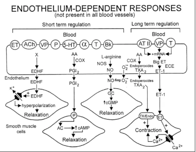
Second Chapter:Figure 1 : Variety of endothelium-derived relaxing and contracting factors.
AA: arachidonic acid; AC: adenylate cyclase; ACh: acetylcholine; ATII: angiotensin II; BK: bradykinin; COX: cyclooxygenase; ECE: endothelin converting enzyme; EDHF: endothelium-derived hyperpolarizing factor; ET: endothelin-1; O2
-: superoxide anions; P-: purines; PGI2: prostacyclin; NO: nitric oxide; NOS: nitric oxide synthase; T: thrombin; TX/Endo: TP-receptor; VP: vasopressin; TXA2: thromboxane A2; HT:
5-hydroxytryptamine (serotonin); α: alpha-adrenergic. [1] ………….. p. 8
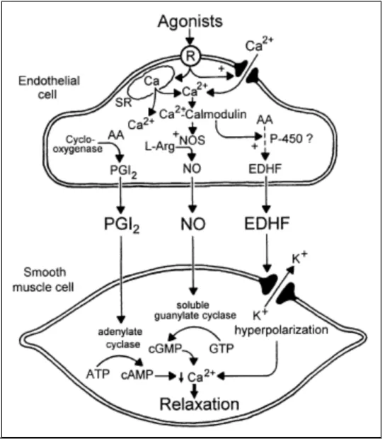
Figure 2 : Release of endothelium-derived relaxing factors and their effects on
vascular smooth muscle cells
AA: arachidonic acid; cAMP: cyclic adenosine monophosphate; cGMP: cyclic
guanosine monophosphate; EDHF: endothelium-‐derived hyperpolarizing factor; L-‐ Arg: L-‐arginine; NO: nitric oxide; NOS: nitric oxide synthase, PGI2: prostacyclin; R:
cell surface receptor; SR: sarcoplasmic reticulum. [2]……….. p. 10
Third Chapter:
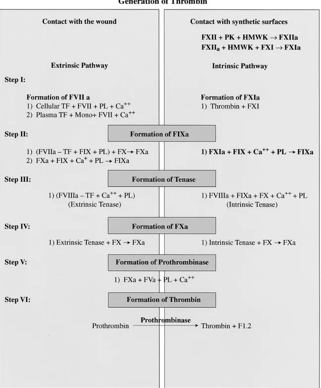
Figure 3 : Steps in the generation of thrombin in the wound and in the CPB circuit via
the extrinsic, intrinsic, and common coagulation pathways
HMWK: high-‐molecular-‐weight kininogen; mono: monocyte; PK: prekallikrein; PL: cellular phospholipid surface; TF: tissue factor; activated coagulation proteins are indicated by the suffix "a." [3] ……… p. 37
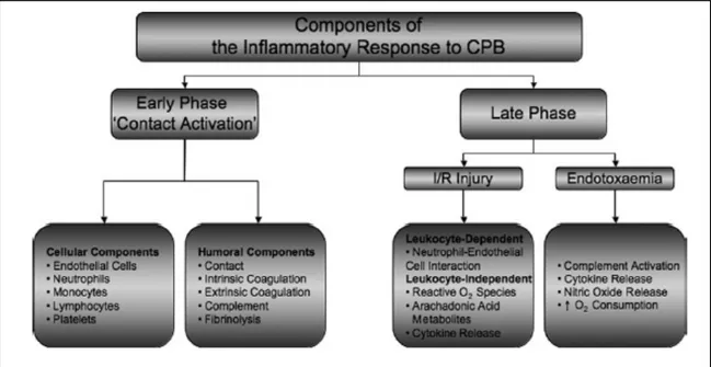
Figure 4 : Summary of the inflammatory response to CPB
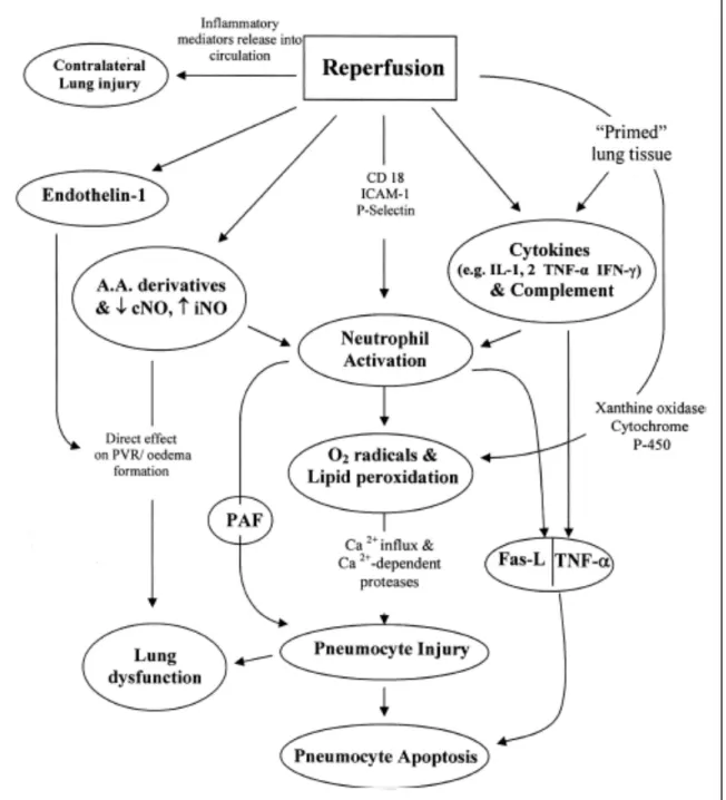
CPB: Cardiopulmonary bypass; I/R: ischemia-‐reperfusion injury. [4] ……… p. 40 Figure 5 : Inflammatory response following reperfusion of ischemic lung
AA: arachidonic acid; ICAM-‐1: intracellular adhesion molecule-‐1; IFN-‐γ: interferon-‐ γ; IL: interleukin; cNO: constitutive nitric oxide; iNO: inducible nitric oxide; PAF: platelet activating factor; PVR: pulmonary vascular resistance; TNF-‐α: tumor necrosis factor-‐α. [5] ………. p. 48
Fourth Chapter:
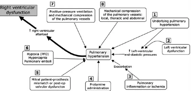
Figure 6 : The most common mechanisms contributing to pulmonary hypertension in cardiac surgery.
PFO: Patent foramen ovale. [6] ……… p. 57
ABBREVIATIONS
5-‐HPETE: 5-‐hydroperoxyeicosatetraenoic acid 12-‐HHT: 12-‐hydroxyheptadecatrienoic acid 15-‐HETE: 15-‐hydroxyeicosatetraenoic acid
20-‐HETE: 20-‐hydroxy-‐5,8,11,14-‐eicosatetraenoic acid AI: Angiotensin-‐I
AII: Angiotensin-‐II
ACE: Angiotensin-‐converting enzyme AMP: Adenosine monophosphate ACh: Acetylcholine
ACT: Activated coagulation time ADH: Antidiuretic hormone ADP: Adenosine diphosphate
ARDS: Acute respiratory distress syndrome AT: Angiotensin
AT1: Angiotensin receptor type 1
AT2: Angiotensin receptor type 2
ATP: Adenosine triphosphate AVP: Arginine vasopressin B1: Bradykinin receptor type 1
B2: Bradykinin receptor type 2
BAL: Bronchoalveolar lavage BET-‐1: Big endothelin-‐1
BH4: Tetrahydrobiopterin
BK: Bradykinin
BKCa: Calcium-‐sensitive potassium channel
cAMP: Cyclic adenosine monophosphate CABG: Coronary artery bypass grafting cGMP: Cyclic guanosine monophosphate COX: Cyclooxygenase
CPB: Cardiopulmonary bypass DAG: Diacyl-‐glycerol
DNA: Deoxyribonucleic acid
DHCA: Deep hypothermic circulatory arrest ECE: Endothelin-‐converting enzyme
EDHF: Endothelium-‐derived hyperpolarizing factor EDRF: Endothelium-‐derived relaxing factor
EET: Epoxyeicosatrienoic acid EFA: Essential fatty acid
EGF: Endothelial growth factor
ELAM: Endothelial leukocytes adhesion molecule eNOS: Endothelial nitric oxide synthase
ERA: Endothelin-‐1 receptor antagonist ET-‐1: Endothelin-‐1
ETA: Endothelin receptor type A
F1.2: Prothrombin fragment FRC: Functional residual capacity GPCR: G-‐protein couple receptor H2O2: Hydrogen peroxide
HMWK: High-‐molecular-‐weight kininogen ICAM-‐1: Intercellular adhesion molecule-‐1 ICU: Intensive care unit
IGF-‐1: Insulin-‐like growth factor-‐1 IκB: inhibitor of κB
IKK: IκB kinase IL: Interleukin
inhNO: Inhaled nitric oxide
iNOS: Inducible nitric oxide synthase IP3: Inositol 1,4,5-‐trisphosphate
IR: Ischemia-‐reperfusion
JGA: Juxtaglomerular apparatus L-‐NMMA: NG-‐monomethyl-‐L-‐arginine
LT: Leukotriene LV: Left ventricle
MAP: Mean arterial pressure N2O3: Dinitrogen trioxide
NADPH: Nicotinamide adenine dinucleotide phosphate NE: Norepinephrine
NF-‐κB: Nuclear factor kappaB
nNOS: Neuronal nitric oxide synthase NO: Nitric oxide
NO2: Nitrogen dioxide
NOS: Nitric oxide synthase O2-‐: Superoxide
OH-‐: Hydroxyl radical
ONOO-‐: Peroxynitrite
PAF: Platelet activating factor
PAH: Pulmonary arterial hypertension PAI-‐1: Plasminogen activator inhibitor-‐1 PAP: Pulmonary artery pressure
PDE: Phosphodiesterase
PDGF: Platelet-‐derived growth factor PDTC: Pyrrolidine dithiocarbamate PE: Phenylephrine
PEEP: Positive end-‐expiratory pressure PGE2: Prostaglandin E2
PGF1: Prostaglandin H2
PGH2: Prostaglandin H2
PGI2: Prostaglandin I2, Prostacyclin
PHTC: Pulmonary hypertensive crisis PIP2: Phosphatidyl 4,5-‐bisphosphate
PMN: Polymorphonuclear cell ppm: Parts per million
PSGL-‐1: P-‐selectin glycoprotein ligand-‐1 PVR: Pulmonary vascular resistance RAP: Right atrial pressure
RAS: Renin-‐angiotensin system RHD: Rel homology domain RNS: Reactive nitrogen species ROS: Reactive oxygen species RV: Right ventricle
RVEF: Right ventricular ejection fraction SNP: Sodium nitroprusside
SVR: Systemic vascular resistance TNF: Tumor necrosis factor
t-‐PA: Tissue plasminogen activator TPG: Transpulmonary gradient TXA2: Thromboxane A2
TXB2: Thromboxane B2
V1: Vasopressin receptor type 1
V1a: Vasopressin receptor type 1a
V2: Vasopressin receptor type 2
VCAM-‐1: Vascular cell adhesion molecule-‐1 VSMC: Vascular smooth muscle cell
Acknowledgements
This project has been a particular challenge, which makes it a particular pleasure to see it come together. The three years that have passed since this project have seen me grow enormously not only as a student and learner but also as doctor and as a person. I could not have made it through the process of obtaining this degree while simultaneously learning the intricacies of my future profession without the help and support of many people.
I would first like to thank my parents for their continuing, enduring, unconditional support. It would have been difficult to succeed in any of my projects without their help and encouragement. I would also like to thank my closest friends Gordan and Nancy for enduring me during the rollercoaster ride that is obtaining a master’s degree while simultaneously becoming a cardiac surgeon.
A big thank you to the team from the lab, specifically Stephanie Blanchet, Marie-‐ Pierre Mathieu, and Célia Sid-‐Otmane, for teaching me the inner workings of the lab, for all their hard work, and for accommodating to my “dual life.” I’d also like to thank the perfusionists without whom none of the experiments would have been possible, Clotilde Perrault-‐Hébert and Thierry Lamarre Renaud.
Last but not least, a special thank you to my research director, Dr Louis P. Perrault. Thank you for bringing me into your lab and allowing me to work with your great team and for your valuable advice.
FIRST CHAPTER
INTRODUCTION
Since the introduction of cardiopulmonary bypass into clinical practice in 1953 with John Gibbon’s successful closure of an atrial septal defect using cardiopulmonary byass (CPB) [7], millions of patients around the world have undergone successful open-‐heart surgery with CPB. However, while permitting the execution of complex and often life-‐saving surgery on the heart and great vessels, cardiopulmonary bypass induces significant disturbances in hemostasis, electrolyte balance and rheology, vascular autoregulation, and generally minor disturbances in most organ systems, as well as inducing a systemic inflammatory response. These effects may lead to grave complications, including post-‐CPB pulmonary hypertension with resultant right ventricular failure, cardiogenic shock, multiorgan dysfunction, and death. This infrequent yet often catastrophic complication is poorly understood and remains a therapeutic challenge.
Vascular endothelium has long been known to play roles in metabolism, coagulation, and transport. Furchgott and Zawadzki’s [8]discovery in 1980 of the obligatory role of the endothelium in the acetylcholine-‐induced relaxation of isolated arteries presented an additional function for these cells as modulators of the contractions and relaxation of the surrounding vascular smooth muscle. Endothelial cells regulate basal vascular tone as well as smooth muscle reactivity in response to mechanical stimuli and a variety of neurohormonal mediators via the production and release of relaxant and contracting factors. In time, physiologists and
pharmacologists discovered not only the significance of endothelial function in normal cardiovascular physiology but also notably the importance of disturbed endothelial function in many cardiovascular disorders, including atherosclerosis,
hypertension, heart failure, cardiac allograft rejection, pulmonary hypertension, and others [9]. Increasing understanding of the role of endothelial dysfunction in post-‐ CPB pulmonary hypertension has slowly lead to more tailored approaches and therapies. A number of pharmacological agents have been developed to combat pulmonary hypertension in this and other contexts including intravenous, and later inhaled, phosphodiesterase inhibitors, inhaled nitric oxide, endothelin inhibitors, and prostaglandins. Their efficacy has often been found to be limited while some have repercussions on the systemic circulation that prevent optimal usage.
Alternative therapies have been sought and glucocorticoids have shown promise in the prevention of post-‐CPB pulmonary hypertension in experimental animals [10]. Glucocorticoid effects are however non-‐selective and may have unintended negative effects and consequently efforts are being made to develop agents that act more selectively downstream in the signal transduction pathway targeted by
glucocorticoids. Several agents, all targeting nuclear-‐factor κB, have shown efficacy in different animal models [11-‐13].
SECOND CHAPTER
Endothelial Cell Anatomy
The internal structure of an endothelial cell is no different from the majority of other human cells. A nucleus and a number of other organelles are found within the cytoplasm and are all contained within a phospholipid bilayer membrane. This cell membrane is crossed by complex proteins that may serve as receptors for blood-‐ borne ligands or as transmembrane channels that allow the passage of substances into and out of the cell. It lies as a single monocellular layer separating the
circulating blood from the deeper layers of the vascular wall. Also, contractile
proteins that give the cell motor activity traverse the cytoplasm. These include actin, myosin, tropomyosim, and α-‐actin and they permit conformational changes of the cell in response to specific stimuli such as shear stress, which causes endothelial cells to flatten and align in the direction of blood flow [14].
Normal Endothelial Cell Functions
Vascular endothelial cells are very active metabolically, producing a variety of vasoactive mediators creating a delicate balance between vasodilatation and vasoconstriction and between thrombosis and anticoagulation. Production and release of nitric oxide (NO) is central to the regulatory role of endothelium, as the final common pathway in a number of important homeostatic mechanisms [9]. Through the production and secretion of NO and other molecules, endothelium inhibits the contraction, migration, and proliferation of vascular smooth muscle cells (VSMC) [9, 15].
The endothelium also plays an important role in the prevention of inappropriate thrombosis, through the following mechanisms: the surface expression of ecto-‐
adenosine phosphatase, which degrades adenosine diphosphate thus inhibiting platelet aggregation, heparan sulfate proteoglycan, a cofactor for antithrombin III, and thrombomodulin as well as the secretion of soluble factors such as tissue-‐factor pathway inhibitor, tissue-‐plasminogen activator, and the platelet-‐inhibitors NO and prostacyclin [16]. Simultaneously, the endothelium produces coagulation factors such as von Willebrand factor and tissue factor and the antifibrinolytic plasminogen activator inhibitor-‐1 (PAI-‐1) in order to maintain an hemostatic equilibrium.
Inflammation is also under the regulatory influence of the endothelium. Leukocyte migration and adhesion to the endothelium is downregulated by NO [9]. The dimension of the intercellular space is determined by contractions and relaxations of the junction-‐associated filament [17] system. Pro-‐inflammatory cytokines, reactive oxygen species, thrombin, platelet activating factor (PAF), increased calcium concentration in ischemic conditions, adenosine triphosphate (ATP)
exhaustion, and other toxic substances all disturb the functioning of the FAU system and increase endothelial permeability [14]. Cyclic adenosine monophosphate
(cAMP) and cyclic guanine monophosphate (cGMP) are intracellular second messengers that prevent intercellular separation, acting in the same manner as nitrates and their derivative NO [14].
Control of Vascular Tone
Endothelial cells also play a major role in the modulation of vascular tone. Many of the substances they synthesize and produce have vasorelaxant or vasoconstrictive properties. These substances, majoritarily vasorelaxant, include NO (the once-‐
unidentified endothelial-‐derived relaxing factor [EDRF]), prostacyclin (also called prostaglandin-‐I2), the still unidentified endothelium-‐derived hyperpolarizing factor
(EDHF), and others. The endothelium can also modulate VSMC activity in response to substances that it is itself reactive to. For example, blood-‐borne serotonin is almost entirely contained within circulating platelets and it is released during platelet aggregation. While most blood vessels will contract in response to serotonin, the endothelium is simultaneously triggered to produce an inhibitory signal to counteract this contraction [18]. Endothelial cells may detect these stimuli through cell membrane receptors, which lead to activation or inhibition of
intracellular signaling pathways. Acetylcholine (ACh) and bradykinin (BK) are other important autacoids that stimulate endothelium-‐dependant relaxation. A number of endothelial-‐derived agents contribute to resting vascular constrictor tone including renin, angiotensin-‐1, and endothelin while neurohormonal agents such as
norepinephrine, epinephrine, and vasopressin, while not synthesized by the endothelium, exert similar effects [19]. Vasocontrictor free radical species such as superoxide anion are also generated.
Figure 1: Variety of endothelium-‐derived relaxing and contracting factors.
(Reproduced with permission from the author) [1] Control of Growth of Vascular Smooth Muscle Cells
Another important function of the endothelium is its regulation of the proliferation of vascular smooth muscle. Its control over this process is through secretion of some of the same substances that are necessary for control of vascular tone: NO and prostacyclin. Nitric-‐oxide donors have been shown in vitro to inhibit smooth muscle cell proliferation, through the cGMP-‐induced activation of cAMP-‐dependent protein kinase [20]. This effect has been shown to be potentiated by sildenafil through its inhibitory action on phosphodiesterase-‐5 [21]. Prostacyclin has also been long known to inhibit vascular SMC proliferation and studies have shown the
antiproliferative effects of prostaglandins to be related to increased intracellular cAMP [22, 23]. Also, certain species of heparan sulfate, a proteoglycan, possess antiproliferative properties and are produced by endothelial cells as well as VSMC [24]. Pro-‐proliferative compounds secreted by endothelial cells include endothelin-‐ 1, angiotensin-‐II, platelet-‐derived growth factor (PDGF), basic fibroblast growth factor, and insulin-‐like growth factor-‐1 (IGF-‐1) [25, 26].
Vasorelaxant Factors
Figure 2. Release of endothelium-‐derived relaxing factors and their effects on
vascular smooth muscle cells. (Reproduced with permission from the author) [2]
Nitric Oxide (NO)
Once thought to be simply a pollutant gas, the identification of nitric oxide as the agent responsible for the activity of EDRF was a major discovery in the field of cardiovascular medicine [27-‐29]. Produced by the endothelial cells, NO is quite
labile in solution and has a short half-‐life (as short as 4 seconds) [30]. However, due to its low molecular weight and lipophilic properties, it is able to diffuse easily across cell membranes [14]. In addition to its role in endothelial function and cardiovascular homeostasis, NO also acts as a neurotransmitter and plays a role in the immune response as a cytostatic/cytotoxic agent. It is synthesized in the endothelial cells from L-‐arginine and molecular oxygen by nitric oxide synthase (NOS), yielding citrulline as a by-‐product. A number of cofactors are required for nitric oxide synthesis, including nicotinamide adenine dinucleotide phosphate (NADPH), flavin mononucleotide, flavin adenine dinucleotide, tetrahydrobiopterin (BH4), and calmodulin [9]. After diffusing from the endothelium into the underlying VSMC, it activates guanylate cyclase, causing an increase in the cGMP concentration [31]. It is this second messenger that mediates many of the effects of NO, inducing vascular smooth muscle relaxation through cyclic-‐GMP dependent phosphorylation of the myosin light chain and decreases in cytosolic calcium concentration [32, 33]. A number of other intracellular molecules may interact with NO, such as heme and other iron-‐containing proteins, DNA, and thiols. Nitric oxide may also affect the enzymes of the respiratory chain, thus altering mitochondrial ATP generation. Superoxide and other oxygen free radicals are powerful inactivators of NO, leading to the generation of peroxynitrite which is rapidly converted to nitrate [9, 34]. Hemoglobin and myoglobin are also potent inactivators of NO, with methemoglobin and metmyoglobin relatively weaker inactivators, and all produce reversible
Three isoenzyme forms of NOS have been identified: neuronal-‐NOS (nNOS or NOS-‐I), inducible-‐NOS (iNOS or NOS-‐II), and endothelial-‐NOS (eNOS or NOS-‐III) [9, 14, 30]. The genes that encode for these enzymes are on chromosomes 12, 17, and 7, respectively [36]. The two constitutive forms of NOS, eNOS and nNOS, are
stimulated by increases in intracellular calcium and calmodulin and produce low levels of NO. The third isoform, inducible NOS (iNOS), is found usually within macrophages but may be found within endothelial cells as well and steadily produces important quantities of NO when stimulated. This enzyme is calcium-‐ independent and its expression is seen mainly in states of inflammation and infection where it is stimulated by cytokines, such as tumor necrosis factor, or bacterial endotoxin. Expression and production of this form of the inducible enzyme is inhibited by glucocorticoids, unlike the constitutive forms, while the enzymes’ activity is unresponsive to this treatment [37, 38]. However, it is eNOS, not iNOS, which is responsible for the continuous basal synthesis of nitric oxide by endothelial cells that maintains normal vascular tone. NG-‐monomethyl-‐L-‐arginine (L-‐NMMA), an
analogue of L-‐arginine that acts as a reversible inhibitor of NOS, has been used in experimental models to demonstrate the importance of basal NO secretion from the endothelium. Intravenous infusion of L-‐NMMA induces a dose-‐dependent increase in blood pressure in experimental animals and infusion of L-‐NMMA into human brachial artery cause significant dose-‐dependent vasoconstriction [9, 39]. Conversely, in the venous system of a variety of animals and of humans, NOS inhibition has little effect on vascular tone, signifying a small role for basal NO production in the maintenance of resting venous tone [40]. eNOS is preferentially
found within caveolae in endothelial cells after it has undergone post-‐translational acylation and it is negatively regulated by caveolin [41]. Stimulation of endothelial cells by agonists that mobilize intracellular calcium leads to dissociation of the NOS-‐ caveolin complex and binding of calcium-‐calmodulin to NOS, thus activating NO synthesis [42].
A number of factors induce endothelium-‐dependent vasodilatation through the production of NO. One of the most important stimuli is shear stress, which is related to blood velocity [43]. Activation of NOS by shear stress occurs through a calcium-‐ independent, protein tyrosine kinase-‐dependent mechanism as well as through more rapid changes in calcium, potassium, and chloride transmembrane currents [9, 44]. A number of chemical substances induce endothelium-‐dependent relaxations through release of NO, such as acetylcholine, bradykinin, substance P, and the calcium ionophore A23187 [9, 29, 45]. The principal pathway for the action of these substrates (other than the calcium ionophore) is through G-‐protein dependent activation of a phospholipase C leading to increases in intracellular free calcium due to mechanisms resulting from hydrolysis of phosphatidylinositol-‐4,5-‐biphosphate [46].
Prostacyclin (PGI2)
Eicosanoids are signaling molecules made by the oxidation of twenty-‐carbon
essential fatty acids (EFAs). They can be classified into four families: prostaglandins, prostacyclins, leukotrienes, and thromboxanes. Arachidonic acid, an omega-‐6 fatty acid, is the most abundant and most important of the three eicosanoid precursor
EFAs. After arachidonic acid has been mobilized from the cellular membrane by phospholipase-‐A2, the enzyme cyclooxygenase oxidizes it into prostaglandin-‐H2
(PGH2) in a two-‐step process. PGH2 is subsequently transformed into prostacyclin
(also known as prostaglandin-‐I2) by the endoplasmic reticulum membrane protein
prostacyclin synthase. While prostacyclin synthase activity is not regulated by intracellular calcium concentration, phospholipase-‐A2 activity, and therefore the
generation of prostacyclin precursors, is calcium-‐dependent [46]. Prostacyclin has potent antithrombotic effects through inhibition of platelet activation and
vasodilatory effects. It is actively generated within endothelial cells in response to sodium arachidonate, thrombin, the ionophore A23187, and trypsin and accounts for the vast majority of the prostanoids produced [47, 48]. Their action, both on platelets and on VSMC, depends on the presence of receptors for this molecule [49]. Prostacyclin-‐receptors within the VSMC are coupled to adenylate cyclase, leading to increased cAMP levels [50]. Elevated cAMP increases calcium extrusion from the cytosol, inhibiting the contractile machinery [51, 52]. Prostacyclin-‐dependent stimulation of ATP-‐sensitive potassium channels produces hyperpolarization of the cell membrane and contributes in small part to the relaxation induced by
prostacyclin [53]. Prostacyclin also contributes to endothelium-‐dependent
relaxation through stimulation of NO release by endothelial cells as well as acting synergistically with NO on VSMC [54]. The mechanism underlying this synergy is cGMP-‐induced inhibition of phosphodiesterase 3, leading to increased levels of cAMP [55].
Endothelium-‐dependent relaxations that are resistant to antagonists of the L-‐
arginine−nitric-‐oxide pathway and cyclooxygenase inhibitors exist and appear to act through membrane hyperpolarization. Acetylcholine was the first compound
demonstrated to induce release of the endothelium-‐derived, albeit unidentified, hyperpolarizing factor (EDHF) responsible for this effect [56]. Bradykinin and shear stress have also been shown to induce EDHF production in endothelial cells. EDHF release in response to acetylcholine is mediated by M1-‐muscarinic receptors,
whereas NO release is mediated by M2-‐muscarinic receptors [57]. This
hyperpolarization appears to be mediated by the opening of K+ channels, given that
its effect is mimicked by the K+ channel opener cromakalim and inhibited by
increasing extracellular potassium concentration [58]. Inhibition of acetylcholine-‐ induced relaxation by tetraethylammonium and not glibenclamide suggests that EDHF acts on calcium-‐sensitive potassium channels (BKCa) rather than ATP-‐
sensitive potassium channels [59]. Relaxation by EDHF is more prominent in arteries with smaller diameters, unlike NO, which is greater in larger vessels [60]. Despite having identified these characteristics of EDHF, its identity remains uncertain, in part due to heterogeneity between species and between different tissues. However several substances have been proposed as EDHF.
Epoxyeicosatrienoic acids [61] are cytochrome P450 metabolites of arachidonic acid and, particularly the 5,6-‐EET metabolite, have been shown to have characteristics and activities very similar to EDHF [62]. The endogenous cannabinoid, or
‘endocannabinoid’, anandamide is another arachidonic acid metabolite formed via the action of a transacylase enzyme that has been suggested as an EDHF [63]. Other
reports suggest however that the physiological properties of anandamide are not identical to those of EDHF [64]. Edwards et al. demonstrated that an increase in potassium in the extracellular space between endothelial and VSMC can mimic the effects of EDHF by stimulating an inwardly rectifying potassium current in vascular myocytes (1998).
Adenosine
Adenosine is generated in metabolically active tissues as the result of hydrolysis of adenosine monophosphate (AMP) in order to generate energy when the more phosphorylated adenosine moieties (adenosine diphosphate [ADP] and ATP) are insufficient to produce the energy required in cellular metabolism. It may also be released from aggregating platelets. Adenosine itself directly causes relaxation of vascular smooth muscle through surface receptor-‐mediated induction of guanylate cyclase [65]. It can also induce endothelium-‐dependent relaxations by stimulating endothelial cell production and release of NO [66]. While adenosine and AMP may cause endothelium-‐independent relaxation, ATP and ADP both require the presence of the endothelium in order to provoke relaxations [67]. Adenosine may also be directly generated by the endothelium, leading to autocrine stimulation [68].
Bradykinin
Bradykinin is a peptide of the kinin group of proteins that are derived from precursor kininogens. Along with acetylcholine, it has been long known to cause endothelium-‐dependent vasodilation [69]. Bradykinin may be generated by
the enzyme kallikrein or from the proteolysis of kallidin by the enzyme
aminopeptidase B [70]. Kallikrein is found both in plasma, where it also plays an important role in hemostasis, and within tissues [71]. Bradykinin degradation is accomplished by three different enzymes: angiotensin-‐converting enzyme (ACE), aminopeptidase P, and carboxypeptidase N. Notably, part of the antihypertensive effect of the ACE inhibitor class of medications is by preventing the loss of the vasodilatory stimulus of bradykinin. Some of the vasorelaxant effect of bradykinin appears to be through generation of the relaxing factor prostacyclin [67]. Synthesis of this molecule by endothelial cells is the result of cytosolic phospholipase A2
activation through an increase in intracellular calcium concentration and through protein kinase C-‐dependent mechanisms [72]. However, much of the endothelium-‐ dependent relaxant activity of bradykinin is through the generation of NO [27]. Bradykinin exerts its activity through the B1 and B2 bradykinin receptors, both of
which are G-‐protein coupled receptors (GPCR). Stimulation of endothelial B2-‐ receptors leads to activation of multiple transduction pathways which increase intracellular calcium, with some pathways doing so directly while other pathways produce the same effect in a more roundabout way [71]. In particular, activation of phospholipase C leads to cleavage of inositol 1,4,5-‐trisphosphate (IP3) from
phosphatidyl 4,5-‐bisphosphate (PIP2), which stimulates calcium channels on the
endoplasmic reticulum to release calcium. Ultimately, the increase in endothelial intracellular calcium is the stimulant for NOS activity.
Histamine is a molecule that participates in the local immune and inflammatory response to cellular infection or injury. It is produced and release at the tissue level by mast cells and by basophils in the blood. Its principal actions on endothelium are through the H1 histamine receptors, which lead to vasodilation and increased
cellular permeability, the purpose of which is to allow leukocyte extravasation.
Vasoconstricting Factors
While the discussion has until now almost exclusively concerned vasorelaxant factors, endothelium also produces vasoconstrictor substances, which are necessary in its principal task of maintaining vascular homeostasis. These vasoconstricting factors include endothelin-‐1, the components of the Renin-‐Angiotensin system (the angiotensins and renin), the arachidonic acid metabolites prostaglandin H2 and
thromboxane A2, and reactive oxygen species such as peroxynitrite, superoxide, and
hydroxyl radical. Of all of these, the physiologically most important is endothelin-‐1, which will be explained below.
Prostaglandins/Thromboxanes
The prostanoids are generated as the result of transformation of arachidonic acid by cyclooxygenase [73] into prostaglandin H2, the precursor to all other prostanoids.
COX exists in two isoforms, COX-‐1 and COX-‐2 also called PGH2 synthase-‐1 and -‐2,
respectively. COX appears to be present in the membranes of all cells [74]. Thromboxane A2 (TXA2) is produced from PGH2 by the enzyme thromboxane A
synthase 1, found in platelets, and is relatively labile, with a half-‐life of 30 seconds [49]. Both TXA2 and PGH2 share the same pharmacological properties of increasing
platelet activation and aggregation as well as constriction of VSMC and bronchial smooth muscle and are thought to share the same GPCR [49]. Notably, TXA2 and prostacyclin can be regarded as antagonist molecules, with many effects that oppose eachother. However, given that prostacyclin is the major product of COX in
endothelial cells and the additional vasodilatory influence of NO and EDHF, vasoconstrictor prostanoids have little influence on vascular tone under physiological conditions [74].
Endothelin
Endothelin-‐1 (ET-‐1) was first described as a then-‐unknown protein vasoconstrictor found within the culture media of bovine aortic endothelial cells [75]. It was soon isolated and identified as an endothelium-‐derived 21-‐residue polypeptide that was also shown to be one of the most potent vasoconstrictors known [76]. ET-‐1 is the only isoform produced by endothelium and may be released in response to a number of stimuli, including hypoxia, low sheer stress, thrombin, interleukin-‐1, angiotensin II, transforming growth factor-‐β, vasopressin, and catecholamines. Other tissues that may produce endothelins are the brain, the lungs, the kidneys, and certain circulating cells such as mononuclear cells [77]. The principal producer of endothelin, however, is the vascular endothelium. The human ET-‐1 gene is encoded on chromosome 6 and the mature protein corresponds to the second exon [78]. The result of transcription and translation of the gene is preproendothelin-‐1, a 203 amino acid long protein, which is then processed into the 39-‐amino-‐acid big endothelin-‐1 (BET-‐1). BET-‐1 is secreted into and circulates in plasma. However, the vasoconstrictive potency of BET-‐1 is 1/100th that of the mature protein and thus
generally does not contribute importantly to the control of vascular tone. BET-‐1 is converted into the active form ET-‐1 by the metalloprotease endothelin converting enzyme (ECE), of which there are two forms, both sensitive to the neutral
metalloprotease inhibitor phosphoramidon [77]. ECE-‐1 is the most important form and is present on the cell surface membrane. The plasma half-‐life of ET-‐1 is 4-‐7 minutes and there is significant clearance by the lungs, with 80% on the first pass [77].
Two other endothelin isoforms exist: endothelin-‐2 and endothelin-‐3. ET-‐1 exerts its actions through the GPCRs ETA and ETB. ETA receptors are mainly found on VSMC
and serve to induce a vasoconstrictive response. Binding of ET-‐1 to this receptor activates phospholipases A2 and C as well as causing an increase in intracellular
calcium concentration, both leading to smooth muscle cell contraction. ETB
receptors are also found on VSMC but are much more frequently found on
endothelial cells and lead to the production of the vasorelaxant molecules NO and prostacyclin [79]. This effect may serve as a negative-‐feedback mechanism that maintains vascular tone. In fact, at physiologically low ET-‐1 concentrations, the secondary vasodilation induced may supercede any vasoconstrictive effect. In addition to its effects on vascular tone, endothelins appear to have mitogenic properties on vascular and airway smooth muscle [77]. While principally acting through autocrine and paracrine effects, increased plasma levels of endothelin may be found in disease states such as heart failure, pulmonary hypertension, and coronary artery disease and was demonstrated to be increased after CPB in a porcine model in a recent paper from our laboratory [80, 81].
The Influence of the Renin-Angiotensin System
The Renin-‐Angiotensin System (RAS) is a cascade of enzymes that culminates in the production of angiotensin-‐II (AII). Its principal roles are in the regulation of blood
pressure and intravascular fluid balance. Renin is produced by the kidney and degrades the liver-‐derived protein angiotensinogen into angiotensin-‐I (AI). This
protein is then hydrolyzed by circulating or tissue ACE into AII. Other enzymes
including Chymase, Carboxypeptidase, Cathepsin G and Tonin can all generate AII
from AI independently of ACE, and angiotensinogen can also be directly hydrolyzed
to AII by non-‐renin enzymes, such as tissue plasminogen activator (t-‐PA), Cathepsin
G and Tonin [14]. Also, substrates other than AI may be hydrolyzed by ACE due to
structural similarities. These substrates include bradykinin (thus further enhancing the vasoconstrictive activity of the RAS), substance P, enkephalins, neurotensin, and takynine.
The stimulus for secretion of renin into the circulation by the kidney is a decreased flow rate of the filtrate received by the macula densa of the distal tubule or by a decreased sodium concentration of the filtrate. The macula densa stimulates the specialized VSMC of the afferent arteriole, known as the juxtaglomerular apparatus [82], to produce and secrete renin. Release of renin by the JGA may also be
stimulated by autonomic nervous system-‐induced activation of β1-‐receptors.
While traditionally considered to be solely a systemic circulatory control mechanism, more recent data has shown that there is a tissue-‐level RAS that complements the plasma RAS in maintenance of homeostasis. The endothelial cells generate and modify angiotensins at the local tissue level [83]. Circulating
components of the RAS may also be taken up by endothelial cells and stored for later use in the tissue RAS [84]. In addition to effects on vascular tone, the tissue RAS has been found in a number of organs and may have a role in cell growth and
differentiation as well as apoptosis.
Angiotensin-II
Angiotensin-‐II, as previously described, is the product of hydrolysis of AI by ACE. AII
is the principle ligand for the angiotensin (AT) receptors, a group of GPCR. While four different subtypes of AT receptors have been described, the most
physiologically important receptors (and also the best characterized) are the AT1
and AT2 receptors. AT1 receptor action is mediated through immediate activation of
phospholipase C with generation of inositol triphosphate and intracellular calcium release, activation of the mitogen-‐activated protein kinase system within minutes, and activation of nuclear transcription factor pathways within hours [85].
Generation of diacyl-‐glycerol (DAG) by phospholipase C leads to activation of the protein kinase C signaling pathway. This pathway has substrates that include proteins important for cellular proliferation. Hence, among the effects of the AT1
receptor are VSMC hypertrophy and cardiomyocyte hyperplasia. These actions are through transactivation of cellular receptors for endothelial growth factor (EGF) and platelet-‐derived growth factor) [86]. Other effects include vasoconstriction, aldosterone synthesis and secretion, increased vasopressin secretion, increased peripheral noradrenergic activity, decreased renal blood flow, renal renin inhibition, renal tubular sodium reuptake, modulation of central sympathetic nervous system activity, cardiac contractility, a role in central osmocontrol, and extracellular matrix
formation [87]. AT1 receptors are principally found in cardiovascular cells (VSMC
and cardiac myocytes) as well as several other organs, including the brain, adrenal cortex, kidneys, and lungs. AT2 receptors are found principally in the fetus and
demonstrate significantly decreased expression after birth. In the adult, AT2
receptor activity seems to be limited to the coronary microcirculation [88]. While its effect are less well known, they generally antagonize those of the AT1 receptors,
specifically vasodilatation, inhibition of cellular proliferation, and apoptosis [14]. Significantly less is known about the effects of AII on endothelial cells compared to
those on VSMC. AII increases production of reactive oxygen species (ROS),
specifically superoxide, through AT1 and AT2 receptor-‐mediated activation of
NADPH oxidase [89]. Through this increase in ROS generation and other indirect mechanisms, AII induces endothelial cell apoptosis. AT1 receptor activation also
directly alters eNOS function by binding to membrane-‐localized eNOS [90].
Conversely, AT2 receptor activation in endothelial cells and VSMC leads to increased
generation of bradykinin, which binds to B2 receptors on both endothelial cells and
vascular smooth muscle cells [90]. In the endothelial cells, the B2 receptor increases
eNOS activity leading to NO generation and VSMC relaxation. NO also leads to downregulation of transcription of AT1 receptors [91].
Humoral Control of Circulation
Epinephrine and Norepinephrine (NE)
Epinephrine and norepinephrine are catecholamines principally produced by the chromaffin cells of the adrenal medulla and also by the postganglionic fibers of the