Comparison of the Sequences of Class A 1-Lactamases and of the
Secondary Structure Elements of
Penicillin-Recognizing
Proteins
B. JORIS,' P. LEDENT,' 0. DIDEBERG,' E. FONZE,'J. LAMOTTE-BRASSEUR,' J. A. KELLY,2 J. M. GHUYSEN,1 ANDJ. M. FRERE1*
Laboratoire d'Enzymologie etLaboratoire de Cristallographie, Centre d'Ingenierie des Proteines, Institut de Chimie, B6, Universite de Liege, B4000 Sart Tilman (Liege 1), Belgium,' and Biochemistry
andBiophysics, University ofConnecticut, Storrs, Connecticut 062682
Received 15April 1991/Accepted26August 1991
Thesequences of class A ,I-lactamaseswere compared. Four maingroupsofenzymesweredistinguished: those from the gram-negative organisms and bacilli and two distinct groups of Streptomyces spp. The
StaphylococcusaureusPC1enzyme,although somewhat closertotheenzymefrom the Bacillusgroup,did not
belongtoanyof thegroupsof I-lactamases. Thesimilaritiesbetweenthesecondarystructureelements ofthese enzymesandthose of the class C
P-lactamases
and of the Streptomycessp. strainR61 DD-peptidasewerealso analyzed andtentatively extendedtothe class D ,I-lactamases. A unified nomenclatureofsecondarystructureelements is proposed for all the penicillin-recognizing enzymes.
In the last few years, many different 13-lactamase
se-quences have become available. Most of them are active
serineenzymes,andthey have been divided into three major
classes(classes A, C, and D)onthe basis of theirsequences.
Crystallographic studies have revealed details of the three-dimensional structures of four class A and one class C
13-lactamases
and ofarelatedpenicillin-recognizing
enzyme,the Streptomyces sp. strain R61 DD-peptidase. These data
indicate, as expected, that most of the major structural featuresof the four classAenzymes arenearly
superimpos-able and, in addition, that the peptidase and the class C
enzyme appeartobe built accordingtoverysimilarpatterns.
Class A
P-lactamases,
for which themostdetailed structuralinformation is available, have been used as models for the wholegroupof active serine penicillin-recognizingenzymes.
Inthis study wecompared the sequences of class A
1-lac-tamases and searched for corresponding areasin the other
serine 1-lactamases and the Streptomyces sp. strain R61
DD-peptidase.
Origins of compared sequences. (i) Class A
P-lactamases.
Class A
P-lactamase
sequences were those from Bacillus licheniformis 749/C,Bacillus cereus569/H(type I), TEM-1, StaphylococcusaureusPC-1 (1), PSE-3 encoded by plasmid Rmsi49, and Rhodopseudomonas capsulata (8); SHV-1 (also called PIT-2) (5); Bacillus cereus 5/B (50); Bacillus cereus569/H(type III) (24); LEN-1 (Klebsiella pneumoniae) (3); Klebsiellaoxytoca E23004(4); PSE-4 (7); Actinomadura sp.strainR39(22); Streptomyces albus G(11);Streptomyces lavendulae,Streptomyces badius, and Streptomyces fradiae (15); andStreptomyces aureofaciens (2).Two different enzymes secreted by Streptomyces cacaoi
have been described. In this report, to distinguish the two
sequences, we added the abbreviation of the institution
wherethegene was sequencedtothenameof the strain, as follows: ULg and NDRE for Lenzinietal. (33) and Forsman
etal. (15), respectively. A large number of
extended-spec-trum ,-lactamases (SHV-2 and all the TEM enzymes) have been described recently. Sequencing of the corresponding
genes often showed that theywerederived from the parent
*Correspondingauthor.
enzymesbyaverysmall number of amino acid substitutions
(see, for instance, references 6 and 36). Similarly, the OHIO-1 and the Citrobacter diversus ULA-27
P-lactamases
are closely related to the SHV-1 (47) and K. oxytoca (43)
enzymes, respectively. Thus, only the TEM-1, SHV-1, and
K. oxytocaenzymes were included in the comparison.
Unless otherwise stated, the ABL consensus numbering
scheme of class A 1-lactamases (2) is used throughoutthis report.
(ii) Class C ,B-lactamases. Class C r-lactamase sequences were those from Escherichia coli K-12 (26); Citrobacter
freundii 0S60 (34); Enterobacter cloacae P99, Q908R, and MHN1 (16); SerratiamarcescensSR50 (38);and Pseudomo-nas aeruginosa (35).
(iii) ClassD ,3-lactamases. The aligned sequences of class
D,3-lactamaseswerethose from OXA-1 (41), OXA-2 (9), and
PSE-2 (23) were used. A consensus numbering scheme for
classD,-lactamases is proposed in thisreport.Theputative active Ser residue was arbitrarily for class D ,B-lactamases assigned the number 70 (as in the ABLconsensusnumbering scheme).
(iv) Penicillinreceptor. Thepenicillin-binding, C-terminal domain of the blaRgenehas been expressedas a26,000-Mr soluble protein in E. coli (28). It is referred to as the BLAR-CTD protein, and its numbering is accordingto the class Dconsensus scheme.
(v) DD-Peptidase. The DD-peptidasewasthatfrom
Strepto-mycessp.strainR61(14).Thenumberingsystemusedis that of thematureprotein.
MATERIALS AND METHODS
The comparison matrix (Table 1) was constructed by comparing pairwise the sequences of the 3-lactamases of
class A with the help of two algorithms. The distances algorithm requires aligned sequences and compares them
residue by residue (12). The alignments were those of Ambler et al. (2), but the C- and N-terminal portions were deleted so that all compared sequences extended from
residues 30to285(ABLconsensusnumbering scheme).The
finalscorerepresentsthe number of matches divided by the length of the shorter sequence, excluding the gaps. In the
2294 Copyright ©D1991, American Society for Microbiology
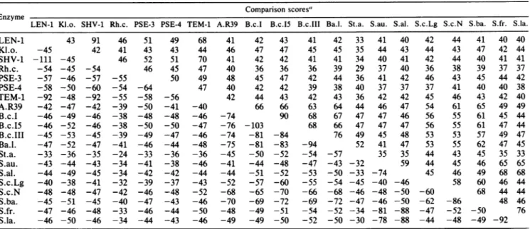
TABLE 1. Comparison of 20 class A ,-lactamases
Comparison scores"
Enzyme
LEN-1 Kl.o. SHV-1 Rh.c. PSE-3 PSE-4 TEM-1 A.R39 B.c.1 B.c.15 B.c.III Ba.l. St.a. S.au. S.al. S.c.Lg S.c.N S.ba. S.fr. S.la.
LEN-1 43 91 46 51 49 68 41 42 43 41 42 33 41 40 42 44 41 40 40 Kl.o. -45 42 41 43 43 44 46 47 47 45 45 35 44 43 44 43 47 42 44 SHV-1 -111 -45 46 52 51 70 41 42 42 41 41 34 40 41 42 44 40 41 41 Rh.c. -54 -45 -54 46 45 47 40 36 36 36 39 29 37 40 36 38 39 37 37 PSE-3 -57 -46 -57 -55 50 49 48 45 47 42 44 36 41 42 46 43 45 44 42 PSE-4 -58 -50 -60 -54 -64 47 40 42 42 39 38 40 37 37 37 41 40 40 38 TEM-1 -92 -48 -92 -55 -58 -56 42 44 43 42 43 36 42 42 45 46 43 42 40 A.R39 -42 -47 -42 -39 -50 -41 -40 66 66 63 64 44 46 47 54 61 65 49 49 B.c.I -46 -49 -46 -38 -48 -48 -46 -74 90 68 67 47 47 46 56 55 61 45 44 B.c.15 -46 -52 -46 -38 -50 -50 -47 -76 -103 68 66 47 47 47 56 55 61 47 44 B.c.III -45 -53 -45 -39 -49 -47 -46 -74 -81 -84 76 49 45 48 53 53 57 49 47 Ba.l. -47 -52 -47 -41 -46 -44 -48 -75 -81 -83 -94 52 41 47 53 55 62 47 45 St.a. -33 -36 -35 -24 -33 -36 -36 -45 -50 -52 -54 -57 35 35 44 43 45 35 33 S.au. -43 -44 -43 -34 -41 -38 -46 -41 -44 -48 -47 -43 -32 59 44 45 46 65 65 S.al. -44 -49 -45 -34 -42 -42 -44 -44 -51 -52 -53 -50 -33 -74 45 46 49 68 68 S.c.Lg -40 -38 -41 -32 -39 -37 -43 -52 -57 -60 -55 -54 -45 -40 -46 58 60 46 44 S.c.N -48 -48 -47 -42 -46 -48 -52 -68 -65 -70 -66 -68 -46 -48 -50 -60 68 44 44 S.ba. -45 -51 -45 -40 -47 -43 -46 -70 -69 -72 -69 -72 -47 -46 -50 -62 -86 48 46 S.fr. -47 -46 -48 -33 -46 -44 -50 -48 -49 -51 -54 -52 -34 -81 -88 -47 -52 -50 76 S.la. -46 -50 -46 -34 -44 -43 -46 -49 -49 -50 -52 -50 -30 -78 -88 -44 -48 -49 -92
"Thepositive values above the diagonal were calculated on the basis of the distances algorithm. They represent the percentage of very similar or identical residues (see text). The negative values below the diagonal were obtained with the help of the SEQDP algorithm. The gross values were divided by 10.KI.o.,
Klebsiella oxytoca; Rh.c., Rhodopseudomonas capsulata; A.R39, Actinomadura sp. strain R39; B.c.1, B.c.I.5, and B.c.III: P-lactamases I, I5b, and III,
respectively,of Bacillus cereus;Ba.l.,Bacillus licheniformis; St.a., Staphylococcusaureus; S.au.,Streptomyces aureofaciens; S.al., Streptomyces albus G; S.c.Lg, Streptomyces cacaoi ULg; S.c.N, Streptomvces cacaoi NDRE; S.ba., Streptomyces badius; S.fr., Streptomycesfradiae;S.la.,Streptomyces lavendulae.
present analysis, a match was scored when the substitution had a value greater than 1 in the table of Gribskov and Burgess (19), which is a normalized form ofthe table of
Dayhoff
(10). The chosen value correspondsto ahigh degree of structural conservation, since identical residues yield a scoreof 1.5, whilethe mean value iszero.The SEQDP algorithm givesa scorefor the best alignment oftwosequences, as described by Goad and Kanehisa (18) and Needleman and Wunsch(39). The finalscoreis thesum of the individual scores obtained for each pair of residues minusagappenalty of 8. The original table of Dayhoff(10) was used, and the scores varied from 0 to -17, with the lower value
representing
identical residues. The sequences that were compared were the same as those above, from residues 30to285(ABLconsensusnumbering scheme), but thegaps wereintroducedby theprogramitself, incontrast to thedistancesalgorithm,
in whichthe gaps were imposed apriori.
Secondary structure predictions were performed as de-scribed by Robson and Gamier (44).
RESULTS AND DISCUSSION
Amino acid sequencecomparisons. (i)Class A,-lactamases. The
alignment
of the class AP-lactamase
sequences has been described by Ambler et al. (2). Table 1 presents the comparison matrices whichwere obtainedwith residues 30 to285 ofthe ABLnumberingscheme. It shouldbe stressed thatthedeletionofthe N-andC-terminal residues increased the similarities between the various enzymes, since thealignments
show that these areas exhibithigher degrees
of variation(2).Both comparison methods yielded very similar results (Table 1). The SEQDP algorithm generally yields
higher
scores, sinceit uses areplacement
matrix,
while the otherprocedure
counts matches only above a certain level. Twopairs
ofenzymes, SHV-1-LEN-1 and B. cereusI-B. cereusSb,
exhibitedparticularly high
scores(greater
than90). They
were notconsideredtobe different
proteins
inthefollowing
analysis.
The enzymes ofS. aureus and R.capsulata
con-sistently
exhibited poor scores.Moreover,
they
wereex-tremely
different from eachother; indeed,
thescoreobtainedby
comparing
themwith each otherwasthe lowestscoreof all.Toposition
eachenzymeinthecomplete
population,
wecomputed
anaveragescoreasthemeanof allthescores(the
distancesprogram was
used)
for agiven
enzyme, with the standard deviationrepresenting
theheterogeneity
of its behavior. Those averagesranged
from 39.5 ± 5 for the R.capsulata
and S. aureus enzymes to 51 ± 10 for the B.licheniformis
enzyme and weregenerally
higher
for theStreptomyces
andBacillusP-lactamases
thanfor thoseorig-inating
fromgram-negative species.
Thosescores wereprob-ably
somewhat biased because of the presence of sevenStreptomyces
andfour Bacillusenzymesin thesample.
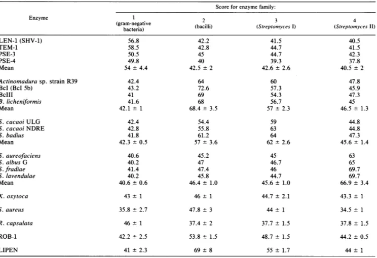
Asaconsequence, we triedtogroup the various enzymesintoa limited number ofenzymefamilies. Thebasic ideawasthat
a
given
enzymeshould exhibitasignificantly higher
averagescore
against
itsownenzymefamily
thanagainst
theothers. This resulted in theclassification shown in Table2compris-ing
fourenzymefamilies andthree"loners,"
inwhich each enzymenicely
fulfillsthatcondition. Themajor
surprise
was thattheStreptomyces
enzymesfell intotwodistinctenzyme families and thatoneofthemwasmuch closertothe enzymefamily
ofthe bacilli thanto thatofthe otherStreptomyces
spp.
Moreover,
theStreptomyces
I enzymefamily
couldnot even be considered as an intermediate between the bacilli andtheStreptomyces
II enzymefamilies since the averagescoreofthe latter enzyme
family
washigher
with the bacilli than with theotherStreptomyces
spp. enzymefamilies(46.5
versus
45.6, although
the difference isprobably
notmean-ingful).
Thethreelonerswerethe S. aureus,theK. oxytoca, and theR.capsulata
enzymes.Although
theS. aureusandTABLE 2. Classificationof the enzymes and scoreforeach family
Score for enzymefamily:
Enzyme 1 2 3 4
(gram-negative (bacilli)
(Streptomyces
I)
(Streptomyces II)bacteria) LEN-1(SHV-1) 56.8 42.2 41.5 40.5 TEM-1 58.5 42.8 44.7 41.5 PSE-3 50.5 45 44.7 42.3 PSE-4 49.8 40 39.3 37.8 Mean 54 + 4.4 42.5 ±2 42.6 ± 2.6 40.5 ±2 Actinomadura sp. strain R39 42.4 64 60 47.8 BcI(BcI 5b) 43.2 72.6 57.3 45.9 BcIII 41 69 54.3 47.3 B.licheniformis 41.6 68 56.7 45 Mean 42.1 ± 1 68.4 ± 3.5 57 ± 2.3 46.5± 1.3 S.cacaoi ULG 42.4 54.4 59 44.8 S. cacaoiNDRE 42.8 55.8 63 44.8 S. badius 41.8 61.2 64 47.3 Mean 42.3 ± 0.5 57 ± 3.6 62 ± 2.6 45.6± 1.4 S. aureofaciens 40.6 45.2 45 63 S. albusG 40.2 47 46.7 65 S.fradiae 41.4 47.4 46 69.7 S.lavendulae 40.2 45.8 44.7 69.7 Mean 40.6 ± 0.6 46.4 ± 1.0 45.6± 1.0 66.9 ± 3.4 K.oxytoca 43 ± 1 46 ± 1 44.7 ± 2.1 43.3 ± 1 S. aureus 35.8 ± 2.7 47.8± 3 44± 1 34.5 ± 1 R.capsulata 46± 1 37.4 ± 2 37.7 ± 1.5 37.8± 1.5 ROB-1 42.2 ± 2.5 53.8 ± 1.5 48.7 ± 1.5 44.2 ± 0.5 LIPEN 41±2.3 69±8 55±1.7 44±1
theK. oxytoca enzymes
appeared
tobe closertothose ofthe bacilli andthe R.capsulata
enzymeappeared
tobe closerto those of thegram-negative
bacteria,
because of their low scores,they
could notbe included inthosefamilies withoutdestroying
thehomogeneity
ofthe bacilliandgram-negative
bacteria enzyme families. As noted
above,
the three loners were alsoextremely
differentfrom each other. It should benoted, however,
that the K. oxytoca enzyme was not arealloner,
since the enzyme of Citrobacter diversuscertainly
belongs
tothe same enzymefamily.
The Actinomadura sp. strainR39 enzymewasmuch closertothebacillithantotheStreptomyces
enzymes, which is a somewhatsurprising
behavior for a member of the
Actinomycetales
taxonomicfamily. Finally,
in thegram-negative
bacteria,
the enzymesLEN-1, SHV-1,
and TEM-1 formeda ratherhomogeneous
group.Onthe contrary, the enzymesPSE-3andPSE-4 could
notbe considered tobe morerelated toeach other than to
the enzymesfromother
gram-negative
bacteria.We further tested our classification with two other
en-zymes which were
inadvertently
omittedduring
the firstanalysis.
Data for those two enzymes, ROB-1 (29) and LIPEN(30),
appearatthebottomofTable 2,wheretheyarecompared
with the variousenzymefamilies.Clearly,
LIPEN presents all the characteristics of a member ofthe bacilli enzymefamily,
including
agood similarity
withthe Strepto-myces I enzymefamily. Although
the ROB-1 enzyme was more similar to the bacilli enzymefamily,
it cannot be included in thatfamily
withoutdrastically decreasing
itsaverage internal score. It could be considered a distant relative, but it also exhibits the same preference for the StreptomycesI enzyme
family
asdothe truemembersof the bacilli enzyme family.At present, it is not
possible
to build aphylogenetic
treewith the proteins compared here. Pastor et al. (42) have recently proposed a tree, some aspects of which are at variance with ourclassification. The reasons forthose dif-ferencesare notclear, but itremains difficult to understand how the K. oxytoca and S. albus G enzymes, which,when compared by ourcriteria, exhibited a very low score of 43, could be considered to be closely related by those investi-gators.
(ii) Class C ,-lactamases and the Streptomyces sp. strain
R61 DD-peptidase. Alignment of the five known distinct sequences ofclass C
P-lactamases
(16, 35, 38) reveals 93 invariant residues (26%), and this does not allow an easy pinpointing offunctionally
important conserved residues. However, by introducing the Streptomyces sp. strain R61 DD-peptidaseinto thecomparison,a moresignificant identi-fication of such conserved areas could be performed (27). Recent three-dimensional data show that Y159 in theDD-peptidase superimposes
onY150 in the ,-lactamases, which suggests that the YSN sequence of the peptidase corre-sponds to the YAN sequence ofthe class CP-lactamase
enzymes. Moreover, the hydroxyl group of the Y residue superimposesonthatofS130in class AP-lactamases.
40 ....MKNTIH INFAIF.LII ANIIYSSASA STDISTVASP LFEGTE...G
...MAIR IFAILFSIFS LATFAHAQEG TLERSDWRK. FFSEFQA.KG
...M KTFMYVIIA CLSSTALAGS ITENTSWNKE .FSA.EAVNG ... ...PGT NVEYEDYST. FFDKFSAS.G
--- --- --- --E--- -F---- A--G
70 90
CFLLYD.AST NAEIAQFNKA KCATOMAPDS TFKIALSLMA FDAEII.DQK TIVVADEROA DRAMLVFDPV RSKKRYSPAS TFKIPHTLFA LDAGAVRDEF VFVLCKSSSK SCATN..DLA RASKEYLPAS TFKIPNAIIG LETGVIKNEH GFVLFN.SNR KKYT.IYNRK ESTSRFAPAS TYKVFSALLA LESGIITKND -FVL--- --- ---PAS TFKI---L-A
L--G-I----140 TIFKWDKTPK GMEIWNSNHT PKTWMQFSVV WVSQEITQKI RLNKIKNYLK QIFRWDGVNR GFAGHNCIJDDLRSAMRNSTV WVYELFAKEI GDDKARRYLK QVFKWDGKPR AMKOWERDLT LRGAIQVSAV PVFQQIAREV GEVRMOKYLK SHMTWDGTOY PYKEWNOQO LFSAMSSSTT WYFQKLDROI GEDHLRHYLK --F-WDG--- ----WN-D-- L--AM--S-V WV-Q----I 6---YLK 190 DFDYGNQOFS GDKERNNGLT EAWLESSLKI SPEEQIQFLR KIINHNLPVK
KIDYGNADPS TSNG... DYWIEGSLAI SAQEQIAFLR KLYRNELPFR
KFSYGNQNIS GG...ID KFWLEGOLRI SAVNOVEFLE SLYLNKLSAS SIHYGNEDFS VP...A DYWLDGSLOI SPLEQVNILK KFYDNEFDFK ---YGN-D-S ---LEGSL-I S--EQ--FL- K-Y-N-L---240 NSAIENTIEN MYLQODNST KLYGKTGAGF TANRT...LONGWFEGFI
.VEHORLVKDLMIVEAGRNW ILRAKTGWEG RMG... WWGN E K.ENQLIVKE ALVTEAAPEY LVHSKTGFSG VGTESNPGVA WWVGWVE... QSNIE.TVKD SIRLEESNGR VLSGKTGTSV INGELHAG.. WFIGYVE... ---\VK- ----E--- -L--KTG--- ---
W--GWVE---290 OXAI ISKSGHKYVF VSALTGNLGS NLTSSIKAKK NAITILNTLN L ... OXA2 ..WPTGSVFF ALNIDTPNRM DDLFKREAIV RAILRSIEAL PPNPAVNSDA AR PSE2 ..KETEVYFF AFNMDIDNES KLPLRKSIPT KIMESEGIIG G. BLAR-CTD ..TADNTFFF AVHIQGEKRA AGSSAAEIAL SILDKKGIYP SVSR Consensus ---FFA---
--FIG. 1. Aligned sequences of BLAR-CTD and class D ,B-lactam-ases. Strictly invariant residues are underlined. A residue was considered asaconsensus when it was present in at least three of the proteins.
high degreeofsimilarityhasbeen found between the OXA-2
1-lactamase
andtheC-terminaldomainof theproduct
ofthe blaRgene(BLAR-CTD) (51), although BLAR-CTD exhibits onlypenicillin-binding
properties and an exceedingly low hydrolytic activity. Figure 1 shows the aligned sequences, and Table 3 provides results of the comparisons that were performed as described above for the class A enzymes. It was remarkable to observe that the BLAR-CTD fragment exhibited higherscoreswith OXA-2andPSE-2than OXA-1. Infact,
thosescorescharacterize by farthehighest degree ofsimilarity
everfound betweenaP-lactamase
anda penicillin-binding protein. Bycontrast, byusingtheSEQDP algorithm and the samescale, the comparisonbetween the Streptomy-ces sp. strainR61
DD-peptidase and a class C3-lactamase
TABLE 3. Copmparisonmatrixfor the class D enzymes Comparison scores'
Enzyme 1-Lactamase BLAR-CTD
OXA-1 OXA-2 PSE-2 B.licheniformis TnS52
OXA-1 30 30 33 31
OXA-2 -25 43 44 33
PSE-2 -28 -43 39 35
B. licheniformis -32 -42 -40 47
Tn552 -31 -30 -31 -57
aFordetails regardingthescores,seefootnoteaof Table1.
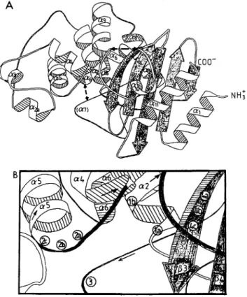
FIG. 2. Generalarrangementof the secondarystructure(A) and the four conserved structural elements (B)in classA,3-lactamases.
(A) A ribbon representation of the polypeptidechain of the S.albus ,B-lactamase.Thedashed lines indicate possible direct connections (seetext)in class Denzymes.(B) The circlesindicate thepositions of the a carbon of the conserved residues composing the four
structuralelements described in thetext.la is Ser-89 and ABL-70 and lb is Lys-92 andABL-73ofstructural element1. 2a isSer-153 andABL-130, 2bis Asp-154andABL-131, and2c is Asn-155 and ABL-132 of structural element 2. 3 is GIU-191 and ABL-166 of structuralelement3.4a isLys-259andABL-234,4b is Thr-260and ABL-235, and4cisGly-261 and ABL-236 of structural element 4. The numbering of the S. albus G residues includes the signal peptide. It should be noted that two errors were found in the
sequencepublished by Dehottayetal.(11): residueGly-128 should be eliminated and thetripeptideAla-Gln-Leuatresidues 140to142 should be replaced by the dipeptide Gly-Met. The complete se-quence is thus two residues shorter (312 residues). Taking into
accountthesecorrections,thesecondarystructureassignmentsare asfollows (numbersareresiduenumbers): al,48to60(ABL28to
40); [31,63to70(ABL43to50); [32,76to79(ABL56to60);a2, 89
to102(ABL70to83); a2a,118to124(ABL 99-103);a3,133to138
(ABLlllato115);a4, 142to151(ABL119to128);aS, 155to165 (ABL132to 142);a6, 168 to 177(ABL145to 154);a8 206to 217 (ABL185to194);a9,224to235(ABL201to212); alO,244to247 (ABL221 to224); 13,253to260(ABL230to237); [4, 266to273 (ABL244to251); ,B5,280to287(ABL259to269); all,297to310 (ABL276to289).
yielded ascoreof -12.4 (27) which, in thatprevious analy-sis, was the highest inter-enzyme familyscore.
A gene homologous tothe blaR gene has been found in
Tn552, a transposable element from S. aureus (45). Its
sequencehasbeendetermined,andits C-terminalportionis
included in thecomparison presented onTable 3.
General comparison. Thepresently available X-ray struc-turesshow that four class A [B-lactamases (13, 21, 37, 46) and
oneclass C [-lactamase (40)andthe R61DD-peptidase (31) arecomposed oftwodomains,oneao/[ andonealla(Fig. 2).
OXAI OXA2 PSE2 BLAR-CTD Consensus OXAI OXA2 PSE2 BLAR-CTD Consensus OXAl OXA2 PSE2 BLAR-CTD Consensus OXAI OXA2 PSE2 BLAR-CTD Consensus OXAI OXA2 PSE2 BLAR-CTD Consensus
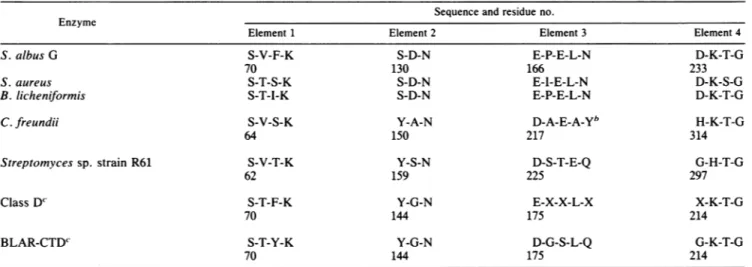
TABLE 4. Fourstructuralelements thatlimit the active site" Sequenceand residue no. Enzyme
Element 1 Element 2 Element 3 Element 4
S. albus G S-V-F-K S-D-N E-P-E-L-N D-K-T-G
70 130 166 233
S. aureus S-T-S-K S-D-N E-I-E-L-N D-K-S-G
B. licheniformis S-T-I-K S-D-N E-P-E-L-N D-K-T-G
C.freundii S-V-S-K Y-A-N D-A-E-A_Yb H-K-T-G
64 150 217 314
Streptomyces sp. strain R61 S-V-T-K Y-S-N D-S-T-E-Q G-H-T-G
62 159 225 297
Class D' S-T-F-K Y-G-N E-X-X-L-X X-K-T-G
70 144 175 214
BLAR-CTDc S-T-Y-K Y-G-N D-G-S-L-Q G-K-T-G
70 144 175 214
" The numbering of the various proteins isasfollows: classA 3-lactamases,ABL (2),C.freundii,consensusclassC(16);Streptomycessp. strain R61:mature protein sequence (14);classD andBLAR-CTD,seeFig.1.
b The identification of element 3 in the class C ,-lactamase ofC.freundii is tentative since theC,,,coordinates oftheenzymeare notavailable.
FortheclassD,B-lactamases and theBLAR-CTD.theidentification of elements1and4 cannotbeeasily challenged,but thatof elements 2 and 3 is based onlyonsequencealignments.
The penicillin-binding site is in a crevice at the interface between thetwodomains. In the sixenzymes,the active site
is limited by four structural elements whichoccupy similar
positions (Table 4).
(i) Element 1. Element 1 is the sequence
-S*-X-X-K-,
where S* is the active serine, atthe N terminus of the a2 helix.
(ii) Element 2. Element 2 is the SDN loop of class A P-lactamases, corresponding, respectively, to YAN and
YSN sequences in class C enzymes and the DD-peptidase.
This loop is between helices a4 andaSin class A 1-lactam-ases. However, inclass AP-lactamases,that serineappears
to be involved in maintaining the functional positioning of the two domains (25) and in the protonation of the sub-strate's leaving group (32), roles which might be very
dif-ferent from that of the tyrosine in class C 1-lactamases, which could actas ageneral base in thecatalytic
phenome-non(40).
(iii) Element 3. Element 3 isan acidic residue situatedjust before orat the N terminusofaone-turn helixorofaloop whose first three residuespresenthelix-like characteristics. The residue is E in class A P-lactamases and D inclass C P-lactamases and the peptidase. In class A 1-lactamases, site-directed mutagenesis indicates that E166 (ABL
consen-sus numbering scheme) playsanimportant role in catalysis (17). Accordingly, its side chain points into the substrate-binding cavity. In class C P-lactamases and the Streptomy-ces sp. strain R61 DD-peptidase, the side chain does not point into the active site, and modifications of that residue do
notappearto alter the enzymeactivity (20, 49). Even if, in
those cases, it plays no functional role in catalysis, the reproducible presence of that residue might still represent
anotherelement that iscommontoall the structures. (iv)Element 4. Element 4is the KT(S)G sequence onthe
P3
strandof class AP-lactamasesfacing the SDN looponthe other side of the active serine. The same KTG sequence isfoundon the 17strand of the class C enzyme and HTGon
the 13 strand of the DD-peptidase, in similar positions. Finally, it is interestingto remember that the SXXK and
KTG elements are also found in all penicillin-binding
pro-teins that have been described so far.
Moreover;
a SXN sequence,whichmight correspondtoelement2is also found in allpenicillin-binding protein sequences (48).Those four elements can serve as markers to compare the folding ofthe four classes of enzymes discussed here. By using the numbering of the helices and ,B strands as they occur in the sequence in class A
P-lactamases
(Fig. 3), the following conclusionscan be made.(i) The residues preceding the active Ser contribute one
helix and two , strands to the
at/I
domain.(ii) The active serine is atthe N terminus ofthe hydro-phobic helix a2, which is followedbyaloop containing one short helix (a3) in theB.-
licheniformis
and S.aureusclassAP-lactamases
and in the Streptomyces sp. strain R61DD-peptidase. The S. albus G ,-lactamase contains one addi-tional helix (a2a), and the class C enzyme contains two additionalshorthelices. This is alsoa
hydrophilic
partofthe moleculewhere ashort(12-residue) insertionoccursinclass CP-lactamases
and the DD-peptidase.(iii) Thereafter, six helices occur successively in the
P-lactamases
(a4toa9).Whenit ispresent, thefourth helix (a7)is very short (oneturn)andsometimes reduces to aloop where two or threeresiduespresenthelix-likeangle values. Active-site element 2 is between a4 and aS in all the enzymes, and element3 isjustatthebeginning ofthe short helix a7. Helixa8isattheborder betweentheallaandthe oa/3 domains. Itis incontactwiththe,3 sheet andmight play an important role in the stability ofthe whole structure. In this all-a part of the molecules, two insertions appear to occur in the class C enzyme and the DD-peptidase, one between a4 and aS and theother between a6 and a7. This mightexplain why the orientation ofthe a4 helix is some-whatdifferent in the class A enzyme and the DD-peptidase. (iv)The C-terminal part ofthepolypeptide chainbelongs to theaIl3
domain. Structures common to all the enzymes are, in the class A nomenclature, helices otlO andall;
D strandsP3, P4,
andP5;
and loops betweena9 andalOand between alO andP3.
In addition to these structures, the class C enzymes contain three short ,B strands between a9 and alO and another one between alO andP3.
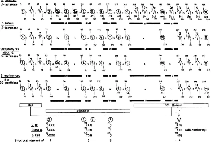
This couldCLASS A 1-LACTAMASE SEQUENCE COMPARISON 2299 C.freundi i /-lactamae 5 27 37 66 89 9 106
1?8
152170
217 227 246 ©19171Az26V195®2> 6 7 ®2 ''Q73! £r4~ S:~ a X , x' aiam!
25 34 45 79 93 103 110 137 163 177 220 238 255 S.aureus WE -'9- --lactamase 33 42 5 71 107 119 132 1iS 166 1803 m 1 +3 +,10% 24 X ®2®10 11®7 40 S0 60 82 113 127 142 155 171 193 213 Streptomyces 36 U * _27_ _ albus G. /3- lactamase 48 63 76 91 118 193 142 115 148 2?6 2?4 29 9 1 1 8 3 93!2 ® 26 6 60V70 * 1 1 60 70 79 102 1. 138 151 166 177 217 2jS 258 26s 22 280 299 311 3fl 334 34 :2a i2b ,2c 2d 3 4 5A3
>
AS: 042,
6,
07!
>
anB'
262 26 275 292 305 316 340 360 2 16 218 225 230 242 258 278 3 ':4 5 238 282 268 20 _ 4 XN- m 244 253 266 280 297 ,. I 27 28 247 260 273~ 2fi 110 Streptomyces 9m *3 28 - 8 5 R61peptids 36 56 65 93 IS0 157 192 212 23 296 312 327 36 346 359DD-peptidase
I)3I9() 4 :I I~ 3 2 19t i1 m 2 Q18®5® 59 S A 2 A2A3© a7 (0'1 a S1 62 73 108_48*18-
154 173 206 218 207 307 321 333 343 3$ 377 aso_Sm_; a, 64 C.fr. SXXK 70 ClassA. SXXK 62 S.R61 SXXK Structural element n° 1FIG. 3. Analysisofthe secondarystructuresof theStreptomyces sp. strain R61DD-peptidaseandthe class A and C,B-lactamases. The
distances (in residues) betweeneachsecondarystructureelementaregiven. The circlesrepresentahelices,andthejaggedlinesrepresent
the strands. Heavy bars indicatetheareaswhereimportantinsertionsareobserved in theStreptomycessp. strain R61(S. R61)and class
Cenzymes.Theconsensuselements borderingthe active sitearehighlightedin the lowerpartof the figure.TheproposedABLnumbering
of thesecondarystructureelements, correspondingtothat ofthe S.aureusenzyme,isusedforthe fourenzymes.W4'ndifferent,theoriginal numberingisalso shown underthe corresponding value of the ABL consensg numbering scheme. In the proposed unified nomenclature,a
secondarystructurewhich is absent in the referenceproteinisassignedthenuhiberof theprecedinghelixorstrand withanadditional letter.
To illustrate thispoint, twoclass A,B-lactamases areincluded. In the S. albusGenzyme, anadditional helix is found between S. aureus
helices 2 and3. Itisreferredtoashelix a2a. Conversely,in theS.albus Genzyme,helix a7 reducestoaloopandcannotbe consideredto
beahelix. The additionali strands in the class C 1-lactamaseareaccordinglynumbered,B2a-, i2b; ,B2c,and ,B2d,since theyareinserted between 132 and 133. In theupperpartofthefigure, thenumberings
of,
theS.R61DD-peptidaseand of the S. albus 13-lactamase include thesignal peptides.
explain one major difference between the class C 1-lacta-mases and the enzymes of the other classes. In class C
13-lactamases, helix alO appears to run parallel to al and
antiparalleltoall,while it has the opposite orientationinthe DD-peptidase and in class A (Fig. 4). The a/l domain
containsonlyoneactive-site element(KTG), which ison 13
in allcases.
Atthispoint, wewishto stressagain that,onthe basis of
the available knowledge, the role of structural elements 2
and 3might be quite different in class A and C 13-lactamases. Startingwiththe rather safeassumption thatageneralbase
is necessary for activating the hydroxyl of the active Ser
residue, that general base would be E166 (element 3)in class
A 1-lactamases and theanion ofY150(element 2)inclass C
1-lactamases.
One can also attempt to fit the sequence of class D
,B-lactamases to the general model, on the basis of the
conserved active-site elements andsecondarystructure
pre-FIG. 4. Organization of the a113 domains in the class A and
Streptomycessp.strain R61enzymes(A)andin class Cenzymes(B).
I aDomain 150 YAN 130 SON 159 YSN 2 21~ 166 E 225 3 WA Domain_ -r 3 315 KTG 234 KTG 296 HTG 4 (ABLnumbering) VOL. 35,1991
dictions. These
predictions
show thatthe activeserine is atthe endofan
ao/1
areaandatthebeginning
ofahydrophobic
helix. There are 74 residues between theactive serine and theYGN element;
thus,
between those elements thereare30moreresidues in theclassD 1-lactamases than there arein class A
1-lactamases,
but this is also an insertion area in both theDD-peptidase
and the class C,B-lactamases. By
contrast, the distance between the YGN sequence and the thirdelement
(E140)
isonly
15residues,
and nosecondary
structure is
predicted
in that area. The class A structureshows that a direct passage from a5 to cx7
involving
the deletion of a6might
bepossible. Similarly,
the distance between E140 and the KTG sequence isonly
49residues,
i.e.,
19 residues shorter than in class A 3-lactamases.Assuming
that helix(x8
is necessary for thestability
of themolecule,
onemight
predict, again
on thebasis oftheclass A structure, adirectpassagefromtheC-terminal endofa9to the Nterminus of
13,
thusinvolving
the deletionof(o10. Between the active Ser andresidue187,
just
beforeKTG,
only
helices arepredicted.
In the C-terminalportion
(resi-dues 187 to
255),
predictions
indicate an(x/1
domain. Theproposed
shortcutsin class D enzymesareshowninFig.
2A. Inconclusion,
the class Af3-lactamase
modelcanbe usedas a reference for all serine 1-lactamases and
probably
for manyDD-peptidases.
We proposethat,
in addition to the ABL consensusnumbering
scheme(2),
thenumbering
of class Asecondary
structureelementsalsobeadopted
for all active serinepenicillin-recognizing
enzymes, as we have done inFig.
3.ACKNOWLEDGMENTS
This workwassupported, in part,bytheBelgianProgrammeon
InteruniversityPoles of Attraction initiated by the Science Policy ProgrammingPrime Minister's Office, Belgian State (PAI 19); an
action concertee with the Belgian government (convention
86/91-90); the fonds de la Recherche Scientifique Medicale (contract 3.4537.88); and a convention tripartite between the Region Wal-lonne, SmithKline Beecham, United Kingdom, and the University
of Liege. B.J. is chercheur qualifie of the Fonds National de la Recherche Scientifique (Brussels). P.L. is research fellow of the Institut pour l'Encouragement de la Recherche Scientifique dans l'Industrie etl'Agriculture(Brussels).
REFERENCES
1. Ambler, R. P. 1980. The structure of 1-lactamases. Philos. Trans. R.Soc. London B Biol. Sci. 289:321-331.
2. Ambler,R.P.,A. F. Coulson,J. M.Frere,J. M.Ghuysen,M.
Forsman,B. Joris, R. Levesque, G. Tiraby,and S. G. Waley. 1991. A standard numbering scheme for the class A beta-latamases. Biochem. J. 276:269-270.
3. Arakawa,Y., M. Ohta,N. Kido,Y. Fujii,T.Komatsu, andN. Kato. 1986. Close evolutionary relationshipbetween the
chro-mosomallyencoded1-lactamasegeneofK.pneumoniae and the Tem 13-lactamase gene mediated by R plasmid. FEBS Lett. 207:69-74.
4. Arakawa,Y.,M.Ohta,N. Kido,M.Mori,H.Ito, T.Komatsu, Y.
Fujii,
and N. Kato. 1989. ChromosomalP-lactamase
ofKlebsiella oxytoca, a new class A enzyme that hydrolyzes
broad-spectrum ,-lactam antibiotics. Antimicrob. Agents Che-mother. 33:63-70.
5. Barthelemy, M., J. Peduzzi, and R. Labia. 1988. Complete
amino acid sequence ofp453-plasmid-mediated PIT-2
,-lacta-mase. Biochem. J.251:73-79.
6. Barthelemy,M., J. Peduzzi,H. B.Yaghlane,and R.Labia. 1988.
Single
amino-acid substitution between SHV-1 ,-lactamase andcefotaxime-hydrolysing
SHV-2enzyme. FEBS. Lett. 231:217-220.7. Boissinot, M.,and R.C.Levesque.1990. Nucleotide sequenceof thePSE-4 carbenicillinasegeneand correlationswith the
Staph-ylococcusaureusPC1 beta-lactamase crystal structure. J. Biol. Chem. 265:1225-1230.
8. Campbell, J. I. A., S. Scahill, T. Gibson, and R. P. Ambler. 1989. The phototrophic bacterium Rhodopseudomonas capsulata splO8 encodes an indigenous class AP-lactamase. Biochem.J. 260:803-812.
9. Dale, J. W., D. Godwin, D. Massakowska, P. Stephenson, and S. Wall. 1985. Sequence of the OXA2 ,-lactamase: comparison with otherpenicillin-reactive enzymes. FEBS Lett. 191:39-44. 10. Dayhoff, M. 0. 1979. Atlas of protein sequence and structure, vol 5,suppl.3. National Biochemical Foudation, Silver Spring, Md.
11. Dehottay, P., J. Dusart, F. De Meester, B. Joris, J. Van Beeu-men, T. Erpicum, J. M. Frere, and J. M. Ghuysen. 1987. Nucleotide sequence of the gene encoding the Streptomyces albus GP-lactamaseprecursor. Eur. J. Biochem. 166:345-350. 12. Devereux, J., P. Haeberli, and 0. Smithies. 1984. A comprehen-sive set of sequence analysis programs for the VAX. Nucleic Acids Res.12:387-395.
13. Dideberg, O., P. Charlier, J. P. Wery, P. Dehottay, J. Dusart, T. Erpicum, J. M. Frere, andJ. M. Ghuysen. 1987. The crystal structure of the ,B-lactamase Streptomyces albus G at 0.3 nm resolution. Biochem. J. 245:911-913.
14. Duez, C., C. Piron-Fraipont, B. Joris, J. Dusart, M. Urdea, J. Martial, J.M. Frere, and J. M. Ghuysen. 1987. Primary struc-ture of theStreptomycesR61 extracellularDD-peptidase.Eur.J. Biochem. 162:509-518.
15. Forsman, M., B. Haggstrom, L. Lindgren, and B. Jaurin. 1990. Molecularanalysisof,B-lactamases from four species of Strep-tomyces: comparison of amino acid sequences with those of otherP-lactamases. J. Gen.Microbiol. 136:589-598.
16. Galleni, M., F. Lindberg, S. Normark, S. Cole, N. Honore, B. Joris,andJ. M. Frere. 1988.Sequenceandcomparative analysis of three Enterobacter cloacaeampCbeta-lactamase genes and their products. Biochem. J. 250:753-760.
17. Gibson, R. M., H. Christensen, andS. G. Waley. 1990. Site-directed mutagenesisof ,-lactamaseI. Single and double
mu-tantsofGlu-166 andLys-73.Biochem. J. 272:613-619. 18. Goad, W. B.,and M. I. Kanehisa. 1982. Patternrecognitionin
nucleic acid sequences. I. Ageneral method for finding local homologiesandsymetries. Nucleic Acids Res. 10:247-263. 19. Gribskov, M.,and R. R. Burgess. 1986. Sigma factors from E.
coli, B. subtilis, phage T4 are homologous proteins. Nucleic AcidsRes. 14:6745-6763.
20. Hadonou,A.M.,and B.Joris. Unpublisheddata.
21. Herzberg, 0. 1991. Refined structure of ,B-lactamase from
Staphylococcusaureus PC1at 2.0 A resolution. J. Mol. Biol. 217:701-719.
22. Houba, S., S. Willem, C. Duez, C. Molitor, J. Dusart, J. M. Frere, andJ. M. Ghuysen. 1989. Nucleotide sequence ofthe gene encodingtheactive-site serine P-lactamasefrom Actino-maduraR39. FEMSMicrobiol. Lett.65:241-246.
23. Huovinen, P., S. Huovinen,and G.Jacoby. 1988. Sequenceof PSE-2 ,-lactamase. Antimicrob. Agents Chemother. 32:134-136.
24. Hussain, M.,F.J. Pastor,and J. 0. Lampen. 1987.Cloning and
sequencing of the blaZgene encodingP-lactamase III, a
lipo-proteinofB. cereus569/H.J.Bacteriol. 169:579-586. 25. Jacob, F.,B.Joris, 0. Dideberg,J. Dusart, J. M. Ghuysen, and
J.M.Frere. 1990.Engeneeringanovel 3-lactamasebyasingle
pointmutation. Protein Eng.4:79-86.
26. Jaurin, B., and T.Grundstrom. 1981.AmpC cephalosporinase of Escherichia coliK12has adifferent evolutionary origin from thatof ,-lactamases of penicillinasetype. Proc.Natl. Acad.Sci. USA 78:4897-4901.
27. Joris, B.,J. M. Ghuysen, G. Dive, A. Renard,0.Dideberg, P. Charlier, J. M.Frere, J. Kelly, J. Boyington,P.Moews, and J. Knox. 1988. The active-site-serine penicillin-recognizing en-zymes as members of the Streptomyces R61 DD-peptidase
family.Biochem. J.250:313-324.
28. Joris, B., P. Ledent,T. Kobayashi, J. 0. Lampen, and J. M. Ghuysen. 1990. ExpressioninEscherichia coli ofthecarboxy terminaldomainof BLARsensory-transducer protein of
Bacil-lus licheniformis as a water-soluble Mr-26000 penicillin-binding protein. FEMS. Microbiol. Lett. 70:107-113.
29. Juteau, J.-M., and R. C. Levesque. 1990. Sequence analysis and evolutionary perspectives of ROB-1 1-lactamase. Antimicrob. AgentsChemother. 34:1354-1359.
30. Kato, C., Y. Nakano, and K. Horikoshi. 1989. The nucleotide sequence of the lipo-penicillinase gene of alkalophilic Bacillus sp. strain 170. Arch. Microbiol. 151:91-94.
31. Kelly, J. A., J. R. Knox, H. Zhao, J. M. Frere, and J. M. Ghuysen.1989.Crystallographic mapping of ,B-lactams bound to a D-alanyl-D-alanine peptidase target enzyme. J. Mol. Biol. 209:281-295.
32. Lamotte-Brasseur, J., G. Dive, 0. Dideberg, P. Charlier, J. M. Frere, and J. M. Ghuysen. 1991. Mechanism of acyl transfer by the class A serine 1-lactamaseof Streptomyces albus G. Bio-chem. J. 279:213-221.
33. Lenzini, M. V., H. Ishihara, J. Dusart, H. Ogawara, B. Joris, J. Van Beeumen, J. M. Frere, and J. M. Ghuysen. 1988. Nucleotide sequence of the geneencoding the active-site serinef-lactamase from Streptomyces cacaoi. FEMS Microbiol. Lett. 49:371-376. 34. Lindberg, F., and S. Normark. 1986. Sequence of the Citrobac-ter freundii chromosomal ampC [B-lactamase gene. Eur. J. Biochem. 156:441-445.
35. Lodge, J. M., S. D. Michin, L. J. V. Piddock, and S. J. W. Busby. 1990. Cloning and analysis of the structural gene and regulatory region of the Pseudomonas aeruginosa chromosomal ampC1-lactamase. Biochem. J. 272:627-631.
36. Mabilat, C., S. Goussard, W. Sougakoff, R. C. Spencer, and P. Courvalin. 1990. Direct sequencing of the amplified structural gene and promoter for the extended-broad-spectrum
13-lacta-mase TEM-9 (RHH-1) of Klebsiella pneumoniae. Plasmid 23: 27-34.
37. Moews, P. C., J. R. Knox,0. Dideberg, P. Charlier, and J. M. Frere. 1990. P-lactamase of Bacilus licheniformis 749/C at 2 A resolution. Proteins Struct. Funct. Genet. 7:156-171.
38. Momura, K., and T. Yoshida. 1990. Nucleotide sequence of the Serratia marcescens SR50 chromosomal ampC gene. FEMS Microbiol. Lett.70:295-300.
39. Needleman,S. B.,andC. D.Wunsch. 1970.Ageneral method
applicable to the search for similarities in the amino acid sequence oftwo proteins. J. Mol. Biol. 48:443-453.
40. Oefner, C.,A.Darcy, J. J. Daly,K.Gubernator,R. L.Charnas, I.Heinze, C. Hubschwerlen, and F. K. Winkler. 1990. Refined
crystal structure of beta-lactamase from Citrobacter freundii indicates a mechanismfor beta-lactam hydrolysis. Nature (Lon-don) 343:284-288.
41. Ouellette, M.,L.Bissonnette,and P.Roy. 1987. Precise insertion of antibiotic resistance determinants into Tn2l like transposons: nucleotide sequence of the OXA-1 ,B-lactamase gene. Proc. Natl.Acad. Sci. USA84:7378-7382.
42. Pastor, N., D. Piniero, A. M. Valdes, and X. Sober6n. 1990. Molecular evolution of class A
P-lactamases:
phylogeny and patterns of sequence conservation. Mol. Microbiol. 4:1957-1965.43. Perilli, M., N. Franceschini, B. Segatore, G. Amicosante, A. Oratore,C.Duez,B.Joris,andJ.M. Frere. FEMS Microbiol. Lett., in press.
44. Robson, B.,andJ. Garnier. 1986. Introductiontoproteins and proteinengineering. Elsevier, Amsterdam.
45. Rowland, S.-J., and K. G. H. Dyke. 1990. Tn552, a novel transposable element fromStaphylococcusaureus.Mol. Micro-biol.4:961-975.
46. Samraoui, B., B. Sutton, R.Todd, P. Artimyuk, S. G. Waley, and D. Phillips. 1986. Tertiary structure similarity between a class A,B-lactamase andapenicillin-sensitive D-alanyl-D-alanine carboxypeptidase-transpeptidase. Nature (London) 320:378-380.
47. Shlaes,D.M., C.Currie-McCumber,A.Hull, I.Behlau,andM. Kron. 1990.OHIO-1 ,B-lactamase isapart of the SHV-1family.
Antimicrob. Agents Chemother. 34:1570-1576.
48. Spratt, B. G., and K. D. Cromie. 1988. Penicillin binding proteins of gram negative bacteria. Rev. Infect. Dis. 10:699-711. 49. Tsukamoto, K., R. Kikura, R. Ohno, and T. Sawai. 1990. Substitution ofaspartic acid-217 ofCitrobacterfreundii
cepha-losporinase and properties of themutantenzymes.FEBS Lett. 264:211-214.
50. Wang, W.,P.S.F.Mezes,Y.Q. Yang,R. W.Blacher,andJ. 0. Lampen. 1985. Cloning and sequencing of the P-lactamase I gene of Bacillus cereus 5/B and its expression in Bacillus subtillis. J. Bacteriol. 163:487-492.
51. Zhu,Y.F., I.H. A.Curran,B.Joris, J. M.Ghuysen,andJ. 0. Lampen. 1990. Identification ofBlaR, the signal transducer for
P-lactamaseproduction in Bacilluslicheniformis,as a
penicillin-bindingproteinwith stronghomologytothe OXA-2P-lactamase
(class C) of Salmonella typhimurium. J. Bacteriol. 172:1137-1141.