The structure at 2 Å resolution of Phycocyanin from Gracilaria chilensis and
the energy transfer network in a PC
–PC complex
☆
Carlos Contreras-Martel
b, Adelio Matamala
d, Carola Bruna
a, German Poo-Caamaño
c,
Daniel Almonacid
a, Maximiliano Figueroa
a, José Martínez-Oyanedel
a, Marta Bunster
a,⁎
aLaboratorio de Biofísica Molecular, Grupo de Biología Estructural, Depto. de Bioquímica y Biología Molecular, Facultad de Ciencias Biológicas,
Universidad de Concepción, Chile
bInstitut de Biologie Structurale, CEA-CNRS-UJF, Grenoble, France cUniversidad del Bío-Bío, Concepción, Chile
d
Laboratorio de Química Teórica y Computacional, Facultad de Ciencias Químicas, Depto. de Físico-Química, Universidad de Concepción, Chile Received 5 August 2006; received in revised form 28 September 2006; accepted 29 September 2006
Available online 10 November 2006
Abstract
Phycocyanin is a phycobiliprotein involved in light harvesting and conduction of light to the reaction centers in cyanobacteria and red algae. The structure of C-phycocyanin from Gracilaria chilensis was solved by X-ray crystallography at 2.0 Å resolution in space group P21. An interaction model
between two PC heterohexamers was built, followed by molecular dynamic refinement. The best model showed an inter-hexamer rotation of 23°. The coordinates of a PC heterohexamer (αβ)6and of the PC–PC complex were used to perform energy transfer calculations between chromophores pairs
using the fluorescence resonance energy transfer approach (FRET). Two main intra PC (Iβ382→Iα184→Iα584→Iβ682andIβ1533 →Iβ5153) and two main inter
PC (Iβ682→IIβ382andIβ5153→IIβ3153) pathways were proposed based on the values of the energy transfer constants calculated for all the chromophore pairs
in the hexamer and in the complex. © 2006 Elsevier B.V. All rights reserved.
Keywords: Structure; Phycocyanin; Gracilaria chilensis; Protein–protein docking; Energy transfer pathway
1. Introduction
Phycobilisomes (PBS) are auxiliary photosynthetic complexes present in cyanobacteria and eukaryotic red algae. They are composed of phycobiliproteins (PBP) and linker polypeptides. Phycobiliproteins are highly fluorescent proteins due to the presence of covalently bonded chromophores to their cysteine residues, which provide them the functional properties to absorb light at a range of the visible spectrum not fully utilized by chlorophyll. Therefore, improving the light harvesting ability of the algae that contain them[1–3].
Phycobilisomes share a general common morphology[4]that consists of a core of face-to-back cylinders formed by stacked
discs of PBP and several rods that radiate from the core composed of stacked back-to-back discs of PBP. Linker polypeptides are responsible for the maintenance of this structure, providing the necessary structural environment for efficient energy transfer and for the interaction with the photosynthetic reaction center[1,5].
The phycobilisome of Gracilaria chilensis contains three phycobiliproteins: Phycoerythrin (PE), phycocyanin (PC) and allophycocyanin (APC) [6,7]. The absorption and emission spectra of these proteins overlap, which allows a non-radiative, direct and efficient transfer of the excitation energy among them (PEλamax= 566 nm PEλemmax= 574 nm; PCλamax= 621 nm
PCλemmax= 640 nm; APCλamax= 651 nm APCλemmax= 660 nm).
This transfer is channeled along an energy gradient from the rods to the core and finally transferred to chlorophyll a in the thylakoid membrane[1,3].
In G. chilensis, the rods are composed of phycoerythrin and phycocyanin[6]. The three dimensional structure of phycoery-thrin from this algae was reported in 2001[7]. The structure of the ☆C.C.-M. and J.M.-O. contributed equally to this work.
⁎ Corresponding author. Tel.: +56 41 203822; fax: +56 41 239687. E-mail address:mbunster@udec.cl(M. Bunster).
0301-4622/$ - see front matter © 2006 Elsevier B.V. All rights reserved. doi:10.1016/j.bpc.2006.09.014
next component of the rods, phycocyanin, is now presented in this paper. The structures of other phycocyanins from Fremyella diplosiphon (1cpc)[8], Spirulina platensis (1gho)[9], Synecho-coccus vulcanus (1i7y) [10,11] and Synechococcus elongatus (1jbo) [12] have been reported previously. The structure of phycocyanin from Polysiphonia ureceolata (1f99) [13] and Cyanidium caldarium (1phn)[14]have also been reported. These structures present a common subunit organization that consists of oneα subunit (with cyanobilin (CB) associated to Cys84) and one β subunit (with cyanobilins covalently attached to Cys82 and Cys153). They interact to form a heterodimer (αβ) or a “pseudo monomer” that aggregates to form an hexameric ring (αβ)6able to
pile up to form the main frame of a rod.
The rods in phycobilisomes normally include two or more phycocyanin hexamers and in this alga there are also one or more PE hexamers. Theoretical and experimental studies have been reported regarding the light transfer among chromophores inside one hexameric ring[8,14–17], including models that represent the energy transfer between hexamers[8,14,16]. In these studies, preferential energy transfer pathways have been proposed from data calculated using the Förster resonance approach [18], considering only the chromophore–chromophore distances and a Förster radius of 50 Å´ as approximation[8]or the orientation among the transition dipole moments in one trimer of phycocyanin[19,20].
This paper reports the three dimensional structure of phycocyanin from G. chilensis, the building of a docking model of two PC hexamers refined by molecular dynamics and the determination of the constants for the energy transfer in resonance between pairs of chromophores. For the latter purpose the orientation factors between pairs of chromophores were calculat-ed from the dipole moments of their aromatic portions. The transfer constants were used to propose preferential pathways for the light conduction intra and inter hexamers of phycocyanin. 2. Materials and methods
2.1. Protein purification and crystallization
Phycocyanin from G. chilensis was purified according to Gantt[21]. Phycobiliproteins were extracted from algae collected at Coliumo Bay, Dichato, after maceration of N2(l)-frozen algae, in
20 mM phosphate buffer pH 7.0. The extract was fractionated with ammonium sulphate (30% and 60% saturation). The pellet was dissolved in distilled water and dialyzed against 5 mM phosphate buffer pH 7.0. The phycobiliproteins were separated by anion exchange chromatography (Fractogel EMD DEAE 650S) in an FPLC system (Merck-HITACHI) using 5 mM phosphate buffer as an equilibrium buffer and eluting the proteins with a linear gradient from 5 to 300 mM sodium phosphate pH 7.0. The phycobiliprotein content of the eluted fractions was analyzed by absorption at 566 nm, 621 nm and 651 nm. The phycocyanin-rich fraction was dialyzed against 900 mM phosphate pH 7.0 to precipitate phycocyanin and separate it from other phycobilipro-tein contaminants. The phycocyanin precipitate was dissolved in 1 ml of 50 mM phosphate buffer pH 7.0. The protein concentration was estimated by the absorption at 280 nm and
the purity was assessed by 12.5% SDS-PAGE and absorption and emission spectra. Crystals were grown by the vapor-diffusion method using sitting-drop insets at 291 K. 1 ml of 50 mM HEPES buffer pH 7.0, 1.2 M ammonium sulphate was used in the reservoir. The 10 μl droplet contained reservoir solution and protein at a concentration of 18 mg ml− 1in a 1:1 ratio.
2.2. Data collection, structure solution and refinement The data was collected at the IMCA-CAT Synchrotron in Argonne with an ADSQ detector with an exposition time of 10 s per frame (λ=1.00 Å) and processed with XDS[22]. The initial phases were obtained by Molecular Replacement[23,24]in the program package CNS [25] using the structure of C-phy-cocyanin from Spirullina platensis (1gho) [9] as a searching model. Sessions of manual rebuilding with TURBO FRODO [26]and refinement with CNS were performed. Lateral chains were assigned according to the residual electron density maps and to multiple sequence alignment of homologous proteins. After several cycles of refinement in the resolution range 60– 2.0 Å the Rworkand Rfreeconverged. The stereochemistry was
verified with PROCHECK [27] and the refined model was
deposited in the Protein Data Bank (code 2bv8). 2.3. Phycocyanin–phycocyanin interaction model
The coordinates of 2bv8 were used for the docking procedure of two phycocyanin molecules performed with the program ZDOCK[28,29], with 6° angular steps. The docking models were scored by the program considering desolvation, electrostatic and hydrophobic contributions[30–32]. The models generated were then analyzed to select those in agreement with the reported electron micrographs images [4]. The selected models were characterized by the number of hydrophobic residues in the interface using the protein–protein interaction server[33]and by the number of H-bonds in the interface using the program HBPLUS [34]. The highest scored rigid model was refined by molecular dynamics using the Force Field OPLS/AA[35]with the program GROMACS[36], with which a simulated annealing protocol for 200 ps produced the convergence of the system. The coordinates of the chromophores in the final PC–PC interaction model were used to evaluate the energy transfer constants. 2.4. Energy transfer calculations
Förster theory has been confirmed to be a good approximation to study the light transfer processes occurring in phycobilisomes [19,20]. Förster defines[18]a transfer constant kDAas a measure
of the frequency of events of energy transfer between a Donor (D) and an Acceptor (A). By introducing appropriate constants, the Förster equation can be expressed as a product of four terms. k
d DA¼ Cd Gd Sd I ð1Þ
where C is a collection of constants that considers a refraction index n = 1.567 [19,20,37,38], S include the spectroscopic properties of the interacting chromophores, I is the overlap
389 C. Contreras-Martel et al. / Biophysical Chemistry 125 (2007) 388–396
integral between the emission and absorption spectra of the donor and acceptor chromophores respectively as described in [20]. Considering the homology among phycocyanins[39]and the structural similarity of the residues in contact with the chromophores (Fig. 1), the experimental values included in the terms S and I were assigned as those reported for Synechococ-cus sp PCC 7002[19,20]. The geometric factor G,
G¼K 2 DA R6 DA ð2Þ includes the distances between the centers of mass for each pair of chromophores RDA, and the dipole orientation coefficientκDA
as described in Eq. (3).
jDA¼ ̂lDd Âl −3ð ̂lDd DÂr Þð ̂lAd DÂr Þ ð3Þ
whereμˆDandμˆAare unit vectors describing the direction of the
transition dipole moments of the donor and acceptor chro-mophores respectively and rˆDAis the unit vector describing the
direction of the line that connects the centers of mass of the interacting chromophores. The transition dipole moments were approximated to the dipole moments of the conjugated frag-ments[40]of the chromophores reported in 2bv8 and calculated by applying the semi empirical method PM3 Hamiltonian im-plemented in the software Gaussian98 [41]. Energy transfer steps with constants higher than 20 ns− 1 and 10 ns− 1(transfer times shorter than 50 ps and 100 ps) were used to define intra and inter phycocyanin preferential light transmission pathways respectively.
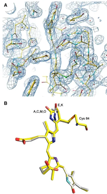
Fig. 2. Conformation of chromophores atα84. A) Fo–Fc electron density map
showing the electron density of chromophore atα84in chain K. B) Superposition
of the chromophores atα84in chains A (also representing chains C, M and O) and E (also representing chain K).
Table 1
Data collection, refinement and Ramachandran plot statistics Data collection Space group P21 Unit cell (Å´ ) a 101.99 b 151.80 c 101.55 α=γ 90.00 β 117.45 Resolution (Å´ ) 2.0
Number of observed reflections 178,325 Number of unique reflections 172,985 Completeness (last bin) (%) 93.6 (80.4) Rsym (last bin) (%) 6 (30) I/σ(I) (last bin) 18.9 (5.8) Last resolution shell 2.0–2.12 Refinement
Protein atoms 15,024
Water molecules 1,008
Heterogenic atoms 780
Rwork/ Rfree 19.9/23.1
Average B-factor, protein (Å´2) 28.4 Average B-factor, solvent (Å´2) 53.4 Rms deviations bond length (Å´ ) 0.022 Rms deviations bond angle (°) 2.3 Rms deviations dihedral angle (°) 18.8
Ramachandran plot
Residues in core (%) 1,707 (95.2) Residues in additional allowed (%) 81 (4.5) Residues in not allowed (%) 6 (0.3) Non-glycine non-proline residues 1,794 Total number of residues 2,004
Fig. 1. Sequence and structural alignment of phycocyanin from G. chilensis. Sequence and structural alignment ofα(A) and β(B) subunits of C-phycocyanin from G. chilensis (2bv8), F. diplosiphon (1cpc), Polysiphonia urceolata (1f 99), S. platensis (1gho), S. vulcanus (1i7y) and S. elongatus (1jbo). The rmsd in Å for theα and β subunits on this comparison are respectively: 1cpc: 0.64–0.78; 1f99: 0.43–0.52; 1gho: 0.62–0.74; 1i7y: 0.50–0.66; 1jbo: 0.51–0.64. The conserved regions are shown with squares. The conserved helical regions are indicated by cylinders drawn at the top of the alignment. The cysteines to which the chromophores are bound are indicated with a full grey inverted triangle. The conserved residues in contact with the chromophores in these structures are indicated by black triangles.
391 C. Contreras-Martel et al. / Biophysical Chemistry 125 (2007) 388–396
3. Results and discussion
Although other phycobiliproteins are purified along with associated linker polypeptides[42], SDS-PAGE analysis of the purified phycocyanin of G. chilensis showed no presence of other polypeptides besides theα and β subunits (data not shown).
Purple crystals appeared in one week and continued to grow for three weeks as rombohedral sheets of 0.1 mm high and 1 mm wide. The diffraction pattern of the crystals identified the P21
space group with a = 101.99 Å, b = 151.80 Å, b = 101.55 Å, α=β=90.00°, γ=117.45°. The structure was solved as de-scribed in Materials and Methods, with data collection up to 2.0 Å. Results on the data statistics, refinement and stereochem-istry are shown inTable 1. The good quality of the molecular replacement allowed fitting the template (1gho) onto the electron density maps obtained and substituting the residues as required by the electron density. The lateral chains of highly conserved regions in the structures were maintained (Fig. 1). One hexamer (αβ)6was identified in the asymmetric unit. As
non-crystallo-graphic symmetry was not used, each subunit was solved independently 6 times providing confidence in the sequences reported for theα and β subunits. The higher similarity obtained with BLAST[43]was, as expected, with PC from Gracilaria tenuistipitata. Nevertheless, there is also a clear sequence and structural similarity with all the other phycocyanins deposited at the Protein Data Bank, as it is shown onFig. 1. The final model converged to a Rwork of 19.9% and Rfree of 23.1%. The
stereochemistry was also adequate and it is reported onTable 1, with the exception of Thr75, which occupied a disallowed region of the Ramachandran plot. This finding is present in all the structures of phycobiliproteins reported until now, regardless the type of phycobiliprotein or the space group. Asn 72 are methylated in all solved PC, including the one reported in this paper. The crystallographic data has been deposited at the Protein Data Bank under the accession code 2bv8.
Phycocyanin from G. chilensis is formed by sixα subunit (162 residues and one phycocyanobilin covalently bound to C84) and sixβ subunit (172 residues and two phycocyanobilins associated to C82and C153).Fig. 2A shows the 2Fo–Fc electron density map corresponding to phycocyanobilin associated to Cys 84 in chain K. Both subunits contain a globulin like fold and they interact to form a heterodimer (αβ) that oligomerises to a hexamer (αβ)6as is
shown on Fig. 3. The structure allows the piling up and organization in phycobilisome rods. The hexamer is stabilized by hydrogen bonding and hydrophobic interactions. The structural analysis shows that H-bonds that involveα subunits are participating in the stabilization of the hexamer and those between atoms of theβ subunits are responsible for the lateral stabilization of the trimer (αβ)3. Aspartic acids (Dα13, Dβ13, Dβ39)
interact with A and D rings of the respective chromophore. The involvement of Aspartic acids maintaining the curvature of phycobilins has been reported in all the phycobiliproteins[7,44] and it may be a relevant feature for the biological activity.
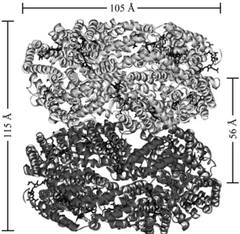
Fig. 4. PC–PC docking model. Ribbon representation of the refined PC–PC model. The general dimensions are indicated. The chromophores are shown in black.
Fig. 3. Representation of the structure of phycocyanin from G. chilensis. A) Ribbon representation of theα (light blue) and β (blue) subunits of C-phycocyanin of G. chilensis. The chromophores are shown in ball and stick representation. B) The molecule in the asymmetric unit. Ribbon representation of the heterohexamer (αβ)6.
Theα subunits are chains A, C, E, K, M, O and the β subunits are chains B, D, F, L, N, P according to the structure deposited in the Protein Data Bank.
The docking procedure performed by ZDOCK[29]involved a rigid body interaction of two hexamers of phycocyanin (PC). The models generated were evaluated by the program using desolvation, electrostatic properties and hydrophobic interac-tions. The top models ranked by ZDOCK reproduced the piling of PBPs as proposed by electron micrographs of the rods[4]. Even though it has been reported that for surface recognition a rigid body approach is adequate, a molecular dynamics procedure with the whole structure was performed in order optimize contacts and to obtain better packed interaction surfaces [45]. The docking complex with the highest hydro-phobic character in its interaction surface was refined by molecular dynamics as described in Materials and Methods. This produced modifications in the surfaces while preserving the relative position of the chromophores. The refined model presents a surface with less hydrophobic patches and an in-creased number of H-bonds (30 to 35) and salt bridges (3 to 7) between hexamers. The final complex is shown on Fig. 4. A rotation between hexamers was detected, which has previously been reported by Stec et al. [14] in a docking model of phycocyanin from C. caldarium. Thus, the best packing of the hexamers in a rod should be accomplished by a rotation of one over the other. This model allows a better packing of the side chains, increasing the number of interactions and improving the stability of the complex. This stable model was used to establish the coordinates of every chromophore for the determination of the energy transfer network in a PC–PC complex. Previous studies performed by our group[40]for phycocyanin from F. diplosiphon, show that a 20.5° rotation not only improves the packing of hexamers in a PC–PC complex, but also allows the participation of all the chromophores in the light transfer pro-cess, explaining the high efficiency of the system.
The calculation of kDAbetween all chromophores pairs was
performed using the extended Eq. (1) with the orientation factors calculated from the dipole moments as described in Materials and Methods.
Our results confirm that the transfer constants are very de-pendent on the geometric and spectroscopic factor of the chromophores, but the dependence is stronger for the distances between chromophore pairs as it is shown onTable 2. At iden-tical distances, the geometric factor becomes more important. Energy transfer steps for all chromophore pairs in our system were calculated and the value of the inverse of the transfer constant expressed in ps for the intra- or inter-phycocyanin steps are shown onTable 2. As hexamers of phycocyanin present high symmetry, in previous works, the analysis of possible light transfer pathways were performed in 1/3 of the biological unit. In our case, as the structure was solved with one hexamer per asymmetric unit, slight differences in the conformation of equivalent chromophores were detected, specially for the chro-mophores at chain E(α384) and K(α684), which present a rotation of
ring D as shown onFig. 2B. This conformational diversity also contributes to the differences in the values of kDA or kADfor
equivalent pairs of chromophores. The most significant dif-ferences are observed in the inter-hexamer steps of the pathway shown on Table 2. Differences of distances and orientation factors explain the different times obtained for equivalent steps.
These effects may be additive considering that the PC–PC complex is the result of a docking procedure.
The analysis of the energy transfer constants suggests two main intra-phycocyanin pathways: Iβ382→Iα184→Iα584→Iβ682
andIβ3153→Iβ5153(these pathways are reproduced in each third
of the hexameric ring in equivalent subunits). Two preferential
Table 2
Characteristics of energy transfer steps between pairs of chromophores Intrahexamer
Pair Distance(Å) Angle (°) Kappa 1/kDA(ps) 1/kAD(ps)
Internal energy transfer pathways
α184–β382 20.6 61.33 −1.26 6.27 4.07 α384–β282 20.5 93.81 −1.17 7.17 4.66 α284–β182 20.7 64.70 −1.27 6.39 4.15 bα84–β82N 20.6 6.61 4.29 α184–α584 25.6 66.75 1.18 16.90 16.90 α384–α484 25.6 88.71 1.37 13.10 13.10 α284–α684 25.6 81.03 1.26 15.00 15.00 bα84–α84N 25.6 15.00 15.00 α584–β682 20.7 74.45 −1.33 5.87 3.81 α484–β582 20.6 64.13 −1.34 5.48 3.56 α684–β482 20.6 93.04 −1.12 7.84 5.09 bα84–β82N 20.6 6.39 4.15 External energy transfer pathways
β1 153 –β6 153 26.2 158.08 1.52 5.63 5.63 β3 153 –β5 153 26.0 161.90 1.55 5.22 5.22 β2 153 –β4 153 26.1 162.65 1.50 6.06 6.06 bβ153–β153N 26.1 5.63 5.63 Interhexamer
Pair Distance(Å) Angle (°) Kappa 1/kDA(ps) 1/kAD(ps)
Internal energy transfer pathways
I β4 82 –II β1 82 22.02 72.24 0.98 12.67 12.67 I β5 82 –II β2 82 22.78 64.65 1.06 13.39 13.39 I β6 82 –II β3 82 21.23 74.14 0.98 10.21 10.21 bIβ82–IIβ82N 22.03 12.09 12.09 External energy transfer pathways
I β1534 –IIβ2153 39.37 151.30 1.47 65.30 65.30 I β1535 –IIβ3153 38.38 148.87 1.49 57.15 57.15 Iβ 6 153–IIβ 1 153 36.91 147.15 1.51 46.32 46.32 bIβ153–IIβ153N 38.22 56.26 56.26 Other possible connections
Iα 4 84–IIβ 2 82 33.20 13.04 −1.65 41.93 64.55 Iα 5 84–IIβ 3 82 31.66 10.21 −1.75 28.02 43.19 Iα 6 84–IIβ 1 82 32.16 29.92 −1.86 27.18 41.86 bIα84–IIβ82N 32.34 33.71 49.85 Iα 4 84–IIα 3 84 31.57 76.12 1.34 47.52 47.52 I α5 84 –II α1 84 29.83 96.98 1.45 29.19 29.19 I α6 84 –II α2 84 31.18 98.73 1.47 36.62 36.62 bIα84–IIα84N 30.86 37.77 37.77 I β4 82 –II α2 84 32.64 10.94 −1.72 53.49 34.74 I β825–IIα843 32.81 11.96 −1.63 61.36 39.85 I β826–IIα841 31.60 29.28 −1.89 36.53 23.73 bIβ82–IIα84N 32.35 50.46 32.77 Distance: Distance between the donor acceptor mass centers, Angle: Measured between the dipolar moments, Kappa: Geometric factor, 1/kDA: Donor–Acceptor
transfer rate, 1/kAD: Acceptor–Donor transfer rate. The identity of the hexamers
of the PC–PC complex is indicated by the roman numbers in superscripts: I for the upper hexamer and II for the lower hexamer.
393 C. Contreras-Martel et al. / Biophysical Chemistry 125 (2007) 388–396
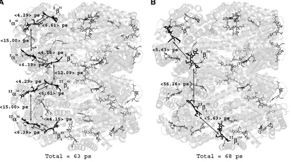
energy transfer pathways from one PC hexamer to the next are also describedIβ682→IIβ382andIβ5153→IIβ3153. These pathways
are indicated onFig. 5. The total average transference time of the internal and external preferential pathways shown inFig. 5 is 63 ps and 68 ps respectively. Experimental values in the range of 1 ps to 20 ps for the excitation transfer between trimers and 45 to 130 ps between hexamers in a rod have been reported [15,20,46,47]. In addition, calculated values using comparable approaches in other C-PC show a similar ps time-scale[14,16].
Most of the calculations performed with phycocyanin hexamers indicate that the transfer between trimers in one phycocyanin ring occurs through α84 and identify β82 as the donor between two PC hexamers. A complementary external pathway can be also described through β153. As the chromo-phore groups are distributed along the antenna in the biological structure, every chromophore will receive light continuously either from other chromophores or from the environment, which validates the pathways throughβ153.
4. Conclusions
Literature describes the analysis of the energy transfer pathways using heterodimers (αβ) trimers (αβ)3 or
subcom-plexes of phycobilisomes. In cases where the hexamer was used, it was built using crystallographic symmetry. In this article, the hexameric structure of phycocyanin from G. chilensis was solved experimentally. As the asymmetric unit was the hexamer, it was possible to detect slight differences between equivalent subunits, representing more accurately the biological functional unit.
The structure allowed to build a reliable model of the PC–PC complex, a minimum unit of a rod in phycobilisomes and to
describe the light transfer pathways along the rod. The theoretical approach used the Förster equation, previously validated for monomers and trimers, was applied to the higher complex PC–PC, describing two main transfer pathways: An internal pathway of 63 ps that involves β82 and α84 and an external of 68 ps that involvesβ153.
The existence of two preferential independent pathways, which repeat three times within the complex, explains the high efficiency of the energy transfer in the phycobilisome. This assures that every chromophore that absorbs light has an acceptor, minimizing energy loss, constituting one of the designs of nature that could be imitated for biotechnological purposes.
Acknowledgements
We thank Jorge L. Ríos for his help with the data collection. This work was supported by Dirección de Investigación, Universidad de Concepción, DIUC Grant N° 205.036.002-1.0. References
[1] A.N. Glazer, Phycobilisomes, structure and dynamics, Annu. Rev. Microbiol. 36 (1982) 173–198.
[2] C. Lipschutz, E. Gantt, Association of Phycoerythrin and Phycocyanin: in vitro formation of a functional energy transferring phycobilisome complex of Porphyridium sordidum, Biochemistry 20 (1981) 3371–3376. [3] D. Lundell, A.N. Glazer, Molecular architecture of a light harvesting
antennae. Core substructure in Synechococcus 6301 phycobilisomes: two new allophycocyanins and allophycocyanin B complexes, J. Biol. Chem. 258 (1983) 902–908.
[4] A. Ducret, W. Sidler, E. Wehrli, G. Frank, R. Huber, Isolation, char-acterization and electron microscopy analysis of a hemidiscoidal phycobilisome type from the cyanobacteria Anabaena sp PCC7120, Eur. J. Biochem. 236 (1996) 1010–1024.
Fig. 5. Energy transfer pathways in a PC–PC complex. A) Representative internal energy transfer pathway. B) Representative external energy transfer pathway. The protein is shown as a transparent matrix in which the chromophores are represented as sticks in different shades of grey in the three dimensional context. The darker chromophores show the pathways. The average acceptor–donor transfer rates for each pair of chromophores are indicated.
[5] N. Tandeau de Marsac, G. Cohen-Bazire, Molecular composition of cyanobacterial phycobilisomes, Proc. Natl. Acad. Sci. U. S. A. 74 (4) (1977) 1635–1639.
[6] M. Bunster, J. Tellez, A. Candia, Characterization of phycobiliproteins present in Gracilaria chilensis, Bol. Soc. Chil. Quím. 42 (1997) 449–455. [7] C. Contreras-Martel, J. Martínez-Oyanedel, M. Bunster, P. Legrand, C. Piras, X. Venerde, J.C. Fontecilla-Camps, Crystallization and 2.2 Å resolution structure of R-phycoerythrin from Gracilaria chilensis: a case of a perfect hemihedral twinning, Acta Crystallogr., D Biol. Crystallogr. 57 (2001) 52–60.
[8] M. Duerring, G.B Schmidt, R. Huber, Isolation, crystallization, crystal structure analysis and refinement of constitutive C-phycocyanin from the chromatically adapting cyanobacterium, Fremyella diplosiphon at 1.6 Å resolution, J. Mol. Biol. 217 (1991) 577–592.
[9] X.-Q. Wang, L.-N. Li, W.R. Chang, D.C. Liang, Structure of C-phycocyanin from Spirulina platensis at 2.2 Å resolution: a novel monoclinic crystal form for phycobiliproteins in phycobilisomes, Acta Crystallogr., D Biol. Crystal-logr. 57 (2001) 784–792.
[10] N. Adir, Y. Dobrovetsky, N. Lerner, Structure of c-phycocyanin from the thermophilic cyanobacterium Synechococcus vulcanus at 2.5 A: structural implications for thermal stability in phycobilisome assembly, J. Mol. Biol. 313 (2001) 71–81.
[11] N. Adir, R. Vainer, V. Lerner, Refined Structure of C-phycocyanin from the Cyanobacterium Synechococcus vulcanus at 1.6 Å: insights in the role of solvent molecules in thermal stability and co-factor structure, Biochim. Biophys. Acta 1556 (2002) 168–174.
[12] J. Nield, P.J. Rizkallah, J. Barber, N.E. Chayen, The 1.45 Å three dimensional structure of C-phycocyanin from the thermophylic Cyano-bacterium Synechococcus elongatus, J. Struct. Biol. 141 (2003) 149–155. [13] W.R. Chang, T. Jiang, Z.L. Wang, Z.X. Yang, D.C. Liang, Crystal structure of R-phycoerythrin from Polisiphonia urceolata at 2.0 Å resolution, J. Mol. Biol. 262 (1996) 721–731.
[14] B. Stec, R.F. Troxler, M.M. Teeter, Crystal structure of C-phycocyanin from Cyanidium caldarium provides a new perspective on phycobilisome assembly, Biophys. J. 76 (1999) 2912–2921.
[15] J. Zhang, F. Zhao, X. Zheng, H. Wang, Direct measurement of excitation transfer dynamics between two trimers in C-phycocyanin hexamer from Cyanobacterium Anabaena variabilis, Chem. Phys. Lett. 304 (1999) 357–364. [16] A.A. Demidov, A.Y. Borisov, Computer simulation of energy migration on C-phycocyanin of the blue green algae Agmenellum quadruplicatum, Biophys. J. 64 (1993) 1375–1384.
[17] S.A. Pizarro, K. Sauer, Spectroscopic study of the Light harvesting protein C-phycocyanin associated with colorless linker peptides, Photochem. Photobiol. 73 (2001) 556–563.
[18] T. Förster, in: M. Florkin, E. Stotz (Eds.), Comprehensive Biochem-istry, Mechanism of Energy Transfer, vol. 2, Elsevier, Amsterdam, 1967, pp. 261–280.
[19] M.-P. Debreczny, K. Sauer, J. Zhou, D.A. Bryant, Comparison of calculated and experimentally resolved rate constants for excitation energy transfer in C-phycocyanins, 1. Monomers, J. Phys. Chem. 99 (1995) 8412–8419.
[20] M.-P. Debreczny, K. Sauer, J. Zhou, D.A. Bryant, Comparison of calculated and experimentally resolved rate constants for excitation energy transfer in C-phycocyanins, 2. Trimers, J. Phys. Chem. 99 (1995) 8420–8431. [21] E. Gantt, C.A. Lipschutz, Phycobilisomes from Porphyridium cruemtum,
J. Cell Biol. 54 (1972) 313–324.
[22] W. Kabsch, Automatic processing of rotation diffraction data from crystals of initially unknown symmetry and cell constants, J. Appl. Crystallogr. 26 (1993) 795–800.
[23] M. Rossman, The molecular replacement method, Acta Crystallogr., A 46 (1990) 73–82.
[24] J. Navaza, AmoRe: an automated package for molecular replacement, Acta Crystallogr., A 50 (1994) 157–163.
[25] A.T. Brunger, P.D. Adams, G.M. Clore, W.L. Delano, P. Gros, R.W. Grosse-Kuntsleve, J.-S. Jiang, J. Kuszewski, N. Nilges, N.S. Pannu, R.J. Read, L.M. Rice, T. Simonson, G.L. Warren, Crystallography and NMR System (CNS): a new software system for macromolecular structure determination, Acta Crystallogr., D Biol. Crystallogr. 54 (1998) 905–921.
[26] A. Roussel, C. Cambillau, Silicon Graphics Geometry Partners Directory, The Turbo-Frodo Graphics Page, Silicon Graphics, Mountain View, CA, USA, 1991.
[27] R. Laskowsky, M. McArthur, D. Moss, J. Thornton, PROCHECK: a program to check the stereochemical quality of protein structures, J. Appl. Crystallogr. 26 (1993) 283–291.
[28] R. Chen, Z. Weng, Docking unbound proteins using shape complementarity, desolvation and electrostatics, Proteins 47 (2002) 281–294.
[29] R. Chen, L. Li, Z. Weng, ZDOCK: an initial stage protein docking algorithm, Proteins 52 (2003) 80–87.
[30] A. Archarov, V. Govorum, A. Dubanov, Y. Ivanov, A. Veselovsky, P. Lewi, O. Jansen, Protein–protein interactions as a target for drugs in proteomics, Proteomics 3 (2003) 380–391.
[31] B. Ma, T. Elkayam, H. Wolfson, R. Nussinov, Protein–protein interactions: structurally conserved residues distinguish between binding sites and exposed protein surfaces, Proc. Natl. Acad. Sci. U. S. A. 100 (2003) 5772–5777.
[32] B. Vallone, A. Miele, P. Vecchini, E. Chiancone, M. Brunori, Free energy of burying hydrophobic residues in the interface between protein subunits, Proc. Natl. Acad. Sci. U. S. A. 95 (1998) 6103–6107.
[33] S. Jones, J.M. Thornton, Progress in Biophysics and Molecular Biology, Protein–Protein Interactions: A Review of Protein Dimer Structures, vol. 63, 1995, pp. 31–165,http://www.biochem.ucl.ac.uk/bsm/PP/server/index. html.
[34] I.K. McDonald, J.M. Thornton, Satisfying hydrogen bonding potential in proteins, J. Mol. Biol. 238 (1994) 777–793.
[35] W.L. Jorgensen, J. Tirado-Rives, The OPLS force field for proteins. Energy minimizations for crystals of cyclic peptides and crambin, J. Am. Chem. Soc. 110 (1988) 1657–1666.
[36] W.F. Van Gunsteren, P.H. Hünenberger, A.E. Mark, P.E. Smith, I.G. Tironi, Computer simulation of protein motion, Comput. Phys. Commun. 91 (1995) 305–319.
[37] J. Grabowsky, E. Gantt, Photophysical properties of phycobiliproteins from phycobilisomes: fluorescent lifetimes quantum yields and polariza-tion spectra, Photochem. Photobiol. 28 (1978) 39–45.
[38] F.J. Kleima, E. Hofmann, B. Gobets, I.H. van Stokkum, R. Grondelle, K. Diederichs, H. van Amerongen, Förster energy transfer in peridinin-chlorophyll-a-protein, Biophys. J. 78 (1) (2000) 344–353.
[39] K.E. Apt, J.E. Collier, A.R. Grossman, Evolution of phycobiliproteins, J. Mol. Biol. 248 (1995) 79–96.
[40] A.R. Matamala, D.E. Almonacid, M.F. Figueroa, J. Martínez-Oyanedel, M.C. Bunster, A Semiempirical Approach to the Intra-Phycocyanin and Inter-Phycocyanin FRET Pathways in Phycobilisomes, J. Comput. Chem. (2006) Manuscript accepted.
[41] M.J. Frisch, G.W. Trucks, H.B. Schlegel, G.E. Scuseria, M.A. Robb, J.R. Cheeseman, V.G. Zakrzewski, J.A. Montgomery Jr., R.E. Stratmann, J.C. Burant, S. Dapprich, J.M. Millam, A.D. Daniels, K.N. Kudin, M.C. Strain, O. Farkas, J. Tomasi, V. Barone, M. Cossi, R. Cammi, B. Mennucci, C. Pomelli, C. Adamo, S. Clifford, J. Ochterski, G.A. Petersson, P.Y. Ayala, Q. Cui, K. Morokuma, D.K. Malick, A.D. Rabuck, K. Raghavachari, J.B. Foresman, J. Cioslowski, J.V. Ortiz, A.G. Baboul, B.B. Stefanov, G. Liu, A. Liashenko, P. Piskorz, I. Komaromi, R. Gomperts, R.L. Martin, D.J. Fox, T. Keith, M.A. Al-Laham, C.Y. Peng, A. Nanayakkara, C. Gonzalez, M. Challacombe, P.M.W. Gill, B. Johnson, W. Chen, M.W. Wong, J.L. Andres, C. Gonzalez, M. Head-Gordon, E.S. Replogle, J.A. Pople, Gaussian 98, Revision A.7, Gaussian, Inc., Pittsburgh PA, 1998.
[42] W. Reuter, G. Wiegand, R. Huber, M. Than, Structural analysis at 2.2 Å of orthorhombic crystals presents the asymmetry of allophycocyanin-linker complex AP-L7.8C, from phycobilisomes from
Mastigocladus laminosus, Proc. Natl. Acad. Sci. U. S. A. 96 (1999) 1363–1368.
[43] S.F. Altschul, W. Gish, W. Miller, E.W. Myers, D.J. Lipman, Basic local alignment search tool, J. Mol. Biol. 215 (1990) 403–410.
[44] J. Martínez-Oyanedel, C. Contreras-Martel, C. Bruna, M. Bunster, Structural-functional analysis of the oligomeric protein R-phycoerythrin, Biol. Res. 37 (2004) 733–745.
395 C. Contreras-Martel et al. / Biophysical Chemistry 125 (2007) 388–396
[45] C.J. Camacho, Modeling side-chains using molecular dynamics improve recognition of binding region in CAPRI targets, Proteins 60 (2005) 245–251.
[46] A.R. Holzwarth, J. Wendler, G.W. Suter, Studies on chromophore coupling in isolated phycobiliproteins: II. Picosecond energy transfer
kinetics and time resolved fluorescence spectra of C-phycocyanin from Synechococcus 6301 as function of the aggregation state, Biophys. J. 51 (1987) 1–12.
[47] A.R. Holzwarth, Structure-function relationships and energy transfer in phycobiliprotein antennae, Physiol. Plant. 83 (1991) 518–528.