by
Vanessa PRINSEN
MANUSCRIPT-BASED THESIS PRESENTED TO ÉCOLE DE
TECHNOLOGIE SUPÉRIEURE
IN PARTIAL FULFILLMENT OF A MASTER’S DEGREE
WITH THESIS, WITH A PERSONALIZED CONCENTRATION
M.A.Sc.
MONTREAL, JUNE 4, 2020
ÉCOLE DE TECHNOLOGIE SUPÉRIEURE
UNIVERSITÉ DU QUÉBEC
BY THE FOLLOWING BOARD OF EXAMINERS
Mrs. Rita Noumeir, thesis supervisor
Department of Electrical Engineering, École de technologie supérieure
Mr. Philippe Jouvet, co-supervisor
Pediatric Intensive Care Unit, CHU Sainte-Justine
Mr. José Dolz, president of the board of examiners
Department of Software and IT Engineering, École de technologie supérieure
Mrs. Catherine Laporte, member of the jury
Department of Electrical Engineering, École de technologie supérieure
THIS THESIS WAS PRESENTED AND DEFENDED
IN THE PRESENCE OF A BOARD OF EXAMINERS AND THE PUBLIC ON MARCH 12, 2020
RÉSUMÉ
L’apparence et le comportement de la région oculaire jouent un rôle important dans le diagnostic de la douleur et de l’état de conscience d’un patient, surtout lorsqu’il ou elle est trop jeune pour parler. Toutefois, la localisation et le suivi des yeux dans un environnement hospitalier pédiatrique constitue toujours un défi majeur pour les applications d’aide à la décision clinique et de surveillance des patients. L’objectif global de ce projet est de développer un système d’aide à la décision clinique qui utilise des caméras pour détecter les signes de conscience et de détresse chez les enfants. Ce travail de recherche se concentre sur le premier défi, à savoir comment localiser les yeux dans une image d’un patient pédiatrique dans un lit d’hôpital. Les solutions existantes pour la localisation des yeux atteignent d’excellentes performances sur les images d’adultes, mais sont médiocres dans l’environnement hospitalier pédiatrique, où l’apparence du visage peut varier en raison de l’âge, de la position du patient, et de la présence d’équipements médicaux. Peu d’études ont examiné l’application de la vision par ordinateur et l’analyse faciale dans ce milieu.
Pour développer une solution performante pour la localisation des yeux, un nouvel ensemble de données d’entrainement, constitué d’images de jeunes enfants issues des recherches sur l’Internet, est ajouté aux données images d’adultes pour entrainer des classifieurs cascade et des réseaux de neurones convolutifs. Un autre ensemble de données innovant, constitué de 59 enregistrements de patients dans une unité de soins intensifs pédiatrique, est utilisé pour évaluer la performance de ces modèles. Cet ensemble de données servira également aux travaux futurs sur ce projet et d’autres projets de recherche sur les applications pédiatriques de la vision par ordinateur.
Le réseau de neurones convolutifs entrainé avec l’ensemble de données des jeunes enfants atteint un taux de localisation des yeux de 79,7%, ce qui est bien supérieur aux modèles entrainés uniquement avec des données adultes. Ce modèle surpasse également les modèles cascade. L’amélioration significative de performance après l’ajout des images des jeunes enfants souligne le besoin de données et modèles personnalisés pour des applications spécialisées telles que la surveillance des patients en pédiatrie. Les projets cliniques de vision par ordinateur ne peuvent pas s’appuyer sur des modèles et des ensembles de données existants pour couvrir leurs besoins. Par contre, la taille raisonnable de l’ensemble de données d’entrainement supplémentaire utilisé pour ce projet suggère que le développement d’une base de données d’entrainement interne est à la portée d’un grand hôpital.
L’efficacité du réseau de neurones convolutifs, compte tenu les défis du milieu hospitalier pédiatrique, fait de lui une approche puissante pour la localisation et le suivi des yeux dans l’environnement hospitalier. La capacité du réseau de neurones convolutifs à apprendre des
caractéristiques d’image uniques lui permet de s’adapter à la localisation des yeux dans un environnement atypique où les approches basées sur des règles ou suppositions sur l’apparence du visage ne s’appliquent pas toujours. Les faiblesses actuelles du modèle, telles que des performances médiocres sur des yeux avec apparences atypiques et la lenteur de traitement des images, vont s’améliorer avec la croissance des ensembles de données d’entrainement et les améliorations technologiques.
Mots-clés: vision par ordinateur, réseau de neurones convolutifs, classifieur cascade, pédiatrie,
ABSTRACT
The appearance and behaviour of the eye region are important windows into a patient’s condition and level of consciousness, particularly for patients too young to speak. Unfortunately, reliable localization and tracking of the eye region in the pediatric hospital environment is a significant challenge for clinical decision support and patient monitoring applications. The overall aim of this research project is to develop a clinical decision support system that uses bedside cameras to detect signs of consciousness and distress due to pain. This work focuses on the first problem to be solved, namely how to locate the eyes in an image of a pediatric patient in a hospital bed. Existing work in eye localization achieves high performance on adult datasets but performs poorly in the busy pediatric hospital environment, where face appearance varies because of age, position and the presence of medical equipment. Few studies have examined the application of computer vision and facial analysis techniques to young children in a hospital environment. To develop an appropriate solution for eye localization, a new training dataset, formed of images of young children from internet searches, is added to adult facial images to train cascade classifiers and convolutional neural networks. Another novel dataset, consisting of 59 recordings of patients in a pediatric intensive care unit, is used to evaluate the performance of these models. This dataset will also serve future work on this and other research projects in pediatric computer vision.
The convolutional neural network trained with the added image data of young children achieves a 79.7% eye localization rate, much higher than models trained on adult data alone. This model also outperforms the cascade models. The dramatic performance improvement gained from adding task-specific images to the training data highlights the need for custom-trained models for specialized applications like pediatric patient monitoring. Existing models and datasets are not sufficient, but the moderate size of the task-specific training dataset used here suggests that developing an internal training dataset is within reach of a typical large hospital.
The effectiveness of the convolutional neural network, given the challenges of this setting, makes it a powerful approach for eye localization and tracking in the hospital environment. The convolutional neural network’s ability to learn unique features allows it to adapt to the challenges of eye localization in an atypical setting where usual assumptions about facial appearance do not necessarily apply. The present weaknesses of the model, like poor recognition of uncommon eye appearances and slow image processing times, will improve with larger training datasets and technological improvements.
Keywords: computer vision, convolutional neural network, cascade classifier, pediatrics, hospital
INTRODUCTION . . . 1
CHAPTER 1 LITERATURE REVIEW . . . 5
1.1 Object detection . . . 5
1.2 Eye detection for pediatrics . . . 7
1.3 The eyes as a measure of mental activity . . . 8
1.4 Machine learning from eye movements . . . 9
1.5 Eye features . . . 9
1.6 Consciousness in pediatrics . . . 9
1.7 Pediatric pain recognition . . . 10
CHAPTER 2 ORGANIZATION OF THE DOCUMENT . . . 17
CHAPTER 3 METHODOLOGY . . . 19
3.1 Training Datasets . . . 19
3.2 Algorithms . . . 20
3.2.1 Cascade classifiers . . . 20
3.2.2 Convolutional neural networks . . . 21
3.2.3 Evaluation . . . 23
CHAPTER 4 AUTOMATIC EYE LOCALIZATION FOR HOSPITALIZED INFANTS AND CHILDREN USING CONVOLUTIONAL NEURAL NETWORKS . . . 25 4.1 Introduction . . . 26 4.2 Technical challenges . . . 28 4.3 Existing Work . . . 30 4.4 Methodology . . . 31 4.4.1 Test data . . . 32 4.4.2 Training data . . . 33 4.4.3 Cascade classifiers . . . 34
4.4.4 Convolutional neural networks . . . 36
4.5 Results . . . 38
4.6 Discussion . . . 41
4.6.1 Task-specific training dataset . . . 41
4.6.2 Model weaknesses . . . 42
4.6.3 Convolutional neural networks vs. cascade classifiers . . . 42
4.6.4 Limitations . . . 43
4.7 Conclusion . . . 43
CONCLUSION AND RECOMMENDATIONS . . . 47
APPENDIX I SELECTED PEDIATRIC EVALUATION SCALES . . . 49
APPENDIX II USER GUIDE: TRAINING DATASET . . . 59
APPENDIX III USER GUIDE: TEST DATASET . . . 63
APPENDIX IV IMPLEMENTATION GUIDE: CONVOLUTIONAL NEURAL NETWORKS USING LUMINOTH . . . 67
Table 1.1 Quantitative eye features . . . 14 Table 1.2 Eye states by level of wakefulness . . . 15 Table 1.3 Summary of pediatric evaluation scales . . . 16 Table 4.1 Performance of existing open-source face localization tools on 42
test images . . . 29 Table I-1 Coma and sedation assessment tools for children . . . 49 Table I-2 Delirium assessment tools for young children . . . 56
Figure 4.1 Series of frames from one recording at CHU Sainte-Justine . . . 32
Figure 4.2 Hard negative mining . . . 36
Figure 4.3 Accuracy of eye localization with cascade classifiers and CNNs . . . 39
AU Action Unit
CAPD Cornell Assessment of Pediatric Delirium CAPP Canonical-normalized Appearance
CERT Computer Expression Recognition Toolbox CHUSJ Centre hospitalier universitaire Sainte-Justine CLAHE Contrast Limited Adaptive Histogram Equalization CNN Convolutional Neural Network
COMFORT-B Comfort Behavioural
FACS Facial Action Coding System
FLACC Face Legs Activity Cry Consolability GPU Graphical Processing Unit
ICU Intensive Care Unit KNN k-Nearest-Neighbours LBP Local Binary Pattern LTP Local Ternary Pattern
PCAM Pediatric Confusion Assessment Method PICU Pediatric Intensive Care Unit
SPTS Similarity-normalized Shape SVM Support Vector Machine
Consciousness–that is, the patient’s wakefulness and awareness of his or her surroundings– is an important indicator of health and neurological state. Altered consciousness, ranging from confusion and lethargy to loss of consciousness or even coma, may indicate many problems, including neurological disorders, poisoning, or brain injury (Avner, 2006). In cases when consciousness is purposefully altered by sedation or anesthesia, it is important to track the level of consciousness and ensure that patients are not oversedated or undersedated. Oversedation "will often result in prolonged mechanical ventilation and hemodynamic instability" (Johansson & Kokinsky, 2009), while undersedation may cause patients undue stress and pain.
It is therefore common practice to evaluate patient consciousness and distress regularly in intensive care units, using well-known tools like the Glasgow Coma Score (Teasdale & Jennett, 1974). In pediatric units, tools like the COMFORT behavioural (COMFORT-B) scale (Van Dijk, Peters, Van Deventer & Tibboel, 2005) and the Face Legs Activity Cry Consolability (FLACC) scale (Merkel, Voepel-Lewis, Shayevitz & Malviya, 1997) attempt to quantify consciousness and distress in children by observing their behaviour. These scales are typically evaluated by nursing staff using visual observation and require less than 5 minutes to perform, but the parameters of these scales are not always easy to score. For example, when determining the facial tension score for the COMFORT-B scale, it may be difficult for a nurse to distinguish between "facial muscle totally relaxed" and "facial muscle tone normal", especially for a child he or she has not seen before. As a result, different people may calculate different scores for the same patient.
The assessment of consciousness and distress in children under two years of age presents a particular challenge because these patients do not have the ability to respond to commands or explain what they are feeling. A study by Johansson & Kokinsky (2009) demonstrated that in
20% of cases there was a disagreement between nurses’ bedside assessments of the level of sedation of young patients, highlighting the necessity of pediatric assessment tools to support their work.
The pediatrics research team at the Justine University Hospital Centre (CHU Sainte-Justine) in Montreal is interested in developing a clinical decision support system that uses bedside cameras to detect signs of consciousness and distress due to pain.
Visual inspection of patients is an important part of clinical work and evaluation, yet the use of video for patient care remains rare in the hospital setting. Where video is used, it is often for retrospective analysis or remote monitoring by care staff rather than automated, real-time patient monitoring.
The development of compact, high-resolution cameras allows detailed and discreet monitoring directly at the point of care, and together with automated video image analysis would enable standardized, round-the-clock surveillance of patient state. This could be particularly valuable in situations where continuous bedside monitoring by trained staff is not possible, such as in remote regions or during medical transport. Such a system would also reduce human subjectivity in the evaluation of the COMFORT-B and FLACC scales and ensure the continuous and accurate recording and transmission of information throughout the patient’s care.
The appearance and behaviour of the eye region are of particular interest in such a system because it can provide a wealth of information about a patient’s condition and consciousness. Computer analysis of images using machine learning techniques has already allowed the identification of infant facial expressions and pain, as in Parodi, Melis, Boulard, Gavelli & Baccaglini (2017) where infant facial features like "eye squeeze" were detected in order to automatically score pain scales, and Fotiadou, Zinger, Ten, Oetomo & With (2014) where changes in facial appearance and expression were used to train a classifier to distinguish between pain and no-pain states.
The subjects in these studies were generally healthy with no feeding or breathing tubes or other facial occlusions, and they were recorded facing the camera.
Reliable localization and tracking of the eye region become more challenging when continuously monitoring directly in a hospital room, where patient age, condition, position and surroundings can vary widely. Patients may not be looking directly at the camera, and their faces may be partially occluded by medical equipment like nasal tubes or bandages. In addition, many existing approaches are developed and tested on adult facial datasets, which do not generalize well to infants and young children. Existing approaches and best practices for eye localization must therefore be re-examined for application in a pediatric clinical environment.
Research objectives
The goal of this work is to develop effective techniques for extracting the eye region from images of hospitalized pediatric patients, taking into account the challenges and constraints of the setting. Existing state-of-the-art computer vision and machine learning algorithms will be evaluated and refined for the task of detecting and tracking the eye region of children in a live clinical environment. What are the challenges and constraints of these techniques? How can they be refined to detect the eye region in the pediatric hospital setting? What datasets and technologies must be developed to support this project?
In parallel with technological development, a dataset is developed comprising videos of patients in the pediatric intensive care unit at CHU Sainte-Justine. These videos will be recorded with a photography camera pointed at the patient’s upper body to acquire clear, high-resolution video footage of the facial region. Patients of all ages and medical conditions will be targeted for recording, so long as their eye region is at least partly visible. This dataset will fill an important gap for several research projects at CHU Sainte-Justine, as there are currently no
publicly available datasets of hospitalized children available to researchers wishing to train and evaluate computer vision and machine learning models.
Achievements
The outcomes of this work demonstrate the potential of convolutional neural networks (CNNs) for eye localization in a pediatric hospital setting and the significant performance improvements that are gained by including a moderately sized sample of task-specific data during training. The convolutional neural network trained in this way performs better at eye localization than other methods on the novel dataset of 59 videos recorded of patients in the pediatric intensive care unit at Sainte-Justine Hospital. This real-world dataset demonstrates the challenges of working in a pediatric hospital environment, where faces are often partially occluded by positioning or medical devices.
1.1 Object detection
Cascade classifiers, as first proposed in Viola & Jones (2001), are a widely used method for face and eye localization due to the availability of pre-trained models and the ease of training new models using limited data. Cascade classifiers are trained using manually defined features, such as Haar features or a local binary pattern (LBP), and use a sliding window approach to locate objects in new images.
The "cascade" refers to the narrowing of the search region as the classifier is applied to an image. Rather than calculate all relevant features for each window, a window is evaluated with a subset of features. If the object is not detected using those features, the window is discarded and not evaluated further. If, on the other hand, the results are promising, another subset of features is applied, and so on until the window is classified. This narrowing of the search region allows a classifier trained with a large number of features to run efficiently. Cascade classifiers are much faster than convolutional neural networks, making it easy to achieve real-time object localization. The use of cascade classifiers is illustrated in a study to detect fatigue in bus drivers (Mandal, Li, Wang & Lin, 2017) which shares similar constraints to our project: images are recorded using a camera placed off-centre and above the subject, who may move freely and even face away from the camera. A chain of cascade detectors is used to identify first the upper body, then the face and its orientation, and finally the eye region. This gradual narrowing of the region of interest improves performance and accuracy by shrinking the candidate search region and eliminating false positives in the background.
Mingxin, Yingzi & Xiangzhou (2016) also use a trained classifier to search for eyes but add some rules: the search is limited to the upper half of the face, and this upper region is further divided into left and right halves (which should have one eye each). A variance filter is used to
eliminate non-eye regions, then a support vector machine (SVM) is used to check the remaining candidate regions for eyes. El Kaddouhi, Saaidi & Abarkan (2017) eschewed the usual classifier and instead detected corner points using the Shi-Tomasi corner detector and grouped these points (by k-means) to produce candidate eye regions, which were then analyzed by template matching. One approach not based on machine learning is proposed by Chen & Liu (2011). The facial image is converted to YUV colour space and the U component is thresholded to highlight the eye, which has a lower intensity than the surrounding region. Projection functions are then used on this binary image to find the eye boundaries. Chun-Ning, Tai-Ning, Pin & Sheng-Jiang (2012) describe another similar approach, without the thresholding, where rapid changes of intensity around the eyes are detected. However, both these approaches work best if the eyes are open, limiting their usefulness in practice.
Nevertheless, colour can be useful for narrowing the search region for other face and eye localization algorithms. For example, masking parts of an image based on skin colour can be used as a pre-processing step before a cascade classifier or other machine-learning-based approach, as in Mutneja & Singh (2017). It can also be used after face localization to discard face candidates that do not contain a sufficient number of skin colour pixels, as suggested by Ge, Han & Quan (2015). The accuracy of skin localization has been demonstrated on adults using the HSV and YCbCr colour spaces (Shaik, Ganesan, Kalist, Sathish & Jenitha, 2015), the RGB, YCbCr, and HSV colour spaces (Kolkur, Kalbande, Shimpi, Bapat & Jatakia, 2017), and the YCbCr colour space alone (Emmanuel & Ibiyemi, 2017; Ge et al., 2015).
Convolutional neural networks (CNNs), a class of deep neural network particularly well suited to computer vision tasks, grew in popularity through the 2010s as computation power increased to permit faster training and predictions.
CNNs can be trained to sort images of objects into different classes, but there is an additional problem to solve in real-world situations: how to identify which objects are of interest in a crowded scene. Many algorithms have been proposed to solve this problem of object detection, among them Faster R-CNN (Ren, He, Girshick & Sun, 2015), which sacrifices the speed of
other algorithms like YOLO (Redmon, Divvala, Girshick & Farhadi, 2016) for greater accuracy. Faster R-CNN builds on previous work in the R-CNN and Fast R-CNN algorithms, which used slow selective search algorithms to identify regions of interest in an image. Faster R-CNN speeds up this procedure by training a "region proposal network" during the training process, which identifies these regions of interest. Once identified, these subsections of a larger image can be classified by a conventional CNN.
CNNs underlie the most powerful facial and eye detection systems now available, and recent work has applied them in the hospital setting to detect adult patients exiting their beds (Chwyl, Chung, Shafiee, Fu & Wong, 2017), to identify the pose of adult patients in hospital beds (Liu, Yin & Ostadabbas, 2019a), and to detect infants in bed and segment their skin region (Chaichulee, Villarroel, Jorge, Arteta, Green, McCormick, Zisserman & Tarassenko, 2017). Nevertheless, the application of CNNs to real-world problems remains limited by the need for large quantities of training data and the computational cost of analyzing images through complex, many-layered networks.
1.2 Eye detection for pediatrics
Many of the approaches described above perform poorly when applied to young children in a hospital setting. As discussed by Saeijs, Tjona Ten & De With (2018), not only is there a lack of "in-the-wild" datasets of young hospitalized children with which to train models, but infant faces differ in appearance from those of older children and adults, complicating the use of models trained on adult datasets. Infant faces have less prominent features like eyebrows and skin folds, and facial proportions and morphology are different, making template and rules-based approaches more difficult to apply. Additionally, in a hospital setting faces may be partially occluded by items like bandages and tubes, and may move quickly and unpredictably.
Past studies interested in the facial features of babies and young children have largely avoided the problem of face and eye localization by manually selecting facial landmarks, as in Zamzami, Ruiz, Goldgof, Kasturi, Yu & Ashmeade (2015), or by using laborious alternative approaches
like custom active appearance models, as in Fotiadou et al. (2014). Recent work in Chouinard, Scott & Cusack (2019) uses the Amazon Rekognition system to analyse infant facial images. Amazon Rekognition is a cloud-based computer vision platform that allows users to submit their own images for facial detection and analysis using pre-trained models. While results were promising, submitting patient data to an online service such as Amazon Rekognition raises privacy concerns that make this approach inappropriate for a medical setting.
1.3 The eyes as a measure of mental activity
Chen & Epps (2013) proposed several features that could be used to measure cognitive load in adults, such as pupil size, blink number, and saccade amplitude. They observed that the number of blinks and pupil size increased with more demanding tasks. Chen, Epps & Chen (2013) identified 29 possible eye features and used them to detect task transition (for which the most sensitive features were pupil response and blinks), perceptual load (for which the most sensitive features were blinks and saccades/fixations), and cognitive load (for which the most sensitive feature was average pupil size).
Several studies have tried to detect consciousness and alertness in car drivers. Kojima, Kozuka, Nakano & Yamamoto (2001) found that it is possible to detect alertness from the duration of blinks (longer blinks indicate drowsiness) and concentration from the variance of gaze movements (variance decreases during mental work). Jo, Lee, Park, Kim & Kim (2014) measured drowsiness based on percentage of frames with eyes closed in a given period. Tokuda, Obinata, Palmer & Chaparro (2011) showed that saccadic eye movements increase as mental work increases.
Blink rate decreases during visually demanding tasks (e.g., reading) but increases if a task is prolonged or made more difficult. (Stern, Boyer & Schroeder, 1994)
De Rivecourt, Kuperus, Post & Mulder (2008) showed that fixation duration and dwell time decreased as task demands increase during a flight simulation exercise.
1.4 Machine learning from eye movements
Lee, Ojha & Lee (2015a) fed four eye features into a one-class SVM classifier to determine concentration: number of eye blinks, duration of "open eye" state, pupil size variation, and rate of missing data (user looking away from eye tracker). The classifier was able to differentiate between concentration and non-concentration states for the small group of adult test users. Jang, Mallipeddi, Lee, Kwak & Lee (2014) used fixation length, fixation count and pupil size variation to classify whether users were searching for an object or not when viewing images of indoor and outdoor scenes. They achieved a recognition rate of 85.26% using an SVM classifier.
1.5 Eye features
Table 1.1 presents a list of measurable features that can be detected in the eye region using computer vision techniques, given images or video of sufficient illumination and resolution. The exact resolution required varies depending on the feature: for example, a higher resolution is needed to detect changes in pupil size than to detect the opening and closing of the eye.
1.6 Consciousness in pediatrics
The pediatric age range presents a particular challenge for determining consciousness and alertness because infants and young children lack the ability to speak or follow instructions, their motor control is still poor, and it can be difficult to differentiate reflex responses from conscious responses.
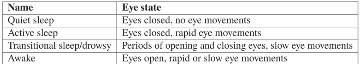
Four major states of wakefulness are typically identified in newborns, each with unique eye behaviour. Table 1.2 lists these states and a brief description of the associated eye behaviour. (Yasova Barbeau & Weiss, 2017)
Various scales have been developed to quantify the neurological state of young children in a hospital setting, many using information from the face and eyes. Table 1.3 summarizes some of
the most well-known coma, sedation, and delirium scales for infants and young children, along with a brief description of each and some examples of their criteria in and around the eye region. These scales are reproduced in full in Appendix I: Selected pediatric evaluation scales.
Unlike the measurable eye features proposed in Table 1, these pediatric evaluation scales rely heavily on qualitative measures–for example, whether the eyes are "bright" or "dull", as in the Vancouver Sedative Recovery Scale. Evaluation is also limited by the abilities of infants and young children. For example, a newborn’s verbal response to stimuli, which is a criterion measured by the Glasgow Coma Score, would be limited to cries.
Nelson, Lachman & Gold (2017) compared bedside diagnoses of delirium using the Cornell Assessment of Pediatric Delirium (CAPD) and Pediatric Confusion Assessment Method for the ICU (PCAM) to diagnoses performed by psychiatrists and found wide gaps between assessments by the two groups. This demonstrates the great difficulty of diagnosing cognitive state in pediatric patients, and the need for more accurate tools to assist clinicians.
Several studies have used eye tracking systems to gain insight into the cognitive development of young children (Corbetta, Guan & Williams, 2012; Saez de Urabain, Nuthmann, Johnson & Smith, 2017), but these studies are performed with alert children and eye trackers that require system calibration, something impossible to do with unconscious, distressed, or very young patients in a pediatric intensive care (PICU) environment.
1.7 Pediatric pain recognition
Parodi et al. (2017) proposed a tool that could automatically recognize infant facial features (for example, "eye squeeze" or "mouth stretch") that are used to calculate three traditional pain scores. The scores obtained from the system were compared to those calculated manually by users. There were high mismatch rates between the automatic and manual evaluation of scores (averaging near 50% mismatch for many parameters), but the study did not try to validate whether this was due to system error or user error.
Brahnam, Chuang, Sexton & Shih (2007a) photographed 26 neonates undergoing three unpleasant but non-painful stimuli (transport from one crib to another, a puff of air to the nose, friction on the outside of the heel) and one painful stimulus (a heel lance). The grayscale pixel intensity of the face region was extracted and the resulting feature vector was processed by four different machine learning techniques to classify the images as pain or nonpain. The highest classification accuracy achieved was 90.2%, but it was observed that certain subjects were harder to classify than others: all four algorithms performed poorly on the same subset of faces.
Nanni, Brahnam & Lumini (2010a) used the same dataset as the study above (Brahnam et al., 2007a) to extract the following texture descriptors:
• Local Binary Pattern (LBP): compares a pixel to each of its 8 neighbours and assigns 0 if
its value is greater than the neighbour and 1 otherwise, generating an 8-digit binary number for each pixel.
• Local Ternary Pattern (LTP): similar to LBP but assigns 0 if a pixel is the same value
as its neighbour (within a threshold t), and -1 if it is greater than or -1 if it is less than the neighbour. This descriptor is less sensitive to noise than LBP.
• ELBP/ELTP (Elongated LBP/LTP): uses an elliptical neighbourhood instead of circular.
Not rotation invariant but can work better with images that have anisotropic structures (like faces).
• ILBP/ILTP (Improved LBP/LTP): compares pixel neighbours to the local mean instead of
the central pixel of a region. This descriptor is less sensitive to noise.
These texture descriptors were also extracted from images preprocessed using Gabor filters, a Laplacian of Gaussian filter, and the Illumination Normalization method, but the best classification results were obtained using grey levels. The best classification performance with an SVM (0.926 AUC) is obtained using the ELTP descriptor.
The above two approaches are limited by the static images used and cannot take into account facial movement and dynamic facial expressions. (Zamzmi, Pai, Goldgof, Kasturi, Sun & Ashmeade,
2016) Furthermore, pixel intensity, as a feature, is sensitive to illumination changes and occlusion (for example, by an infant’s hands). Texture descriptors like local binary patterns are less sensitive to illumination and noise (though still affected by occlusion) and may be well suited to detecting the wrinkles and furrows on the facial expressions of newborns. (Nanni et al., 2010a) Zamzami et al. (2015) collected video sequences for 9 neonates experiencing acute (heel lance) and chronic (post-operative recovery) pain. Facial landmarks were identified manually due to problems with existing detection algorithms, which are developed and trained on adult faces and do not deal well with unpredictable infant movements and occlusion by pacifiers and hands. The identified face is divided into four regions and an optical flow vector is generated for each of these regions and used to estimate optical strain. The estimated strain values for each region are added together to generate overall strain magnitude, which is used to classify the expression as pain or no-pain using k-Nearest Neighbours (KNN) and SVM classifiers. The KNN algorithm with k=3 produced the best classification accuracy of 96%.
Fotiadou et al. (2014) filmed video of 10 infants in a neonatal intensive care unit experiencing painful (heel lance) and non-painful (diaper change, hunger, resting, sleeping) experiences. During initialization, the infant’s face is detected, its pose estimated (frontal or rotated), and it is fit to a pre-trained facial model (the Active Appearance Model, or AAM). This shape is then used to initialize future frames. The AAM allows the face’s geometry to be manipulated to extract the following features:
• SPTS (similarity-normalized shape): the shape/geometry of the face in the current frame compared to the mean/default shape.
• SAPP (similarity-normalized appearance): the pixels of the facial image warped to the similarity-normalized shape.
• CAPP (canonical-normalized appearance): the pixels of the facial image warped to the mean shape.
Preprocessing techniques like Illumination Normalization and Laplacian of Gaussian filtering are used on the two appearance representations (SAPP and CAPP) before extracting features using various texture descriptors (including LBP and ELBP as described earlier), and an SVM classifier is trained to classify pain vs. no-pain. The best performance (0.98 AUC) is obtained using no preprocessing and ELBP texture descriptors on the appearance data. Performance with this approach is highly dependent on successful tracking of the face, which is challenging for infants whose movements are unpredictable and whose faces may be occluded by pacifiers or breathing tubes. Furthermore, custom AAMs were constructed for each infant in the study, but this is not a feasible approach for a real-world application given the time and effort required to create the models.
Sikka, Ahmed, Diaz, Goodwin, Craig, Bartlett & Huang (2015) recorded video of 50 post-operative youth (5-18 years old) experiencing pressure at a surgical site. Patients, parents, and nurses simultaneously rated pain using the Numerical Rating Scale. Videos were analyzed using the Computer Expression Recognition Toolbox (CERT) to extract standardized facial component movements known as facial action units (AUs), as described in the Facial Action Coding System (FACS). (Examples of AUs used in this study are "eye closure" and "upper lip raiser".) A 42-feature AU data vector was used to train two models:
• Binary classification (pain or no-pain), using a logistic regression model to learn mappings between features and binary pain labels.
• Pain-intensity estimation, using a linear regression model to learn a linear combination of features to predict pain self-ratings. A second pain-intensity model was trained using not only the AU vector but also "time since surgery" as a feature.
For binary classification, the trained model performed similarly to nurse and parent assessments of pain (>0.8 AUC). Performance was poorer for pain intensity estimation but comparable to the nursing assessment.
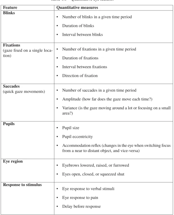
Table 1.1 Quantitative eye features
Feature Quantitative measures Blinks
• Number of blinks in a given time period • Duration of blinks
• Interval between blinks
Fixations
(gaze fixed on a single loca-tion)
• Number of fixations in a given time period • Duration of fixations
• Interval between fixations • Direction of fixation
Saccades
(quick gaze movements) • Number of saccades in a given time period
• Amplitude (how far does the gaze move each time?)
• Variance (is the gaze moving around a lot or focusing on a small area?)
Pupils
• Pupil size
• Pupil eccentricity
• Accommodation reflex (changes in the eye when switching focus from a near to distant object, and vice-versa)
Eye region
• Eyebrows lowered, raised, or furrowed • Eyes open, closed, or squeezed shut
Response to stimulus
• Eye response to verbal stimuli • Eye response to pain
Table 1.2 Eye states by level of wakefulness
Name Eye state
Quiet sleep Eyes closed, no eye movements Active sleep Eyes closed, rapid eye movements
Transitional sleep/drowsy Periods of opening and closing eyes, slow eye movements Awake Eyes open, rapid or slow eye movements
Table 1.3 Summary of pediatric evaluation scales
Name of scale Description Examples of face- and eye-related criteria
Modified Glasgow Coma Scale for Infants and Children
(Teasdale & Jennett, 1974)
Widely used coma scale, adapted for infants and children
• Eye response to stimulus COMFORT scale
(Ambuel, Hamlett, Marx & Blumer, 1992)
Assesses sedation/distress
in PICU patients • Facial tension • Physical movements Vancouver Sedative Recovery
Scale
(Macnab, Levine, Glick, Phillips, Susak & Elliott, 1994b)
Originally developed to assess recovery from ma-jor surgery
• "Bright" vs. "dull" eyes • Looks "at you" vs. "through
you"
• "Alert" vs. "flat" facial ex-pression
Blantyre coma scale
(Newton, Chokwe, Schellen-berg, Winstanley, Forster, Peshu, Kirkham & Marsh, 1997)
Modified GCS developed to assess malarial coma in children
• Directed vs. not directed eye response
FLACC scale (Merkel et al., 1997)
Pediatric pain assessment tool for children unable to communicate
• Facial expression Richmond Agitation Sedation
Scale
(Sessler, Gosnell, Grap, Brophy, O’neal, Keane, Tesoro & El-swick, 2002)
Measures the agitation or
sedation level of a patient • Ability to maintain eyes open • Eye contact
Cornell Assessment of Pediatric Delirium
(Traube, Silver, Kearney, Patel, Atkinson, Yoon, Halpert, Augen-stein, Sickles & Li, 2014)
Assessment of pediatric delirium with develop-mental anchor points pro-vided for scoring children under 2
• Awareness of surroundings • Eye contact
Preschool Confusion Assess-ment Method for the ICU (Smith, Gangopadhyay, Goben, Jacobowski, Chestnut, Savage, Rutherford, Denton, Thompson, Chandrasekhar et al., 2016)
Version of the Confu-sion Assessment Method adapted to young (non-verbal) children
• Response to pictures/mirrors • Ability to maintain eyes open
The remainder of this document is structured as follows:
Chapter 3 describes the methodology of the work, providing background information and technical details about the algorithms and datasets that will be used in chapter 4.
Chapter 4 contains an article submitted for publication in the IEEE Journal of Biomedical and Health Informatics, covering the first phase of work done for CHU Sainte-Justine’s broader project of developing a clinical decision support system that uses bedside cameras to detect signs of consciousness and distress due to pain. The article describes the development of a solution for eye localization, which involved assembling relevant training and test datasets, training cascade classifier models and convolutional neural networks for the task of eye localization in the pediatric hospital environment, and evaluating the results of these models against real-world data.
Chapter 5 discusses the implications of the results to the project at CHU Sainte-Justine. Finally, the Conclusion summarizes the outcomes of this work and suggests avenues for future research and development.
Appendices provide supplementary material about the datasets and models created in this work: the new training dataset in Appendix II, the new test dataset in Appendix III, and the framework used to train convolutional neural networks in Appendix IV.
This project required the creation of new models for eye localization using machine learning methods. Before training these eye localization models, we also had to develop new datasets of training and test data since there were no suitable databases of images of babies and children available. We begin by introducing the datasets used or developed for this project, then the cascade classifiers and convolutional neural networks trained for the eye localization task, and finally the procedure and dataset used to evaluate the results.
3.1 Training Datasets
A total of five datasets were used for training the cascade classifiers and convolutional neural networks.
Four of these datasets are freely available on the internet for research purposes: 1. The Closed Eyes in the Wild dataset (Song, Tan, Liu & Chen, 2014)
• 4848 images of open and closed eyes.
2. The annotated subset of the 10k US Adult Faces dataset (Bainbridge, Isola & Oliva, 2013) • 2222 annotated images of adult faces primarily with open eyes, cropped oval to remove
a majority of hair and background.
3. The BioID Face Database (retrieved at https://www.bioid.com/facedb/)
• 1521 annotated images of adult upper bodies in an interior setting, with varied facial expressions and lighting conditions.
4. Portions of the CIFAR-10 dataset (Krizhevsky & Hinton, 2009) • 4000 images of non-organic objects like ships and trucks.
We created a fifth dataset, consisting of 664 task-specific images of babies and young children in a medical setting, gathered using Google Images searches using the following search terms: • Baby breathing tube,
• Baby eye hospital, • Baby eyes,
• Baby hospital, • Baby intubated, • Baby NG tube.
This custom dataset adds a pool of setting- and task- specific images to the training dataset. No data recorded at CHU Sainte-Justine was used for training.
3.2 Algorithms
3.2.1 Cascade classifiers
Cascade classifiers have a long history of use as object detectors, especially for face and eye localization. Their efficiency and speed make cascade classifiers an obvious solution to explore for object detection problems.
To measure the effectiveness of cascade classifiers for our problem, we trained two cascade classifiers: one using LBP features and one using Haar features. All five datasets described in Section 3.1 were used, for a total 12,617 positive training images and 47,499 negative training images.
For the 10k US Adult Faces, BioID and custom datasets, positive training examples (images of eyes) were cropped from the full images using the eye annotations provided with the datasets, and
negative training examples (images of non-eye regions and objects) were generated by randomly cropping sections of the face and background located outside the annotated eye regions. All images were resized to 24x24 pixels, which corresponds to the smallest resolution of training images, those from the "Closed Eyes in the Wild" dataset. The images are in black and white, because training with colour features is not supported by the baseline OpenCV cascade classification algorithm.
The classifiers were trained using OpenCV’s opencv_traincascade function until reaching precision >99.5%.
3.2.2 Convolutional neural networks
Convolutional neural networks, or CNNs, have grown in popularity in the past decade as hardware and architectural improvements have improved performance and reduced training time. However, as with all deep neural networks, large amounts of training data are needed to avoid overfitting. In our case, we have large amounts of training data of adult faces, but very little data related to children or to the hospital environment.
To measure the impact of different training data on performance and accuracy, we trained two convolutional neural networks.
The first CNN was trained using two datasets of adult faces, for a total of 3743 images: 1. The annotated subset of the 10k US Adult Faces dataset (Bainbridge et al., 2013)
• 2222 images of adult faces primarily with open eyes, cropped oval to remove a majority of hair and background.
2. The BioID Face Database (retrieved at https://www.bioid.com/facedb/)
• 1521 images of adult upper bodies in an interior setting, with varied facial expressions and lighting conditions.
The second CNN was trained using the above two datasets, plus a third dataset of children’s faces, for a total of 4407 images:
3. The custom dataset of images of babies and young children gathered from Google Images searches
• 664 task-relevant images of babies and young children.
The two adult datasets were selected for the quality of their images and annotations. The high resolution of the full facial images ensures that the eye region will be sufficiently large and detailed for the CNN to extract good information during training. It also approximates the high resolution of the videos that make up our test dataset. The detailed annotations provided with these datasets identify the location of all major facial landmarks, including the eyes, allowing us to easily extract relevant regions for training.
Both CNNs were trained using Luminoth, a framework for training convolutional neural networks for object detection using the Faster R-CNN algorithm. (Ren et al., 2015) This algorithm uses the training images and labels provided to produce two networks: a region proposal network that identifies objects of interest in a given image, and a classification network that classifies those objects of interest.
A ResNet-101 base network is used as a starting point for training the classification network. Deep residual networks, or ResNets, are a type of convolutional neural network whose architecture supports very deep (100+ layer) networks. (He, Zhang, Ren & Sun, 2016) ResNet-101 is a 101-layer network pre-trained on ImageNet (http://www.image-net.org), an image database currently containing links to more than 14 million images representing over 20,000 objects. The last two layers of this base network were fine-tuned during training with our provided images and annotations.
Images were used as-is during training, accompanied by the coordinates of the facial features in the image that the model will learn to locate. The thoroughness of the available facial landmark annotations led us to include both mouth and eye regions in our training. We thought that
training the CNN to recognize and differentiate between these two facial features could improve eye localization performance, since it was observed when testing other eye localization solutions that many false positives occurred when mouths were mistaken for eyes. Mouths and eyes are typically the darkest regions on a light-coloured face. This, combined with the similar ellipsoid shape of the two facial features, can easily cause false detections, particularly in low lighting conditions or with low-quality images.
The training was allowed to run for 50 epochs, which was found in initial testing to minimize training loss without overfitting data or unduly extending training time.
3.2.3 Evaluation
Evaluation of the models produced was done using the dataset of babies and children recorded at CHU Sainte-Justine hospital. This dataset was not used during the training and validation of the developed models, making it ideal to verify the performance of the trained models and their ability to generalize to the real-world hospital setting.
Fifty-nine patients were recorded in their hospital rooms in CHU Sainte-Justine’s pediatric intensive care unit between September 2018 and May 2019. A standard photography camera was used to capture videos with a resolution of 1920 x 1080 in RGB colour. In most cases, a single five-to-ten-minute video was recorded for each patient. Patients range from 9 days to 19 years of age, with approximately half (32) under the age of 2. Where possible, the camera was fixed at the lower-left or lower-right corner of the patient’s bed, looking down at the upper half of the bed, and zoomed in to capture the full width of the bed.
Parental consent was obtained for all recordings and for the publication of the images included in this paper (CHU Sainte-Justine research ethical board approval number: 2016-1242). Of the 59 patients recorded, one was discarded from the test set because the face was completely occluded by a breathing mask and one was discarded because the video was recorded while the patient was in a parent’s lap resulting in multiple faces visible in the frame. Three patients under
6 months of age were filmed on different days or from different angles and contributed multiple recordings to the dataset.
Five still frames were randomly selected from each remaining video, for a total of 300 frames used for evaluation. The randomly selected frames were reviewed manually and those frames where the eye region was out of frame or completely occluded (for example, by the patient’s hands) were discarded and replaced with another randomly selected frame. Frames with poor lighting, blurring, or other picture quality issues were kept, as long as the eye region was in-frame and visible.
Before evaluating images with the cascade classifiers, a skin colour filter was applied to mask areas of the image not likely to be the face or body. No such filter was applied to the images evaluated by the CNNs.
A test was considered successful if at least one eye was detected. This threshold was selected because both eyes were not visible in all images in the dataset, either due to patient positioning or occlusion by medical equipment. Given the dataset’s small size, subdividing the data based on patient position and facial occlusion was not feasible, and automated identification of patient head position was outside the scope of this project.
CHILDREN USING CONVOLUTIONAL NEURAL NETWORKS
Vanessa Prinsen1,2 , Rita Noumeir1, Philippe Jouvet2, Sally Al Omar2, Gabriel Masson2, and Armelle Bridier2
1Département de génie éléctrique, École de Technologie Supérieure,
1100 Notre-Dame Ouest, Montréal, Québec, Canada H3C 1K3
2Groupe de recherche clinique en soins intensifs pédiatriques, CHU Sainte-Justine,
3175 Chemin de la Côte-Sainte-Catherine, Montréal, Québec, Canada H3T 1C5 Paper submitted to the IEEE Journal of Biomedical and Health Informatics in January 2020.
Abstract
Reliable localization and tracking of the eye region in the pediatric hospital environment is a significant challenge for clinical decision support and patient monitoring applications. Existing work in eye localization achieves high performance on adult datasets but performs poorly in the busy pediatric hospital environment, where face appearance varies because of age, position and the presence of medical equipment.
A new training dataset, developed by gathering images of young children from internet searches, is used to train new cascade classifiers and convolutional neural networks for eye localization. Another new dataset, consisting of 59 recordings of patients in a pediatric intensive care unit, is used to evaluate the performance of these models. The convolutional neural network trained with the added image data of young children achieves a 79.7% eye localization rate, much higher than models trained on adult data alone. This model also outperforms the cascade models. The effectiveness of convolutional neural networks, given the challenges of this setting, make it our preferred approach for eye localization and tracking in the hospital environment. The dramatic performance improvement gained from adding task-specific images to the training data highlights the need for custom-trained models for specialized applications like pediatric patient
monitoring. The moderate size of this added dataset is promising for future work, suggesting that it is feasible to develop an internal training dataset for clinical computer vision applications.
4.1 Introduction
Consciousness–that is, the patient’s wakefulness and awareness of his or her surroundings– is an important indicator of health and neurological state. Altered consciousness, ranging from confusion and lethargy to loss of consciousness or even coma, may indicate many problems, including neurological disorders, poisoning, or brain injury (Avner, 2006). In cases when consciousness is purposefully altered by sedation or anesthesia, it is important to track the level of consciousness and ensure that patients are not oversedated or undersedated. Oversedation "will often result in prolonged mechanical ventilation and hemodynamic instability" (Johansson & Kokinsky, 2009), while undersedation may cause patients undue stress and pain. It is therefore common practice to evaluate patient consciousness and distress regularly in intensive care units, using well-known tools like the Glasgow Coma Score (Teasdale & Jennett, 1974). In pediatric units, tools like the COMFORT-B scale (Van Dijk et al., 2005) and the FLACC scale (Merkel et al., 1997) attempt to quantify consciousness and distress in children by observing their behaviour. These scales are typically evaluated by nursing staff using visual observation and require less than 5 minutes to perform, but the parameters of these scales are not always easy to score. For example, when determining the facial tension score for the COMFORT-B scale, it may be difficult for a nurse to distinguish between "facial muscle totally relaxed" and "facial muscle tone normal", especially for a child he or she has not seen before. As a result, different people may calculate different scores for the same patient.
The assessment of consciousness and distress in children under two years of age presents a particular challenge because these patients do not have the ability to respond to commands or explain what they are feeling. A study by Johansson & Kokinsky (2009) demonstrated that in 20% of cases there was a disagreement between nurses’ bedside assessments of the level of
sedation of young patients, highlighting the necessity of pediatric assessment tools to support their work.
The pediatrics research team at the Justine University Hospital Centre (CHU Sainte-Justine) in Montreal is interested in developing a clinical decision support system that uses bedside cameras to detect signs of consciousness and distress due to pain.
Visual inspection of patients is an important part of clinical work and evaluation, yet the use of video for patient care remains rare in the hospital setting. Where video is used, it is often for retrospective analysis or remote monitoring by care staff rather than automated, real-time patient monitoring.
The development of compact, high-resolution cameras allows detailed and discreet monitoring directly at the point of care, and together with automated video image analysis would enable standardized, round-the-clock surveillance of patient state. This could be particularly valuable in situations where continuous bedside monitoring by trained staff is not possible, such as in remote regions or during medical transport. Such a system would also reduce human bias in the evaluation of the COMFORT-B and FLACC scales and ensure the continuous and precise recording and transmission of information throughout the patient’s care.
The appearance and behaviour of the eye region are of particular interest in such a system because it can provide a wealth of information about a patient’s condition and consciousness. Computer analysis of images using machine learning techniques has already allowed the identification of infant facial expressions and pain, as in Parodi et al. (2017), where infant facial features like "eye squeeze" were detected in order to automatically score pain scales, and Fotiadou et al. (2014), where changes in facial appearance and expression were used to train a classifier to distinguish between pain and no-pain states. The subjects in these studies were generally healthy with no feeding or breathing tubes or other facial occlusions, and they were recorded facing the camera. Reliable localization and tracking of the eye region become more challenging when we are continuously monitoring directly in a hospital room, where patient age, condition, position
and surroundings may vary widely. The effectiveness of existing eye localization and tracking solutions must be re-examined for application in this environment.
In this paper, we present work done using a novel dataset of 59 videos recorded of patients in the pediatric intensive care unit at Sainte-Justine Hospital. Using this dataset, we demonstrate the potential of convolutional neural networks (CNNs) for eye localization in a pediatric hospital setting and the significant performance improvements that are gained by including a moderately sized sample of task-specific data during training. We also compare the performance of CNNs with that of cascade classifiers.
Our approach yields better results than other methods on our real-world dataset of pediatric patients, where faces are often partially occluded by positioning or medical devices. We discuss this outcome and the challenges and possibilities for eye localization techniques in a live clinical setting.
4.2 Technical challenges
Existing work in eye localization has achieved high performance on datasets of adults in controlled settings who exhibit a narrow range of facial variations such as glasses and facial hair. In a live pediatric hospital setting, we must contend with greater differences in:
• Age: patients range from newborn to adult, which affects body and facial proportions. For
example, the eyes are located lower in the face of an infant. These differences make facial landmark identification more challenging.
• Position: patients may be lying down, sitting up, facing the camera, or turned on their side.
• Appearance: in addition to typical variations in appearance, such as glasses, faces may also
look different due to a medical condition or medical equipment, and facial features may be partially occluded by bandages, feeding tubes, or caregivers around the bed.
• Environment: although hospital rooms provided a more consistent environment than the
real world, the area surrounding a patient can be visually complex, populated by medical equipment, toys, caregivers and visitors.
Additionally, any monitoring system must not impede clinical work. Mounting a camera in a suitable location, such as the ceiling or the foot of the bed, necessarily limits the resolution and quality of facial images that can be obtained.
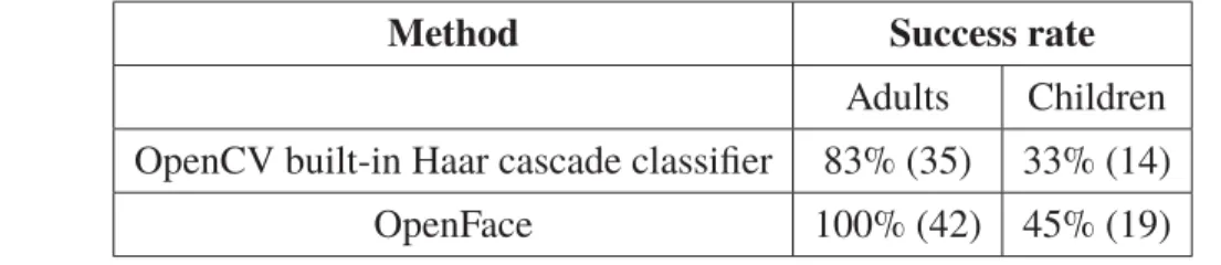
As a result, we found very poor results when applying existing face and eye localization solutions, which are trained and tested primarily on controlled adult facial datasets, to real-world images of hospitalized pediatric patients. As shown in Table 4.1, the performance of existing face detection tools is significantly worse on a set of images of hospitalized children drawn from the 59 recordings made at Sainte-Justine Hospital: OpenCV’s Haar cascade (Bradski & Kaehler, 2000) detected 83% of adult faces but only 33% of children’s, while OpenFace (Baltrusaitis, Zadeh, Lim & Morency, 2018) correctly detected 100% of adult faces but only 45% of children’s faces. The 42 test images of children were taken from the 59 video recordings made at CHU Sainte-Justine’s pediatric intensive care unit over the course of this project. The 42 test images of adults come from the 10k US Adult Faces dataset.
Table 4.1 Performance of existing open-source face localization tools on 42 test images
Method Success rate
Adults Children OpenCV built-in Haar cascade classifier 83% (35) 33% (14) OpenFace 100% (42) 45% (19)
The development of a custom model is necessary, but the lack of publicly available datasets with annotated facial images of children and babies makes it difficult to train and test a new model. Acquiring such a dataset at the hospital is a slow process, limited by the unit’s admission rate and parental consent. Additionally, while common wisdom is that large quantities of data are needed to train "deep" models such as CNNs, there are no precise guidelines on the quantity
of data needed for good results. This makes it difficult to plan the development of a suitable training dataset.
4.3 Existing Work
Cascade classifiers, as first proposed in Viola & Jones (2001), are a widely used method for face and eye localization due to the availability of pre-trained models and the ease of training new models using limited data. Cascade classifiers are trained using manually defined features, such as Haar features or a local binary pattern (LBP), and use a sliding window approach to locate objects in new images.
The "cascade" refers to the narrowing of the search region as the classifier is applied to an image. Rather than calculate all relevant features for each window, a window is evaluated with a subset of features. If the object is not detected using those features, the window is discarded and not evaluated further. If, on the other hand, the results are promising, another subset of features is applied, and so on until the window is classified. This narrowing of the search region allows a classifier trained with a large number of features to run efficiently. Cascade classifiers are much faster than convolutional neural networks, making it easy to achieve real-time object localization, but their accuracy is limited because they are unable to learn new features and must use a predetermined set of features such as Haar edge features.
The use of cascade classifiers is illustrated in a study to detect fatigue in bus drivers (Mandal
et al., 2017) which shares similar constraints to our project: images are recorded using a camera
placed off-centre and above the subject, who may move freely and even face away from the camera. A chain of cascade detectors was used to identify first the upper body, then the face and its orientation, and finally the eye region. This gradual narrowing of the region of interest improves performance and accuracy by shrinking the candidate search region and eliminating false positives in the background.
Convolutional neural networks (CNNs), a class of deep neural network particularly well suited to computer vision tasks, grew in popularity through the 2010s as computation power increased
to permit faster training and predictions. Their application in real-world settings remains limited by the need for large quantities of training data and the computational cost of analyzing images through complex, many-layered networks. Nevertheless, CNNs underlie the most powerful facial and eye detection systems available today, and recent work has applied them in the hospital setting to detect adult patients exiting their beds (Chwyl et al., 2017), identify the pose of adult patients in hospital beds (Liu et al., 2019a), and detect infants in bed and segment their skin region (Chaichulee et al., 2017).
Studies interested in the facial features of babies and young children have largely avoided the problem of face and eye localization by manually selecting facial landmarks, as in Zamzami
et al. (2015), or by using time- and effort-intensive alternative approaches like custom active
appearance models, as in Fotiadou et al. (2014). Recent work in Chouinard et al. (2019) uses the Amazon Rekognition system to analyse infant facial images. Amazon Rekognition is a cloud-based computer vision platform that allows users to submit their own images for facial detection and analysis using pre-trained models. While results were promising, submitting patient data to an online service such as Amazon Rekognition raises privacy concerns that make this approach inappropriate for a medical setting.
4.4 Methodology
This project required the creation of new models for eye localization using machine learning methods. Before training these eye localization models, we also had to develop new datasets of training and test data since there were no suitable databases of images of babies and children available. We begin by introducing two new datasets that we developed for this project, then describe the cascade classifiers and convolutional neural networks trained for the eye localization task.
4.4.1 Test data
Fifty-nine patients were recorded in their hospital rooms in CHU Sainte-Justine’s pediatric intensive care unit between September 2018 and May 2019. A standard photography camera was used to capture videos with a resolution of 1920 x 1080 in RGB colour. In most cases, a single five-to-ten-minute video was recorded for each patient. Three patients under 6 months of age were filmed on different days or from different angles and contributed multiple recordings to the dataset. Parental consent was obtained for all recordings and for the publication of the images included in this paper (CHU Sainte-Justine research ethical board approval number: 2016- 1242). Patients range from 9 days to 19 years of age, with approximately half (32) under the age of 2. Where possible, the camera was fixed at the lower-left or lower-right corner of the patient’s bed, looking down at the upper half of the bed, and zoomed in to capture the full width of the bed.
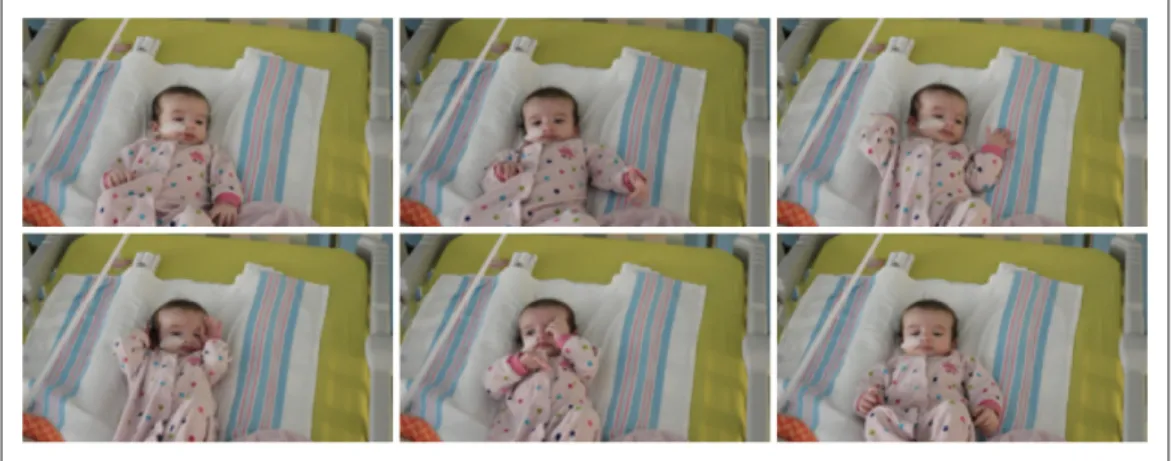
An example sequence of frames from one such video is shown in Figure 4.1.
Figure 4.1 Series of frames from one recording at CHU Sainte-Justine
Of the 59 patients recorded, one was discarded from the test set because the face was completely occluded by a breathing mask and one was discarded because the video was recorded while the patient was in a parent’s lap resulting in two faces in the frame.
A set of still images was randomly extracted from the recordings of the remaining fifty-seven patients to form a test set for eye localization, hereafter referred to as the CHUSJ test set. Five frames were randomly selected from each recording, for a total of 300 still frames (including three patients with more than one recording available).
The randomly selected frames were reviewed manually and those frames where the eye region was out of frame or completely occluded (for example, by the patient’s hands) were discarded and replaced with another randomly selected frame. Frames with poor lighting, blurring, or other picture quality issues were kept, as long as the eye region was in-frame and visible.
4.4.2 Training data
We also created a new dataset to help train our eye localization models, consisting of 664 relevant images of babies and young children gathered using Google Images searches using the following search terms:
• Baby breathing tube, • Baby eye hospital, • Baby eyes,
• Baby hospital, • Baby intubated, • Baby NG tube.
The eye and mouth regions in these dataset images were manually labelled using labelImg, an image annotation tool downloaded from Github (Git, 2015). The resulting images and annotations were included in our training data.
4.4.3 Cascade classifiers
Cascade classifiers have a long history of use as object detectors, especially for face and eye localization. Their efficiency and speed make cascade classifiers an obvious solution to explore for object detection problems.
To measure the effectiveness of cascade classifiers on our test data of children in a hospital setting, we trained a pair of cascade classifiers: one using LBP features and one using Haar features. Both cascade classifiers were trained using OpenCV’s opencv_traincascade application with default parameters.
Five datasets were used for training:
1. The Closed Eyes in the Wild dataset (Song et al., 2014) • 4848 images of open and closed eyes.
2. The annotated subset of the 10k US Adult Faces dataset (Bainbridge et al., 2013)
• 2222 images of adult faces primarily with open eyes, cropped oval to remove a majority of hair and background.
3. The BioID Face Database (retrieved at https://www.bioid.com/facedb/)
• 1521 images of adult upper bodies in an interior setting, with varied facial expressions and lighting conditions.
4. Portions of the CIFAR-10 dataset (Krizhevsky & Hinton, 2009) • 4000 images of non-human objects like ships and trucks.
5. The custom dataset of images of babies and young children gathered from Google Images searches
• 664 task-relevant images of babies and young children.
The complete training dataset for the cascade classifiers consisted of 12,617 positive (eye) and 47,499 negative (non-eye) training images, resized to 24x24 pixels. This corresponds to the