Université de Montréal
Correlates of Long-term Immune Protection in Hepatitis C Virus (HCV)
Exposed Non-reinfected Individuals
Par Asiyah Siddique
Département de Microbiologie et Immunologie Faculté de Médecine
Mémoire présenté à la Faculté des études supérieures en vue de l’obtention du grade de maîtrise (M.Sc.)
en microbiologie et immunologie
Décembre 2016
Université de Montréal Faculté des études supérieures
Ce mémoire intitulé:
Correlates of Long-term Immune Protection in Hepatitis C Virus (HCV) Exposed Non-reinfected Individuals
Présenté par: Asiyah Siddique
A été évalué par un jury composé des personnes suivantes:
Dr. Michel Roger, Président-rapporteur Dre. Naglaa Shoukry, Directrice de recherche
Résumé
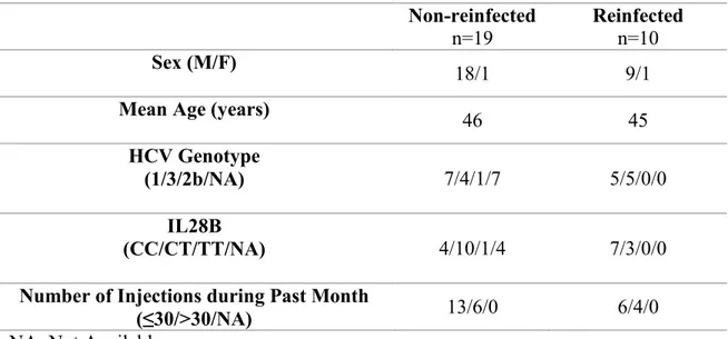
La majorité des personnes infectées par le virus de l’hépatite C (VHC) développent une virémie persistante. Malheureusement, notre connaissance des paramètres de l'immunité protectrice est limitée. La génération et la maintenance des cellules T CD4+ et T CD8+ spécifiques au VHC et polyfonctionnelles, caractérisées par la sécrétion de plusieurs cytokines comme l’IFN-γ, le TNF-α et l’IL-2, sont associées à la résolution spontanée du virus. De plus, la présence d’anticorps neutralisants est associée à une charge virale plus faible et à une durée de virémie réduite suivant une réinfection. Dans ce projet, nous avons l'intention de définir les paramètres de l'immunité protectrice contre le VHC chez les personnes s’injectant des drogues (PID) qui ont déjà été infectés par le VHC. Ces personnes sont exposées au virus à répétition, mais sont résistantes à une seconde infection. En outre, nous avons l’intention de comparer l’immunité protectrice chez les personnes ayant résolu leur infection spontanément versus ceux qui ont reçu un traitement antiviral. Nous émettons l'hypothèse que les PID résistantes à la réinfection développent une réponse immunitaire supérieure, caractérisée par une augmentation de la fréquence de cellules T mémoires spécifiques au VHC; du nombre d'épitopes ciblés par ces cellules; du nombre de fonctions effectrices par cellule; ainsi qu’une réponse élevée des anticorps neutralisant. En utilisant les techniques « Enzyme-linked immunospot » (ELISPOT), le marquage intracellulaire des cytokines (ICS), ainsi qu’un test de neutralisation de pseudoparticules (HCVpp), nous avons examiné la réponse cellulaire et humorale chez les PID exposées mais non-réinfectées versus les PID réinfectées. Nous avons observé une plus grande fréquence des cellules T spécifiques au VHC chez les PID qui n’ont pas été réinfectées pendant ≥ 2 ans. Nous n’avons pas observé une différence significative pour la neutralisation par les anticorps, ni pour la fonctionnalité de cellules T spécifiques. Nos résultats suggèrent que la protection contre la réinfection chez les PID est corrélée à une haute fréquence de cellules T spécifiques au HCV soutenue dans le temps.
Mots-clés : virus de l’hépatite C (VHC), réinfection, immunité protectrice, vaccins, personnes s’injectant des drogues (PID)
Abstract
The majority of HCV infected individuals develop persistent viremia. Correlates of long-term protective immunity remain undefined. Generation and maintenance of polyfunctional CD4 and CD8 HCV-specific T cells that produce multiple cytokines like IFN-γ, TNF-α and IL-2 correlates with spontaneous resolution of acute primary HCV and can be predictive of long-term protection. The presence of neutralizing antibodies (nAb) was associated with reduced magnitude and length of viremia upon reinfection. The aims of this project were: 1) to define correlates of protective immunity in HCV-resolved high risk people who inject drugs (PWIDs) repeatedly exposed to but resistant to reinfections; and 2) to compare protective immune responses in PWIDs with spontaneous versus treatment-induced clearance. We hypothesized that PWIDs resistant to reinfections harbor an immune response of high magnitude, breadth and quality of the HCV-specific memory T cells as well as higher nAb responses. Using IFN-γ enzyme-linked immunospot (ELISPOT), intracellular staining for cytokines, and pseudoparticle neutralization assays, we examined cellular and humoral immune responses in long-term protected versus reinfected PWIDs. We observed long-lived HCV-specific T cell responses in spontaneously resolved and treatment resolved PWIDs who are protected against reinfection. We did not observe a difference in the nAb responses nor in the functionality of HCV-specific T cells. Our results suggest that protection from reinfection in PWIDs is associated with a sustained high frequency of HCV-specific cellular immune responses.
Table of Contents
Résumé ... iii
Abstract ... iv
List of tables ... vii
List of figures ... viii
List of abbreviations ... ix
Acknowledgements ... xiii
Chapter 1: Literature Review ... 1
1.1. Historical Introduction ... 2
1.2. The virus ... 3
1.2.1. Genome ... 3
1.2.2. The viral proteins ... 5
1.2.3. Classification and Genetic variability ... 6
1.2.4. HCV Life Cycle ... 7
1.2.4.1. Virus Composition and Viral Entry ... 8
1.2.4.2. Translation and replication ... 9
1.2.4.3. HCV Virion Assembly and Release ... 10
1.2.5. Difficulties in Studying HCV and Experimental Models ... 12
1.2.5.1. In vitro Models ... 12
1.2.5.1.1. Huh-7 cells and replicons ... 12
1.2.5.1.2 HCV pseudoparticles (HCVpp) ... 13
1.2.5.1.3. Primary human hepatocytes (PHH) ... 13
1.2.5.2. In vivo Models ... 13
1.2.5.2.1. Mouse models ... 14
1. 3. The disease ... 16
1.3.1. Epidemiology ... 16
1.3.2. Transmission ... 17
1.3.3. Natural history of HCV infection ... 18
1.4. The Immune Response to HCV ... 19
1.4.1. Innate Immunity ... 19
1.4.1.1. Natural Killer (NK) Cells ... 21
1.4.1.2. Dendritic Cells (DC) ... 22
1.4.2. Adaptive immunity ... 23
1.4.2.1. Humoral Responses ... 23
1.4.2.2. Cell-Mediated Immunity (CMI) ... 25
1.4.2.2.1. CD8+ T cell response in HCV infection ... 25
1.4.2.2.2. CD4+ T cell response in HCV infection ... 28
1.4.2.2.3. Memory cell-mediated immunity and reinfection ... 29
1.4.3. Genetic Factors and Outcome of Acute HCV ... 30
1.4.5. Viral Evasion of Adaptive Immune Responses ... 33
1.5. HCV Treatment ... 35
1.5.1. Interferon Ribavirin Therapy ... 35
1.5.1.1. Patterns of Response to IFN/RBV Therapy ... 36
1.5.1.2. Factors Determining Outcomes of Therapy ... 37
1.5.1.2.1. IL28B SNP and outcome of therapy ... 38
1.5.1.3. Mechanisms of Action of IFN/RBV Therapy ... 38
1.5.2. Direct-acting antivirals ... 40
1.5.2.1. NS3/4A Protease Inhibitors ... 41
1.5.2.2. NS5B Inhibitors ... 42
1.5.2.3. NS5A Inhibitors ... 43
1.5.2.4. Success and Remaining Challenges of DAAs ... 43
1.5.3. Vaccine against HCV: Need and Challenges ... 44
1.5.3.1. Vaccine Trials ... 45
1.6. Hypothesis and Objectives ... 47
Chapter 2: Article Correlates of Long-term Immune Protection in Hepatitis C Virus (HCV) Exposed Non-reinfected Individuals ... 49
2.1. List of Abbreviations ... 52
2.2. Abstract ... 53
2.3. Introduction ... 54
2.4. Materials and Methods ... 57
2.5. Results and Discussion ... 62
2.6. Acknowledgements ... 69 2.7. References ... 70 2.8. Tables ... 73 2.9. Figures ... 75 Chapter 3: Discussion ... 84 Future Directions ... 93 Bibliography ... 94 Appendix I: The candidate’s contribution to the article ... I
List of tables
Table 1. Clinical Characteristics of Spontaneously Resolved Subjects………...73 Table 2. Clinical Characteristics of Treated (PEG-IFN & RBV) Subjects…………...….74
List of figures
Chapter 1
Figure 1.1. Genomic Organization of the HCV virus ... 4 Figure 1.2. Summary of HCV replication cycle ... 7 Figure 1.3. The estimated prevalence of HCV infection and the global distribution of HCV genotypes ... 16 Figure 1.4. Increasing cure rates with newer antiviral therapies over the years ... 40
Chapter 2
Figure 1. Long-lived HCV-specific T cell responses correlate with protection from reinfection ... 75 Figure 2. Increased breadth of long-lived HCV-specific T cell responses in non-reinfected (≥2 years) individuals compared to reinfected ≥ 2 years as well as non-reinfected < 2 years ... 76 Figure 3. Increased magnitude and breadth of HCV-specific T cell responses in SVR non-reinfected individuals compared to SVR non-reinfected ... 77 Figure 4. Increased cytotoxic profile in CD4+ T cells in non-reinfected PWIDs ... 78 Figure 5. Increased percentage of neutralization in individuals non-reinfected for ≥2 years ... 79
Supplementary Figure S1. Number of regions targeted (breadth) and magnitude of HCV-specific T cells ... 80 Supplementary Figure S2. Comparison of HCV-specific T cell response and number of injection in the past month before time point tested ... 81 Supplementary Figure S3. Representative flow cytometry plot of stimulated and non-stimulated CD8+ T cells ... 82 Supplementary Figure S4. Correlation between HCV-specific T cell responses and % neutralization ... 83
List of abbreviations
+ss Positive single stranded RNA
Ab Antibody
Ago Argonaute
CLDN1 Claudin-1
CMI Cell-Mediated Immunity
CTL Cytotoxic T lymphocyte
CTLA-4 Cytotoxic T lymphocyte associated antigen-4
DAA Direct-acting antiviral
DC Dendritic cell
E1-E2 Envelope glycoprotein 1- envelope glycoprotein 2
EC50 Half maximal effective concentration
eIF2α Eukaryotic initiation factor 2α
ELISPOT Enzyme-linked ImmunoSpot
ESCRT Endosomal-sorting complex required for transport
EVC Early virological clearance
EVR Early virological response
HCC Hepatocellular carcinoma
HCVpp Hepatitis C virus pseudoparticle
HIV Human immunodeficiency virus
HLA Human leukocyte antigen
HVR Hypervariable region
IL Interleukin
IRES Internal ribosome entry site
ISDR Interferon sensitivity-determining region
ISG Interferon-stimulated gene
JFH Japanese fulminant hepatitis 1
Kb Kilobase
LCMV Lymphocytic choriomeningitis virus
LDL Low-density lipoproteins
LDs Lipid droplets
MAVS Mitochondrial antiviral signalling protein
mDC Myeloid dendritic cell
MHC I,II Major histocompatibility complex class I, class II
miR MicroRNA
MTP Microsomal triglyceride transfer protein
nAb Neutralizing antibody
NI Nucleotide inhibitor
NK Natural killer
NNI Non-nucleotide inhibitor
NR Non-responder
NS Non-structural
OCLN Occludin
ORF Open reading frame
PBMC Peripheral blood mononuclear cells
PD-1 Programmed death 1
pDC Plasmacytoid dendritic cell
PEG Polyethylene glycol
PHH Primary human hepatocyte
PI Protease inhibitor
PKR Protein-kinase R
PRR Pattern recognition receptor
PWID People who inject drugs
RdRp RNA-dependent RNA-polymerase
RIG-I Retinoic-acid-inducible gene I
RLR RIG-I-like receptor
SCID Severe combined immunodeficiency
SNP Single nucleotide polymorphism
SOCS3 Suppressor of cytokine signalling 3
SR Spontaneous Resolver
SRB1 Scavenger receptor B1
STING Stimulator of interferon gene
SVR Sustained virologic response
Tfh T follicular helper cell
Th1,2 Helper T cell subtype 1,2
Tim-3 T cell immunoglobulin and mucin domain 3
TNA Tumor necrosis factor
TRAIL TNF-related apoptosis-inducing ligand
Treg Regulatory T cell
TRIF TIR domain-containing adapter inducing IFN
UTRs Un-translated regions
Acknowledgements
Dr. Shoukry: Thank you for your unwavering guidance and support; you accepted me in your lab when I had no previous lab experience and showed me every step of the way. You are a true leader and I will try to always follow your example. Without a doubt, these past 2 years under your guidance will serve me well in my future career.
Dr. Bruneau and the HEPCO Team: Thank you for your support and all the hard work you put into establishing the cohort.
The Donors: I would like to thank each individual donor for their generous donation of time and blood. With their help, we are closer to understanding immunity to HCV and hopefully putting an end to this disease.
The Team:
Nathalie: Merci pour ton aide et tes précieux conseils qui ont rendus ces 2 dernières années agréables et sans embûches. Tu es le pilier de ce laboratoire et un exemple en matière d’organisation et de productivité. Je te souhaite le meilleur pour l’avenir. Sarah: I could not have done my project without your samples! Thank you for all your hard work and positive energy in the lab.
Maude: You are so kind to always answer my questions and help me with my computer related issues. You are a great scientist! Good luck in your career. Mohamed: I was the ELISPOT girl in the lab thanks to your great mentorship in the very beginning of my internship. Good luck in your research career, you deserve the best!
Thomas: Thank you Thomas for always being there to answer my endless questions and for giving me valuable advice. I wish you the best for your future in research. Manuel: You are definitely someone I will miss talking to about psychology, history, and anything not related to science. I wish you the best and I am sure you will be a great researcher. Thank you for showing me what it means to be honest and trustworthy.
Sabrina: Thank you for being my life coach, my French dictionary, and my personal pastry chef. You are so talented, and I wish you nothing but the best in this life and the next. I can’t wait to attend your conferences iA!
Marion: Thank you Marion for your support, especially at the beginning when I had no idea how to thaw cells properly. All the best for the future!
Elsa: Even though you are not officially part of our lab, you are an important part of my time at the CRCHUM. I will really miss seeing your smiling face every morning. You will be a great researcher and I look forward to attending your talks!
1.1. Historical Introduction
The hepatitis C virus (HCV) is the causative agent of approximately 185 million chronic infections worldwide [1]. The first traces of this virus were documented in 1975 when twenty-two patients showed symptoms of transfusion-associated hepatitis without exhibiting serological markers of hepatitis A and B viruses [2], hence adopting the term, non-A, non-B viral hepatitis (NANBH). In the majority of patients, NANBH resulted in persistent liver damage and caused liver cirrhosis in 20% of chronically infected patients [3]. Following an arduous screening process of millions of bacterial cDNA clones derived from experimentally infected chimpanzees, a single HCV clone was discovered by a team of scientists lead by Michael Houghton at the Chiron Corporation in 1989 [3]. This discovery unveiled the 50-80 nm enveloped positive sense single stranded RNA virus of about 10,000 nucleotides [4]. Within the past 25 years, from its discovery to the development of effective therapies, significant breakthroughs have been accomplished, however HCV prevails as a major global problem as an astounding ≈ 3% of humans remain chronically infected [5].
1.2. The virus
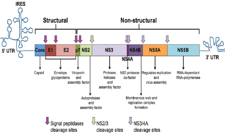
1.2.1. GenomeHCV is a non-cytopathic virus infecting only hepatocytes of humans and chimpanzees. The HCV genome is comprised of an uncapped positive single-stranded RNA (+ssRNA) of about 9.6 kb-pairs [4]. The genome consists of an uninterrupted open reading frame (ORF) encoding a 3000 amino acid polyprotein precursor [4]. Following co- and post-translational modifications by cellular and viral proteases, the polyprotein is cleaved into three structural (Core, E1, and E2) and seven non-structural (NS) proteins (P7, NS2, NS3, NS4A, NS4B, NS5A, NS5B) (Figure 1.1). The ORF is flanked by 5’ and 3’ un-translated regions (UTRs) that harbour secondary RNA structures important for viral replication. The 5’ UTR contains an internal ribosome entry site (IRES) that facilitates the initiation of protein translation [6].
Figure 1.1.: Genomic Organization of the HCV Virus. HCV RNA is translated into a
precursor polyprotein molecule that undergoes further post-translational processing into HCV proteins and enzymes. (Adapted from Abdel-Hakeem et al., 2014 [7])
1.2.2. The viral proteins
The core protein is the scaffolding unit of the viral nucleocapsid. The E1 and E2 glycoproteins make contacts with receptors on target cells and mediate entry of the virus. E2 harbours hypervariable regions (HVR) that are the targets of neutralizing antibodies (nAbs). P7, a small hydrophobic polypeptide, acts as a viroporin or ion channel. The NS2 protein bears an autoprotease activity essential for the cleavage of the HCV polyprotein between NS2 and NS3. NS3 is a multifunctional protein, and together with the cofactor NS4A, act as an NS3-NS4A serine protease that catalyzes the processing of the HCV polyprotein. NS3 also possesses RNA helicase/NTPase activity that loosens RNA-RNA substrates and is vital for RNA replication. The functions of NS4B and NS5A are not well defined. However, some evidence suggests that NS4B causes the formation of a membranous web wherein viral replication is allowed to take place [8]. NS5A was suggested to have an important role in enhancing viral replications [9, 10] and was also shown to contain a region important for alpha interferon (IFN-α) therapy susceptibility known as the interferon sensitivity-determining region (ISDR) [11]. NS5B is the viral RNA-dependent RNA-polymerase (RdRp) that allows HCV-RNA replication [6]. The HCV RdRp enzyme has an absence of proofreading ability and is consequently error prone, resulting in the emergence of varying viral populations present in a patient’s blood as a mosaic of related sequences, collectively termed “quasispecies” [6]
1.2.3. Classification and Genetic variability
The HCV virus is classified under the Hepacivirus genus within the Flaviviridae family, which also encompasses the yellow fever, dengue and the west Nile virus [4]. HCV shows extensive genetic diversity and is classified into seven genotypes (1-7) based on phylogenetic and sequence analysis of the viral genome [12]. These different genotypes diverge at approximately 30-35% of nucleotide sequences. In addition, each genotype is further subdivided into 67 confirmed subtypes, and strains in the same subtype differ at less than 15% of nucleotide sites [13]. Interestingly, it is well established that genotypes show geographically variance, and certain genotypes such as subtypes 1a, 1b, 2a and 3a are extensively spread across the globe and are endemic in high-income countries [14].
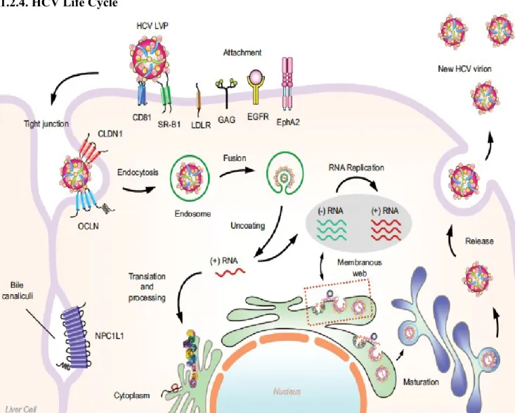
1.2.4. HCV Life Cycle
Figure 1.2. Summary of HCV replication cycle. The HCV lipoviral particle (LVP) anchors to SRB1 and CD81 as well as claduin-1 and occluding and enters the cell via receptor-mediated endocytosis. Following internalization and fusion of the viral envelope with endosomes, the viral genome is uncoated and released into the cytoplasm. In the ER, the viral RNA is translated and the HCV polyprotein is produced and cleaved into mature structural and non-structural (NS) proteins. In collaboration with host factors, viral NS proteins form a membranous web wherein viral replication takes place. Viral assembly is suggested to take place in proximity the ER. Lastly, HCV particles are secreted into the extracellular environment through the secretory pathway. (Adapted from Wong et al.. 2016 [15])
1.2.4.1. Virus Composition and Viral Entry
HCV virions are 50-80 nm in diameter and contain a single-stranded (ss) RNA genome [16]. The genome is enclosed by a nucleocapsid composed of core protein. The nucleocapsid is then enclosed within a lipid membrane that constitutes the viral envelope. The envelope glycoproteins E1 and E2 are anchored within the lipid-laden viral envelope. The HCV virion is closely associated with lipoproteins and apolipoproteins such as apoE and apoB. The lipid composition of the viral envelope is similar to very-low density lipoproteins (VLDL) and low-density lipoproteins (LDL) and cholesteryl esters make up for approximately half of total HCV lipids [17]. The E1 and E1 envelope glycoproteins play an essential role in receptor attachment and mediate the fusion between the viral envelope and the target cell membrane.
During the onset of an HCV infection, HCV virions are transported by the blood and after crossing the endothelium of liver sinusoids, they establish direct contact with the basolateral surface of hepatocytes in the space of Disse. The first attachment of HCV particles onto hepatocytes is made possible by the heparin sulfate proteoglycan syndecan-1 or syndecan-4 [syndecan-18, syndecan-19] or by the scavenger receptor Bsyndecan-1 (SRBsyndecan-1) [20]. The steps immediately following the initial attachment of the virion are partially understood and are thought to involve four important cellular factors. Among these are SRB1, tetraspanin CD81, tight-junction protein claudin-1 (CLDN1) and occludin (OCLN) [21]. It has been suggested that SRB1 may be the first entry factor that interacts with the virion after cell attachment due to its dual interaction with E2 and lipoproteins. An interesting hypothesis the remains to be confirmed is that SRB1 modifies the lipid composition of the virion enabling the unveiling of the CD81 binding site on the E2 glycoprotein [20, 22]. HCV is
endocytosed in a clathrin-dependent process into Rab5a positive early endosomes where fusion takes place. Once released into the cytosol, the HCV genome is translated to produce viral proteins and start viral replication. Interestingly, in the liver of HCV patients, infected cells are found in clusters, therefore suggesting that cell-to-cell spread accounts for the main mechanism of HCV transmission. The exact mechanisms governing this mode of transmission remain to be investigated, but may implicate the role of exosomes [23].
1.2.4.2. Translation and replication
HCV RNA translation begins with the aid of cellular factors [24]. The main outcome of HCV ORF translation is a single large polyprotein that is subsequently cleaved and processed into 10 mature proteins mentioned earlier. The junctions between the structural proteins are cleaved by host signal peptidases from the ER [6]. NS3 performs the cleavage of NS4A from itself and NS4B. NS4A then partners with NS3 resulting in the NS3/NS4A complex ready to cleave at the NS4B/5A and NS5A/NS5B junctions. The NS2/3 autoprotease achieves the cleavage between NS2 and NS3 [25].
Once translation is complete, the HCV proteins are associated with endoplasmic reticulum-derived membranes. Collectively, the NS3/4A, NS4B, NS5A, NS5B proteins comprise the viral proteins of the replication machinery, that is responsible for replicating the positive sense RNA genome from a negative strand intermediate [26]. The NS5B RdRp is the principal enzyme for RNA synthesis and HCV replication is dependent on a liver-specific microRNA (miR) called miR-122 that recruits the Argonaute (Ago) 2 protein to the 5’ end of the viral genome [27]. This attachment of Ago to the viral genome retards the degradation of the HCV genome by the 5’ exonuclease Xrn1 [28].
1.2.4.3. HCV Virion Assembly and Release
The morphogenesis of the HCV virion requires the aggregation of viral structural proteins and genomic RNA that are brought to proximity in a timely and spatially organized condition [29]. It is difficult to detect assembling, budding, or egressing virions in infected cells, implying that these mechanisms are either rare or extremely rapid. An interesting peculiarity of HCV assembly is its close connection to the lipid metabolism pathway. An important component of the viral particle is the core protein, which constitutes the nucleocapsid harbouring the HCV genome. Upon synthesis and cleavage in the ER membrane, the core protein homodimerizes [30] and is then displaced to lipid droplets (LDs) [31, 32]. This interaction between the core protein and the LDs is suggested to be imperative for the recruitment of other key components in HCV assembly [33].
Another crucial component of the HCV virion is the envelope glycoprotein complex whereby E1 and E2 glycoproteins form a non-covalent heterodimer in the ER [34] that needs to migrate close to the LDs where assembly occurs [33]. It has been reported that NS2 interacts with E1, E2 and p7, establishing essential interactions for the migration of the E1E2 heterodimer to the virion assembly site [35-38]. The disulphide bridges between E1 and E2 at the surface of the HCV virion are suggested to play an active role in the budding step of the HCV particle [39].
HCV particle biogenesis is believed to have a close connection the VLDL assembly pathway. It has been reported that blocking microsomal triglyceride transfer protein (MTP), a protein implicated in VLDL biogenesis, inhibits the formation of HCV viral particles [40-42].
The endosomal-sorting complex required for transport (ESCRT) pathway has been suggested to have a key role in HCV budding [43-45]. The ESCRT pathway is involved in the budding and the fission of vesicles out from the cytoplasm, and is exploited by many enveloped viruses for their release from infected cells [46].
Once assembly and budding have been completed in the ER, HCV particles transit through the secretory pathway and are released from infected cells [47]. In this process, the HCV virions attain their particular low buoyant density [40, 48]. Lastly, during egress, it is believed that HCV virions are dependent on p7 for neutralizing the acidic compartments within the secretory pathway [49].
1.2.5. Difficulties in Studying HCV and Experimental Models
Humans and chimpanzees are the only two species that can be infected with HCV. Early studies of HCV immunity used the chimpanzee model to generate important findings as the timing and dynamics of infection can be controlled, and hepatic tissues could be isolated. However, research using chimpanzees is now restricted, forcing scientists to innovate newer models to probe further investigations.
1.2.5.1. In vitro Models
1.2.5.1.1. Huh-7 cells and replicons
The HuH-7 cell line, isolated from an HCC tumour of a Japanese man, proved to be instrumental in the early HCV research [50]. This cell line was used to create a replicon that could express and replicate HCV RNA continuously. This development of a stable cell line expressing the HCV RNA genome was indeed a remarkable achievement as it allowed for drug screening and discovery of potent direct-acting antivirals against HCV [51]. Despite this major advancement, this replicon model lacked the ability to be a fully infectious model in vitro. It was reported that the increased replication rate of the replicon was conferred by mutations in the HCV genome, which rendered it incapable of producing infectious virions in chimpanzees [52]. Consequently, it was agreed upon that the optimal method to develop an in vitro model would focus on wild type HCV genomes. Finally in 2005, the first successful attempt at generating an infectious in vitro model of HCV infection was achieved with the discovery of the Japanese fulminant hepatitis 1 (JFH1) genotype 2a clone. This viral strain was obtained from a Japanese patient having developed fulminant hepatitis [53]. Upon transfection of this newly isolated strain into HuH-7.5 cells,
infectious HCV virions were produced and could be used to infect chimpanzees [54]. In 2006, more infectious isolates were developed for HCV genotype 1a [55] as well as for genotypes 1b, 3a and 4a [56, 57].
1.2.5.1.2 HCV pseudoparticles (HCVpp)
Lentiviruses easily forms pseudotypes with the envelope proteins of countless viruses [58]. Accordingly, HCV pseudoparticles (HCVpp) developed from an HIV backbone expressing HCV glycoproteins are infectious for hepatocytes and hepatoma cell lines [58, 59], and facilitated research on HCV entry as well as measuring nAb responses for the first time. The infectivity of HCVpp is pH-dependent and can be neutralized by many E2-specific mAbs.
1.2.5.1.3. Primary human hepatocytes (PHH)
PHHs are considered the gold standard for studying hepatocyte function. These normal cells are isolated from adjacent tumour tissue in patients undergoing liver resection. PHHs can also be obtained from the fetal livers of aborted embryos and can serve as the substrate for infection with HCV [60]. Once plated, PHHs do not divide and have a limited lifespan of approximately 1-2 weeks. Several studies have observed productive infection of PHHs after culture, including cells from HCV-infected patients [61-63].
1.2.5.2. In vivo Models
Due to the asymptomatic nature of acute HCV infection, a very small number of patients seek medical consultation in the acute phase. Consequently, it is not possible to determine the exact date of infection or exposure and to figure out the exact infecting strain of virus. The majority of the early knowledge of acute infection is derived from chimpanzee
experiments or from studies following high-risk exposures such as needle-stick accidents in health care workers and blood transfusions, and the rare cases of symptomatic acute HCV. There have also been important efforts in establishing cohorts for the monitoring of high-risk individuals such as people who inject drugs (PWIDs) who represent the main reservoir of HCV in developed countries.
1.2.5.2.1. Mouse models
The generation of a successful mouse model for the study of HCV has been slow and difficult as compared to other hepatic viruses such as HBV. Mouse hepatocytes are a poor model for HCV infection due to the differences in many host factors present in human hepatocytes [64]. To overcome the hurdle of this species-specific phenomenon, humanized mice were generated through the xenotransplantation of PHHs. For this system to be effective, the mouse has to be immunodeficient for it to accept human PHHs and replace the normal mouse hepatocytes. The first successful use of this strategy was attained in the severe combined immunodeficiency (SCID) mice carrying a transgene activated by a liver-specific albumin promoter [65]. Expression of this transgene allows for acute hepatotoxicity, which can then be rescued by the transplantation of injected PHHs, the target for inducing HCV infection [65]. Another model wherein hepatotoxicity can be controlled consists of using Fah-knockout mice where the liver stays healthy with the administration of a molecule called NTBC. When this molecule is no longer administered, mice hepatocytes develop toxicity and PHHs can be implanted into the mice liver. Moreover, another strategy to develop humanized mice requires genetically adding human genes to express human entry factors required for HCV infection. With the discovery of HCV entry factors CD81 and occludin, mice were engineered to express human
homologues of these proteins and viral entry was reported for the first time in a proof-of-concept study [66]. Subsequently, HCV infection was successfully reported in a follow-up study [67], however, viral replication was low and the mice failed to develop liver disease, indicating that this model does not fully exemplify an HCV infection. Another group used a similar approach in mice with a different genetic background and were able to achieve sustained viremia with progression to fibrosis and cirrhosis [68], suggesting a more promising model.
Humanized mouse models may constitute a reasonable substitute to chimpanzees and additional efforts are required to replicate the natural history of human immune responses to HCV [69]. These mouse models may also be useful to test vaccine efficacy and more efforts are needed to expand their use in the vaccine community [70].
1. 3. The disease
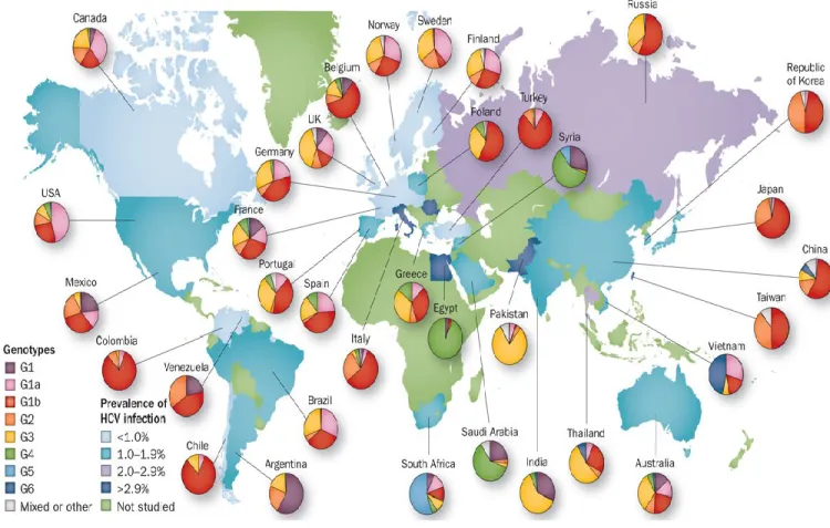
1.3.1. Epidemiology
Figure 1. 3: The estimated prevalence of HCV infection and the global distribution of
HCV genotypes. The highest prevalence rates of HCV are in Egypt, Pakistan, Greece, and
The HCV virus infects approximately 180 million individuals are worldwide, which translates to a 3% prevalence worldwide [1]. HCV distribution across countries can range from <1 % to more than 10% [72, 73]. The highest number of infections is in the developing countries of Africa and the Middle East, whereas higher income countries such as the Americas, Australia, Northern and Western Europe have a lower prevalence [72, 73]. Egypt and Cameroon have the highest prevalence of HCV at > 10% of the population [74, 75]. The countries with the highest absolute numbers of HCV infections are China (29.8 million), India (18.2 million), Egypt (11.8 million), Pakistan (9.4 million) and Indonesia (9.4 million) [72].
1.3.2. Transmission
HCV is most efficiently transmitted by percutaneous exposure to contaminated blood, such as by blood donation, hemodialysis and injection drug use [76, 77]. HCV transmission from mother to child is rare (2% to 10%) [78]. The exact details are not known, however maternal HIV co-infection, ruptured membranes and/or high HCV RNA levels augment the risk of transmission [78, 79]. The predominant mode of transmission in developed countries such as USA, Australia and Europe is through intravenous drug use [80]. In Canada, between 70 and 80% of newly acquired acute HCV infections are attributable to injection drug use [81]. Conversely, in developing countries, the majority of transmissions of HCV occur through iatrogenic exposure. For example, the main source of the HCV surge in Egypt was due to unsafe mass parenteral therapy campaigns against schistosomiasis from 1920s to 1980s [82].
1.3.3. Natural history of HCV infection
An unfortunate feature of HCV infection is its predisposition to establish a chronic infection. Nearly 70% (55-85%) of acute infections progress to a persistent infection [83]. Moreover, HCV has an extremely rapid turnover with a half-life of 3 hours, generating up to 1012 virions daily [84], resulting in exponential serum titres by the end of the first week of infection [85, 86]. Furthermore, physical manifestations such as jaundice only result in one third of acute infections, leaving the rest of the patients largely asymptomatic and undiagnosed for years until serious liver disease symptoms become apparent [87]. Due to its asymptomatic nature, the spread of infection and loss of opportunity for early intervention are major consequences.
Approximately 20% of chronically infected HCV patients develop end-stage cirrhosis, liver failure and hepatocellular carcinoma (HCC) [88], comprising a quarter of the worldwide cirrhosis and HCC cases [89]. Accordingly, HCV is the most frequent cause of liver transplantation (40-50%) [90]. As HCV is a non-cytopathic virus, several reports investigated its contribution to the immunopathogenesis of the liver. Proposed mechanisms include direct cytotoxic T lymphocyte (CTL)-mediated killing of hepatocytes [91], and continuous secretion of inflammatory cytokines resulting in tissue impairment [92]. Chronic HCV is also linked to metabolic dysfunction such as insulin resistance, type 2 diabetes, lipid disorder, and steatosis [93]. Suggested mechanisms for metabolic dysfunction consist of the down-regulation of hepatocyte insulin receptor substrate 1 [94] as well as the glucose transporter, and the increased expression of PP2A [95].
1.4. The Immune Response to HCV
1.4.1. Innate ImmunityThe innate immune system is the host’s first line of defense against viral infections whereby the interferons (IFNs) are produced for mounting an antiviral state in infected cells [96]. Depending on their antiviral properties, IFNs are divided into three classes: type I, type II and type III IFNs [97]. In humans, type I IFNs consist mainly of α and IFN-β [98]. IFN-α and IFN-IFN-β target viruses directly by blocking viral replication or indirectly by generating innate immune responses [97]. IFN-γ is the only member of the type II IFN group and unlike type I IFNs that are induced directly in response to viral infection; IFN-γ is secreted by NK cells as well as mitogenically activated T cells [97]. It has been demonstrated that IFN-γ downregulates claudin-1 and CD81, thereby inhibiting HCV infection [99]. Type III IFNs are comprised of three members including IFN-λ1 (IL-29), IFN-λ2 (IL-28A) and IFN-λ3 (IL-28B). Similar to Type I IFNs, type III IFNs are also directly activated by viral infection and are secreted mainly in the liver during HCV infection [97]. In addition to infected hepatocytes, type I and type III IFNs can also be secreted by Kupffer cells [100], BDCA3+ myeloid cells [101] and as plasmacytoid DCs (pDCs) [102, 103].
The innate immune response begins with the recognition of foreign RNA molecular patterns shared by related pathogens, called pathogen-associated molecular patterns (PAMPs) by pattern recognition receptors (PRRs) including TLRs and intracellular nucleic acid binding proteins [104-106]. Upon HCV entry into hepatocytes, the main PRRs that are activated are TLR3, protein-kinase R (PKR), and retinoic-acid-inducible gene I (RIG-I). Signalling through TLR3 is relayed through the TIR domain-containing adapter
inducing IFN (TRIF), while PKR and RIG-I signalling is relayed via the mitochondrial antiviral signalling protein (MAVS) [107]. These adaptor proteins lead to signalling cascades ultimately inducing the secretion of type I IFNs [107]. The auto- and paracrine binding of type I IFNs to their cognate receptors relays signalling via the JAK-STAT pathway which ultimately leads to the expression of hundreds of ISGs within the infected and neighbouring cells [108]. The expression of ISGs establishes a general antiviral state in the liver whereby HCV RNA replication and cell-to-cell viral spread is restricted [106]. Certain ISGs expressed in response to HCV are proteins with known antiviral effects; however, certain others are known to promote HCV replication in vitro, such as ISG15 and USP18 [109, 110]. The induction of ISGs in the liver is observed early following HCV infection regardless of the outcome of infection, implying that most HCV strains are resistant to antiviral effects of this primitive innate response [111-114]. Type III IFNs also generate ISGs similar to those induced by Type I IFNs in addition to distinct ISGs [115, 116].
1.4.1.1. Natural Killer (NK) Cells
NK cells constitute one of the earliest defenses of the innate immune response. Their main function consists of killing virally infected cells through the secretion of cytotoxic molecules such as granzymes and perforin, or via TNF-related apoptosis-inducing ligand (TRAIL)-mediated killing. In addition, NK cells can also secrete cytokines regulating innate and adaptive immunity such as IFN-γ, TNFα, IL-10 and IL-21. The activation of NK cells is controlled by the calibration of inhibitory and activating signals. The strength of the interactions between inhibitory KIRs expressed on NK cells and their MHC class I ligands expressed on target cells is the main determinant of NK activity [117].
NK cells are ubiquitous within the liver and execute an important role in defending against hepatotropic infections [118, 119]. The early activation of NK cells following an accidental exposure to HCV in healthcare workers was suggested to contribute to protection in 11/12 workers who remained aviremic [120]. In high risk PWIDs, the activation of NK cells and expression of the activating receptor NKp30 was associated with protection from HCV infection [121]. Furthermore, genetic studies revealed that KIR2DL3 expressing NK cells secreted more IFN-γ and that homozygosity for this allele correlated with spontaneous resolution of HCV infection [122]. Another report demonstrated that PWIDs resistant to HCV infection have an enrichment in KIR2DL3+NKG2A- NK cells. NKG2A is an inhibitory receptor and binds to HLA-E which is highly expressed during HCV infection. This suggests that low expression of NKG2A is advantageous in the presence of high HLA-E expression. Overall, KIR2DL3+NKG2A- NK cells are not susceptible to HLA-E-mediated inhibition and may explain the ‘natural resistance’ to HCV in PWIDs [123].
1.4.1.2. Dendritic Cells (DC)
Dendritic cells are the dominant antigen-presenting cells (APCs) in humans. They play a crucial role in bridging innate and adaptive immunity and also impact the priming of HCV-specific immune responses. DCs promptly differentiate into mature DCs following the sensing of danger signals through PAMPs, particularly TLR ligands, interactions with innate lymphocytes (NK and NKT cells), cytokines and other mediators of inflammation [124]. Myeloid DCs (mDCs) and plasmacytoid DCs (pDCs) are the two major subsets of DCs and both play a role in HCV infection. mDCs account for the majority of DCs and are responsible for antigen processing and presentation, whereas pDCs sense viral infections and produce type I and type III IFNs. Additionally, pDCs can sense HCV RNA in a TLR-7 dependent manner when presented by an infected cell [102]. Therefore, DCs are regarded as a principal coordinator of the HCV innate and adaptive immunity.
The functional role of DCs in acute and chronic HCV continues to be controversial. Certain studies have demonstrated that the frequencies of mDCs and pDCs correlate with the outcome of infection whereby decreased frequencies were associated with chronic infection [125-128]. It was also reported that constant hyper-responsive DCs correlate with spontaneous clearance of HCV, implying a superior priming of HCV-specific T cells [129]. Moreover, DCs have been shown to be defective in chronic HCV particularly in response to TLR ligands [130-133] and may cause the proliferation of Tregs [134].
1.4.2. Adaptive immunity
Unlike the innate arm of the immune system which is induced within a short period of time (hours to days) after Hepatitis C infection, the adaptive arm requires 6-8 weeks for its induction, a phenomenon that is still poorly understood. The adaptive immune system comprises of two important components involved in viral clearance, namely T cell responses and humoral antibody responses [135].
1.4.2.1. Humoral Responses
Despite HCV RNA reaching high blood titres by 2 weeks post-infection, anti-HCV antibodies (seroconversion) are largely absent before week 8 [136, 137]. Initial reports showed that antibodies (Abs) directed at the HVR-1 region of the HCV E2 glycoprotein are neutralizing both in vitro and in vivo [138, 139]. Meanwhile, studies in chimpanzees demonstrated that Ab responses did not inherently correlate with viral clearance [140, 141]. In humans who spontaneously cleared HCV infection, Ab responses were reported to be of delayed onset, bearing low titers and fading rapidly [142-144]. One interesting study described how neutralizing Abs (nAbs) emerged in patients only once HCV had established a chronic infection, consequently failing to eradicate the virus and selecting for escape mutants [145]. Conversely, another study demonstrated that the early appearance of nAbs correlated with spontaneous clearance of a primary HCV infection [146].
A major obstacle in elucidating humoral immunity against HCV remains that lack of optimal tools to measure accurate levels of nAbs. The current approach determines neutralization of HCV pseudoparticles (HCVpp) displaying HCV E1-E2 envelope glycoproteins that correspond to small number of HCV reference sequences, and
unfortunately do not represent all the autologous E1-E2 sequences present in a patient [58, 59]. An HCVpp library consisting of 19 distinct sequences representing the natural variability of E1-E2 glycoprotein of genotype 1 strain was developed to show the evolution of the HVR-1 sequences in response to nAbs [147]. Applying this HCVpp library, a study demonstrated that clearance of HCV infection correlated with robust induction of nAb response mounted early in the infection [148].
There are three main hurdles in inducing a protective humoral response against HCV. Firstly, HCV envelope proteins (E1-E2) are not strongly immunogenic, leading to weak and late Ab response in primary infection [147]. Secondly, due to most Abs targeting the HVR of E2, a mutation-prone region, there is a high tendency for the selection of viral sequences bearing resistance to Ab neutralization [149]. Lastly, high glycosylation and association with host lipoproteins of epitopes targeted by nAbs, shield their visibility and restrict their efficacy in vivo [150].
1.4.2.2. Cell-Mediated Immunity (CMI)
The significance of CMI in the clearance of HCV is made evident by the correlation of specific HLA class I and class II alleles and spontaneous clearance and emphasized by depletion studies in the chimpanzee model demonstrating that both CD4+ and CD8+ T cells are necessary for viral clearance [151, 152]. CD8+ T cells mediate target cell killing by displaying viral antigens on the MHC class I molecules whereas CD4+ T cells mediate a helper role in priming and assisting cytotoxic CD8+ T cell (CTL) responses [152].
1.4.2.2.1. CD8+ T cell response in HCV infection
The number of epitopes targeted by CD8+ T cells, known as breadth, is an important factor in the spontaneous resolution of HCV infection. Up to nine unique epitopes were recognized by CD8+ T in the acute resolving HCV in humans as well as in chimpanzees, whereas few epitopes were targeted in chronic progressors [153, 154]. The NS proteins were shown to be immunodominant and correlated with clearance of the virus [155].
The magnitude of HCV-specific CD8+ T cells correlates with spontaneous resolution. Using MHC class I tetramers, it was reported that T cells specific for one epitope can attain up to 8% of total CD8+ T cells in spontaneous resolvers [153, 156]. Ex vivo phenotypic characterization of HCV-specific CD8+ T cells was also accomplished using MHC class I tetramers. It was demonstrated that the primitive expression of the IL-7 receptor alpha (CD127) on the surface of HCV-specific CD8+ T cells was a key predictor of viral clearance and the lack thereof was correlated with viral persistence [157, 158]. Meanwhile, during acute infection, PD-1 is variably expressed HCV-specific CD8+ T cells, indicating that it is an activation marker instead of an exhaustion marker [159, 160].
Additional exhaustion markers such as T cell immunoglobulin and mucin domain 3 (Tim-3), cytotoxic T lymphocyte associated antigen-4 (CTLA-4), CD160, KLRG-1 and 2B4 are expressed at varying levels in acute and chronic HCV indicating a range of exhaustion that is associated with chronic infection [161, 162].
The presence of HCV-specific CD8+ T cells in the liver and blood and presence of IFN-γ, CD3, CD4, and CD8 mRNA levels in the liver are kinetically associated to a decrease in viremia [113, 114]. However, they become challenging to detect in the peripheral blood in chronically progressing infections, albeit being easily detectable in the liver, displaying an exhausted and activated phenotype [163, 164]. Furthermore, peripheral blood HCV-specific CD8+ T cells are suggested to be somewhat defective in their proliferative and cytokine producing potential upon initial appearance in the blood [165]. Nevertheless, screening of simultaneous effector functions revealed the existence of a polyfunctional population of HCV-specific CD8+ T cells concurrently secreting IFN-γ, T cell growth factor, IL-2, and expressing the degranulation marker CD107a, a substitute marker of cytotoxicity [156]. Further experiments on sorted cells revealed that polyfunctionality was specific to CD127+ tetramer-positive CD8+ T cells, reiterating the significance of this T cell subset in resolving viremia [156]. Furthermore, it was reported that early administration and subsequent SVR to IFN therapy recovers CD127+ long-lived memory T cells [156, 166].
As HCV infection progresses, noteworthy differences in the CD8+ T cell functions are observed coupled with the loss of helper CD4+ T cell responses. In chronically progressing patients, continuous loss of function, diminished polyfunctionality and decreased proliferative capacity are manifest in CD8+ T cells [156, 167]. CD8+ T cell loss
of function is concomitant with their extent of exhaustion and is reversible upon in vitro blockade of inhibitory pathways such as PD-1- CTLA-4, and/or TIM-3 [168]. Nonetheless, there is a limited efficiency upon in vivo blockade of PD-1 in chronic humans and chimpanzees, indicating that a threshold of functional virus-specific T cells is necessary for the aforementioned approaches to be successful [169, 170]. This loss of function was suggested to be a result of antigen persistence, as demonstrated in the LCMV model whereby continuous exposure to viral antigens was the key source of diminished frequency and defective effector functions of LCMV-specific CD8+ T cells [171, 172].
1.4.2.2.2. CD4+ T cell response in HCV infection
The importance of HCV-specific CD4+T cells was first reported when patients with spontaneously clearing HCV mounted broad CD4+ T cell responses during acute infection with increased T cell proliferation and IL-2, IFN-γ, and TNF-α production compared to patients who developed a persistent infection [173-176]. The significance of CD4+ T cell help in sustaining a functional CD8+ T cell response was proven by in vivo chimpanzee studies whereby the depletion of CD4+T cells resulted in a gradual decline in the frequency of CD8+ T cells and their cytokine production capacity as well as an increase in escape mutation is targeted CD8+T cell epitopes [151]. Furthermore, it has been demonstrated that the specific expansion of CD161hiCCR6+CD26+ CD4+ T cells expressing 17A, IL-21 and Th17 cell lineage-specific transcription factors have an important correlation with the course of the infection [161]. The plasma concentration of IL-17A are higher during the acute phase of infection in patients who spontaneously resolve their infection compared to their chronic counterparts [161]. A similar trend in IL-21 concentration is apparent a few weeks later and correlates with increased HCV-specific CD8+T cells, rescuing them from Tim-3/galectin-9 (Gal-9) -mediated apoptosis [161]. Furthermore, IL-21 is a signature cytokine of T follicular helper (Tfh) cells, a subtype of CD4+ T cells involved in providing maturation signals to B cells for antibody production [177]. Interestingly, HCV-specific-Tfh cells have been reported to accumulate in the liver and produce more IL-21 compared to their peripheral blood counterparts in HCV infected patients. However, the functional role of liver-resident HCV-specific Tfh cells in HCV immunity requires further investigation [177].
When broad CD4+ T cells are detectable in the acute phase in chronically progressing patients, these T cells suffer rapid exhaustion followed by consecutive loss of IL-2 production, proliferation and IFN-γ production [178, 179] as well as higher expression of TIM-3, PD-1 and CTLA-4 [180]. Lastly, it has been demonstrated that chronically evolving patients have an expansion of Gal-9 expressing regulatory T (Treg) cells as well as higher Gal-9 plasma concentrations, suggesting that binding of Gal-9 to Tim-3 blocks IL-21 production by HCV-specific Th17 cells [161].
1.4.2.2.3. Memory cell-mediated immunity and reinfection
A landmark study by Mehta et al. reported that patients who were previously infected with HCV and spontaneously cleared the virus were 12 times less prone to develop persistent viremia [181]. This may be attributable to the robust memory T cell population generated in subjects who successfully resolve their infection [182]. Another important study followed up a group of women many years after an accidental exposure to an identical strain of virus. Interestingly, HCV-specific CD4+ and CD8+ T cell responses were detectable up to 20 years after the successful clearance of primary infection in these women [183]. Among spontaneous resolvers, phenotypic characterization of HCV-specific CD4+ and CD8+ T cells identified the expression of CCR7+, a lymphoid homing marker, and CD45RO, both markers associated with memory T cells [184, 185]. Furthermore, in spontaneously resolving chimpanzees re-challenged with heterologous HCV isolates, duration of viremia was significantly decreased and associated with a high frequency IFN-γ secreting CD4+ and CD8+ T cells [185-188]. Additional investigation into the role of memory T cells was carried out using antibody-mediated depletion of either CD4+ or CD8+ T cells in chimpanzees. Depletion of CD4+ T cells led to low viremia levels where
CD8+ T cells were able to partially control the infection [151]. However, depletion of CD8+ T cells resulted in a considerable delay in the control of viremia, wherein re-appearance of CD8+ T cells coincided with viremia control [152]. In summary, the aforementioned studies highlight the significance of memory T cells in mediating protective immunity in reinfection. Importantly, this protective immunity acts to mainly reduce the duration and level of viremia in lieu of conferring ‘sterilizing-immunity’ [137].
1.4.3. Genetic Factors and Outcome of Acute HCV
In addition to the genetic influence of NK cell receptors mentioned earlier, three separate genome-wide association studies (GWAS) published reported an association between several single-nucleotide polymorphisms (SNPs) near the IFNλ3 (IL28B) gene locus and response to IFN therapy and spontaneous clearance of infection [189-192]. The C/C genotype at the SNP rs12979860 strongly correlates with spontaneous clearance of primary HCV infection among patients of both European and African backgrounds [189]. Furthermore, an RNA sequencing study performed in PHHs revealed a new gene variant upstream of IL28B called ss469415590 which creates a new gene called IFNλ4. The IFNλ4 was reported to be more predictive of HCV viral clearance compared to IFNλ3 in African subjects. [193].
The exact mechanism for how the IL28B SNPs impact HCV outcomes are unknown, however, it is well established that IL28A, IL28B and IL29, also known as type III interferons, are induced by viral infections and harbour antiviral activity [194]. Type III IFNs are suggested to have similar intracellular response as IFN-α but with greater specificity as their receptors have restricted expression. It is also reported that IL28B SNP may have an effect on NK cell functions or the interaction between the innate and adaptive
immune systems. For example, the KIR2DS3 and IL28B may be predictive of chronic progression of HCV infection [195]. Furthermore, it was reported that the IL28B polymorphism, HLA-C and KIRs additively predict HCV therapy outcomes [196]. Moreover, it was recently reported the CC genotype is associated with higher IFN-γ production by NK cells during acute HCV infection, although it does not prevent chronicity [197].
1.4.4. Viral evasion of Innate Immunity
As HCV infections can be spontaneously resolved in the acute phase, the innate immune response triggered by HCV PAMPs seems to be able to control the acute infection [106, 198]. However, 70% of acute infections progress to chronicity, implying that the virus has figured out strategies in order to escape or to counteract host defenses. Numerous studies have shown how several HCV proteins are capable of blocking host antiviral responses, ultimately leading to a chronic HCV infection. The HCV core, E2, NS3/4A, NS4B and the NS5A proteins all have unique evasion mechanisms to fight the host immune response [15].
Core protein: Expression of the core protein blocks IFN signaling by preventing STAT1
tyrosine phosphorylation, which in turn blocks STAT1 dimerization with STAT2, preventing its translocation to the nucleus and ultimately stopping IFN signal transduction and ISG expression [199]. Moreover, the core protein induces the expression of suppressor of cytokine signalling 3 (SOCS3) which is a repressor of the JAK-STAT pathway given its ability to block STAT1 phosphorylation [200, 201]. It has been described that that SOCS3 expression is increased in chronic HCV patients who are unresponsive to IFN therapy [201].
E2 : The HCV E2 protein utilizes the phenomenon of molecular mimicry as an evasion
strategy to bypass host defense mechanisms [202]. Molecular mimicry is a mechanism whereby viral proteins structurally resemble host defense proteins and can behave as immune modulators [202]. The E2 protein contains a 12-amino acid sequence that is similar to eukaryotic initiation factor 2α (eIF2α) and PKR [203]. This identical domain blocks the phosphorylation of eIF2α ultimately repressing protein synthesis and conferring resistance to type I IFN treatment [204].
NS3/4A: In addition to its essential role in the maturation of NS proteins, in RNA
replication and virus morphogenesis, the NS3/4A protease also possesses mechanisms to suppress the host antiviral system [106, 107, 198, 205, 206]. The protease contains the NS4A transmembrane domain and the amphipathic α-helix at the NS3 N-terminus which allow for the cleavage of their respective targets, MAVS and TRIF, which are fundamental proteins involved in the signaling cascade leading to type I IFN production [207, 208]. MAVS plays a critical role in the RLR pathway and is cleaved by NS3/4A at cysteine 508, causing the dislocation of the N-terminal part of MAVS from the mitochondria and therefore suppressing downstream IFN synthesis [209]. In chronically infected HCV patients, the cleavage of MAVS and therefore the decrease in IFN levels have been reported [210].
NS4B: Stimulator of interferon gene (STING) plays an important role in the initiation of
transcription pathways that are crucial for innate immune signalling. When stimulated with dsDNA, STING polymerises and facilitates the connection of TBK1 with IRF3, phosphorylating IFR3 and therefore activating downstream transcription of type I IFNs
[211]. The HCV NS4B confines STING on the ER, preventing its association with TBK, thereby suppressing downstream IFN signalling [212, 213].
NS5A: This pleiotropic protein regulates the host environment such that it favours virus
replication and persistence [214]. Moreover, NS5A binds to MyD88, a key player in the TLR pathway, and blocks the recruitment of IRAK1 to MyD88, thereby weakening TLR signalling and ultimately decreasing cytokine production [215].
1.4.5. Viral Evasion of Adaptive Immune Responses
Cellular immune responses are present during early HCV infection regardless of outcome and may even continue in chronic infection [216]. Several reports have elucidated that the immune response responses developed during early infection decline in chronically progressing patients [86, 153, 216, 217]. The majority of subjects with appreciable cellular immune responses during early infection exhibit loss of both breadth and magnitude of responses during the chronic phase of infection. The deterioration of T cell responses is not well understood and is believed to be a result of escape mutations. As pathogens replicate on the scale of hours and days and immune responses are generated over a period of weeks, it is suggested that escape mutations contribute to blunting the immune response efficacy [218]. Viral kinetic models show that up to 1012 virions are produced daily in persistent HCV infections [84]. This incredibly high rate of virion output, paired with the absence of proofreading activity of the HCV RNA polymerase amounts to common mutations in the viral genome. Consequently, mutations within the class I or II MHC restricted T cell epitopes lead to the delay in clearance of infected hepatocytes [219]. Escape mutations within CD8+ T cell epitope are correlated with chronicity and accounts for a key viral evasion mechanism. Mutated epitopes induce defective new T cell responses as they lose
binding and recognition capacity to their restricting MHC [220, 221]. Decreased recognition can result either from mutations in the epitope sequence itself or in flanking residues that are implicated in antigen processing [222]. Escape mutations are also influenced by the interaction between the host genetics and the virus. The host HLA alleles drive selective pressure on their corresponding epitopes. This is demonstrated when escape mutations convert to their wild type sequences when transferred to another person carrying a different HLA allele and therefore the epitope lacking selection pressure [223]. Importantly, the extent of epitope mutations is constrained by viral fitness cost [224-228]. Consequently, some HLA alleles such as HLA*B27 are regarded as protective since they prime responses to extremely constrained epitopes that due to high fitness cost, have rare tendency to mutate [226, 227].
1.5. HCV Treatment
1.5.1. Interferon Ribavirin Therapy
In the mid-1970’s, following the identification of hepatitis A, non-A, non-B hepatitis (NANB) was acknowledged [2], and it was initially thought that it would have a negligible health impact. However, it was soon reported that NANB hepatitis mostly exhibited progressive disease leading to cirrhosis and potentially to liver cancer [229]. Therefore, coupled with efforts to identify the causative agent of NANB hepatitis, significant dedication was invested into discovering efficient drug therapies to hamper disease progression.
The noticeable success of IFN-α therapy for hepatitis B spurred a pilot study at the National Institutes of Health in 1986 where NANB hepatitis patients were treated with recombinant IFN-α, well before the discovery of the causative agent, the hepatitis C virus [230]. Promising results from this study encouraged further controlled trials with IFN for NANB hepatitis [231, 232], and partial efficacy was concluded. The first approved regimen consisted of IFN-α at three doses per week for 6 months, achieving sustained virological response (SVR) rates of approximately 6% [231, 232]. Prolongation of the treatment to 12 months only marginally improved SVR rates to 16%. The combination of IFN-α with a nucleoside-analogue antiviral, ribavirin (RBV), improved SVR rates to 34% after 6 months, and to 42% after 12 months of treatment [233, 234]. Further steps in improving the half-life of IFN-α consisted in covalently coupling it to polyethylene glycol (PEG) to generate PEG-IFN-α. In combination with RBV, PEG- IFN-α achieved SVR rates of up to 56% [235, 236].
Unfortunately, the majority of patients treated with combination IFN/RBV therapy experienced adverse side effects. In a trial of 1,000 patients treated with the standard dose, the following side effects were reported: fatigue (66%), headache (50%), nausea (42%), insomnia (41%), pyrexia (35%), anemia (34%), myalgia (27%), neutropenia (31%), depression (25%), irritability (25%) and rash (28%) [237]. Consequently, these adverse reactions resulted in the early interruption of therapy in 13% of participants and a dose decrease in 43% [237].
1.5.1.1. Patterns of Response to IFN/RBV Therapy
The outcomes of IFN-α based therapy can be divided into three categories: SVR, relapse and non-response (NR). The main goal of therapy is SVR, characterized as undetectable HCV RNA 6 months after completion of therapy. SVR seems to represent a cure of infection and is correlated with lack of intrahepatic RNA and histologic recovery [238, 239]. Conversely, in relapsers, viremia rebounds upon completion of therapy. Relapse patients can be re-treated and achieve SVR often with prolonged and increase dose of therapy. Lastly, non-responders fail to exhibit a decrease in viral load below detection levels throughout and after the course of therapy [240].
More comprehensive characterization of treatment outcomes include early virological responses (EVR) and early virological clearance (EVC) defined as negative or ≥ 2 logs decrease in HCV RNA after 12 weeks of treatment [241]. In patients who achieve SVR, the decline in viral load is biphasic: an early rapid reduction in HCV RNA within 2-24 weeks due to direct blocking of virus replication [2-242], followed by a delayed secondary phase with slower reduction due to death of infected cells by immune-mediated and other mechanisms [243, 244].
1.5.1.2. Factors Determining Outcomes of Therapy
The key viral factors influencing the outcome of therapy are the genotype and the delay in prescribing therapy [240]. In comparison to genotype 2 infection, genotype 1a and 1b progress to more severe liver disease and have low cure rates to IFN therapy. Among genotype 2 patients, 72% respond to IFN therapy, whereas only 28% of genotype 1a and 26% of genotype 1b exhibit a response [245]. Starting treatment early during the course of HCV infection remarkably increases SVR rate to 88% regardless of genotype [246, 247]. Additional factors associated with increased SVR rates are low baseline viral loads [240] and increased diversity of quasispecies before start of therapy [248].
Relevant host circumstances affecting the outcome of therapy include ethnicity and the lack of co-morbidities such as HIV infection, alcohol abuse, and renal diseases [240]. Accordingly, African Americans were reported to display fractionally lower SVR rates compared to Caucasians [249], and therapy was evidently less effective in patients suffering from HCV/HIV co-infections compared to infections with HCV alone. Additional factors associated with improved SVR rates include the female gender, young age, low body weight and better liver function [240].
1.5.1.2.1. IL28B SNP and outcome of therapy
It was long observed that treatment outcomes with the standard PEG-IFN and RBV therapy had distinct outcomes among different ethnic populations. For example, African Americans had approximately 50% reduction in SVR rates compared to non-Hispanic Europeans after accounting for socio-demographic characteristics and adherence to treatment [249, 250]. Several GWAS studies reported SNPs surrounding the IL28B region to be associated with treatment outcomes [189-192]. The rs12979860 SNP is the most clinically relevant locus as it was strongly associated with SVR among both individuals of European as well as African descent. The C/C genotype is considered the favourable allele as about 80% of patients with this allele achieved SVR compared to 30% with the T/T allele. This difference in allele frequency between African and European populations accounts for about half of the variation in response rates to therapy [251].
1.5.1.3. Mechanisms of Action of IFN/RBV Therapy
The mechanism of action of IFN-α is similar to endogenous IFN signaling as demonstrated by experiments wherein in exogenous IFN-α administration to cell-cultures lacking IFN signaling pathways resulted in induction of many ISGs [252-254].
The exact mechanisms of action of ribavirin remain to be understood. However, a proposed explanation is its direct inhibition of viral replication due to an increase in the error rate of RdRp, resulting in early chain termination [255]. Additional suggestions include its action as a viral mutagen, resulting in viruses with reduced infectious capacity [256], modifying the Th1/Th2 ration in favour of Th1 [257], and lastly, inhibiting IL-10 production [258].
Due to the considerable amount of side effects of PEG-IFN-α and ribavirin, and the varying efficacy of this treatment across genotypes, the success of this therapy was indeed limited. The combination of PEG-IFN-α and RBV was the standard therapy for HCV infection until the recent discovery of direct-acting antivirals (DAAs).
1.5.2. Direct-acting antivirals
Figure 1.4: Increasing cure rates with newer antiviral therapies over the years. Graph
indicating the percentage of cures among patients treated with different regiments from 1989 to 2014. (Adapted from Rehermann, 2016 [259])
Due to the remarkable advances in the understanding of the structure and molecular biology of the HCV viral life cycle, coupled with the suboptimal response rates to IFN/RBN therapy, intense efforts were undertaken towards the discovery of novel therapies targeted at the virus itself. In the past decade, new DAAs have been developed and are currently revolutionizing therapy standards for HCV. Initially, DAAs were used in combination with IFN and ribavirin to improve response rates, however this regimen was accompanied by toxic side effects [260]. Recently, the combination of DAAs that target different mechanisms in the viral life cycle have shown promising results without the need of IFN and ribavirin, and thereby reducing negative side effects. The cure rates achieved