Faculté de génie Département de génie chimique
DÉVELOPPEMENT DE SURFACES DE POLY(ACRYLIQUE ACIDE) POUR ÉTUDIER LES PROPRIÉTÉS ANTI-ADHÉSIVES À DES FINS BIOMÉDICALES
Mémoire de maîtrise es sciences appliquées Spécialité: génie chimique
Sherbrooke (Québec), Canada
Library and Archives Canada Bibliothèque et Archives Canada Published Héritage Branch 395 Wellington Street Ottawa ON K1A 0N4 Canada
Your file Votre référence ISBN: 978-0-494-31366-4 Our file Notre référence ISBN: 978-0-494-31366-4 Direction du Patrimoine de l'édition 395, rue Wellington Ottawa ON K1A 0N4 Canada
NOTICE:
The author has granted a non-
exclusive license allowing Library
and Archives Canada to reproduce,
publish, archive, preserve, conserve,
communicate to the public by
télécommunication or on the Internet,
loan, distribute and sell theses
worldwide, for commercial or non-
commercial purposes, in microform,
paper, electronic and/or any other
formats.
AVIS:
L'auteur a accordé une licence non exclusive
permettant à la Bibliothèque et Archives
Canada de reproduire, publier, archiver,
sauvegarder, conserver, transmettre au public
par télécommunication ou par l'Internet, prêter,
distribuer et vendre des thèses partout dans
le monde, à des fins commerciales ou autres,
sur support microforme, papier, électronique
et/ou autres formats.
The author retains copyright
ownership and moral rights in
this thesis. Neither the thesis
nor substantial extracts from it
may be printed or otherwise
reproduced without the author's
permission.
L'auteur conserve la propriété du droit d'auteur
et des droits moraux qui protège cette thèse.
Ni la thèse ni des extraits substantiels de
celle-ci ne doivent être imprimés ou autrement
reproduits sans son autorisation.
In compliance with the Canadian
Privacy Act some supporting
forms may have been removed
from this thesis.
While these forms may be included
in the document page count,
their removal does not represent
any loss of content from the
thesis.
Conformément à la loi canadienne
sur la protection de la vie privée,
quelques formulaires secondaires
ont été enlevés de cette thèse.
Bien que ces formulaires
aient inclus dans la pagination,
il n'y aura aucun contenu manquant.
i*i
Canada
RÉSUMÉ
Le présent mémoire est la somme de travaux touchant principalement le sujet de la physico-chimie des surfaces, ayant pour objectif principal l’élaboration de surfaces de poly(acrylique acide) (PAAC) résistant à l’adsorption de protéines non spécifiques. Le contrôle in vitro de la différenciation/croissance de cellules à l’aide de facteurs de régulations spécifiques immobilisés sur un substrat solide est pratiquement impossible sans l’utilisation de surfaces empêchant l’adhésion de molécules non-désirées. L’objectif initial était donc de développer des surfaces de PAAC résistantes à l’adsorption de protéines non spécifiques, de greffer ensuite sur ces surfaces des molécules spécifiques pour la culture des cellules souches hématopoïétiques et enfin de cultiver les cellules sur ces surfaces bioactives. Ce mémoire comporte donc deux chapitres : une revue de littérature sur les cellules souches hématopoïétiques tout d’abord, puis une deuxième section présentant les effets des conditions d’immobilisation sur la physico-chimie des couches minces et l’adsorption de protéines sur ces couches.
La revue de littérature sur les cellules souches hématopoïétiques constitue une partie de chapitre soumis dans un livre à être publié par Landes Biosciences (Cell - Mate rial
Interactions: Molecular to Biomolecular Events related to Material Properties. Editors: Patrick Vermette, Yves Martin, Charles J. Doillon). Cette revue de littérature s’intitule "The
rôle of regulatory and environmental factors in self-renewal and différentiation of hematopoietic stem cells". Le titre français est "Le rôle de facteurs environnementaux et de régulation dans la survie et la différenciation de cellules souches hématopoïétiques". Le deuxième chapitre présente la méthodologie utilisée et les résultats expérimentaux obtenus dans le cadre du projet de maîtrise. Des couches minces de PAAC ont ainsi été élaborées et l’influence des conditions d’immobilisation sur la physico-chimie des couches minces et l’adsorption de protéines sur ces couches a également été déterminée. Les paramètres étudiés étaient : 1) le poids moléculaire du PAAC, 2) la concentration en solution du PAAC ainsi que 3) le ratio de catalyseurs par rapport aux groupements carboxylique du PAAC.
REMERCIEMENTS
De nombreuses personnes m ’ont apporté leur soutien durant ce projet, soit par leurs conseils techniques ou scientifiques, ou tout simplement par leurs encouragements. Premièrement, je tiens à remercier Patrick Vermette de m ’avoir accordé sa confiance pour réaliser ce projet ainsi que pour m ’avoir fourni de nombreux conseils scientifiques. Yves Martin, qui en plus d’avoir tenu avec moi de nombreuses discussions sur les hypothèses possibles expliquant les nombreuses embûches de ce projet, a su me divertir grâce à son goût éclairé de la mode. Félix Dupont m’a également beaucoup aidé lors de nombreuses expériences par ses maintes connaissances en chimie ainsi qu’au niveau technique.
Merci également aux autres membres du laboratoire: Anne Danion, Pierre Sarrasin, Davod Mohebdi Khalori, Afra Hajizadeh, Valérie Centis, Emmanuelle Monchaux, Heïdi Brochu, Hanna Bramfeldt et Julie Chouinard. Sonia Biais, Alain Lévesque, Serge Gagnon et Denis Turcotte m ’ont également apporté un soutien indispensable par leur aide technique lors de l’utilisation (et réparation) de nombreux appareils.
Finalement, un merci à des gens moins impliqués mais qui ont servi de source d’inspiration: Cornélius St-Pierre, Hector Dandavino, Rémi Gillespie et Lucas Budry (vive la bière au gingembre). Hugh Mignault, Greg Lebel, Stephen Lévesque et Alex Rondo pour leur amitié, ainsi que Maxime Crosby et Jean-Sébastien Gagné pour leurs talents de hockeyeurs.
Ce travail est dédié à mes parents, Sylvie et Raymond, ainsi qu’à Réal et André, Yvonne et Laura mes nombreux grands-parents! Jean, Michèle, Alexis, Françoise “Frank with an accent” et Gabriel ainsi que Mr. et Mme Legault (ma deuxième famille), je vous remercie de m ’avoir supporté.
TABLE DES MATIÈRES
Résumé...i
Remerciements... ii
Table des matières... iii
Liste des figures... v
Liste des tableaux... vi
Introduction...1
Résumé français du chapitre 1...5
Chapitre 1 : The rôle of regulatory and environmental factors in self-renewal and différentiation of hematopoietic stem cells 1. Abstract...6
2. Introduction... 7
3. Characterization methods to identify hematopoietic stem cells... 10
4. Sources of hematopoietic stem cells... 20
5. Clinical u ses... 24
6 Stem cell niche... 24
7. Growth factors and cytokines... 28
7.1 Introduction and nomenclature...28
7.2 Importance of ligand concentration on hematopoietic stem cell behaviour... 37 7.3 Genetic factors... 40 8. Négative modulators...44 9. Physico-chemical factors... 45 10. Plasticity... 46 11. Conclusion... 53 12. References... 56
Résumé français du chapitre 2 ... 74
Chapitre 2 : Effects of immobilisation conditions on PAAC layers fouling properties 1. Abstract... 75
2. Introduction... 76
3. Expérimental section...77
3.2 Methods...77
3.2.1 Surface Immobilization of PAAC... 77
3.2.2 Factorial design... 79
3.2.3 Atomic Force Microscopy Colloidal Probe Force Measurements...79
3.2.4 Quartz Crystal Microbalance... 81
3.2.5 X-ray Photoelectron Spectroscopy... 81
4.Results and discussion... 82
4.1. Effects of immobilisation conditions on PAAC layer Chemical composition by XPS...82
4.2 Effects of the immobilisation conditions on PAAC layers fouling properties by QCM... 88
4.3 PAAC graft layers relative thickness and structure by AFM colloidal probe force measurements...97 5. Conclusions...105 6. Références...106 7. Conclusions générales... 111 8 Appendix...114 8.1 Glossary... 114
8.2 Frequency shift caused by the injection of RPMI on a PAAC layer... 124
iv
LISTE DES FIGURES
FIGURE 1.1 : Sequential steps of the hematopoiesis...14
FIGURE 1.2 : FACS (fluorescence-activated cell sorting) adapted fro m ...16
FIGURE 1.3 : Identification of cell surface markers with fluorescent tags...18
FIGURE 1.4 Human hematopoiesis in the bone marrow... 23
FIGURE 1.5 : Stem cell niche... 26
FIGURE 1.6 : Diagram of hematopoiesis and cytokines involved in its régulation...29
FIGURE 1.7 : Stem cell mobilization: mechanisms and interaction between certain mobilizing agents...33
FIGURE 1.8: Examples of plasticity... 48
FIGURE 2.1 High-resolution XPS C ls spectra of HApp surfaces MW : 5 kDa, [ ]PAAC :1%, EDC+NHS/COOH : 0.01/1, MW : 5 kDa, [ ]PAAC : 0.01%, EDC+NHS/COOH : 0.01/1 and of HApp film...84
FIGURE 2.2A: Effect of RPMI and FBS 10% solution on the A frequency shift of QCM electrode coated with PA A C ... 90
FIGURE 2.2B: Effect of RPMI and FBS 10% solution on the A HBHW shift of QCM electrode coated with PAAC... 91
FIGURE 2.3A: Frequency shift of PAAC immobilisation conditions (constant parameters are EDC+NHS/PAAC: 0.05/1 and PAAC molecular weight: 5kD)...95
FIGURE 2.3B: Frequency shift of PAAC immobilisation conditions (constant parameters are [ ] PAAC: 1% and PAAC molecular weight: 5kD)... 96
FIGURE 2.4A: Effect of PAAC MW on the apparent thickness of the PAAC graft layers and the work necessary for the cantilever to compress these layers (i.e., Riemann sum)... 99
FIGURE 2.4B: Effect of PAAC solution concentration on the apparent thickness of the PAAC graft layers and the work necessary for the cantilever to compress these layers (i.e., Riemann sum)...100
FIGURE 2.4C: Effect of EDC+NHS/COOH ratio on the apparent thickness of the PAAC graft layers and the work necessary for the cantilever to compress these layers (i.e., Riemann sum)... 101
LISTE DES TABLEAUX
TABLE 1.1: In vitro and in vivo assays used to study hematopoietic progenitors and stem
cells... 12
TABLE 1.2 : Markers used to identify bone marrow and blood stem cells and to characterize differentiatied cell types... 17
TABLE 1.3: Ex vivo génération of primitive hematopoietic progenitors/stem cells from human CD34+ cells...31
TABLE 1.4: In vivo studies with ex vivo-expanded hematopoietic stems or progenitor cells... 32
TABLE 1.5: Effects of différent cytokines on HSCs... 34
TABLE 1.6: Concentration-related effects of some cytokines and growth factors on HSCs behaviour... 38
TABLE 1.7: Some transcriptional factors and their rôle in hematopoiesis...41
TABLE1.8: Genes and proteins implied in the régulation of HSCs fate...42
TABLE 1.9: Plasticity assays... 49
TABLE 2.1 : Elemental composition of polymer surfaces and HApp film on borosilicate surface derived from the XPS survey spectra... 85
TABLE 2.2: Elemental composition of polymer surfaces and HApp film on borosilicate surface derived from the XPS survey spectra (fixed parametrers of PAAC immobilisation conditions)... 86
TABLE 2.3: Statistical analysis of XPS O/C ratio... 87
v i
Introduction
Bien que la médecine moderne soit parvenue à faire de grandes avancées dans les domaines de la chirurgie, notamment dans le cas de greffes d ’organes, le manque de donneurs et les risques immunologiques associés à cette technique ont contribué à l’élaboration de nouvelles stratégies. Une de celles-ci est le génie tissulaire qui a pour objectif la reconstruction d ’organes in vitro ou
in vivo. Les principaux avantages du génie tissulaire sont d’éliminer non seulement les risques de
rejet d ’organes évitant ainsi l’utilisation de médicaments anti-rejets qui peuvent induire de nombreux effets secondaires chez les patients, mais également l’utilisation de prothèses et autres organes artificiels dont l’efficacité, le coût ainsi que la durée de vie sont problématiques. L ’accès à des méthodes de culture cellulaire permettant de contrôler le développement de cellules afin de recréer les différents types de tissus composant un organe fonctionnel représente le principal défi. Il faut de plus sélectionner des cellules capables de recréer le développement d ’organes entiers in vitro sous l’influence de divers stimuli (facteurs de croissance). Les chercheurs ont depuis longtemps identifié les cellules souches embryonnaires comme étant la source de cellules pouvant permettre au génie tissulaire d ’atteindre cet objectif de construction d ’organes in vitro.
Cependant, avec tous les débats éthiques entourant l’utilisation de ces cellules souches embryonnaires, d ’autres sources de cellules doivent être considérées. Les cellules souches adultes représentent un excellent substitut en limitant les conflits moraux. Les cellules souches hématopoïétiques sont parmi les cellules souches adultes les plus utilisées en contexte clinique (traitement de leucémie, infarctus) et les plus documentées, tant au niveau de leur potentiel clinique que de leur physiologie (marqueurs de surface). Des protocoles permettant de les isoler, de les cultiver ainsi que de les utiliser dans des contextes thérapeutiques existent et les connaissances des facteurs influençant la différenciation et la prolifération de cellules souches hématopoïétiques in vitro et in vivo sont de plus en plus étoffées. Cependant, il existe encore de nombreuses interrogations quant aux mécanismes permettant de contrôler efficacement la prolifération et la différenciation de ces cellules in vitro. Une meilleure connaissance de ces mécanismes permettrait le développement de méthodes rapides et efficaces pour établir des réserves de cellules souches hématopoïétiques. Ces réserves fourniraient des cellules autologues
pouvant être transplantées à des patients sans avoir à faire de prélèvements de cellules pour chaque intervention. De plus, le nombre de cellules disponibles pour des études deviendrait plus considérable, ce qui faciliterait et accélérerait le rythme des recherches en génie tissulaire.
De nombreux articles et revues de littérature répertorient les différents facteurs contrôlant la différenciation et la prolifération de cellules souches hématopoïétiques. Cependant, une synthèse regroupant les différentes molécules de régulation (cytokines, facteurs de croissance), les récepteurs membranaires, la transduction de signaux à l’intérieur de la cellule (cascades de signalisation), s’avérait nécessaire. Cette synthèse est présentée dans le chapitre de revue de littérature intitulée « Le rôle de facteurs environnementaux et de régulation dans la survie et la différenciation de cellules souches hématopoïétiques ». Cette revue, permet de cerner les grandes familles de facteurs influençant les cellules souches hématopoïétiques. Cependant, beaucoup d’interrogations demeurent quant aux interactions possibles entre ces différents facteurs, car ils semblent pour la plupart agir de façon synergique. Cela rend l’identification des facteurs environnementaux et de régulation et leurs rôles précis dans la survie et la différenciation de cellules souches hématopoïétiques très complexe.
La connaissance des facteurs influençant la survie et la différenciation de cellules souches hématopoïétiques doit cependant être couplée à un système de culture adéquat. En effet, il est très difficile d ’analyser les phénomènes biologiques liés à la différenciation in vitro et in vivo, souvent par manque d ’outils d ’analyses appropriés. Les micro-puces pourraient ainsi être un outil très utile en culture cellulaire, en permettant de déterminer les conditions de culture optimales. Les micro-puces développées au départ pour des appariements ADN sont maintenant utilisées dans le cadre de la culture cellulaire. Cette méthode permet d’analyser de nombreux échantillons biologiques avec peu de matériel dans un délai très rapide grâce au regroupement de différentes conditions de culture sur un seul support physique. D ’autre part, ces micro-puces permettent de contrôler la concentration des facteurs de croissance et le site de greffage de ces derniers de façon automatique.
Il est donc possible de contrôler encore plus efficacement les conditions dans lesquelles les cellules peuvent être cultivées sur une micro-puce. En effet, un des problèmes inhérent à la
2
culture cellulaire est que les cellules ont la capacité de créer une matrice de protéines sur laquelle elles vont se fixer, leur permettant ainsi d’adhérer à un matériau ou à une surface. L ’utilisation de surfaces de polymères aux propriétés anti-adhésives (low-fouling) devient alors un outil important, puisque ces surfaces ont la propriété de repousser les protéines, ce qui permet de contrôler plus précisément où ces cellules vont pouvoir se fixer sur une micro-puce et de vérifier par le fait même le véritable impact de facteurs de croissance et de cytokines immobilisés sur cette surface.
Donc, le but de ce projet de maîtrise était d ’optimiser des surfaces de poly(acrylique acide) (PAAC) en étudiant l’impact des paramètres d ’immobilisation (poids moléculaire du PAAC, concentration de la solution de PAAC et le ratio des catalyseurs carbodimiides par rapport aux groupements COOH sur le PAAC) de ce polymère sur des substrats. De nombreux polymères ayant des propriétés anti-adhésives ont été étudiés. Le choix du PAAC s’est fait sur la base de nombreux critères : coût peu élevé, bonne biocompatibilité, facilité d ’utilisation en chimie de surface. De plus, peu d ’articles sur son utilisation à des fins anti-adhésives existent dans la littérature, ce qui rendait son étude encore plus intéressante. Le deuxième chapitre «L’effet des conditions d ’immobilisation de couches de PAAC sur ces propriétés de diminution d ’interactions surface-molécules» présente les résultats expérimentaux obtenus dans le cadre de ce projet de maîtrise. Ce chapitre démontre tout d ’abord l’efficacité des paramètres utilisés pour lier de façon covalente le PAAC aux groupements aminés (HApp) fixés par le réacteur au plasma sur les substrats. L ’effet des facteurs d ’immobilisation du polymère sur la capacité de ce dernier à diminuer les interactions surface-molécules est ensuite présenté. Ainsi, les différentes conditions d’immobilisation du PAAC sélectionnées semblent moduler les interactions surface-molécules, bien qu’il n ’ait pas été possible de démontrer quels facteurs affectent de façon statistiquement significative la diminution des interactions surface-molécules.
Les deux chapitres de ce mémoire sont donc complémentaires. Premièrement, il est essentiel de connaître les mécanismes fondamentaux des facteurs de régulation des cellules souches hématopoïétiques, ainsi que les effets cliniques de ces cellules souches, afin de mieux distinguer quels sont les meilleurs combinaisons à utiliser afin d ’optimiser la culture in vitro des cellules souches hématopoïétiques. De plus, il faut également connaître les propriétés anti-adhésives de 3
certains biomatériaux (dans ce cas-ci le PAAC), afin de mieux contrôler les interactions surfaces- cellules. Ces interactions peuvent influencer grandement les comportements de cellules cultivées
in vitro, et leur compréhension et leur contrôle sont tout aussi importants que de connaître les
facteurs de régulation des cellules souches hématopoïétiques.
Chapitre 1: Le rôle de facteurs environnementaux et de régulation dans la
survie et la différentiation de cellules souches hématopoïétiques
Ce chapitre est une revue détaillée des méthodes et des outils utilisés pour maintenir, réguler et contrôler la survie, la prolifération et la différenciation des cellules souches hématopoïétiques. Ces outils et ces méthodes reposent sur l ’utilisation de molécules (molécules uniques,
combinaisons de molécules ou des variations de leurs concentrations) et de facteurs
environnementaux (type de culture). Une vue d ’ensemble sur l ’état actuel des connaissances concernant les cellules souches hématopoïétiques est incluse et permettra une meilleure
compréhension des différents domaines de recherche et des applications cliniques de ces cellules.
Chapter 1: The rôle of regulatory and environmental factors in self-renewal
and différentiation of hematopoietic stem cells (HSCs)
1. Abstract
This chapter consists of an in-depth analysis of methods and tools used to maintain, regulate and control the différentiation, prolifération and survival of hematopoietic stem cells. The tools and methods can involve the use of molécules (single molécules, combinations, and variation of their concentrations), environmental factors (type o f culture). A wide overview o f the knowledge of HSCs will also be provided to allow a better compréhension of the différent fields of research and the clinical applications of those adult stem cells. So, clinical uses, characterization methods, the source o f the cells, behaviour of HSCs and the différent key factors affecting their behaviour will be covered.
2. Introduction
Since Till and McCulloch’s paper (1) rose the possibility that cells in the bone marrow could provide protection to lethally irradiated mice (i.e., bone marrow transplants from healthy donors could rescue lethally irradiated mice from death), researchers have been on the lookout to discover which cells possess such ability. Thus, the concept o f stem cells has emerged - a définition that is now known to include various kinds of cells. The report o f the NIH(2) on stem cells indicates that a stem cell is a kind of cell with the unique capacity to renew itself and to give rise to specialized cell types. A stem cell is also uncommitted and remains so until it gets a signal triggering its transformation into a specialized cell.
Here, a brief reminder o f the concept of hematopoiesis could be useful. It is first established early in embryonic development within the blood islands o f the yolk sac. Then, it is moved to the fetal liver, then to the spleen and after that, the final localization of hematopoiesis is the bone marrow, where the cell populations that sustain formation o f blood cells throughout life are generated. Hematopoiesis deals not only with the replacement of millions of mature cells that are expanded daily (steady-state hematopoiesis), but also with sudden requirements such as infection or acute blood loss (emergency hematopoiesis). Two mechanisms are involved in the control o f hematopoiesis: cell-to-cell interactions, predominantly involving specialized stromal cell elements that form the microenvironment of the hematopoietic tissues and HSCs (hematopoietic stem cells) by way of, among others, the c-kit receptor, and the soluble molecular regulators produced by the hematopoietic microenvironment and other tissues that are able to act from remote sites.(3)
The clinical use o f the stem cells has been reviewed quite so, even more when the HSCs are concemed.(4) It would seem that they are the adult stem cells which possess the most potential for clinical applications, since they would appear to be the cells (apart from the embryonic stem cells) that have the more versatility when it cornes to the pluripotency (see the plasticity section).
The scientifïc literature addressing HSCs research tends to point out towards two main challenges: i) HSCs are difficult to identify and ii) it is diffïcult to multiply them in vitro in high density. HSCs tend to behave and look in culture like standard white blood cells. So far, the best
way to identify HSCs is by their surface proteins (surface markers) such as CD 34 (see Table 1.2). Another issue that complicates the study of HSCs is their rarity: 1/10 000 to 1/15 000 cell in the bone marrow is an HSC and 1/100 000 cell in the bloodstream is an HSC.(2) These cells do not easily stay in a quiescent State for a long period of time, neither are they considered as a robust family of cells. One of the major challenge in HSCs research is the lack of an adéquate culture System to expand HSCs numbers in a reproducible manner and in large-scale in such a way that they can be used in clinical applications.
The following sections aim to review the rôle of regulatory and environmental factors in self-renewal and différentiation of hematopoietic stem cells. In fact, we will review the known natural molécules that are of importance in maintaining in vitro a quiescent line of HSCs and the ones that can be used in a designed fashion to control the fate of these cells. Since attempts to control the in vitro behavior of HSCs are in a way a reconstitution of the stem cell niche, this article will review methods used for the in vitro growth of HSCs, whether they are combinations of extracellular factors (cytokines, concentration gradients of growth factors, structural proteins of the ECM), and intracellular factors (the injection of a transcription factor) and the use of stromal cells as a feeder layer. An important number of factors, signal-transduction pathways and genes seem to be involved in the HSCs fate décisions, but to precisely which extent, it still remains unknown. There is even a few arguments about whether or not the décision to self-renew without différentiation is stochastic (5-7), and in that case, no factor could affect stem cell décision. Nonetheless, many factors are known to impact on the fate of these stem cells.
This review chapter is divided as followed:
• Characterization methods to identify hematopoietic stem cells; • Sources o f hematopoietic stem cells;
• Clinical uses; • The stem cell niche;
• Growth factors and cytokines; • Négative modulators
• Physichochemical factors • Plasticity.
8
It is essential to have a knowledge of the steps involved in the characterization o f HSCs. A section will thus résumé a few key methods o f HSCs characterization and it will also include the surface markers of the HSCs. Their importance lies in the fact that they are used to identify and isolate the HSCs. Also worthy o f attention is the sources of HSCs because it is important to know their sites of production in the organism. A section on clinical uses is included because the clinical potential o f HSCs is illustrated in several papers showing that HSCs transplanted (injected most of the time) in the circulatory system or directly at the desired site tend to regenerate damaged tissues. Whether or not HSCs fuse with already differentiated cells or differentiate themselves is what makes this part controversial, and it remains to be established.
A division on the stem cell niche will follow. Studies have shown that the environmental factors i.e., 3-D structure and physical stimuli are as important as the molecular signais. Given that the stem cell niche is the starting point in the stem cell cycle, it makes sense to briefly discuss about it. Next, a section on the regulatory molécules including growth factors (e.g., cytokines, the most important of the regulatory molécules) and other molécules that have been regrouped in that category. It will describe individual effects of these regulatory molécules as well as their synergitic effects and the effects o f their concentration on the behaviour of HSCs. The section will also cover genetic factors, mainly about genes coding for proteins that have a direct impact on the genes (up or down-regulation o f genes) and transcriptional factors. Négative modulators i.e., molécules that have deleterious effect on the culture of HSCs have been reviewed since they are as important as the molécules that have bénéficiai effects on the culture of these cells.
A brief section on physicochemical factors will also be included. It concems ail the “non- biological” factors such as oxygen, pH, and lactate production that affects the in vitro culture of HSCs. The last topic is about plasticity. Even though it could be included in clinical uses section, the reviewed literature about this issue was more critical regarding the eventual trans- differentiation o f HSCs into specialized cells instead o f reporting on protocols and reports of clinical experiments made to cure patients or to test on animais. This section therefore reviews reports addressing reasons why the plasticity should be regarded as plausible or, on the other hand, as questionable. A glossary is presented in the appendix to allow a better understanding of the terminology used in this chapter.
3. Characterization methods to identify hematopoietic stem cells
A “gold standard” has been developed to demonstrate that the cells derived from mouse bone marrow are HSC-like.(2) Those cells are injected into a mouse that was previously lethally exposed to radiation (powerful enough to kill its own blood-producing cells). If the mouse can recover and that ail types of blood cells reach back normal numbers, the transplanted cells (with a genetic marker from the donor animal) are considered to have included hematopoietic stem cells.
Studies showed that there is two kinds of HSCs: i) long-term stem cells and short-term progenitors/precursor cells.(2) When regenerated cells from an irradiated transplanted mouse are injected into another lethally irradiated mouse and are able to restore its hematopoietic System over a period of some months, these cells are considered to be long-term stem cells.(2) On the other hand, cells from the bone marrow/bloodstream that can immediately regenerate ail the différent blood cells but cannot regenerate themselves for a long period of time (3-4 months) are considered to be short-term progenitor/precursor cells.(8) Progenitor cells seem to be immature cells that are precursors to fully differentiated cells of the same tissue. They can proliferate, but have a small capacity to differentiate into more than one cell type as HSCs are able to do. For clinical uses, the long-term stem cells seem to have the self-replicating advantage for an efficient and long-term HSC therapy. Unfortunately, researchers have been unable so far to distinguish the long-term stem cells from the precursor cells once taken from the bone marrow or blood. In addition to that, the tests used to identify the short-term progenitors and the long-term stem cells are expensive, cumbersome and cannot be carried out in humans. Figure 1.1 shows the sequential steps of the hematopoiesis.
As for in vitro culture, the préservation o f cell function is the best way to establish the success of the culture System (see Table 1.1). It must be understood that before undergoing the “gold standard” assays, samples collected have to be measured for their functional compositions o f hematopoietic cell populations. These tests can détermine if there are cells contained within the samples taken from a subject with a potential to reconstitute ail types of blood cells. Here, is a brief description of each of the in vitro tests listed in Table 1.1.(9) HPP-CFC (high proliferative potential colony forming cells) compares the proliferative potential of a population of hematopoietic cells cultured in the presence of various cytokines. The HPP-CFC cells are
10
considered as being the most primitive cells because they are able to generate late arising and large colonies. The CFU-C (colony forming unit in culture) is a protocol used to identify progenitor cells by evaluating their ability to differentiate into différent lineages. Différent versions of the CFU-C exist, each one aimed to identify the presence of a spécifie erythroid or myeloid progenitor.(10-12) CAFC (cobblestone area forming cells) form colonies on a supportive stromal layer that has been irradiated.(13) The LTC-IC (long-term colony-initiating cells) and ELTC-IC (extended long-term colony-initiating cells) are used to study the ability of human HSCs to survive for long periods o f time in culture and to later differentiate.(14-17) The cells are grown on a stromal feeder layer between 35 to 60 days for LTC-IC, and aliquots are transferred at many periods to CF-U medium to count the colonies of differentiated cells that will be generated. The cells are grown for a longer time period (60 to 100 days) for the ELTC-IC, which allows for the détection of even more primitive cellular populations.
As for the in vivo tests, the CFU-S (colony forming unit-spleen) allows to measure the number of cells in a bone marrow suspension able to proliferate in a continuous fashion, as analysed by the formation of hematopoietic colonies in the spleen foliowing the injection o f bone marrow into subjects that have been lethally irradiated.(l) The radioprotection assay is a test analysing the capacity o f a cell population to protect a subject from a lethal dose irradiation for a minimum period of 1 month. As it has been mentioned, the best tests to detect HSCs are the ones that can verify the ability of a cell population to give rise to ail the hematopoietic lines over a long time period (4 months or more). The first of these tests (compétitive transplantation) is based on the ability o f an expérimental hematopoietic cell population to compete with a cell population (unmanipulated) to reconstitute the hematopoietic System of an irradiated subject.(18) The second version of this test uses a mouse strain having an inherited defect in HSCs, which is showed by a réduction in ail myeloid tissue and in a macrocytic anémia.(19-21) Since the HSCs injected do not have the same genetic defects than the HSCs of the mutant mouse, it is easy to see if they can really have an effect on the mouse.
Table 1.1: In vitro and in vivo assavs used to studv
hematopoietic progenitors and stem cells.
A ssays S tim u la tio n D u ra tio n C ells detected R esu lts
In vitro HPP-CFC
CSF-1, G-CSF, GM- CSF, IL-la, IL-3, SCF, bFGF
14 days Progenitor Proliferative potential (>5xl 04 cells/colony) CFU-C SCF, GM-CSF, IL-3,
EPO
14 days Myeloid progenitor Number and type of colonies (increase) CFU-E SCF, GM-CSF, IL-3,
EPO
10-12 days Erythroid progenitor Erythroid (small hemoglobinized colonies) BFU-E SCF, GM-CSF, IL-3,
EPO
10-12 days, 18 days Primitive erythroid progenitor
Large hemoglobinized colonies
CFU-GM SCF, GM-CSF, IL-3, EPO
16-18 days Granulocytic and monocytic progenitors
Large colonies with granulocytes and macrophages CFU-GEMM SCF, GM-CSF, IL-3,
EPO
16-18 days Most primitive myeloid progenitor
Large colonies with erythroid, granulocytes and macrophages CAFC Irradiated adhèrent
bone marrow feeder layer
30 days Primitive progenitor Time o f appearance of colonies
LTC-IC Irradiated adhèrent bone marrow feeder layer with IL-3, IL-6, SCF-containing media; followed by CFU-C assay
35-60 days Primitive progenitor andHSC
Ability to detect CFU colonies after long period in culture; time in culture détermines primitiveness o f cell type
12
ELTC-IC Irradiated adhèrent bone marrow feeder layer with IL-3, IL-6, SCF-containing media; followed by CFU-C assay
60-100 days Primitive progenitor and HSC
Ability to detect CFU colonies after long period in culture; time in culture détermines primitiveness of cell type
In vivo CFU-S
Non applicable 5 days and more Progenitors and HSC Macroscopic colonies on spleen
Radioprotection Non applicable 30 days Progenitors and HSC Survival of irradiated hosts
W mouse transplant Non applicable 16-52 weeks Long-term HSC Donor-derived multilineage hematopoiesis Compétitive transplant Non applicable 16-52 weeks Long-term HSC Donor-derived multilineage hematopoiesis C FU -E =colony form ing units erythroid; BFU-E=burst form ing unit-erythroid; C FU -G M =colony form ing units-granulocyte/macrophage; C FU -G EM M =colony forming units granulocyte/erythroid/macrophage/megakaryocyte; LTC-IC=long-term culture-initiating cells; ELTC-IC=enhanced long-term culture-initiating cells; W =w hite mutation (sériés o f mutants with mutations in the c-kit tyrosine kinase gene); C SF=colony stimulating factor; G- C SF=granulocyte-colony stimulating factor; GM -CSF=granulocyte/m acrophage/-colony stimulating factor; IL=interleukin; SC F=stem cell factor; bFGF=basic fibroblast growth factor; EPO=erythropoietin: C AFC= cobblestone area-forming cells ; C FU -S=colony form ing unit spleen.
R éférencés : (9 ;22-24)
Q uioscsnt S tem Csll
CD34 CD34
Cyclinj Stem Ce# (CFU-Blast) CD34+
CFU GEMM (CFU-mixj
CD34 CD34T BFU-E '■ T-i-yfflBnotd CFU-GM , PfOgefuter ^ B-Lympfiotd Progemiof * BFU-MK CFU-ê o CFU-BM . c f u-e
Myetoblast MonoMast Pro-S lym phocyte N K-iym prw d \
i * 4 Prooenilor
CFU-MK i Eryttwobtast *
Myetecyts PiGmonocyty P t« -8 Lymphocyte MegeKeryocyta
fMKvilOCyl*
Erythrocyte PMMXu Eoünopmi B u ap M NeutropW Monacyw B Lymphocyte
Référencé taken ffom: (25)
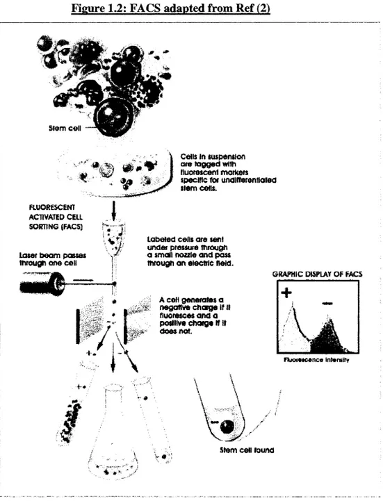
(flt Pra-Thymtc Lymphocyte lymphocyte
Three methods have been developed to sort out populations o f stem cells. The fïrst one makes uses o f the FACS (fluorescence-activated cell sorting) (Figure 1.2), which relies on the tagging of the surface markers o f the HSCs (Table 1.2) with fluorescent tags to individually analyze each cell o f a sample and to sort them by their emitted fluorescence (thus HSCs will be tagged by the fluorescent label and emit more than the other cells). Another technique, which uses also fluorescent tags consists o f tagging the stem cells but to observe them with a microscope in tissues. A slice of tissue is prepared, marked with the fluorescent labels that will specifically bind to the stem cell surface receptors. The tags are then activated by either a Chemical reaction or a spécifie light energy (Figure 1.3). More recently, a genetic engineering technique based on fluorescence has been developed. The différence is that it does not rely on the surface markers of stem cells but rather on their genes as the cell differentiates or becomes specialized.(26) For example, the gene is activated when cells are undifferentiated, directing the cell to produce a protein that emits fluorescence (an intense green color), and is switched off once the cells have become specialized, or differentiated. It is now possible to combine these three techniques to have a broader view of the HSCs behavior and physiology. With flow cytometry and monoclonal antibodies, HSCs can be enriched, and with 20 to 100 o f them, it is possible to reconstitute the lymphohematopoietic System in myeloablated mice.(27-29) Progenitor cells of bone marrow have a limited capacity for différentiation and self-renewal; they can sustain hematopoiesis for only 1-2 months, hence they are called short-term repopulating stem cells. (29; 30)
Figure 1.2: FACS adapted from Ref (2) Cens in suspension ' " » , V ' areto g g ed v rim f e ÿ m orii FLUORESCENT ACTIVATED CELL SORTING (FACS) Laser b o am passes tnrough o i e cell fluorescent markers spécifie for UfldlRerontlalea siem co is.
“yi\
Labeled cells a re sent under pressure through a smon nazzle a n d pass through a n eiectrlc flold.
A coli gén érales a négative c h arg e If II fiuoresces a n d a positive c h arg e if it d o e s not.
GRAPHIC DISPLAY OF FACS
I^ÀJ
Fluorescence InterultyStem c e s found
Table 1.2: Markers used to identify bone marrow and blood stem cells and to
characterize differentiated cell types. Adapted from Ref (2)
Marker name
Cell types
Rôle
Bone morphogenetic Mesenchymal stem and Important for the différentiation of committed mesenchymal cell types protein receptor progenitor cells from mesenchymal stem cells and progenitor cells; BMPR identifies early (BMPR) (osteoblasts) mesenchymal lineages (stem cells and progenitor cells)
CD4 and CD8 White blood cell (WBC) Cell-surface protein markers spécifie for mature T lymphocyte (WBC subtype)
CD34 Hematopoietic stem cell (HSC), satellite, endothélial progenitor
Cell-surface protein on bone marrow cell, indicative of a HSC and endothélial progenitor; CD34 also identifies muscle satellite, a muscle stem cell
CD34+Scal+ Lin- Mesenchymal stem cell Identifies MSCs, which can differentiate into adipocyte, osteocyte, profile (MSC) chondrocyte, and myocyte
CD38 Absent on HSC , présent on WBC lineages
Cell-surface molecule that identifies WBC lineages. Sélection of CD347CD38" cells allows for purification of HSC populations.
CD44 Mesenchymal A type o f cell-adhesion molecule used to identify spécifie types of mesenchymal cells
c-Kit HSC, MSC Cell-surface receptor on BM cell types that identifies HSC and MSC; binding by fetal calf sérum (FCS) enhances prolifération o f ES cells, HSCs, MSCs, and hematopoietic progenitor cells
Colony-forming unit (CFU)
HSC, MSC progenitor CFU assay detects the ability o f a single stem cell or progenitor cell to give rise to one or more cell lineages, such as red blood cell (RBC) and/or white blood cell (WBC) lineages
Fibroblast colony- forming unit (CFU- F)
Bone marrow fibroblast An individual bone marrow cell that has given rise to a colony of multipotent fibroblastic cells; such identified cells are precursors of differentiated mesenchymal lineages
Hoechst dye Absent on HSC Fluorescent dye that binds DNA; HSC extrades the dye and stains lighüy compared with other cell types
Leukocyte common antigen (CD45)
WBC Cell-surface protein on WBC progenitor
Lineage surface HSC, MSC 13 to 14 différent cell-surface proteins that are markers o f mature blood cell antigen (Lin) Differentiated RBC and
WBC lineages
lineages; détection of Lm-negative cells assists in the purification of HSC and hematopoietic progenitor populations
Mac-1 WBC Cell-surface protein spécifie for mature granulocyte and macrophage (WBC subtypes)
Muc-18 (CD 146) Bone marrow fibroblasts, endothélial
Cell-surface protein (immunoglobulin superfamily) found on bone marrow fibroblasts, which may be important in hematopoiesis; a subpopulation of Muc-18+cells
Stem cell antigen (Sca-1)
HSC, MSC Cell-surface protein on bone marrow (BM) cell, indicative of HSC and MSC Bone Marrow and Blood cont.
Stro-1 antigen Stromal (mesenchymal) precursor cells, hematopoietic cells
Cell-surface glycoprotein on subsets o f bone marrow stromal (mesenchymal) cells; sélection o f Stro-1 + cells assists in isolating mesenchymal precursor cells, which are multipotent cells that give rise to adipocytes, osteocytes, smooth myocytes, fibroblasts, chondrocytes, and blood cells
Thy-1 HSC, MSC Cell-surface protein; négative or low détection is suggestive of HSC Adapted from reference(2;25)
Figure 1.3: Identification of cell surface markers with fluorescent tags.
Adapted from Ref (2)
Fluorescent rag atrocheo sa tu iia c e marker
Stem cell
It is now a common practice in the treatment of blood disorders to isolate and transplant CD34+ stem cells, which include progenitors and HSCs. Donnelly et al.(31) have reported that the compartment of HSCs is phenotypically heterogeneous with CD34 populations that are either positive or négative. It appears that CD34 expression can be lost after a transplantation, but if re- transplanted in another lethally irradiated mouse, the cells would either re-express their CD34 marker or produce progeny having the marker.(32;33) In other cases, CD34 expression seems to increase in mice shortly after transplantation in the marrow, which is consistent with this molecule being involved in homing.(34) Perhaps CD34 expression is linked to cell cycle activation(35) and could be réversible in vitro.(36)
A point that was mentioned earlier and that needs to be further discussed is that HSCs do not express spécifie surface markers (more precisely, researchers do not completely agree on these markers); they only share a few characteristics, such as a few antigens (CD34, Thy-1, CD133, Flk-1, Sca-1, c-kit) (37;38) and they are lineage négatives (lin-) for a certain number o f antigens. AC133+, which corresponds to hemangioblast, seems to be another surface marker o f HSC.(39) Table 1.2 récapitulâtes some known cell-surface markers expressed by HSCs. There is also the side population (SP), which represents a small cell population detected by Hoescht fluorescence émission, which is rich in HSCs.(40)
A standard phenotype for HSCs seems to be KLS (Lin' Sca-1+ c-Kit+), which represents 0.08 % o f the nucleated cell population in the bone marrow.(41;42) The use o f CD34+ and CD34' splits KLS population into two catégories: CD34+ contains the short-term repopulating cells and CD34' contains the cells that are the long-term progenitors(43), but this claim has not been proven without any doubt.
Also, the presence of the Flk-2/Flt-3 tyrosine kinase receptor (KLS Flk-2+) indicates cells able to reconstitute the lymphoid lineage, while (KLS F lk-2) is a désignation for the cells o f the hematopoietic lineages (it can sustain multilineage reconstitution) of the récipient mice.(44) The receptor tyrosine kinase (Flt-3) is involved in early hematopoiesis. It would appear that the surface marker tends to switch from being expressed or not. For example, Flt-3' stem cells can be converted to Flt-3+, which seem to be linked to SCF and IL-11.(45) The functional différences between Flt-3' and Flt-3+ hematopoietic stem cells remain to be solved.(45)
Another phenotype has been linked with cells that only generate cells o f the lymphoide lineage (T, B, NK cells) when these cells have been transplanted into adult mice.(46) This phenotype is the CLP (common lymphoid progenitor), which has the following markers: Lin' Sca-1low c-kitlow T h y -l' IL-7R+. Another group o f cells has been identified and shown to generate myeloid lineages (i.e., granulocytes, macrophages, érythrocytes, megakaryocytes). They are named CMP (common myeloid progenitor, Lin' Sca-1' c-Kit+ CD34+ FcyRlow).(46) In vitro studies have clearly shown that CMP and CLP are présent in the human bone m a r r o w .( 3 0 )
Spangrude et al.(47) reported another phenotype o f progenitor cells. Rhodamine-123 low (Rh-123low) cells were quiescent stem cells while Rh-123hl were active progenitor cells. In vitro assays showed that Rh-123hl were able to differentiate into megakaryocytes, while Rh-123low cells were not able to differentiate into as many megakaryocytes. On the other hand, transplantation into irradiated hosts of the two cell populations showed an opposite resuit, revealing that the tissue culture tests were not able to predict in vivo results.(47) Rh-123hl cells do not have the ability to colonize the bone marrow, which would be the element that triggers megakaryocyte commitment while Rh-123low stem cells are able to colonize the marrow, and then migrate and differentiate within the spleen.(48)
As shown in this section, many surface markers for HSCs and progenitor cells are known, and while markers are used to sort HSCs for clinical treatments (CD34+), there is no clear consensus on which are the best HSCs/progenitor cells surface markers. More research needs to be done to identify ail the possible surface markers of HSCs and progenitor cells, which would allow for better efficiancy in collecting HSCs for clinical treatments and for further studies.
4. Sources of hematopoietic stem cells
The fïrst source to obtain HSCs has been the bone marrow, usually by puncturing a bone (hip), drawing out the bone marrow cells with a syringe. Figure 1.4 shows where hematopoiesis takes place in the bone marrow.(2;25) Since a few years, a more convenient and widely used source for HSC transplantation is the peripheral blood.(2;25) Researchers knew that some stem cells and progenitor cells were in circulation in the blood. In addition, ways of getting these cells to migrate from the marrow to the bloodstream in greater quantities have now been developed.
20
Donors are injected with G-CSF a few days before cells are harvested. A few days after the injection, a tube inserted into the donor’s vein is used to pass the blood through a filtering device that keeps CD34+ white blood cells and let red blood cells go back to the patient blood.(2) Only 5-20 percent o f the cells gathered will be HSCs. The CD34+ cells are a mixture o f white blood cells o f varying degrees of maturity, progenitor cells and stem cells.(2;25)
The majority of autologous and allogeneic transplants have been made with cells taken from the peripheral circulation instead o f the bone marrow.(2) Taken ffom the NIH report and other sources(2;25), the harvest of cells from the peripheral blood is easier for the donor (i.e., less pain, no anesthesia, no hospitalisation) and the ratio of the HSCs obtained is better. Patients receiving cells harvested from peripheral circulation have a higher rate of survival than patients receiving bone marrow transplants.(2) These cells would appear twice as numerous, but also to engraft quickly, so the patients recover their platelets, white blood cells and their immune/clotting System many days faster than would have with a bone marrow transplant.(25) Another study (49) claims that cells having CD34+ and Thy-1+ surface markers engraft quickly and easily in patients with breast cancer receiving an autologous transplant of the cells after chemotherapy treatments.
Another source of HSCs is the umbilical cord blood.(2;25) Children are usually the récipients of cord blood transplants, and the results seem to be encouraging and the GVHD (graft versus host disease) is less fréquent with that source of HSCs.(50) Some have suggested that umbilical cord blood contains stem cells capable o f multipotency or of developing cells of multiple germ layers (i.e., pluripotency).(25)
The fetal system is also a source o f HSCs for research purposes (but not for clinical work). Hematopoietic cells are présent early in vertebrate development. For example, they appear in the mouse embryo after 7 days, a fact acknowledged by the presence of blood islands in the yolk sac.(2) It is a point still being debated, but some researchers affirm that the blood production o f the yolk sac is able to generate blood cells for the embryo, but probably not the bulk of the HSCs for the adult animal.(51) However, there is less information about human fetal HSCs, but it has been shown that blood of 12- to 18-week aborted human fetuses was rich in HSCs.(52-54)
Mouse embryonic stem cells can be another source o f precursor cells(55) to différent kind of blood cells. It was also demonstrated that the main lineage of progenitor cells of the
mouse could be obtained from embryoid bodies, even without using growth factors.(56) Mouse embryonic stem cells, coupled with the right growth factors can generate the majority, if not ail, o f the many blood cell types.(57) Even if researchers are studying them, blood-producing cells derived from human embryonic germ cells and embryonic stem cells have not been thoroughly tested for their long-term self-renewal or their capacity to generate ail the différent blood cells(2).
22
Figure 1.4: Human hematopoiesis in the bone marrow. Adapted from reference(25) 8LÜOO StNUSOlO COMPACr BONÏ. 6 0 H B MAFiBOW sthomal CEU 7M 0ECU L P R Û G Ê N IT G H C £ U 6 PBECUR&OH CELLS LEUCOCYTE ESCAPtNG TO THE OSCULATION M E G A X À R V O C V T t R E L E A S iN O F l.A T E L E T S IN T O TH E C IR C U L A T IO N
jr j •
r j t
f k * »* _! f * t. 235. Clinical uses
One of the first uses in clinic of HSCs was for the treatment o f blood cancers (e.g., acute lymphoblastic leukemia and myeloblastic leukemia, chronic myelogenous leukemia, multiple myeloma, Hodgkin’s disease, non-Hodgkin’s lymphoma). In these treatments, the cancerous hematopoietic cells o f the patient were destroyed by way of chemotherapy or radiation, then replaced with a bone marrow transplant or, as it is done now, with a transplant of HSCs gathered from the peripheral circulation of a corresponding donor.(2) Also, many other illnesses are treated with HSCs, such as blood disorders (anémia, genetic disorders characterized by defects in major enzymes needed to generate body components or dégradé byproducts issued o f Chemical reaction, beta-thalassemia, anemiagloboid cell leukodystrophy, Blackfan-Diamond syndrome, sickle-cell anémia, X-linked lymphoproliferative syndrome, severe combined immunodeficiency and Wiskott-Aldrich syndrome.(2) Lesch Nyhan syndrome, Hurley’s syndrome and Hunter’s syndrome can also be treated with HSCs.(2) HSCs are also used to treat patients undergoing chemotherapy and to treat patients that have tumors resisting standard cancer therapy. (2;49;58) Other studies are aimed at différent pathologies such as diabetes, System lupus erythematosis and rheumatoid arthritis.(2)
We can see the différent number of illnesses that can be treated with HSCs with only a minimal knowledge of their true potential. It seems likely that with a better understanding of the mechanisms of the HSCs in curing these illnesses that researchers will be able to apply the use of the cells to other clinical treatments.
6. Stem cell niche
The stem cell niche is the environment in which the HSCs are located. Basically, HSCs are found in the bone marrow in adults, although it is possible to find them in the spleen, a few other tissues and in the peripheral circulation.(59) It would appear that the interstices in the bone marrow allow the engraftment of transplanted cells and the maintenance o f the HSCs as a self- renewing population. The stroma also plays an important rôle because its physical contact allows the prolifération, maturation and différentiation o f the blood cells.(59)
24
The stem cell niche is located in the bone marrow, consisting o f many cell types (e.g., fibroblasts, adipocytes, macrophages), which create an extracellular matrix that is crucial for HSCs. It is a natural scaffold, a physical frame for the HSCs to grow within but at the same time it is also a dynamic environment (60) that offers molecular signaling, whether it is by way of soluble growth factors (SCF or stem cell factor), insoluble extracellular matrix and growth substrates (SCF, VCAM1), or by way of environmental stress, physical eues or cell-cell interactions.(61) These regulatory molécules can have spécifie association with several matrix molécules allowing them to be presented in an adéquate configuration to HSCs within their niche. HSCs express integrins (VLA4) that interact with counter receptors and ECM (extracellular matrix) molécules of the stromal environment to provide an interface allowing adhésion between HSCs and stromal cells.(59)
An important fact to keep in mind is that the bone marrow is vascularized, thus allowing some circulating agents in the peripheral blood to enter in contact with HSCs within their niche, then to trigger their release and their latter fixation in another location followed by their diapesis through the endothélial cells. The complexity o f the niche exemplified by the various natural materials présent within the niche may be part of the explanation why, so far, few artificial materials are used to culture HSCs or to help in the maintenance of HSC in vitro. Différent cells, structural proteins, soluble and linked agents together create an environment that gives a tri- dimensional structure to provide physical and Chemical interactions in a dynamic fashion allowing the cells to respond to the many stimuli of a living organism. But, the science o f biomaterials has evolved to the point where it is now possible to engineer devices from the nanoscopic level to the macroscopic properties that could be used in a near future to modulate the behavior of HSCs without (or with little) need for other cell types. For a schematic model of the hematopoietic stem cell niche, see Figure 1.5.
Figure 1.5: Stem cell niche, taken from (59). Cytokine receptor SDF-1 CXCR4 Differentiating cells gradient renewal Stromal cells VCAM1 Vascular endothélium Extracellular matrix proteins and soluble cytokines. Extracellular matrix
HSCs need précisé biomolecules to be kept in an undifferentiated State, which is difficult to maintain for long. As for in vitro assays, the undifferentiated State can be maintained, at best, up to 7 weeks (62), but this extreme case requires a supportive layer o f stromal cells. The problem with many culture Systems is that stem cell prolifération is almost always accompanied by différentiation events.(63;64) Some problems also arise from the use of pre-established stromal monolayer, even if they can achieve some stem cell renewing and maintenance.(65) The problems linked with stromal cells utilization are the use of unpurifïed input o f stem cells and the heterogeneous nature o f the supportive layer. Other studies have used highly enriched stem cell sources and cloned stromal monolayers (66;67), but except for few systems(66;68), they do not tend to yield good results in terms of survival and réplication rate. On the other hand, the use of AFT024 cell line as a culture system gave good results to support HSCs growth.(68) In fact, this cell line shows: 1) an ability to maintain an arbitrary amount o f stem cell activity no matter the quantity used to start the culture, 2) no remarkable increase or decrease in stem cell activity, and 3) stem cell/AFT024 co-cultures are dynamic i.e., myeloid-erythroid and B cell progenitors were generated during the culture period.(62;68) So it would seem that the AFT024 cell line gives an environment, which allows a balanced state of commitment and of self-renewal while at the same time it generates mature components of the stem cell and progenitor cell hierarchy. Contact with AFT024 seems sufficient to support stem cells without acting through other micro environment elements, the mechanisms maintaining the balance are conserved between mouse and man.(69) Ail these elements seem to point out that this cell line can provide a part of the stem cell niche that seems indispensable for the optimal control o f HSCs in vitro.
The requirement of the stromal cells reminds us that the HSCs are a spécial kind of cells that corne from a peculiar in vivo environment. The rôle o f that environment (stem cell niche) is often considered as being one to facilitate the génération in right proportions of the primitive part of the stem cells and progenitor cells hierarchy.(70) It has even lead some researchers to claim that location rather than spécifie pattems o f genes expression may be what describes better stem cells.(71) Despite some technical difficulties to recreate an artifïcial environment in which the HSCs could survive and multiply, their clinical potential compensate for it since it has been shown that very primitive cells can function for up to 15 months after an hematiopoietic reconstitution and they can even clonally expand during the régénération of the hematopietic 27
System, and they can have 300 to 1000-fold extensive potential when limiting numbers are transplanted in vivo.(72-75) Even if it has been demonstrated that HSCs cultured in vitro can repopulate secondary récipients from the progeny o f a single clone thus showing that HSCs can self-renew in vitro, the identification of the culture conditions supporting a net in vitro expansion remains obscure. Studies show that a little expansion of HSCs can be achieved in adhèrent and non-adherent cultures using différent cytokine combinations added in solution.(76) The numbers of HSCs generated in these studies are reported over 10 days to be approximately 6-fold as compared to culture Systems not using cytokine combinations.(77)
Two major problems seem to have emerged in many studies related to the culture o f HSCs: i) stem cells eventually become extinct due to their différentiation (i.e., lost of their immature character) and ii) it is quite difficult to make them migrate to an accurate in vivo micro-environment.(77)
7 Growth factors and cytokines
7.1 Introduction and nomenclature
Several factors are involved in the régulation o f HSCs différenciation, prolifération and homing into a living organism (see Figure 1.6 for a graphie représentation o f stem cell mobilization). Many molecular regulators (more than twenty) having an effect on the production or function o f hematopoiesis have been listed. On the other hand, not ail o f these regulators affect HSCs; some affect hematopoietic cells that are already committed (3), and others have an effect on HSCs and/or progenitors only when coupled with other regulatory factors. The high quantity of the molecular regulators implies thus some redundancy in the control o f hematopoiesis, where many molécules can share similar effects on the same cell lineage. The combination of the cytokines can also have synergistic effects on the hematopoietic cells (stem cells and commited cells).(3) The overlap of activity of the molecular regulators could be explained by a mechanism aiming at achieving a greater effïciency o f cell production while generating the right amount o f blood cells mixtures required at précisé location in vivo.(3)
Due to the confusion created by the diverging uses of the nomenclature such as cytokines and growth factors, we believe it is important to address some semantic issues. The term growth factor is a collective one which originally included many substances promoting cell growth. (78)
28
Figure 1.6: Diagram of hematopoiesis and cytokines involved in its régulation. Reference
adapted from (25)
ST E M CELLS COM M ITED P R O G E N IT O R S MATURE C ELLS
L y m p h o c y t e B - L y m p ho cy te -'Plasma c©M Ery t h r o cy te M é g a c a r y o c y t e ' P i a te ie t s L y m p h o i O ste m cet; Pr e- B c e t ’ O — H e p o p u ia tm g c e ? B la s t - C F C C F U - s B F U E CF U-E H P P - C F C Muitr-CFC M eg -C F C S e t r e n e w a l B as o p n M as t cell M as t- C F C Eo s m o p h i i Eo - C F C N e u t r o p h i t G -C FC G M- CF C MCFC O c - C F C C?; M on o cy te / M a c r o p h a g e / Ku ptter cell L a n g e r h a n s cell Oenflhtic cell O s t e o c l a s t
YOLK SA C FO ETAL LIVER BO N E M A RRO W P E R IPH ER A L T IS S U E S BLOOD 29 I L - 2 IL-4 1 1 - 7 I L - 1 5 11-2 I L 4 11-5 lt-6 IL 7 *L - 1 3 I L - 3 S C F £F»0 I L - 3 IL 6 IL* 11 LIF TPO IL-3 1 1 - 4 SCF I L - 3 • L-5 GM-CSF I L - 3 I L - 6 GM-CSF G-CSF SCF I L - 3 . 1 L - 4 11-13 GM-CSF M CSF F i K L>g W-CSF