HAL Id: tel-01531903
https://pastel.archives-ouvertes.fr/tel-01531903
Submitted on 2 Jun 2017HAL is a multi-disciplinary open access
archive for the deposit and dissemination of sci-entific research documents, whether they are pub-lished or not. The documents may come from teaching and research institutions in France or abroad, or from public or private research centers.
L’archive ouverte pluridisciplinaire HAL, est destinée au dépôt et à la diffusion de documents scientifiques de niveau recherche, publiés ou non, émanant des établissements d’enseignement et de recherche français ou étrangers, des laboratoires publics ou privés.
nucleic acids into cationic liposomes and to transfect
mammalian cells in vitro
Micaela Vitor
To cite this version:
Micaela Vitor. Droplet-based microfluidic systems to incorporate nucleic acids into cationic liposomes and to transfect mammalian cells in vitro. Micro and nanotechnologies/Microelectronics. Univer-sité Paris Saclay (COmUE); Universidade estadual de Campinas (Brésil), 2017. English. �NNT : 2017SACLX020�. �tel-01531903�
NNT : 2017SACLX020
T
HESE DE DOCTORAT
DE
U
NIVERSIDADE
E
STADUAL DE
C
AMPINAS
(U
NICAMP
)
ET DE
L’U
NIVERSITE
P
ARIS
-S
ACLAY
PREPAREE A
L’
ECOLE POLYTECHNIQUE
E
COLED
OCTORALE N° 573:
Interfaces: approches interdisciplinaires, fondements, applications et innovation
Spécialité de doctorat: Sciences et technologies industrielles
Par
Mme Micaela Tamara Vitor
Droplet-based microfluidic systems to incorporate nucleic acids into cationic
liposomes and to transfect mammalian cells in vitro
Thèse présentée et soutenue au Brésil, le 26 avril 2017: Composition du Jury :
M Hernandes Faustino de Carvalho Professeur, Université de Campinas Président Mme Stephanie Descroix Chargée de recherche, Institut Curie Rapporteur Mme Séverine Le Gac Professeur agrégé, University of Twente Rapporteur Mme Rosiane Lopes da Cunha Professeur, Université de Campinas Examinateur M Charles Baroud Professeur associé, Ecole Polytechnique Directeur de thèse Mme Lucimara Gaziola de la Torre Professeur, Université de Campinas Co-directeur de thèse
Université Paris-Saclay
Titre : Système microfluidique de gouttes pour incorporer des acides nucléiques dans des liposomes
cationiques et pour la transfection de cellules mammifères in vitro
Mots clés : microfluidique des gouttes, liposomes cationiques, acides nucléiques, délivrance de gènes,
cellules dendritiques, cellules CHO.
Résumé : Ce travail a consisté à utiliser deux
systèmes microfluidiques de gouttes pour incorporer d'une part des acides nucléiques dans des liposomes cationiques et d'autre part étudier la dynamique de transfection dans des cellules mammifères. La première micropuce a permis d'insérer de l'ADN dans des liposomes cationiques afin d'obtenir de manière reproductible des lipoplexes appropriés à la transfection de cellules dendritiques (DC). Plusieurs paramètres expérimentaux ont été tout d'abord étudiés. Ensuite, les lipoplexes produits dans une micropuce avec un grand canal en serpentin et une région de division des gouttes, fonctionnant à un rapport de débit eau/huile 0,25 et aux rapports molaire de charge (R+/-) 1,5; 3; 5; 7 et 10; ont été
utilisés pour transfecter des DCs in vitro. Tous les lipoplexes ont transfecté les DCs, tout en offrant une activation des DCs. La seconde étape a consisté à utiliser une micropuce à l'échelle de la cellule unique afin de transfecter des cellules ovariennes de
hamster Chinois (CHO-S) avec lipoplexes au différent chargement d'ADN (R+/- 1,5; 3; 5), dont la
dynamique de transfection a été suivie par la production de protéines vertes fluorescentes (GFP) et par la viabilité cellulaire. Cette micropuce a permis d'évaluer l’hétérogénéité des cellules transfectées, révélant la présence d'une sous-population produisant des niveaux particulièrement élevés de GFP. Ces hautes productrices (HP) ont présenté une taille cellulaire plus importante que celle de la population moyenne. Le lipoplex chargé positif R+/- 5 a produit plus de HPs. Le R+/- 1,5,
avec plus d’ADN, a augmenté la productivité spécifique de GFP des HPs. Cette thèse a été réalisée dans le cadre d'un programme de co-tutelle entre l'Université de Campinas, au Brésil, et l'École Polytechnique, en France. Ce travail a principalement présenté des contributions originales aux domaines de microfluidique et de délivrance de gènes.
Title: Droplet-based microfluidic systems to incorporate nucleic acids into cationic liposomes and to
transfect mammalian cells in vitro
Keywords: droplet-based microfluidic system, cationic liposomes, nucleic acids, gene delivery, dendritic
cells, CHO cells.
Abstract: This work aimed at using one
droplet-based microfluidic systems to incorporate nucleic acids into cationic liposomes and another one to study the mammalian cell transfection process. In the first part of this study we used a droplet-based microfluidic system to complex cationic liposomes with pDNA in order to obtain reproducible and suitable lipoplexes to dendritic cells (DCs) transfection. For this purpose, some experimental parameters were investigated. Lipoplexes produced in a microchip with large serpentine channel and split region, operating at ratio aqueous/oil flow rate 0.25 and molar charge ratios (R+/- ) 1.5, 3, 5, 7 and
10 were used to transfect DCs in vitro. All lipoplexes transfected DCs and resulted in cell activation. In the second part of this study we used a single-cell microfluidic platform to transfect Chinese hamster ovary cells (CHO-S) with
lipoplexes at different DNA loading (R+/- 1.5, 3, 5)
and monitored by green fluorescent protein (GFP) production and cell viability. The single-cell platform enables to assess the heterogeneities of CHO-S population, revealing the presence of a subpopulation producing significantly high levels of GFP. These high producers (HP) showed increased cell size in comparison to the average population. Moreover, the positive charged lipoplex R+/- 5
produced more HPs. Additionally, the R+/- 1.5,
loading more amount of pDNA, increased GFP specific productivity of HPs. This thesis was developed under the joint graduate program of the University of Campinas, Brazil and École Polytechnique, France. In general, this work presents original contributions in the areas of microfluidics and gene delivery.
I dedicate this work to my family and friends, for the understanding and affection.
The development of this work was possible thanks to the collaboration between École Polytechnique (France) and University of Campinas (Unicamp). At first I would like to thank my advisors, Professor Lucimara Gaziola de la Torre and Professor Charles Baroud, for the confidence in my competence, guidance and encouragement to develop this research. I also would like to thank Dr Sebastien Sart, Professor Marcelo Bispo de
Jesus and Professor Ronei Luciano Mamoni for the scientific assistance in biological
field. I would like to acknowledge Professor Rosiane Lopes da Cunha, Professor Maria Helena Andrade Santana, Condensed Matter Physics Laboratory (PMC) at École Polytechnique and Microfabrication Laboratory at Brazilian Center for Research in Energy and Materials (CNPEM) for the availability to use their laboratory structures to the experiments. The financial support of São Paulo Research Foundation (FAPESP), National Counsel of Technological and Scientific Development (CNPq) and European Research Council (ERC).
I specially would like to thanks the researchers and colleagues at Unicamp, Gilson Maia Jr., Andréa Shimojo, Bruna Melo, Amanda Marcelino, Fernanda Lopes, Patrícia Severino, André Lima, Lívia Furquim de Castro, Caroline Sipoli, Aline Oliveira, Ismail Eş, Amanda Pessoa and Franciele Flores for their support and encouragement. École Polytechnique and Hydrodynamics Laboratory (LadHyX) staff, Audrey Lemarechal, Coralie Talet, Caroline Frot, Avin Babataheri, Daniel Guy, Delphine L’huillier, Magali Tutou, Thérèse Lescuyer, Do Chi Toaï Vu for technical support and comprehension. Researchers, colleagues and friends at Hydrodynamics Laboratory (LadHyX), Professor Abdul Barakat, Professor Christophe Clanet, Professor Gabriel Amselem, Professor Julien Husson, Irma Liascukiene, Raphaël Tomasi, Cyprien Guermonprez, Antoine Barizien, Benoît Drogue, Nicolas Taccoen, Carlo Natale, Julie Lafaurie, Elizabeth Antoine, Brenna Hogan, François Cornat, Lionel Guillou, Johanne Mensah for great discussions and friendly reception. At last, I would like to thank my friends from my stay in France, Denize Monaris, Marcela Silva, Zouhaïr Adnani and Meyer family for the caring with me.
“Life is not easy for any of us. But what of that? We must have perseverance and above all confidence in ourselves. We must believe that we are gifted for something and that this thing must be attained.”
FIGURE CAPTIONS
Chapter II
Figure 2.1 - Scheme of liposomes complexation with nucleic acids within droplets in a droplet-based microfluidic system. Arrows represent flow within droplet. ... 36
Chapter III
Figure 3.1 - Microfluidic devices. Cross-junction device for liposome production by a single hydrodynamic flow focusing (A) and droplet-based device for CL complexation with pDNA with serpentine channel and split regions (B). The droplet-based devices designed varying the serpentine width (thin-TC and wide-WC channels). TC: 200 µm of width (D) and 9600 µm of linear length (L); WC: 400 µm of width (D) and 19200 µm of
linear length (L). The channel in the split region have 50 m of width (devices without
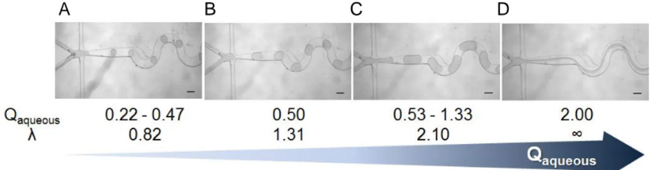
split region were also investigated). ... 62 Figure 3.2 - Images of droplet formation in microfluidic system as function of aqueous
flow rate (Qaqueous): (A) 0.22 - 0.47 μL min−1 with small droplets, (B) 0.50 μL min−1 forms
ideal droplets, (C) 0.53 - 1.33 μL min−1 forms plugs and (C) above 2.00 μL min−1
produces parallel flow streams. The drop size (λ), ratio of the droplet size to the serpentine channel width, varies from λ = 0.82 (A), 1.31 (B) until 2.10 (C). The assays
were developed with fixed Ca = 3x10⁻³, Qoil = 2 μL min−1 and lipids from cationic
liposomes at 2mM. ... 67 Figure 3.3 - Number-weighted size distribution (diameter) of cationic liposomes before (solid line) and after (dashed line) being inserted in droplet-based microfluidic system. Each solid and dashed line represent mean of triplicate from independent experiments. ... 68
Figure 3.4 – Impact of experimental parameters R+/- (A) and microchip design (B) on
lipoplexes characteristics in terms of average diameter (number mean), polydispersity
(PdI) and zeta potential. Volume fraction was set at 0.25 and capillary number at 3x10⁻³.
The droplet-based microfluidic system with serpentine-TC and split region (Figure 3.1B)
was used to investigate R+/- varying of 1.5, 3, 5, 7 and 10. For microchip design
evaluation, R+/- was fixed at 3.0 and tested droplet-based platforms with serpentine
region (thin-TC and wide-WC channels) and in the presence or absence of split region (Figure 3.1B). The error bars represent standard deviation of means (n = 3). Means statistically significant different by Tukey’s test (P<0.10) were flagged with an asterisk
Figure 3.5 – Physico-chemical properties of lipoplexes produced in microfluidic system in terms of average diameter (number mean) (A), polydispersity (PdI) (B) and zeta potential (C). The droplet-based microfluidic system with wide serpentine channel (WC)
and split region was operated with volume fraction 0.25, Ca = 3x10⁻³, Qoil = 6.5 μL min−1
and R+/- varying in 1.5, 3, 5, 7 and 10. The error bars represent standard deviation of
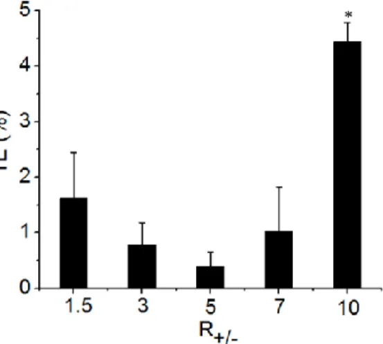
means (n = 4). Means statistically significant different by Tukey’s test (P<0.10) were flagged with an asterisk (*) and non-different means with “ns”. ... 73 Figure 3.6 - In vitro transfection efficacy (TE) of dendritic cells using lipoplexes at
different molar charge ratios (R+/- 1.5, 3, 5, 7 and 10) synthesized by microfluidics
method. The droplet-based microfluidic system with wide serpentine channel (WC) and
split region was operated with volume fraction 0.25, Ca = 3x10⁻³, Qoil = 6.5 μL min−1 and
R+/- varying in 1.5, 3, 5, 7 and 10. The error bars represent standard deviation of means
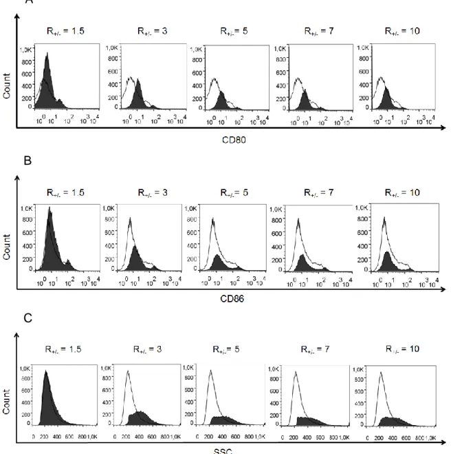
(n = 4). Means statistically significant different by Tukey’s test (P<0.10) were flagged with an asterisk (*). ... 74 Figure 3.7 - DCs activation after transfection with lipoplexes produced by microfluidics method. The droplet-based microfluidic system with wide serpentine channel (WC) and
split region was operated with volume fraction 0.25, Ca = 3x10⁻³, Qoil = 6.5 μL min−1 and
R+/- varying in 1.5, 3, 5, 7 and 10. Histograms of CD80 (A) and CD86 (B) (costimulatory
molecules B7-1 and B7-2, respectively) expressed by DCs is shown. Histograms of DC granulocyte (SSC – side scatter) (C) indicate lipoplex internalization by cells. Histograms are composed of iDC (immature dendritic cells) represented by solid dark lines overlaid by histogram of DCs treated with corresponding type of lipoplex represented by solid-filled background. ... 75
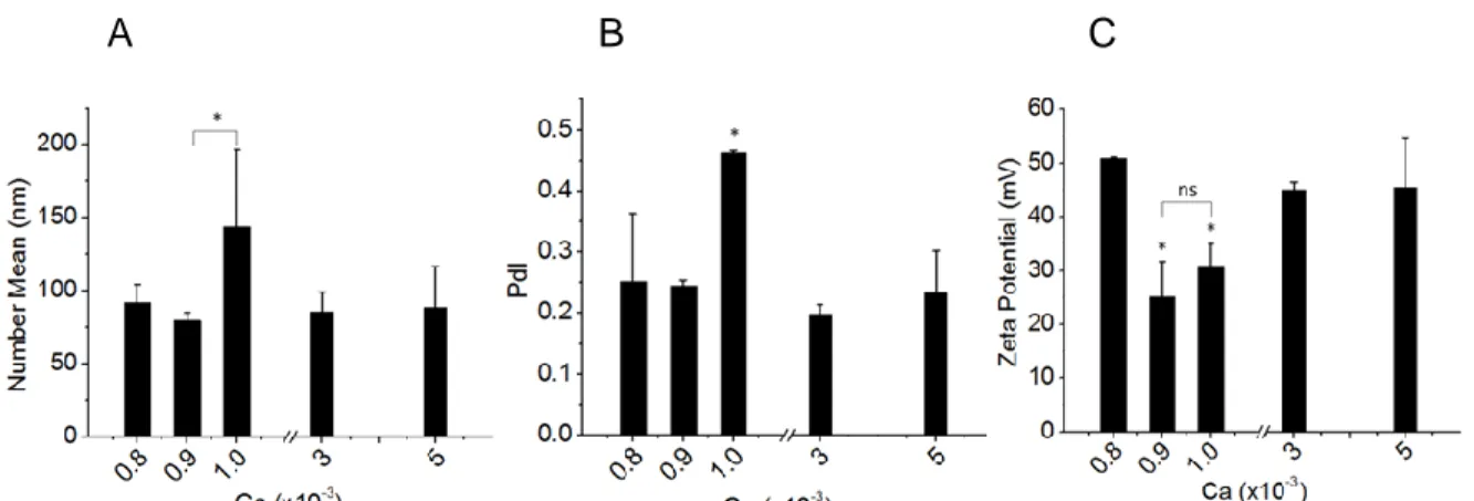
Figure S.1 – Impact of capillary number (Ca) on lipoplexes characteristics in terms of
size (A), polydispersity (PdI) (B) and zeta potential (C). The droplet-based microfluidic system with serpentine-TC and split region (Figure 3.1B) was set at volume fraction
0.25, R+/- = 3 and varying Ca from 8x10⁻⁴ to 5x10⁻³. The error bars represent standard
deviation of means (n = 3). Means statistically significant different by Tukey’s test (P<0.10) were flagged with an asterisk (*) and non-different means with “ns”………….82 Figure S.2 – Strategy of dendritic cells analysis. (A) Dot Plot graph of SSC (side scatter) by FSC (forward scatter) to delimit DCs gate. (B) Graph of CD11c versus HLA-DR to delimit Gate R1, corresponding to cells double-positive………...83
Figure S.3 – Strategy of transfection efficiency analysis. At first, we determined the
negative gate of FITC that is around 99% of this population in the FITC histogram of DCs from Gate R1 treated with liposomes (filled graph with solid line). Then, we overlaid the graph with the FITC histogram of DCs from Gate R1 treated with lipoplexes (empty graph with dot line) in order to define the TE provided by the lipoplexes analyzed…….84
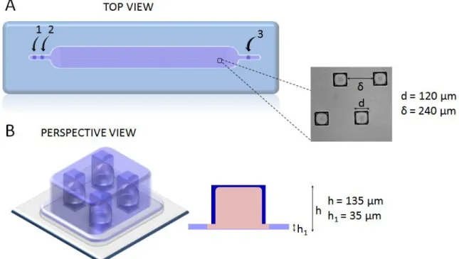
Figure 4.1 - Design of microchip where CHO-S cells were cultivated. The microchip dimension is 0.5 × 4.8 cm with 1495 square anchors (115 × 13). The microchip have two inlets: 1 for oil phase (FC-40/RAN) and 2 for aqueous phase (cells + lipoplexes + agarose), and one exit (3). Microchip top view (A) shows that each square anchor has d = 120 μm of side, spaced by δ = 240 μm. Lateral section (B) shows that the chamber height is h1 = 35 μm and the anchor height h = 135 μm. ... 89 Figure 4.2 - Physico-chemical properties of CL (cationic liposomes EPC/DOTAP/DOPE)
and their lipoplexes R+/- = 5, 3 and 1.5. (A) Size represented by intensity-weighted
distribution disposed in a way to show the increase in ratio pDNA / liposomes when getting down, like represented by arrow in the right. The dotted, dashed and solid lines in each graph represent one independent size distribution. (B) Zeta potential was no
significantly different (ns) between cationic liposome and lipoplexes R+/ 5, and between
lipoplexes R+/- 3 and 1.5, but different among groups by Wilcoxon rank sum test at 5%
significance level. Measures were done in the same conditions as nanoparticles were mixed with cells, i.e., CL and its lipoplexes was diluted in DEPC water. Results represent means ± S.D., n = 3. ... 94
Figure 4.3 – CHO-S cells transfection with lipoplexes at R+/- 5, 3, 1.5 in the microfluidic
device. (A) Large scan at 4x magnification of the whole microchip with 1495 anchored droplets. Scale bar: 200 µm. (B) Distribution of the number of cells per droplet (bars), and best fit to a Poisson distribution (dashed line). (C) View of a typical anchor at 10x magnification, showing an overlay of bright-field and DAPI (live) and TRITC (dead) signals. Images at first (0h) and final (62h) time of experiment are shown. Bar graphs show CHO-S viability for different conditions. There is no significant difference (ns) of cell viability between different conditions by Wilcoxon rank sum test at 5% significance level. Scale bars represent 50 µm. Results represent means ± S.D., n = 2. (D) For transfection analysis, FITC filter was used to quantify GFP production and TRITC to track all cells during the time-lapse. Shown here are overlays of bright field and FITC images at time 0, 24, 48 and 62 h. ... 97 Figure 4.4 - Kinetics of GFP production during the culture period of CHO-S cell in single-cell platform. (A) Boxplots presenting the signal intensity for single-cells transfected with the
lipoplex R+/- 5, 3 and 1.5 at each time step (each 2h). This is how flow cytometry would
present the data. (B) The same data where the signal for each cell at the beginning of the experiment is subtracted from the signal as a function of time. The bold red line represents the evolution of the mean GFP production of the population. ... 99
lipoplexes R+/- 1.5, 3 and 5, during the culture period in single-chip platform. The error bars represent the difference between replicas from each lipoplex condition (n=2). .... 100 Figure 4.6 - Distribution of GFP production on the single-cell level (A) Histograms of the
increase in GFP signal (Δl) for cells transfected with lipoplexes R+/- 5 at culture-times t =
12h (red curve), t = 32h (green curve) and t = 62 h (blue curve). (B) The asymmetry towards the high values is quantified through the Skewness parameter, which is found to increase linearly in time. (C) The skewness of GFP intensity distribution at the final instant (t=62 h) could be used to define a threshold that divides the population into low-producing and high-low-producing cells. (D) Time evolution of the signal due to the high
producers and other cells. Data for other conditions (R+/- 3 and 1.5) are similar. Results
represent means ± S.D., n = 3. ... 102 Figure 4.7 - Characterizing high GFP producers. (A) Percentage of total cells characterized as HP for each of the lipoplex conditions indicate a decreasing number of high producing cells with increased DNA charge. (B) The specific productivity for the three conditions shows that the GFP production per transfected cell increases with increased DNA charge. (C) Size difference between high producers and low producers for different conditions. Means statistically significant different by Kruskal-Wallis (B) or by Wilcoxon rank sum test (A and C) at 5% significance level were flagged with an asterisk (*). Error bars represent the values for the two independent experiments. ... 103 Figure S.1 – GFP production kinetics characterization. An example of linear coefficient
correlation distribution (r2) of GFP produced by CHO-S cells transfected with lipoplexes
R+/- 5 for all single cells (blue columns) and for high GFP producers (red line) is
exhibited……….109 Figure S.2 - High GFP producers evenly distributed within the microchip. HPs are represented as red points on the microchip's map………..109 Figure S.3 - High GFP producers characterization in terms of cell-size. An example of graphs comparing size of HPs (red line) and all cell population (blue columns) for
CHO-S cells transfected with lipoplexes R+/- 5 is shown………..110
Annex I
Figure 1 - Plasmid map of pEGFP-N1 Vector from Clontech. ... 113 Figure 2 - Plasmid map of pmaxGFP Vector from Lonza………...…114
Figure 1 – Images of droplet formation with aqueous phase composed of green and red dye in water and oil phase composed of mineral oil with 2% v/v of surfactant Span 80 in the droplet-based microfluidic system with serpentine-TC and split region (Figure 3.1B). Following droplet behaviors were observed in the microfluidic system: (A) ideal droplets
when flow rates were Qoil = 2 μL min−1 and Qaqueous = 0.67 μL min−1, large droplets when
Qoil = 2 μL min−1 and Qaqueous = 2 μL min−1 (B), until reached a parallel flow when Qoil = 2
μL min−1 and Qaqueous = 3 μL min−1 (C), and small droplets were formed decreasing
Qaqueous to 0.29 μL min−1 with Qoil = 2 μL min−1 (D), until the oil phase invaded aqueous
phase inlet in Qoil = 2 μL min−1 and Qaqueous = 0.10 μL min−1(E)……….117
Figure 2 - Intensity-weighted distribution (A) and number-weighted distribution (B) of cationic liposomes obtained in water, OptiMEM culture medium or PBS buffer solution as aqueous phase. The dashed and solid lines in each graph represent one independent size distribution (n=2)………...119
Annex III
Figure 1 - Droplet-based microfluidic system. ... 122 Figure 2 – Microchamber with 500 cylindrical traps with 250 µm of diameter by 250 µm of height... 125
Figure 3 – Time-lapse images from 0, 5, 16 and 24 hours of smooth muscle cells (Ccell =
4x10⁶ cells/ml) cultivated in collagen hydrogel droplets at 1.2 mg/ml (A, B, C and D) and
6 mg/ml (E, F, G, H). ... 126
Figure 4 – Images of smooth muscle cells (Ccell = 4x10⁶ cells/ml) cultivated in collagen at
6 mg/ml and transfected with lipoplexes (DNA/cationic liposomes at a molar charge ratio of R+/-=5 for 0 day (A), 1 day (B), 2 days (C) and 3 days (D). ... 127
Figure 5 – Images from 0 and 1 day incubation of smooth muscle cells 1x10⁶ cells/ml
cultivated in collagen hydrogel droplets at 1.2 mg/ml (A and B) and 6 mg/ml (C and D). ... 128
Figure 6 – Images incubation of smooth muscle cells 2.5x10⁶ cells/ml cultivated in
collagen hydrogel droplets at 6 mg/ml for 1 day without staining (A) or with cell tracker and lipoplexes (B). ... 129
Figure 7 – Images of smooth muscle cells (Ccell = 2.5x10⁶ cells/ml) stained with cell
tracker, cultivated in collagen at 6 mg/ml and transfected with lipoplexes (DNA/cationic
liposomes at a molar charge ratio of R+/-=5) for 0 day (A), 1 day (B), 7 days (C) and 10
Figure 8 – Images of smooth muscle cells (Ccell = 4x10 cells/ml) added with Y27632, cultivated in collagen hydrogel droplets at 1.2 mg/ml for 0 (A) and 16 hours (B)... 131
Figure 9 – Images of mesenchymal stem cells (Ccell = 4x10⁶ cells/ml) cultivated in
collagen hydrogel droplets at 1.2 mg/ml for 0 and 1 day without inhibitors (A and B), added with Y27632 (C and D) or with Blebbistatin (E and F). ... 133
Figure 10 – Images of lymphoma cells market with cell tracker cultivated in agarose
hydrogel droplets for 0 and 1 day incubation at 2.5x10⁶ cells/ml (A and B) and 5x10⁶
cells/ml (C and D). ... 134
Figure 11 – Images of lymphoma cells (Ccell = 2.5x10⁶ cells/ml) stained with cell tracker,
cultivated in agarose and transfected with lipoplexes (DNA/cationic liposomes at a molar
charge ratio of R+/-=5) for 0 day (A), 7 days (B) and 11 days (C). Detached green
fluorescent images of transfected cells in their respectively day of incubation. ... 135
Figure 12 – Images of lymphoma cells (Ccell = 2.5x10⁶ cells/ml) cultivated in agarose,
transfected with lipoplexes (DNA/cationic liposomes at a molar charge ratio of R+/-=5) for
1 day (A) and 7 days (B), or marked with live/dead staining for 1 day (C) and 7 days (D). Detached green fluorescent images of transfected cells in their respectively day of incubation. ... 136
Chapter II
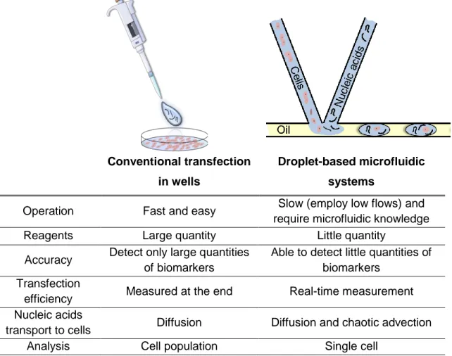
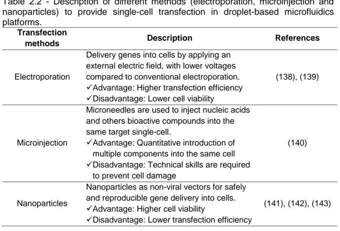
Table 2.1 - Comparative table summarizing differences between in vitro transfection by conventional transfection in wells and by droplet-based microfluidic systems. ... 39 Table 2.2 - Description of different methods (electroporation, microinjection and nanoparticles) to provide single-cell transfection in droplet-based microfluidics platforms. ... 44
Chapter III
Table S.1 - Physico-chemical properties of cationic liposomes before and after droplet-based microfluidic processing. ... 81
Table S.2 - The effect of molar charge ratio (R+/-) and microchip design on
physico-chemical properties of lipoplexes. ... 82
Annex II
Table 1 - Variation in flow rates Qaqueous (aqueous phase composed of green and red
dye in water) and Qoil (oil phase composed of mineral oil with 2% v/v of surfactant Span
80) for droplet formation in the droplet-based microfluidic system with serpentine-TC and split region (Figure 3.1B)………..116 Table 2 - Physico-chemical properties of cationic liposomes obtained by single hydrodynamic focusing in cross-junction microfluidic device using as aqueous phase
water, OptiMEM culture medium or PBS buffer solution………119
ANOVA: analysis of variance;
Ca: capillary number;
CHO-S: Chinese hamster ovary cells;
CL: cationic liposomes;
DCs: dendritic cells;
DNA: deoxyribonucleic acid;
DOPE: 1,2dioleoylsn-glycero-3 phosphoethanolamine;
DOTAP: 1,2-dioleoyl-3-trimethylammonium-propane;
EPC: Egg phosphatidylcholine;
FITC: fluorescein fluorochrome;
GFP: green fluoresce protein;
GFP: green fluorescent protein;
HP: high producer CHO-S cells;
Lipoplexes: cationic liposomes/pDNA complexes;
LP: low producer CHO-S cells;
MFI: median fluorescence intensity;
PdI: Polydispersity Index;
pDNA: DNA plasmidial;
R+/−: cationic lipid:DNA molar charge ratios;
Re: Reynolds number;
S.D.: Standard Deviation;
SSC: side scatter - cells granularity/complexity;
ST: stable transfection;
TC: thin serpentine channel;
TE: transfection efficiency;
TGE: transient gene expression technology;
THP-1: human monocyte cells;
Volume fraction: ratio aqueous/oil flow rate;
ACKNOWLEDGEMENTS ... v
FIGURE CAPTIONS ... vii
TABLE CAPTIONS ... xiii
NOMENCLATURE ... xiv SUMMARY ... xv Chapitre I - Introduction ... 17 1. Introduction ... 17 2. Objectifs ... 20 3. Organization de la thèse ... 21 4. Références ... 23
Chapter II – Literature review ... 26
1. Introduction ... 27
2. Nanoparticles as non-viral vectors for gene delivery ... 28
3. Microfluidic droplet technologies ... 30
4. Droplet-based microfluidic platforms for lipoplexes formation ... 34
5. Droplet-based microfluidic platforms for in vitro transfection ... 37
6. References ... 45
Chapter III – Droplet Microfluidic-Assisted Synthesis of Lipoplexes for DC Transfection ... 57
Abstract ... 58
1. Introduction ... 58
2. Materials and Methods ... 61
3. Results and Discussion ... 66
4. Conclusion ... 76
5. Acknowledgements ... 77
6. References ... 77
Chip ... 85
Abstract ... 86
1. Introduction ... 86
2. Materials and methods ... 88
3. Results and Discussion ... 93
4. Conclusions ... 105 5. Acknowledgments ... 105 6. References ... 106 7. Supplementary data ... 108 Chapter V – Conclusions ... 111 Chapter VI - Perspectives ... 112
ANNEX I – Plasmid vectors ... 113
ANNEX II –Preliminary studies in droplet microfluidic system to synthesize lipoplexes115 1. Study of flow rates for droplets formation ... 115
2. Cationic liposome diluent ... 117
3. References ... 119
ANNEX III – Preliminary studies in droplet microfluidic system to transfect CHO-S cells ... 121
1. Objectives ... 121
2. Materials and methods ... 121
3. Results and discussion ... 124
4. Conclusions ... 137
Chapitre I - Introduction
_______________________________________________________________________________
1. Introduction
La thérapie génique se réfère à la transmission d'un acide nucléique codant un gène d'intérêt dans les cellules ou organes ciblés avec conséquente expression du transgène (1). Un facteur clé dans le succès de la thérapie génique est le développement de systèmes d'administration capables de transférer efficacement les gènes (2). Les nanovecteurs sont généralement classés comme viraux et non-viraux, et dans ces derniers, les liposomes cationiques sont particulièrement prometteurs (1,3). Les liposomes cationiques sont principalement composés de lipides cationiques pour assurer leur charge superficielle positive, ce qui les conduit à interagir électrostatiquement avec les acides nucléiques chargés négativement, pour former des complexes capables d'entrer dans les cellules (3). Les liposomes cationiques sont supérieurs aux vecteurs viraux en termes de reproductibilité, de sécurité d'utilisation, en plus d'être biocompatibles et biodégradables. Cependant, ils sont inférieurs aux vecteurs viraux en termes d'efficacité de transfection (4).
Les progrès récents de la microfluidique ont créés de nouvelles et passionnantes perspectives de thérapie génique. L'environnement à micro-échelle dans les systèmes microfluidiques permet un contrôle et une optimisation précis des processus et techniques multiples utilisés dans le transférer des gènes et dans la production des systèmes d'administration de gènes et de médicaments (5). En particulier, les microgouttes d'eau dans l'huile fournissent un autre format expérimental, car les microgouttes définissent des compartiments de réaction, entraînant une réduction de volume de réaction requis. Il a également été démontré que les microgouttes générées par des dispositifs microfluidiques sont extrêmement uniformes, ce qui est apparemment adéquat pour l'encapsulation de cellules individuelles et pour la synthèse des systèmes non-viraux (6,7).
Les systèmes microfluidiques de gouttes désignés avec certaines géométries peuvent fournir un mélange rapide de réactifs (8). Les microsystèmes fonctionnant
______________________________________________________________
uniquement avec des flux parallèles ont le mélange de fluides favorisé principalement par la diffusion, alors que les microsystèmes de gouttes peuvent ajouter une contribution d'advection chaotique qui augmente le mélange dans le système (9). Les micromélangeurs ont des applications importantes telles que le contrôle de réactions chimiques (10), la contribution à la cristallisation des protéines (11), l’amélioration d’analyses biochimiques (12) et la complexation des nanoparticules avec les acides nucléiques (lipoplexes) (7).
Les méthodes classiques d'obtention de lipoplexes impliquent uniquement le mélange fourni par le « hand shaking » ou par le vortex pour atteindre la complexation des liposomes cationiques avec des acides nucléiques. Cependant, ces méthodes conventionnelles introduisent une grande variabilité dans la formation de lipoplexes et, par conséquent, fournissent des rendements de transfection incompatibles (7). Certaines cellules difficiles à transfecter, comme les cellules dendritiques (DC) (13), utilisent des voies de transfection extrêmement dépendantes de la taille et de la polydispersité des lipoplexes (14). Les cellules dendritiques chargées avec des antigènes tumoraux sont importantes pour des approches immunothérapeutiques (15). Ainsi, l'étude des méthodologies de
formation des lipoplexes est très importante afin d’obtenir lipoplexes avec des
propriétés spécifiques à différentes lignée cellulaire d'une manière reproductible et contrôlable (16). Hsieh et al. (7) ont développé un système microfluidique de gouttes afin de complexer liposomes commerciaux avec ADN. Ils ont montré la robustesse du microsystème pour produire des lipoplexes reproductibles et aussi pour transfecter des cellules, ainsi arrivant à une méthode alternative aux conventionnelles (7).
En outre, les systèmes microfluidiques de gouttes peuvent être utilisés pour analyser des cellules individualisées, car les gouttes permettent la détection rapide de molécules sécrétées par les cellules en raison du faible volume entourant chaque cellule encapsulée. Le risque de contamination croisée diminue et les cellules peuvent être étudiées individuellement par des techniques de fluorescence (17). Les micropuces liées à des équipements de fluorescence permettent l’analyse des cellules de mammifères adhérentes et non adhérentes, la
______________________________________________________________
manipulation et l'étude du contenu de cellules individualisées (18). En plus, les cellules encapsulées dans les gouttes peuvent être surveillées pendant toute la période de culture cellulaire, au lieu de seulement quelques instants, ce qui met en évidence la réponse de chaque cellule même si le signal de la population cellulaire semble relativement homogène pendant l'expérience (19). Cependant, maintenir la viabilité cellulaire dans les gouttes est un défi, car il existe des risques de coalescence, d'appauvrissement des nutriments ou d'accumulation de métabolites toxiques. Ainsi, ces points doivent être considérés avant de faire une analyse robuste sur des longues périodes de culture cellulaire (17). Malgré ces inconvénients, un groupe de recherche a montré que la culture cellulaire dans des plateformes à l’échelle de la cellule unique est possible (20).
L'application d’une plateforme microfluidique pour suivre dynamiquement les réponses cellulaires individualisées ouvre la possibilité de mieux comprendre l'hétérogénéité cellulaire auprès de l'expression transitoire des gènes (21). L'industrie de la biotechnologie utilise largement les cellules ovariennes de hamster Chinois (CHO) pour produire des protéines recombinantes par transfection transitoire. Généralement, ce type de transfection est plus approprié pour la production de protéines en haute quantité et de manière rapide, qui est dont utilisé dans les premières étapes du développement de médicaments (22). Par conséquent, en vue d'optimiser les rendements de production, la transfection par un gène d’expression transitoire à l’échelle de la cellule unique est souhaitable afin de mieux comprendre le processus et de mieux contrôler les conditions de transfection.
Dans ce contexte, l'application de la technologie microfluidique de gouttes pour synthétiser lipoplexes et pour transfecter des cellules de mammifères in vitro de manière transitoire peuvent contribuer aux progrès de la technique de délivrance de gène. A cet effet, dans la première partie de ce travail, certains paramètres qui influencent la synthèse de lipoplexes ont été étudiés dans la plateforme de gouttes. Le but était de synthétiser lipoplexes dans des conditions microfluidiques, avec des propriétés physico-chimiques appropriées pour la
______________________________________________________________
unique, cellules CHO-S ont été transfectées dans une plateforme microfluidique universelle pour des tests biologiques. La transfection transitoire a été réalisée par lipoplexes au différent chargement d'ADN, rapport molaire de charge entre lipides
cationiques et des acides nucléiques (R+/-) 1,5; 3; 5. Avec ce système, c’était
possible de conclure sur l'influence des lipoplexes dans l’hétérogénéité de la
population de cellules en exprimant une protéine recombinante. Cette thèse a été
développée dans le cadre d’une programme de cotutelle entre l'Université de
Campinas - Brésil et de l'Ecole Polytechnique – France, grâce à la collaboration
entre la Professeur Lucimara de la Torre, spécialiste en systèmes non-viraux de transfert de gènes au Brésil, et le Professeur Charles Baroud, spécialiste en microfluidique pour les applications biologiques en France.
2. Objectifs
L'objectif général du projet était de contribuer aux domaines de la nanobiotechnologie, de la microfluidique et du transfert de gènes. En particulier, ce travail visait à appliquer des systèmes microfluidiques de gouttes afin de synthétiser des lipoplexes présentant des caractéristiques physico-chimiques pour transfecter les cellules dendritiques et d’étudier le processus de transfection de
cellules CHO-S par l’analyse de cellules individualisées pendant la période de
culture cellulaire. Ainsi, le premier objectif était de maintenir les propriétés des liposomes cationiques après le traitement dans le système microfluidique de gouttes et de former lipoplexes avec les caractéristiques requises pour transférer les DCs. Ensuite, le deuxième objectif était d’étudier plus profondément l'influence
de lipoplexes au différent chargement d'ADN (R+/-) sur la transfection de cellules
de mammifères. Pour cela, une plateforme microfluidique universelle à l’échelle de la cellule unique a été utilisée pour transfecter les cellules CHO-S par lipoplexes. Avec l’analyse de cellules individualisées fournie par la plateforme, c’était possible d’associer l'influence de lipoplexes au différent chargement d'ADN sur hétérogénéité dans la production de GFP par la population de cellules CHO-S.
______________________________________________________________
• Synthèse de lipoplexes dans un dispositif microfluidique de gouttes pour transfecter les cellules dendritiques:
Investiguer comment les paramètres microfluidiques expérimentaux influencent sur les propriétés physico-chimiques des lipoplexes afin d'obtenir des lipoplexes reproductibles et appropriés à la transfection des DC.
• Transfection des cellules CHO-S dans une plateforme microfluidique à l’échelle de la cellule unique:
Évaluez la manière dont les lipoplexes au différent chargement d'ADN
influencent sur la transfection transitoire des cellules CHO-S par l’analyse de
cellules individualisées. 3. Organization de la thèse
La thèse est organisée en six chapitres, comme décrit ci-dessous. Les résultats expérimentaux ont été présentés sous forme des articles scientifiques (Chapitre III et IV) qui seront soumis à des revues internationales, selon le contenu abordé. Par conséquent, les sections: introduction, matériel et méthodes, résultats et discussion et conclusion; de chaque partie de la thèse sont inclus dans les articles scientifiques de leur respectif chapitre.
Chapitre I – Introduction
Chapitre II – Revue de littérature
Ce chapitre présente une vision globale de la thérapie génique, des liposomes cationiques, de la microfluidique, des systèmes microfluidique de gouttes et leur application dans la synthèse des lipoplexes et dans la transfection des cellules de mammifères in vitro dans l’approche de cellules individualisées. Le texte a été adapté à partir d'un chapitre du livre ("Trends on microfluidic liposome production through hydrodynamic flow-focusing and microdroplet techniques for
gene delivery applications") et d’un article de révision ("Droplet-based microfluidic
systems for production and transfection in vitro of non-viral vectors for gene delivery ") publiés au cours de la thèse, avec Micaela Tamara Vitor comme co-auteur.
______________________________________________________________
Chapitre III – Droplet Microfluidic-Assisted Synthesis of Lipoplexes for DC
Transfection
Ce chapitre montre l'application d'un système microfluidique de gouttes pour étudier la complexation des liposomes cationiques EPC/DOTAP/DOPE avec l’ADN pour obtenir des lipoplexes reproductibles et appropriés à la transfection des cellules dendritiques. À cette fin, certains paramètres expérimentaux ont été étudiés, tels que les débits d'entrée et les propriétés physico-chimiques des liposomes cationiques après le microsystème de gouttes. Avec le système fonctionnant avec un rapport débit aqueux/huile 0,25, les gouttes ont été formées avec une taille de 1,5 fois du canal de serpentin. Les liposomes cationiques maintiennent leurs propriétés après le traitement, même avec un résidu de tensioactif dans la phase aqueuse. Ensuite, les caractéristiques des lipoplexes ont
été étudiées en fonction du rapport molaire de charge (R+/-) et de la géométrie de
la micropuce. Une condition optimale de synthèse des lipoplexes a été obtenue en utilisant le système microfluidique de goutes avec un canal serpentin large et une région de séparation, fonctionnant au rapport débit aqueux/huile 0,25 et dans les
rapports molaire de charge R+/- 1,5 ; 3 ; 5 ; 7 et 10. Ensuite, ces lipoplexes ont été
évalués dans leur capacité à transfecter les cellules dendritiques in vitro, lors de l'activation des cellules. La meilleure efficacité de transfection a été obtenue avec
lipoplexes R+/- 10 qui ont également activé les DCs, un effet important pour les
applications immunologiques.
Chapitre IV – Tracking the Heterogeneities of CHO Cells Transiently
Transfected on a Chip
Ce chapitre montre l'utilisation d'une plateforme universelle pour transfecter
de cellules CHO-S en utilisant lipoplexes au différent chargement d'ADN (R+/- 1,5;
3; 5). En vue d'optimiser les rendements de production de protéines recombinantes dans la transfection transitoire, les hétérogénéités de la population CHO-S ont été
explorées par l’analyse de cellules individualisées fournie par la plateforme. La
cinétique de production de GFP a révélé la présence d'une sous-population produisant des niveaux significativement élevés de protéines recombinantes. Ces
______________________________________________________________
cellules hautes productrices (HP) ont montré une augmentation de la taille par rapport à la population moyenne, ce qui suggère qu’elles étaient dans le cycle de
division cellulaire. Lipoplexes avec une charge positive et moins d’ADN (R+/- 5) ont
produit plus de HP. Lipoplexes avec une charge négative et plus d’ADN (R+/- 1,5)
ont augmenté la productivité spécifique de GFP des HP. Chapitre V – Conclusions
Chapitre VI – Perspectives
ANNEXE I – Vecteurs plasmidiques
ANNEXE II – Etudes préliminaires dans le système microfluidique de gouttes pour
synthétiser les lipoplexes
L’étude des débits sur la formation de gouttes dans une émulsion eau / huile
en utilisant des réactifs moins coûteux et l’étude de la formation de liposomes
cationiques dans différents diluants afin d'obtenir des nanoparticules ayant des caractéristiques physico-chimiques requises pour la transfection des cellules dendritiques.
ANNEXE III – Etudes préliminaires dans le système microfluidique de gouttes pour
transfecter les cellules CHO-S
Avant d'atteindre le système présenté au Chapitre III pour transfecter les cellules CHO-S, d'autres cellules et d'autres microdispositifs ont été explorés. Ils n’ont pas été utilisés pour écrire l’article scientifique en raison de la faible viabilité ou efficacité de transfection. Par contre, la micropuce a montré son potentielle dans la compréhension des paramètres clés qui influent sur le processus de transfection des cellules de mammifères in vitro, et à cause de cela, les expériences ont été présentées dans cette annexe.
4. Références
1. Guang Liu W, De Yao K. Chitosan and its derivatives—a promising non-viral
______________________________________________________________
2. Verma IM, Weitzman MD. Gene therapy: twenty-first century medicine. Annu
Rev Biochem. 2005;74:711–38.
3. Miller AD. Cationic liposomes for gene therapy. Angew Chemie Int Ed.
1998;37(13–14):1768–85.
4. Serikawa T, Kikuchi A, Sugaya S, Suzuki N, Kikuchi H, Tanaka K. In vitro and
in vivo evaluation of novel cationic liposomes utilized for cancer gene therapy. J Control Release. 2006;113(3):255–60.
5. Kim J, Hwang I, Britain D, Chung TD, Sun Y, Kim D-H. Microfluidic
approaches for gene delivery and gene therapy. Lab Chip. The Royal Society of Chemistry; 2011;11(23):3941–8.
6. Kintses B, van Vliet LD, Devenish SRA, Hollfelder F. Microfluidic droplets:
new integrated workflows for biological experiments. Curr Opin Chem Biol. 2010;14(5):548–55.
7. Hsieh AT-H, Hori N, Massoudi R, Pan PJ-H, Sasaki H, Lin YA, et al. Nonviral
gene vector formation in monodispersed picolitre incubator for consistent gene delivery. Lab Chip. The Royal Society of Chemistry; 2009;9(18):2638– 43.
8. Bringer MR, Gerdts CJ, Song H, Tice JD, Ismagilov RF. Microfluidic systems
for chemical kinetics that rely on chaotic mixing in droplets. Philos Trans R Soc London A Math Phys Eng Sci. The Royal Society; 2004;362(1818):1087– 104.
9. Lin B. Microfluidics: technologies and applications. Springer; 2011.
10. Song H, Tice JD, Ismagilov RF. A Microfluidic System for Controlling Reaction Networks in Time. Angew Chemie Int Ed. WILEY-VCH Verlag; 2003;42(7):768–72.
11. Zheng B, Tice JD, Ismagilov RF. Formation of droplets of alternating composition in microfluidic channels and applications to indexing of concentrations in droplet-based assays. Anal Chem. 2004;76(17):4977–82. 12. Brouzes E, Medkova M, Savenelli N, Marran D, Twardowski M, Hutchison JB,
et al. Droplet microfluidic technology for single-cell high-throughput screening. Proc Natl Acad Sci. 2009 Aug 25;106(34):14195–200.
13. Bowles R, Patil S, Pincas H, Sealfon SC. Optimized protocol for efficient transfection of dendritic cells without cell maturation. JoVE (Journal Vis Exp. 2011;(53):e2766–e2766.
14. De Haes W, Van Mol G, Merlin C, De Smedt SC, Vanham G, Rejman J. Internalization of mRNA lipoplexes by dendritic cells. Mol Pharm.
______________________________________________________________
2012;9(10):2942–9.
15. Barbuto JAM, Ensina LFC, Neves AR, Bergami-Santos PC, Leite KRM, Marques R, et al. Dendritic cell–tumor cell hybrid vaccination for metastatic cancer. Cancer Immunol Immunother. Springer Berlin / Heidelberg; 2004;53(12):1111–8.
16. Hsieh AT-H, Pan PJ-H, Lee AP. Rapid label-free DNA analysis in picoliter
microfluidic droplets using FRET probes. Microfluid Nanofluidics.
2009;6(3):391–401.
17. Lindstrom S, Andersson-Svahn H. Overview of single-cell analyses: microdevices and applications. Lab Chip. The Royal Society of Chemistry; 2010;10(24):3363–72.
18. Sims CE, Allbritton NL. Analysis of single mammalian cells on-chip. Lab Chip. The Royal Society of Chemistry; 2007;7(4):423–40.
19. Schaerli Y, Hollfelder F. The potential of microfluidic water-in-oil droplets in experimental biology. Mol Biosyst. The Royal Society of Chemistry; 2009;5(12):1392–404.
20. Clausell-Tormos J, Lieber D, Baret J-C, El-Harrak A, Miller OJ, Frenz L, et al. Droplet-Based Microfluidic Platforms for the Encapsulation and Screening of Mammalian Cells and Multicellular Organisms. Chem Biol. Cell Press; 2008;15(5):427–37.
21. Subramanian S, Srienc F. Quantitative analysis of transient gene expression in mammalian cells using the green fluorescent protein. J Biotechnol. 1996;49(1):137–51.
22. Derouazi M, Girard P, Van Tilborgh F, Iglesias K, Muller N, Bertschinger M, et al. Serum-free large-scale transient transfection of CHO cells. Biotechnol Bioeng. 2004;87(4):537–45.
Chapter II – Literature review
_______________________________________________________________________________
The literature review text was adapted from the book
chapter and the review article co-authored by
Micaela Tamara Vitor
De La Torre LG, Balbino TA, Sipoli CC, Vitor MT, Oliveira AF. Trends on Microfluidic Liposome Production through Hydrodynamic Flow-focusing and Microdroplet Techniques for Gene Delivery Applications. In: Finney L, Eds. Advances in Liposomes Research. New York: Nova Science Publishers; 2014. ISBN: 978-1-63117-074-4.
Republished with permission from Nova Science Publishers, Inc. (confirmation # 11628021)
Vitor, M. T., Sipoli, C.C., & De La Torre, L.G. (2015). Droplet-based Microfluidic Systems for Production and Transfection In Vitro of Non-Viral Vectors for Gene Delivery. Journal of Pharmacy and Pharmaceutical Sciences, 4(4), 1-17.
Republished with permission from Research & Reviews under the terms of the Creative Commons Attribution License.
_______________________________________________________________
1. Introduction
Gene delivery is a promising technique that involves the insertion of nucleic acid inside target cells for curing a disease or at least improving the clinical status of a patient (1). One important step of gene therapy is the transfection process that introduces foreign nucleic acids into cells to produce genetically modified cells (2). In this context, the development of delivery systems capable of efficient and safe gene transfer (3), while protecting the genetic material from different barriers (extracellular matrix, cell membrane, cytosol, nuclear membrane) (4), is necessary. Among the delivery systems, cationic liposomes stand out due to their reproducibility, safety of use, biocompatibility and biodegradability (5). However, besides the promising results of cationic liposomes, the development of methods to insert nucleic acids into cationic liposomes in a control and reproducible way is still a challenge. Depending on cell lines, the production of lipoplexes with specific physico-chemical properties is required for transfection (6).
On the other hand, microfluidics, technology that manipulates small amounts of reactants inside microchannels, appears as a powerful strategy to overcome these drawbacks (7). The microenvironment of microfluidic systems allows precise control and optimization of multiple processes and techniques used in gene therapy (8). Moreover, droplet-based microfluidic systems define microreactors that reduce of many orders the volume manipulated, being attractive for molecular biology assays (9). Droplets moving in some microfluidic systems with specific geometry become micromixers that provide a rapid mixing of reagents (10). Droplet-based microfluidic systems can be also used to transfect cells in vitro. Since microfluidics generates extremely uniform droplets, they are appropriate for single-cell encapsulation and/or for in vitro expression of single genes (11). Molecules secreted by cells are fast detected due to the low volume surrounding each cell, allowing investigation of transfection parameters in culture real time (9,12).
In this literature review, first we present an overview about gene therapy and the role of non-viral vectors. Then, we show the state-of-the-art of current microengineering methods based in droplet microfluidic platforms for the
_______________________________________________________________
production of lipoplexes and mammalian cell transfection. The understanding of this technology and how fluid dynamics influences the microscale for the production of controllable and stable droplets give the insight to understand how to form complexes for gene delivery applications. At last, we show methods of transfection inside droplets comparing with the conventional transfection in wells. 2. Nanoparticles as non-viral vectors for gene delivery
Gene therapy refers to the transfer of genetic material encoding a therapeutic gene of interest into a cell, tissue, or whole organ with consequent expression of the transgene in order to treat a disease. However, for the success of gene therapy, the development of sophisticated and efficient delivery systems capable of transfering genes is a key factor (3,13). In brief, we can describe two classes of efficient nucleic acid carriers: viral and non-viral vectors. These delivery systems should protect nucleic acids from degradation, while providing a safe intracellular delivery (14). Over a decade ago, patients with immunodeficiency-X1 were treated with gene therapy assays based on the use of cDNA in a retrovirus (15,16). After that, gene therapy based on retrovirus vector was used to treat French patients with T cell leukemia, producing aberrant transcription and expression of LMO2 (17). These facts opened an optimistic vision about gene therapy research. Nowadays, gene therapy clinical trials are present in worldwide, mainly in United States of America (62.6%), United Kingdom (10.3%) and Germany (4.1%). These therapies are usually used in the treatment of cancer (63.8%), monogenic diseases (8.9%) and infectious diseases (8.2%) (18). The most commonly vectors used in gene therapy are still virus vectors, highlighting adenovirus (22.5%) and retrovirus (18.8%), but also lipofection (5.2%) and naked pDNA (17.5%) are raising their use (18). Notwithstanding the high efficient transfection provided by viral vectors, they can invoke immune responses or proto-oncogene activations. In this context, the non-viral vectors, particularly cationic liposomes, have a promise and potential future, taking into account their reproducibility and safety of use (5).
Cationic liposomes are non-viral vectors mainly composed of cationic lipids, which guarantee their positive superficial charge. Examples of synthetic cationic
_______________________________________________________________
lipids used in cationic liposome composition are DOTAP
(1,2-dioleoyl-3-trimethylammonium propane), DOTMA
(2,3-bis(oleyl)oxypropyl-trimethylammonium chloride), DDAB (dimethyl dioctadecyl ammonium bromide),
DC-Chol (3 β [N-(N’,N’-dimethylaminoethane)-carbamoyl]cholesterol), DMRIE
(N-(2-hydroxyethyl)-N,N-dimethyl-2,3-bis(tetradecyloxy)-1-propanaminium bromide),
DOGS (dioctadecyl amino glycyl spermine), DOSPA (2,3 dioleyloxy-N-[2(spermine carboxaminino)ethyl]-N,N-dimethyl-1-propanaminium trifluoroacetate). However, the use of cationic lipids can generate cell toxicity, and so the inclusion of others lipids, e.g. egg phosphatidylcholine (EPC), in cationic liposome composition is required to decrease the cytotoxicity (19,20). On the other hand, the positive charge of cationic liposomes provides an electrostatic interaction with negatively charged nucleic acids, forming complexes with net positive charge (21). Hence, these positive charged complexes can enter easily inside cells, whose surface membrane is negatively charged (22). Once into cells, the complexes should release the nucleic acids in cytosol, in case of RNA for rapid synthesis of a target protein (23), or in cell nucleus, in case of DNA to control gene expression for a more long-term. The inclusion of helper lipids, e.g. phosphatidylethanolamines (DOPE), in cationic liposomes composition facilitates the nucleic acid to escape inside the cytosol (24). Felgner et al. (22) were pioneers in the development of cationic liposomes for gene delivery. They produced liposomes composed of 1:1 ratio of DOTMA and DOPE, which were then commercialized as Lipofectin. For example, these cationic liposomes were used in vitro to carrier efficiently hepatitis C virus proteins into a human hepatocyte cell line (HUH7) (25) and in vivo to delivery linamarase gene for the treatment of brain tumors in animals or humans (26). Recently, other commercial cationic liposomes, such as Lipofectamine, DMRIE-C, Oligofectamine, Ambion, 293fectin, Optifecta, Invivofectamine, FuGENE, TransFast, TransFection and CLONfectin, are being used in gene therapy due to their well-established protocols and to provide high efficiency of transfection in some specific cells and with some specific nucleic acids. Our research group (27) also showed the feasibility of dehydrated-rehydrated liposomes composed of EPC, DOTAP and DOPE (50/25/25% molar, respectively)
_______________________________________________________________
carrying polynucleotides encoding HSP65, for prevention and treatment of tuberculosis. And more recently, we obtained the same cationic liposomes produced in a large scale by ethanol injection method to delivery nucleic acid into dendritic cells (DCs) as a potential tool for cancer immunotherapy (28).
Dendritic cells are professional antigen-presenting cells widely used in immunotherapeutic approaches, particularly in immunotherapies against cancer. Since loaded with tumor antigens, mature DCs can induce an immune response against cancer by recruiting patients’ immune system (29). Different strategies are currently used to load DCs with antigens, e.g. peptide pulsing, pulsing with tumor cell lysates, infection with viral vectors, direct nucleic acid loading, or ingestion/fusion with tumor cells (30). Moreover, cationic liposomes stand out in this function as gene carriers, since besides to transfect DCs, they can also activated them (31). Particularly, our research group (32,33) showed that cationic liposomes EPC/DOTAP/DOPE were uptake by DCs, while providing cells stimulation/activation. However, dendritic cells require that these cationic liposomes have a very specific properties (size < 100 nm and polydispersity <0.2) to be internalized (32), due to the use of macropinocytosis and/or phagocytosis as transfection pathways (34). Thus, the study of methodologies that enable to form lipoplexes modulated to specific cells lines in a reproducible and controllable way is very important (35). Conventional methods of obtaining lipoplexes involve just the mixing provided by hand shaking or vortexing to reach cationic liposomes complexation with nucleic acids. However, these conventional methods introduce variability in lipoplexes formation and, as a result, provide inconsistent transfection efficiencies (6). To effectively transfect cells, the physico-chemical properties of lipoplexes should be suitable to the pathway used by the cell line to internalize nanoparticles (36).
3. Microfluidic droplet technologies
The general concept of microfluidics is in the manipulation of small amounts of reactants inside microchannels with the capability to control and manipulate molecules in space and time (37). In microfluidics it is possible to work with small
_______________________________________________________________
amounts of reactants, the period of reactions is short, it is possible to work with parallel operations (38), large surface to volume ratio allows fast diffusion of compounds and fast mass/heat transfer (39). These advantages can be related to the flow in microfluidic devices, which is laminar and corresponds to a low Reynolds number (37,40). Another important consequence of this regime is that the mixture between two parallel flowing streams occurs mainly by diffusion (38).
The origin in microfluidic is in the 90´s for MicroElectroMechanical Systems area (MEMS) (39). Nowadays microfluidics has been exploited in numerous areas as:
Biological analyses - detection of biomolecules (41,42), manipulation and amplification (43) and separation of DNA by capillary electrophoresis (44).
Microbial growth - Screening of variables and kinetic parameters (45,46).
Nanoparticles production – polymeric particles (47,48), liposomes and lipid
vesicles (49–51), metallic nanoparticles (52).
Gene Delivery/Transfection – electroporation (53), hydrodynamic force and
optical energy (8).
Different materials can be used for the construction of microchannels and, for biological application, glass and polymers are detached (54). Glass is considered to be biocompatible, impermeable to gases (54,55), has physic and chemical stability and it is hydrophilic (54). The techniques to prepare microdevices in glass are laser ablation and wet etching (54). Microdevices can also be made by polymers which are not expensive, there is the possibility to change the chemical formulation (55), are stable (37) and hydrophobic (54). The elastomer which has been used extensively for microdevices construction is poly(dimethyl syloxane) well-known as PDMS , moreover the technique employed is soft lithography (56).
In addition, it is important to consider the wettability related to the microchannel material and the droplet system. Since the continuous phase wets the walls faster than the disperse phase and forms a thin film between droplet and walls (57). The droplet breakup occurs when the continuous phase wets the device walls instead the disperse phase, thus the droplet morphology is a result of the interaction between the material which the devices is formed and the continuous
_______________________________________________________________
phase (57). Summarizing hydrophobic channels are required to prepare water in oil systems, inversely hydrophilic channels are necessary for forming oil in in water emulsions (58). Considering the advantages of using PDMS for devices construction, in the literature different strategies are reported to change the wettability of the material, through chemical modifications (58,59).
Microfluidic systems can be classified according to the interfaces created by the fluid flows in microchannel as: pinned interfaces, in which fluids flow in parallel creating vertical interfaces, and floating interfaces, in which immiscible fluids produce droplets of precise shape (60). The use of segmented flow which the reactants are separated in different picoliter/nanoliter droplets has been used in cases when miniaturized systems has to be achieved (61). The segmented flow is the principle of emulsions which are a metastable colloidal systems (62) with two immiscible liquids. In this case there is one continuous phase and one disperse phase in droplets formats (57,61). Some advantages of droplet processes can be described in comparison with parallel flows processes. In parallel flows, in which solute are all distributed over the solvent, the efficiency of chemical reactions and the detection of some molecules inside the channels can be decreased, this phenomenon is called Taylor-Aris dispersion (63,64). The use of droplets processes cut out the contact with solid wall, reducing the probability of reagents adsorption into the channels walls (64). Droplets with samples inside can be seen as micro-reactors which allows the manipulation of small volumes (14,65). In addition, in droplets microfluidics it is possible to carry out many reactions without increasing the number and the channels size (13). Furthermore, considering the relation between the surface area and volume, the reactions inside droplets are faster because the heat and mass transfer times and also diffusion distances are shorter (13,66).
In order to form stable droplets, the use of appropriated surfactants is important. Surfactants are known as amphiphilic molecules with different groups and having affinity for different phases which are immiscible. Due to different groups in the structure, surfactant molecules go to interface and as a result the surface tension between the phases decreases (67). An essential requirement to
_______________________________________________________________
use droplets as microreactors is to avoid the coalescence of them. Thus the surfactant addition provides stabilization in the metastable state (62,67). Furthermore choose biocompatible surfactants is the rule for the success of droplet system application in biological fields (67). Fluorinated oils are promising to be used in biotechnology area; however few surfactants are available to stabilize water in oil interfaces emulsions (67). Different molecules are being studied and developed to microfluidic applications. Nowadays, block copolymer of perfluoropolyether and polyethylenoxide are the most interesting molecules existent. It is known that these molecules reduce protein adsorption or the interactions with cell membranes (67). Another important molecule is a triblock copolymer surfactant composed of perfluoropolyether (PFPE) and polyethylene glycol (PEG) blocks (68). However the problem is the limitation of surfactant modification which can be just by varying the molecular weight or chain-end functionalization. In this way, Wagner et al (68) proposed to synthetize and characterize polyglycerol-based triblock surfactants, and exemplified in droplet-based microfluidics their application in cell encapsulation and in vitro gene expression studies.
The droplet compartments formed in microfluidic systems have many functions. The uniformity and the little volume of these droplets allow them to be used for quantitative assays requiring reduced volumes of reagents, and as a result, providing a low cost for this technique (69). Moreover, droplets compartment design can furnish combined information about molecules function (activity or inhibitory functions), molecules identity and molecules ability to carry out its function by measuring, for example, a fluorescence product. In addition, droplets give the possibility to make several unit operations in a device, like droplets can be divided, fused, incubated, analyzed, sorted and broken up (70). Moreover, the compartments formed by droplets can be used as micromixers, providing efficient and controlled mixing over the reactants inside microcontainers (66).


![Rôle de la protéine kinase C dans la désensibilisation hétérologue des récepteurs [bêta]₁-et [bêta]₂-adrénergiques](data:image/gif;base64,R0lGODlhAQABAIAAAP///wAAACH5BAEAAAAALAAAAAABAAEAAAICRAEAOw==)