HAL Id: hal-03058000
https://hal.archives-ouvertes.fr/hal-03058000
Submitted on 11 Dec 2020
HAL is a multi-disciplinary open access
archive for the deposit and dissemination of
sci-entific research documents, whether they are
pub-lished or not. The documents may come from
teaching and research institutions in France or
abroad, or from public or private research centers.
L’archive ouverte pluridisciplinaire HAL, est
destinée au dépôt et à la diffusion de documents
scientifiques de niveau recherche, publiés ou non,
émanant des établissements d’enseignement et de
recherche français ou étrangers, des laboratoires
publics ou privés.
Molecular Epidemiology of Simian Immunodeficiency
Virus Infection in Wild-Living Gorillas
Neel Cecile, Etienne Lucie, Li Yingying, Takehisa Jun, Rudicell Rebecca S.,
Bass Innocent Ndong, Moudindo Joseph, Mebenga Aime, Esteban Amandine,
Heuverswyn Fran Van, et al.
To cite this version:
Neel Cecile, Etienne Lucie, Li Yingying, Takehisa Jun, Rudicell Rebecca S., et al.. Molecular
Epi-demiology of Simian Immunodeficiency Virus Infection in Wild-Living Gorillas. Journal of Virology,
American Society for Microbiology, 2009, 84, pp.1464 - 1476. �10.1128/jvi.02129-09�. �hal-03058000�
0022-538X/10/$12.00
doi:10.1128/JVI.02129-09
Copyright © 2010, American Society for Microbiology. All Rights Reserved.
Molecular Epidemiology of Simian Immunodeficiency Virus Infection
in Wild-Living Gorillas
䌤
Ce
´cile Neel,
1,2† Lucie Etienne,
1† Yingying Li,
3Jun Takehisa,
3Rebecca S. Rudicell,
3Innocent Ndong Bass,
2Joseph Moudindo,
2Aime
´ Mebenga,
2Amandine Esteban,
1Fran Van Heuverswyn,
1‡ Florian Liegeois,
1Philip J. Kranzusch,
4Peter D. Walsh,
5Crickette M. Sanz,
6David B. Morgan,
7,8Jean-Bosco N. Ndjango,
9Jean-Christophe Plantier,
10Sabrina Locatelli,
11Mary K. Gonder,
11Fabian H. Leendertz,
12,13Christophe Boesch,
13Angelique Todd,
14Eric Delaporte,
1Eitel Mpoudi-Ngole,
2Beatrice H. Hahn,
3and Martine Peeters
1*
UMR145, Institut de Recherche pour le De
´veloppement (IRD) and Universite
´ de Montpellier 1, Montpellier, France
1; Projet Prevention du Sida
au Cameroun (PRESICA), Yaounde
´, Cameroon
2; Departments of Medicine and Microbiology, University of Alabama at Birmingham,
Birmingham, Alabama
3; Department of Microbiology and Molecular Genetics, Harvard Medical School, Boston, Massachusetts 02115
4;
VaccinApe, Bethesda, Maryland
5; Department of Anthropology, Washington University, Saint Louis, Missouri 63130
6;
Lester E. Fisher Center for the Study and Conservation of Apes, Lincoln Park Zoo, 2001 N. Clark Street, Chicago,
Illinois 60614
7; Wildlife Conservation Society, Congo Program, B.P. 14537, Brazzaville, Republic of Congo
8;
Faculties of Sciences, University of Kisangani, Democratic Republic of Congo
9; Laboratoire Associe
´ au
Centre National de Re
´fe
´rence du VIH, CHU Charles Nicolle et Universite
´ de Rouen, Rouen, France
10;
Department of Biological Sciences, University at Albany, State University of New York, Albany,
New York 12222
11; Robert Koch-Institut, Center for Biological Safety, Berlin, Germany
12;
Max Planck Institute for Evolutionary Anthropology, Leipzig, Germany
13;
and WWF-CAR, Bangui, Central African Republic
14Received 8 October 2009/Accepted 2 November 2009
Chimpanzees and gorillas are the only nonhuman primates known to harbor viruses closely related to HIV-1.
Phylogenetic analyses showed that gorillas acquired the simian immunodeficiency virus SIVgor from
chim-panzees, and viruses from the SIVcpz/SIVgor lineage have been transmitted to humans on at least four
occasions, leading to HIV-1 groups M, N, O, and P. To determine the geographic distribution, prevalence, and
species association of SIVgor, we conducted a comprehensive molecular epidemiological survey of wild gorillas
in Central Africa. Gorilla fecal samples were collected in the range of western lowland gorillas (n
ⴝ 2,367) and
eastern Grauer gorillas (n
ⴝ 183) and tested for SIVgor antibodies and nucleic acids. SIVgor antibody-positive
samples were identified at 2 sites in Cameroon, with no evidence of infection at 19 other sites, including 3 in
the range of the Eastern gorillas. In Cameroon, based on DNA and microsatellite analyses of a subset of
samples, we estimated the prevalence of SIVgor to be 1.6% (range, 0% to 4.6%), which is significantly lower than
the prevalence of SIVcpzPtt in chimpanzees (5.9%; range, 0% to 32%). All newly identified SIVgor strains
formed a monophyletic lineage within the SIVcpz radiation, closely related to HIV-1 groups O and P, and
clustered according to their field site of origin. At one site, there was evidence for intergroup transmission and
a high intragroup prevalence. These isolated hot spots of SIVgor-infected gorilla communities could serve as
a source for human infection. The overall low prevalence and sporadic distribution of SIVgor could suggest a
decline of SIVgor in wild populations, but it cannot be excluded that SIVgor is still more prevalent in other
parts of the geographical range of gorillas.
Simian immunodeficiency viruses (SIVs) have been
identi-fied in approximately 40 African primate species, but
chimpan-zees and gorillas are the only nonhuman primates known to
harbor viruses closely related to human immunodeficiency
vi-rus type 1 (HIV-1) (38). These vivi-ruses have been transmitted
to humans on at least four occasions, leading to four different
HIV-1 groups, M to P (14, 26). West central African
chimpan-zees (Pan troglodytes troglodytes) in southern Cameroon are
recognized as the reservoir of the ancestors of HIV-1 group M,
which resulted in the AIDS pandemic, and of HIV-1 group N,
which has been identified in only a few individuals in
Cam-eroon (15). Western lowland gorillas (Gorilla gorilla gorilla) are
infected with SIVgor, which is closely related to the two other
HIV-1 lineages, termed group O, which represents 1% of
HIV-1 infections in west central Africa, and group P, recently
described from a single Cameroonian patient residing in
France (26, 36).
The phylogenetic relationships between SIVcpz, SIVgor,
and HIV-1 show that chimpanzees are the original reservoir of
SIVs found in gorillas and humans (31, 36). Pan troglodytes
troglodytes apes were most likely the original source of SIVgor,
because SIVgor is significantly more closely related to SIVcpzPtt,
* Corresponding author. Mailing address: UMR 145, Institute of
Research and Development (IRD) and University of Montpellier 1,
911 Avenue Agropolis, BP 64501, 34394 Montpellier Cedex 5, France.
Phone: 33 4 67 41 62 97. Fax: 33 4 67 41 61 46. E-mail: martine.peeters
@ird.fr.
† C.N. and L.E. contributed equally to this work.
‡ Present address: BioMaric, Ghent, Belgium.
䌤
Published ahead of print on 11 November 2009.
from Pan troglodytes troglodytes in west central Africa, than to
SIVcpzPts, from Pan troglodytes schweinfurthii in east Africa. In
addition, an ancestral SIVcpzPtt lineage from which SIVgor
and HIV-1 group O viruses are derived has been identified in
the form of mosaic pol fragments in present-day SIVcpzPtt
recombinants (2, 31). However, the ways of transmission and
the exact origin of SIVgor infection in gorillas are not yet
resolved. Because of the extensive overlap in habitat and diet
(6, 23, 29, 33, 40), direct encounters between gorillas and
chimpanzees seem inevitable, but they have rarely been
ob-served and have been described as primarily nonaggressive (17,
28). The primate source of HIV-1 groups O and P also remains
unclear, since current data do not allow one to differentiate
between a chimpanzee and a gorilla reservoir, especially for
HIV-1 group O (26, 31, 36).
To determine the geographic distribution, prevalence, and
species association of SIVgor, we performed a comprehensive
survey of wild gorilla populations in west central (Gorilla gorilla
gorilla) and east (Gorilla beringei graueri) Africa. We found an
overall prevalence of SIVgor of 1.6%, with infection confirmed
at only three field sites. At two of these sites, however, the
prevalence of SIVgor was 4.6%, indicating efficient virus
spread within and between different communities. The
geo-graphic distribution of SIVgor is thus far limited to only a few
sites in Cameroon. However, isolated hot spots of infection do
exist, which could serve as a source of human infection.
MATERIALS AND METHODS
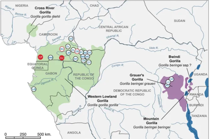
Sample collection and study sites.Fecal samples (2,992) were collected
be-tween June 2006 and June 2009 from wild-living apes in central Africa. In west central Africa, samples were obtained at 18 forest sites, located in the southern part of Cameroon (n⫽ 13), the extreme southwest of the Central African Republic (CAR) (n⫽ 3), the northeast of the Republic of Congo (n ⫽ 1), and western Gabon (n⫽ 1) (Fig. 1). Eight of the 18 sites (CP, MM, LB, DD, ND, DS GT, and LO) were located in national parks or forest reserves, while the re-mainder were in nonprotected areas with considerable hunting pressure. In east central Africa, fecal samples were obtained at three sites, located in the north-eastern part of the Democratic Republic of Congo (DRC). Overall, fecal samples were collected primarily around night nests or feeding sites. For almost all samples, the GPS position and estimated time of deposition were recorded, and the species origin was defined in the field according to nesting sites, prints, vocalizations, and morphological and physical aspects of the samples. Both gorilla and chimpanzee samples were collected at some sites. About 20 mg of dung was collected in a 50-ml tube containing 20 ml of RNAlater (Applied Biosystems/Ambion, Austin, TX). These tubes were kept at base camps at am-bient temperature for a maximum of 3 weeks and subsequently transported to a central laboratory for storage at⫺20°C or ⫺80°C.
Detection of SIVgor and SIVcpz antibodies in ape fecal samples.All gorilla
and chimpanzee fecal samples were tested for the presence of HIV-1 cross-reactive antibodies, using the Inno-LIA HIV I/II score confirmation test (Inno-genetics, Ghent, Belgium) and/or Western blot analysis (Maxim Biotech, Inc., Rockville, MD) as reported previously (15, 36). RNAlater-precipitated immu-noglobulins were resolubilized by diluting the feces-RNAlater mixture (2 ml) with phosphate-buffered saline (PBS)–Tween 20 (7 ml), followed by an incuba-tion for 1 h at 60°C, centrifugaincuba-tion (3,900⫻ g for 10 min) to clarify the solution, and then dialysis against PBS overnight at 4°C. The reconstituted extracts were then subjected to immunoblot analysis.
FIG. 1. Locations of study sites of wild gorillas and/or chimpanzees in central Africa. Red filled circles indicate sampling sites where positive
gorillas were identified in the current study. Red open circles indicate sites where SIVgor infection was previously identified (BQ) (36) or suspected
(GB) (see Materials and Methods) and where no additional positive animals were identified in this survey. KE, LU, and OP are located in the range
of Gorilla beringei graueri (purple), while all other sites are located in the range of Gorilla gorilla gorilla (dark green).
Nucleic acid extraction from fecal samples.Total RNA was extracted from all antibody-positive fecal samples by use of an RNAqueous Midi kit (Applied Biosystems/Ambion, Austin, TX) as described previously (15). When the reverse transcription-PCR (RT-PCR) amplifications were negative using these RNA extracts, nucleic acid extraction was repeated using a NucliSens magnetic extrac-tion kit (bioMe´rieux, Craponne, France), which utilizes magnetic silica particles to purify RNA (4). A 1.5-ml fecal sample was mixed for 1 min with 5 ml of PBS solution and centrifuged at 3,900⫻ g for 30 min. The supernatant was collected in a fresh tube and centrifuged again at 3,900⫻ g for 5 min. The new supernatant was passed through a gauze filter and incubated with 7 ml of NucliSens lysis buffer at room temperature for at least 10 min, followed by the magnetic extrac-tion procedure according to the manufacturer’s instrucextrac-tions, with a final eluextrac-tion volume of 50l. Fecal DNA was extracted using a QIAamp Stool DNA Mini kit (Qiagen, Valencia, CA) as previously described (15). Each extraction was per-formed using 2 ml of feces-RNAlater mixture and eluted in a final volume of 100 l DNA solution.
Amplification of SIVgor and SIVcpz sequences from fecal RNA.Fecal RNA
was subjected to RT-PCR amplification using SIVgor/SIVcpz/HIV-1 consensus primers for env (gp41 ectodomain) (⬃440 or 315 bp) and pol (⬃330 bp or ⬃245 bp) (Table 1). cDNA was synthesized using the R1 primer, followed by nested PCR using primers F1-R1 and F2-R2. All RT-PCRs were performed with Ex-pand reverse transcriptase and an ExEx-pand long-template PCR system (Roche Diagnostics, Indianapolis, IN) according to the manufacturer’s instructions. Briefly, 10l of fecal viral RNA was first incubated with 40 pmol of the outer reverse R1 primer (1l) for 10 min at 65°C, rapidly cooled on ice, and then added to the RT-PCR components, including 20 U of RNase inhibitor (Applied Biosystems/Ambion, Austin, TX), in a 20-l reaction volume. The mixture was finally incubated for 60 min at 42°C, followed by 5 min at 95°C to inactivate the enzyme. For samples that could not be amplified with this protocol, a second type of RT-PCR was performed, using Superscript Retro-Transcriptase III (Invitro-gen, Carlsbad, CA) according to the manufacturer’s instructions. Briefly, 10l of fecal RNA was incubated with 2 pmol of R1 primer and 2l of deoxynucleoside triphosphates (dNTPs) (10 mM) at 65°C for 5 min before being cooled on ice for 1 min. Buffer reagent (1⫻), 200 U of Superscript Retro-Transcriptase III, 20 U of RNase inhibitor, and 5 mM dithiothreitol were added to a final volume of 20 l and incubated at 50°C for 90 min, followed by 15 min at 70°C to inactivate the enzyme. Ten microliters of cDNA was used for first-round PCR amplifications, and 5l of the first-round reaction product was used for nested PCR with the second-round primers, F2 and R2, by using the appropriate thermocycling con-ditions. Mostly, PCR amplifications included 45 cycles of denaturation (94°C, 20 s), annealing (50°C, 30 s), and elongation (68°C, 1 min) in a Peltier thermal cycler (PTC-200). For some amplifications, PCR conditions were slightly modi-fied (with annealing temperatures varying from 45°C to 55°C and/or a touch-down PCR strategy). The resulting amplification products were gel purified using
a GeneClean Turbo kit (Qbiogene, Inc., Carlsbad, CA) and directly sequenced using an automated sequencer (model 3130xl genetic analyzer; Applied Biosys-tems, Foster City, CA).
Phylogenetic analyses of SIVgor and SIVcpz sequences.Newly derived SIVgor
and SIVcpzPtt nucleotide sequences were compared to previously published SIVgor, SIVcpz, and HIV-1 reference sequences. Sequences were aligned using MEGA4 (32), and where necessary, minor manual adjustments were performed. Sites that could not be aligned unambiguously or that contained a gap in any sequence were excluded from the analyses. For the analyses of partial pol and env sequences, the number of nucleotides examined was 280 for pol and 355 or 266 for gp41. Maximum likelihood (ML) trees were constructed using PhyML (http: //www.atgc-montpellier.fr/), with 1,000 bootstrap replicates (11). Phylogenies were also inferred by the Bayesian method (41), implemented in MrBayes, version 3.1 (27), run for 2,500,000 and 2,000,000 generations for partial pol and env fragments, respectively. Trees were sampled every 10 generations, with the first 25% being discarded as burn-in. Parameters were examined with the Tracer program (http://tree.bio.ed.ac.uk/software/tracer). The GTR model with a gamma distribution across sites was the most appropriate model according to TOPALI (22) and was used for both ML and Bayesian analyses.
GenBank accession numbers for additional partial pol and gp41 sequences used in comparative analyses are as follows: for SIVcpzPts, ANT (U42720), TAN1 (AF447763), TAN2 (DQ374657), and TAN3 (DQ374658); for SIVcpzPtt, MB897 (EF535994), LB7 (DQ373064), MB66 (DQ373063), EK505 (DQ373065), CAM5 (AJ271369), DP943 (EF535993), CAM3 (AF115393), US (AF103818), GAB2 (AF382828), GAB1 (X52154), CAM13 (AY169968), MT145 (DQ373066), CP1973 pol (FJ424869), and CP2680 pol (FJ424870); for SIVgor, BQ664 gp41 (AM296484), BQ664 pol (AM296488), CP684 (FJ424871), CP2135 (FJ424863), CP2139.2 (FJ424865), and CP1434 env (AM296487); for HIV-1 group M, subtype A, U455 (M62320); for HIV-1 group M, subtype B, HXB2 (K03455); for HIV-1 group N, YBF106 (AJ271370) and YBF30 (AJ006022); for HIV-1 group O, MVP5180 (L20571) and ANT70 (L20587); and for HIV-1 group P, RBF168 (GQ328744).
Species and subspecies determinations.For all fecal samples positive by one
of the serological assays, the species origin was also determined by mitochondrial DNA (mtDNA) analysis as described previously (15, 36). mtDNA analysis was also performed for a subset of negative samples from Cameroon and CAR, as well as for all samples from the DRC, Republic of Congo, and Gabon. Briefly, 5 l of extracted fecal DNA was used for mitochondrial DNA amplification. An ⬃450- to 500-bp mtDNA fragment spanning the hypervariable D loop was amplified from fecal DNA, using primers L15997 (5⬘-CACCATTAGCACCCA AAGCT-3⬘) and H16498 (5⬘-CCTGAAGTAGGAACCAGATG-3⬘). Phyloge-netic analysis of these D loop sequences allowed identification of all chimpanzee samples and their subspecies classification (P. troglodytes troglodytes or P. troglo-dytes vellerosus). Whereas the majority of gorilla samples could also be identified
TABLE 1. Primer sets used to amplify partial SIVcpz and SIVgor pol and env sequences
Fragment name Gene Primera Primer sequenceb Amplicon size
(bp) Reference
spol
pol
CPZ-pol-F1
CCAGCNCACAAAGGNATAGGAGG
823
37
pol
CPZ-pol-R1
ACBACYGCNCCTTCHCCTTTC
37
pol
CPZ-pol-F2
GGAAGTGGATACTTAGAAGCAGAAGT
340
37
pol
CPZ-pol-R2
CCAATYCCYCCYYTTYKYTTAAAATT
37
polmini
pol
CON-POLmini-F1
CATGTRGCHAGTGGNTWCMTAGARGCAGARGT
518
31
pol
CON-POLmini-R1
ACBACYGCNCCTTCHCCTTTC
31
pol
CON-POLmini-F2
AYAAYCCHCAAAGTCAAGGAGTRGT
285
31
pol
CON-POLmini-R2
GTCCTTTCCAAATDGGRTCTCTGCTGTC
31
gp41
env
CPZ-gp41-F1
TCTTAGGAGCAGCAGGAAGCACTATGGG
594
37
env
CPZ-gp41-R1
AACGACAAAGGTGAGTATCCCTGCCTAA
37
env
CPZ-gp41-F2
ACAATTATTGTCTGGTATAGTGCAACAGCA
453
37
env
CPZ-gp41-R2
TCCTACTATCATTATGAATATTTTTATATA
37
env
GOR-gp41-F1
AGCARGAATTGCTGAGACTYTCTG
359
env
GOR-gp41-R1
CCANTNTGTTATGTCAAGCCAAC
env
GOR-gp41-F2
GGCATAAGACARCTCMGAGCTCGC
315
env
GOR-gp41-R2
AAGCCAACTCCAAAGRTCTGC
aCPZ primers were designed according to SIVcpz/HIV-1 consensus sequences, GOR primers were designed according to SIVgor/HIV-1 group O consensus
sequences, and CON primers were designed according to SIVcpz/SIVgor/HIV-1 consensus sequences. F1, first-round forward primer; F2, second-round forward primer; R1, first-round reverse primer; R2, second-round reverse primer.
with this approach, some samples yielded amplification products of poor quality and were reanalyzed by amplifying a 386-bp fragment spanning the 12S gene (using primers 12S-L1091 [5⬘-AAAAAGCTTCAAACTGGGATTAGATACCC CACTAT-3⬘] and 12S-H1478 [5⬘-TGACTGCAGAGGGTGACGGGCGGTGT GT-3⬘]) (35).
Microsatellite analyses.Fecal DNA was extracted from all SIVgor and SIVcpz
antibody-positive samples for microsatellite analysis to determine the number of infected individuals. Samples were genotyped at 8 loci (D18s536, D4s243, D10s676, D9s922, D2S1326, D2S1333, D4S1627, and D9S905) as previously described (15). For gender determination, a region of the amelogenin gene that contains a deletion in the X but not the Y chromosome (30) was amplified with a set of primers (AmelA-label [5⬘-CCCTGGGCTCTGTAAAGAATAGTG-3⬘] and AmelB [5⬘-ATCAGAGCTTAAACTGGGAAGCTG-3⬘]). All PCRs were initially performed in duplicate. Samples from individuals whose genotype ap-peared to be homozygous were amplified a minimum of four times to exclude allelic dropout. The resulting amplification products were analyzed using an automated sequencer (model 3130xl genetic analyzer; Applied Biosystems, Fos-ter City, CA). Amplification products were visualized and sized using Genemap-per 3.7 software (Applied Biosystems).
Estimation of individuals present in nonhabituated gorilla groups.At the CP
site in Cameroon, gorilla night nests were counted and the approximate time of construction was evaluated following the classification system of Tutin et al. (34). All nests belonging to the same category and distant from one another by less than 20 m were considered to be from the same group (8, 34). The number of individuals per group was estimated by nest counting and/or microsatellite anal-ysis of samples collected around the nesting site. Since weaned gorillas generally make one nest every night, the number of nests corresponds to the number of weaned individuals present in the group; however, more than one nest per individual can occasionally be observed (5, 13). By performing microsatellite analysis, we were able to infer the number of individuals at a given nest site and to identify samples that were collected more than once from the same individual. However, the number of individuals detected is likely to be an underestimation of the total number of gorillas in a social group. The number of individuals estimated by fecal microsatellite analysis represents a minimal number of
indi-viduals in a group because it is likely that fecal samples are not collected for all gorillas.
Nucleotide sequence accession numbers.All of the new SIVgor and SIVcpzPtt
sequences are available at GenBank under accession numbers FN554923 to FN554939 for env (gp41), FN5554940 to FN554958 for the pol mini fragment, and FN554959 to FN554963 for the spol fragment.
RESULTS
Noninvasive sampling of wild-living gorillas and
chimpan-zees in central Africa.
Among the 2,992 fecal specimens
ana-lyzed in this study, 2,367 samples were collected from western
lowland gorillas (G. gorilla gorilla) in west central Africa and
183 samples were from Grauer’s gorillas (G. beringei graueri) in
east central Africa. The remaining 442 samples were from
chimpanzees in Cameroon and CAR. Collection sites are
shown in Fig. 1, and numbers of collected samples are
sum-marized in Table 2. In Cameroon, our survey included three
sites where SIVgor infection had previously been documented
(CP and BQ) (36) or suspected to be present (GB). At the
latter site, a dried blood spot from a dead male gorilla,
con-fiscated after being poached, was collected by an agent of the
ministry of forestry and fauna in Cameroon in 2004. This
sample was antibody positive, as indicated by a strongly
posi-tive Inno-LIA HIV test, but several attempts to amplify viral
RNA remained unsuccessful.
For all samples collected in CAR, Gabon, Congo, and DRC
(n
⫽ 424), the species origin was confirmed by mtDNA
anal-ysis. A subset of the 2,321 samples from Cameroon (n
⫽ 662
TABLE 2. SIV infection in wild gorilla and chimpanzee populations from different field sites in Central Africa
Country Collection sitea No. of gorilla
samplesb No. of SIVgor antibody-positive samples No. of SIVgor-infected gorillasc No. of chimpanzee samplesb No. of SIVcpz antibody-positive samples No. of SIVcpz-infected chimpanzeesc
Cameroon
LM
259
0
0
62
0
0
BQ
27
0
0
50
0
0
MM
152
0
0
0
CP
473
29
11
180
9
2
GB
88
0
0
4
0
0
MB
117
0
0
14
2
1
KK
18
0
0
1
0
0
LB
135
0
0
4
0
0
DJ
206
13
5
17
4
1
DI
0
55
0
0
AL
46
0
0
0
BY
147
0
0
1
1
1
DD
260
0
0
5
0
0
CAR
ME
21
0
0
22
0
0
ND
171
0
0
27
0
0
DS
93
0
0
0
Congo
GT
122
0
0
0
Gabon
LO
32
0
0
0
DRC
KE
128
0
0
0
OP
51
0
0
0
LU
4
0
0
0
Total
2,550
42
16
442
16
4
aLocations of sites are shown in Fig. 1.
b
Determined by mitochondrial DNA analysis of fecal DNA and/or species identification in the field (see Materials and Methods).
c
TABLE
3.
Characteristics
of
SIVgor-infected
gorillas
in
Cameroon
a Individual ID b Fecal sample Date of collection (day/mo/yr) Group c Location Size of amplified SIV virion RNA (bp) d Alleles in locus by microsatellite analysis e Sex f Spol Minipol gp41 FL D18S536 D4S243 D10S676 D9S922 D2S1326 D2S1333 D4S1627 D9S905 CPg-ID1 CP456 18/04/04 CP-GR ⴚⴚ ⴚ 146/150 ⴚⴚⴚⴚ ⴚ 236/236 274/278 F CP684 18/04/04 CP-GR 330 220 394 9,143 146/150 185/193 ⴚⴚ 251/251 298/318 236/236 274/278 F CP685 19/04/04 280 ⴚ 394 146/150 185/193 ⴚⴚ 251/251 298/318 236/236 274/278 F CPg-ID2 CP1434 31/03/06 CP-GR ⴚⴚ 388 150/158 181/193 ⴚⴚ 255/267 294/294 236/244 278/278 F CP1436 31/03/06 ⴚⴚ ⴚ 150/158 181/193 ⴚⴚ 255/267 294/294 236/244 278/278 F CPg-ID3 CP2071 10/02/07 CP-GR ⫺⫺ ⫺ 146/150 181/193 196/200 268/280 ⫺ 314/334 236/240 278/278 F CPg-ID4 CP2072 10/02/07 A CP-GR ⫺ 202 394 146/146 185/193 196/200 268/268 267/275 322/334 232/232 ⫺ F CP2074 11/02/07 ⫺ 202 394 146/146 185/193 196/200 268/268 267/275 322/334 ⫺⫺ F CP2109 11/02/07 ⫺ 202 ⫺ 146/146 185/193 196/200 268/268 267/275 322/334 232/232 274/278 F CP2117 11/02/07 306 202 442 146/146 185/193 196/200 268/268 267/275 322/334 232/232 ⫺ F CP2118 11/02/07 ⫺ 202 412 146/146 185/193 196/200 268/268 267/275 322/334 232/232 274/278 F CPg-ID5 CP2110 11/02/07 A CP-GR ⫺⫺ 290 146/150 177/185 192/200 260/280 251/267 322/326 232/244 278/278 F CP2111 11/02/07 ⫺⫺ ⫺ 146/150 177/185 192/200 260/280 251/267 322/326 232/244 278/278 F CP2119 11/02/07 ⫺⫺ ⫺ 146/150 177/185 192/200 260/280 251/267 322/326 232/244 278/278 F CP2123 11/02/07 ⫺⫺ ⫺ 146/150 177/185 192/200 260/280 251/267 322/326 232/244 278/278 F CPg-ID6 CP2094 11/02/07 B CP-GR ⫺ 202 ⫺ 146/150 ⫺ 196/200 266/280 267/275 322/334 244/ 278/278 F CP2095 11/02/07 ⫺⫺ ⫺ 146/150 ⫺ 196/200 266/280 267/275 322/334 ⫺⫺ CPg-ID7 CP2098 11/02/07 B CP-GR ⫺⫺ ⫺ 146/150 185/185 196/200 ⫺ 251/ ⫺⫺ 240/244 278/278 F CPg-ID8 CP2087 11/02/07 B CP-GR ⫺⫺ ⫺ 146/150 ⫺ 196/200 ⫺ 267/271 354/374 232/236 278/278 CPg-ID9 CP2139 11/02/07 C CP-GR 330 220 394 9,252 146/146 177/189 192/200 276/280 251/271 318/334 228/240 278/278 F CPg-ID10 CP2126 11/02/07 C CP-GR ⫺⫺ ⫺ 146/154 173/193 180/180 276/280 255/271 298/322 228/232 278/278 F CP2132 11/02/07 306 202 412 146/154 173/193 180/180 276/280 255/271 298/322 228/232 278/278 F CP2133 11/02/07 286 202 394 146/154 173/193 180/180 276/280 255/271 298/322 228/232 278/278 F CP2134 11/02/07 ⫺ 202 ⫺ 146/154 173/193 180/180 276/280 255/271 298/322 228/232 ⫺ F CP2135 11/02/07 330 220 394 9,246 146/154 173/193 180/180 276/280 255/271 298/322 228/232 278/278 F CP2141 11/02/07 ND ND ND 146/154 173/193 180/180 276/280 255/271 298/322 228/232 278/278 F CPg-ID11 CP2740 08/11/07 CP-OV ⫺⫺ ⫺ 146/150 181/189 196/200 264/280 ⫺ 314/318 236/236 278/278 CP2744 08/11/07 ⫺⫺ ⫺ 146/150 181/189 196/200 264/280 ⫺ 314/318 236/236 278/278 M CP2746 08/11/07 ⫺⫺ ⫺ 146/150 181/189 196/200 264/280 ⫺ 314/318 236/236 278/278 M CP2747 08/11/07 ⫺⫺ ⫺ 146/150 181/189 196/200 264/280 ⫺ 314/318 236/236 278/278 M CP2749 08/11/07 ⫺⫺ ⫺ 146/150 181/189 196/200 264/280 ⫺ 314/318 236/236 278/278 M CPg-ID12 CP2994 21/04/08 CP-MV ⫺⫺ ⫺ 150/154 185/185 184/196 268/280 267/267 298/306 ⫺ 278/278 F CPg-ID13 CP3018 23/04/08 CP-MV ⫺⫺ ⫺ 150/154 185/185 180/180 268/280 ⫺⫺⫺ ⫺ M CPg-ID13? CP3019 23/04/08 ⫺⫺ ⫺ 150/154 185/185 ⫺ 268/280 ⫺⫺⫺ ⫺ M DJg-ID1 DJ3795 28/05/08 DJ ⫺⫺ 409 146/146 ⫺ 200/200 ⫺ 271/279 318/318 236/236 294/294 F DJg-ID2 DJ4099 09/04/09 DJ 328 240 287 146/146 185/189 196/204 272/280 271/275 318/322 232/244 278/278 F DJ4229 30/05/09 ⫺⫺ 323 146/146 185/189 196/204 272/280 271/275 318/322 232/244 278/278 F DJ4230 30/05/09 ⫺⫺ ⫺ 146/146 185/189 196/204 272/280 271/275 318/322 232/244 278/278 F DJ4237 30/05/09 ⫺ 285 435 146/146 185/189 196/204 272/280 271/275 318/322 232/244 278/278 F DJ4240 30/05/09 ⫺⫺ ⫺ 146/146 185/189 196/204 272/280 271/275 318/322 232/244 278/278 F DJ4258 30/05/09 ⫺ 257 377 146/146 185/189 196/204 272/280 271/275 318/322 232/244 278/278 F DJ4259 31/05/09 ⫺ 252 299 146/146 185/189 196/204 272/280 271/275 318/322 232/244 278/278 F DJ4273 31/05/09 ⫺⫺ ⫺ 146/146 185/189 196/204 272/280 271/275 318/322 232/244 278/278 F DJg-ID3 DJ4102 09/04/09 DJ ⫺⫺ ⫺ 146/146 189/193 196/204 264/280 271/275 293/318 232/244 278/278 M DJ4112 09/04/09 ⫺ 245 ⫺ 146/146 189/193 196/204 264/280 271/275 293/318 ⫺ 278/278 DJg-ID4 DJ4114 09/04/09 DJ ⫺⫺ ⫺ 146/146 ⫺ 192/200 280/280 275/275 298/314 ⫺ 282/282 M DJg-ID5 DJ4257 30/05/09 DJ ⫺⫺ ⫺ 146/146 189/193 196/204 264/280 271/275 318/318 232/240 278/278 F BQg-ID1 BQ664 09/08/04 BQ 892 ⴚ 394 150/154 177/193 ND ND 263/263 314/322 240/248 278/278 F aData in bold represent previously reported samples (CPg-ID1, CPg-ID2, and BQg-ID1) (36). All samples tested positive in two serological assays, nam ely, the fecal Inno-LIA HIV test and fecal HIV Western blot analysis. bIndividuals are numbered in accordance with a previous survey (36) in which three SIVgor-positive gorillas were identified. Overall, the first two let ters refer to the collection site, followed by “g” for gorilla and the ID number. cGorilla groups were identified as described in Materials and Methods. dSpol, partial pol fragment; minipol, second partial pol fragment; gp41, partial env fragment; FL, full-length genome; ⫺ , RT-PCR negative; ND, not done. eEight STR loci were amplified from fecal DNA. Two alleles per locus are shown. ⫺ , repetitively negative; ND, not done. fF, female; M, male.[28.5%]) was also tested by mtDNA and/or 12S DNA analysis.
These samples were selected to confirm the species derivation
for at least one sample per nesting group or collection site with
different GPS coordinates. The results showed 97.3%
concor-dance between field observations and sequence analysis. Thus,
for the remainder of the samples, the species origin
deter-mined by the field assistants was assumed to be correct.
SIVgor infection in wild gorillas.
The immunoblot results for
the 2,550 gorilla samples are summarized in Table 2. New
SIVgor antibody-positive samples were identified only in
Cam-eroon. These included 29 samples from the CP site, in the
extreme southwest, where two SIVgor-positive gorillas were
initially discovered (36), and 13 samples from the DJ site,
located just south of the Dja reserve in south-central
Cam-eroon. All positive samples exhibited strong cross-reactivity
with HIV-1 gp41 and/or p24 antigen in the Inno-LIA assay and
with HIV-1 p24, p31, and/or one or more Env proteins on
HIV-1 Western blots. Microsatellite analyses revealed that the
29 antibody-positive CP samples represented 11 different
go-rillas (Table 3), none of whom corresponded to the two initially
identified SIVgor-infected individuals (36). The 13
immuno-blot-positive samples from the DJ site corresponded to 5
dif-ferent individuals.
For all antibody-positive samples, RNA was extracted and
subjected to RT-PCR amplification, using consensus env (gp41
region) and pol primers. SIVgor sequences were amplified
from 12 CP samples, corresponding to 5 new gorillas, and from
7 DJ samples, corresponding to 3 infected gorillas (Table 3).
For 2 of the 5 CP gorillas, a full-length SIVgor sequence
(SIVgorCP2135 and SIVgorCP2139) has previously been
re-ported (31). Despite strong antibody reactivity, samples from
the remaining six CP (n
⫽ 11) and two DJ (n ⫽ 2) gorillas were
repeatedly virion RNA negative. To compare the evolutionary
relationships of the newly derived SIVgor viruses to each other
and to previously characterized SIVgor, SIVcpz, and HIV-1
strains, phylogenetic trees were constructed using partial pol
(286 bp) and gp41 (355 bp) sequences (Fig. 2). In both trees,
the new SIVgor strains from the CP and DJ sites fell into a
well-supported clade which included the previously
character-ized SIVgor strain, HIV-1 group O, and the recently described
HIV-1 group P sequence. All new SIVgor strains from the CP
site clustered with the previously reported CP strains,
SIVgorCP684 and SIVgorCP1434. The two new DJ viruses
also formed an independent lineage. Thus, as previously
re-ported for SIVcpzPtt, SIVgor viruses cluster according to their
field site of origin. Interestingly, HIV-1 group P and SIVgor
formed a sister clade to HIV-1 group O.
During this survey, we also identified antibody-positive
samples from chimpanzees at the CP (n
⫽ 2), DJ (n ⫽ 1),
MB (n
⫽ 1), and BY (n ⫽ 1) sites (Tables 1 and 4). Although
SIVcpzPtt-infected chimpanzees were identified within 10
km of SIVgor-infected gorillas at both the CP and DJ sites,
FIG. 2. Phylogenetic analysis of partial pol (polymerase gene; 286 bp) (a) and env (gp41 envelope transmembrane region; 355 bp) (b) sequences
of newly identified SIVgor and SIVcpzPtt strains. Representative HIV-1 group M (U455 and HXB2), N (YBF30 and YBF106), O (Ant70 and
MVP5180), and P (RBF168) sequences are included, as well as SIVcpzPts sequences (ANT and TAN1-3), which form the outgroup. Trees were
inferred by maximum likelihood phylogeny (PhyML) with previously characterized SIVcpz/SIVgor/HIV-1 strains (31, 36, 37). The support values
in black above the branches are from 1,000 maximum likelihood bootstraps (shown as ‰; only values above 700‰ are shown), and posterior
probabilities (only values above 0.80) from nucleotide Bayesian analysis are represented by gray asterisks below the branches. The scale bar
represents the number of substitutions per site. HIV-1 strains are shown in gray italic letters. New SIVgor strains identified in this epidemiologic
survey are highlighted in black boxes. For SIVgor strains highlighted by a pound sign, the full-length genome sequence has been reported (31).
New SIVcpzPtt strains identified in this study are highlighted in gray boxes.
their molecular characterization showed that the new
vi-ruses formed distinct lineages within the SIVcpzPtt
radia-tion that were not closely related to SIVgor or HIV-1 group
O and P viruses (Fig. 2).
Prevalence and genetic diversity of SIVgor strains in
non-habituated gorilla groups with overlapping home ranges.
In
Campo Ma’an National Park (CP), where SIVgor was initially
discovered (36), a total of 473 gorilla fecal samples were
col-lected at six different locations (Fig. 3b). The 11 infected CP
gorillas were found at just three sites (Table 5; Fig. 3b), in the
center of the park. The other collection sites were separated
from these three focal sites by either the Ntem river (NG and
ML) or a distance of at least 50 km (ON).
To examine the transmission pattern of SIVgor, we
com-pared the prevalence rates and genetic diversity of SIVgor
strains among gorilla groups with overlapping home ranges at
the CP-GR site (Fig. 3c). Field data indicated that two nesting
groups (groups A and B in Fig. 3c and Table 3) corresponded
to two different gorilla groups, since nests were built on the
same day (nests of category 1) and were 2 km apart. In
addi-tion, microsatellite analyses of positive fecal samples from both
groups confirmed that they were from different individuals.
Microsatellite analyses of fecal samples from the third nesting
site showed that they also belonged to a different group, group
C (Fig. 3c). However, for the fourth group of nests, the positive
sample (CP2074) corresponded to a member (ID4) of group
A. Thus, nest sites A and D most likely represented the same
gorilla group. We estimated the prevalence of SIVgor infection
in groups A, B, and C. In group A, 2 gorillas were positive
among 8 sampled, indicating an infection rate of 25%;
assum-ing that none of the other individuals of that group of 12
(based on nest counts) were infected, the prevalence in group
A was at least 17% (2/12 individuals). In group B, 3 gorillas
were positive in a group of 12 (based on nest counts),
indicat-ing a prevalence of at least 25%. Finally, in group C, 2 gorillas
were positive among 9 sampled, suggesting that 22% of this
group could be infected. At the CP site, SIVgor infection was
identified in at least two other groups, but at the CP-OV site,
prevalence could not be estimated because this constituted a
feeding site where different groups passed through during the
day. At the CP-MV site, samples were collected only around a
single nest.
SIVgor sequences from members of different gorilla groups
formed separate clusters in phylogenetic tree analyses (Fig. 4),
whereas those from members of the same group were closely
related, suggesting epidemiologically linked infections. The
tree in Fig. 4 also identified two relatively more divergent
viruses in a single individual (CPg-ID10), suggesting a case of
coinfection with epidemiologically linked viruses. A similar
case was also identified at the DJ collection site (DJg-ID2).
Interestingly, most new SIVgor infections were identified in
females: at both the CP and DJ sites, only 2 of 10 and 2 of 5
positive gorillas for whom gender could be determined were
males, respectively.
Comparison of SIVgor and SIVcpzPtt prevalence rates in
Cameroon.
To compare the prevalence rates of SIV infection
in gorillas and chimpanzees, we compiled all data from this
study with results from previous studies on wild ape
popula-tions in Cameroon (15, 36, 37). Since 2003, ape fecal samples
have been collected at 27 different sites in Cameroon, which
are depicted in Fig. 5, with sample numbers shown in Table 6.
A total of 1,217 chimpanzee samples were collected at 25 sites,
TABLE 4. Characteristics of SIVcpz-infected chimpanzees identified in Cameroon
aIndividual IDb Fecal sample Date of collection (day/mo/yr) Location
Size of amplified SIV
virion RNA (bp)c Alleles in locus by microsatellite analysisd
Sexe Spol Minipol gp41 D18S536 D4S243 D10S676 D9S922 D2S1326 D2S1333 D4S1627 D9S905
CPc-ID1
CP1973
17/12/06
CP-MV
232
202
ⴚ
154/166 225/225 168/180 273/295
ⴚ
ⴚ
ⴚ
ⴚ
CP1974
17/12/06
232
202
ⴚ
154/166 225/225 168/180
ⴚ
ⴚ
ⴚ
ⴚ
ⴚ
CPc-ID2
CP2678
28/08/07
CP-MV
⫺
202
⫺
171/171 221/245 168/180 292/304 223/251 318/326 224/232 286/286 F
CP2679
28/08/07
⫺
⫺
⫺
171/171 221/245 168/180 292/304 223/251 318/326 224/232 286/286 F
CP2680
28/08/07
⫺
202
⫺
171/171 221/245 168/180 292/304 223/251 318/326 224/232 286/286 F
CP2685
28/08/07
⫺
202
⫺
171/171 221/245 168/180 292/304 223/251 318/326 224/232 286/286 F
CP2686
28/08/07
⫺
202
⫺
171/171 221/245 168/180 292/304 223/251 318/326 224/232 286/286 F
CP2687
28/08/07
⫺
202
1,260 171/171 221/245 168/180 292/304 223/251 318/326 224/232 286/286 F
CP2688
28/08/07
⫺
⫺
⫺
171/171 221/245 168/180 292/304 223/251 318/326 224/232 286/286
CP2689
28/08/07
⫺
⫺
⫺
171/171 221/245 168/180 292/304 223/251 318/326 224/232 286/286 F
DJc-ID1
DJ3259
28/05/08
DJ
⫺
⫺
⫺
158/170 197/229 184/184 288/292 235/243 314/318 216/224 286/290 M
DJ3260
28/05/08
⫺
⫺
⫺
158/170 197/229 184/184 288/292 235/243 314/318 216/224 286/290 M
DJ3261
28/05/08
⫺
245
429
158/170 197/229 184/184 288/292 235/243 314/318 216/224 286/290 M
DJ3262
28/05/08
⫺
⫺
⫺
158/170 197/229 184/184 288/292 235/243 314/318 216/224
⫺
M
MBc-ID1 MB2334
11/04/07
MB
⫺
ND
404
146/154 225/225 176/188 296/304
ND
ND
ND
ND
ND
MBc-ID2 MB2340
11/04/07
MB
328
ND
381
158/158 245/245 168/176 304/316
ND
ND
ND
ND
ND
aData in bold represent previously reported samples (CPc-ID1) (31). All samples tested positive in two serological assays, namely, the fecal Inno-LIA HIV test and fecal HIV Western blot analysis.
b
The first two letters refer to the collection site, followed by “c” for chimpanzee and the ID number.
c
Spol, partial pol fragment; minipol, second partial pol fragment; gp41, partial env fragment;⫺, RT-PCR negative; ND, not done.
d
Eight STR loci were amplified from fecal DNA. Two alleles per locus are shown.⫺, repetitively negative; ND, not done.
e
and 2,239 gorilla samples were collected at 21 sites. Prevalence
rates were estimated based on the proportions of
antibody-positive fecal samples, while also adjusting for repeat sampling,
sample degradation, and potential misidentification of species.
Microsatellite analyses of a subset of samples revealed that
each chimpanzee was sampled approximately 1.7 times (15)
and each gorilla was sampled 1.8 (118 samples/64 gorillas)
times. This suggests that, on average, 649 chimpanzees and
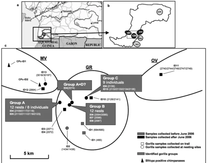
FIG. 3. Localization of the Campo Ma’an Reserve (a) and the six different collection sites (b). Black circles represent the three sites where new
SIVgor-infected gorillas were discovered (MV, GR, and OV). (c) The positions of the 13 identified SIVgor-positive gorillas at the MV, GR, and OV areas within
the CP site are shown in detail. Each individual was identified by microsatellite analysis and is represented by an ID, corresponding to CPg-IDx in Table 3. The
numbers in parentheses correspond to the samples collected from each individual. Previously collected samples are represented with striped gray symbols, and
new samples are shown with black symbols. Circles represent samples collected at nesting sites, while squares represent those collected on trails. The three
different identified groups (A, B, and C) are highlighted in gray boxes. Triangles represent SIVcpz-positive chimpanzees (Table 4).
TABLE 5. SIV infection in wild gorilla and chimpanzee populations from different prospected areas in the CP site, southwest Cameroon
Collection sitea No. of gorilla
samplesb No. of SIVgor antibody-positive samples No. of SIVgor-infected gorillasc No. of chimpanzee samplesb No. of SIVcpz antibody-positive samples No. of SIVcpz-infected chimpanzeesc
CP-GR
111
21
8
5
0
0
CP-OV
83
5
1
15
0
0
CP-MV
119
3
2
95
9
2
CP-ML
7
0
0
49
0
0
CP-ON
116
0
0
12
0
0
CP-NG
37
0
0
4
0
0
Total
473
29
11
180
9
2
aLocations of sites are shown in Fig. 3.
b
Determined by mitochondrial and 12S DNA analysis of fecal DNA and/or species identification in the field (see Materials and Methods).
c
1,194 gorillas were sampled. Table 6 lists the SIVcpzPtt and
SIVgor prevalence rates for each field site as well as for the
entire sampled population. The mean SIVcpzPtt prevalence in
chimpanzees was 5.9% (ranging from 0% to 32%), while the
mean SIVgor prevalence in gorillas was 1.6% (ranging from
0% to 4.6%). Thus, there are three times more
SIVcpz-in-fected chimpanzees than SIVgor-inSIVcpz-in-fected gorillas. Moreover,
positive gorillas were identified at only 3 of the 21 sites
sur-veyed, whereas positive chimpanzees were found at 10 of 25
collection sites. Finally, we did not find sites where SIVgor
infection rates exceeded 5%, although in some gorilla groups
at least one-fourth of group members were infected.
DISCUSSION
The objective of this study was to determine to what extent
gorillas are infected with SIVgor. We thus performed a
com-prehensive survey of wild gorilla populations in west central
Africa and, to a lesser extent, eastern Africa. Our results show
a low and uneven distribution of SIVgor infection in western
lowland gorillas (G. gorilla gorilla) and an absence of infection
in eastern lowland gorillas (G. beringei graueri). We confirmed
the presence of SIVgor at the CP site, where SIVgor was
initially discovered, and identified a new site, DJ, with at least
five infected gorillas. No additional positive gorillas were
iden-tified at the previously reported BQ site and in an area, GB in
Fig. 1, where we suspected the presence of SIVgor infection
based on serological cross-reactivity of a dried blood spot from
a dead male gorilla in 2004. It should be noted that since the
discovery of the first positive gorilla at the BQ site, the gorilla
population decreased in that area due to an anthrax outbreak
in 2004 (12, 18). In addition, intensive poaching and logging
made ape tracking at the GB site extremely difficult. Thus, it is
possible that infected individuals were missed. However, by
combining data from this and previous studies (15, 36, 37), we
have now screened more than 1,200 western lowland gorillas in
Cameroon and adjacent areas and found an overall prevalence
of only 1.6%, which is significantly lower than the overall
prev-alence of SIVcpz in chimpanzees. Moreover, SIVgor is less
evenly distributed, since we found evidence of infection at only
three sites. Thus, SIVgor infection of gorillas is much less
widespread than SIVcpz infection of chimpanzees, at least in
Cameroon. However, the fact that SIVgor infection was
ob-served at three (or possibly four) sites, located 60 to 400 km
apart, and the observation that at some sites SIVgor
preva-lence was almost 5% indicate that this infection is not new in
gorillas. It is also possible that SIVgor infection may be
declin-ing in gorilla populations in Cameroon. However, because of
FIG. 4. Phylogenetic analysis of partial env (gp41; 266 bp) nucleotide sequences of new SIVgor strains (in gray) identified in this survey. The
strains for which the full-length genome has been reported previously (31) are highlighted with a pound sign. The tree was inferred by maximum
likelihood phylogeny (PhyML) with previously characterized SIVcpzPts/SIVgor/HIV-1 strains (26, 31, 36). The support values in black above the
branches are from 1,000 maximum likelihood bootstraps (shown as ‰; only values above 700‰ are shown), and posterior probabilities (shown
as proportions; only values above 0.80 are shown) from nucleotide Bayesian analysis are shown in gray below the branches. The scale bar represents
the number of substitutions per site. Groups A and C correspond to identified groups (Fig. 3). Samples identified in this study are highlighted
in gray.
the sporadic distribution of SIVgor and the unknown
influ-ences of gorilla population decline on the spread of this virus,
it remains possible that SIVgor is still more prevalent in other
parts of the geographical range of western lowland gorillas.
A total of 19 SIV-positive gorillas have now been identified,
and SIVgor strains have been characterized for 11 of them,
from three different locations in southern Cameroon. All
SIVgor strains form a monophyletic lineage closely related to
HIV-1 group P, and the SIVgor/HIV-1O/HIV-1P clade falls
within the SIVcpz radiation. SIVgor strains also cluster
accord-ing to their collection site of origin. This is not surprisaccord-ing, since
the movement of gorilla groups, like that of chimpanzees, is
influenced by major geographical barriers (15). The
impor-tance of these geographical barriers for the spread of SIVgor is
particularly obvious in the Campo Ma’an Reserve, where we
found a concentration of positive gorillas in the center of the
park but not at other sites separated by the Ntem river.
Al-though our sampling size of positive gorillas is limited, we did
not observe cocirculation of divergent SIVgor strains at single
field sites. This differs from our observations in chimpanzees,
where we previously reported cocirculation of genetically
highly diverse SIVcpz strains within a single site (16, 37).
The newly identified SIVgor strains confirm that gorillas
acquired their infection after a single cross-species
transmis-sion from chimpanzees, followed by intraspecies transmistransmis-sions
within the gorilla population (31). Our data are also in line
with previous calculations of the most recent common ancestor
(MRCA) of the current SIVgor clade, which estimated that
SIVgor strains started to diverge at least 100 to 200 years ago
(31). Given our new data, it is clear that cross-species
trans-missions from chimpanzees to gorillas are rare events. Gorillas
do not harbor local SIVcpzPtt strains, and thus far, eastern
gorillas seem to be free of SIVgor infection. This also
corre-sponds with observations that encounters between gorillas and
chimpanzees are rare and seem to be primarily nonaggressive
(28). Western lowland gorillas and central chimpanzees are
sympatric species. Some fruits are eaten by both species, but
they differ in their foraging strategies (17, 23, 33, 40) as well as
in their choice of nesting sites (6, 29, 34). This has led to niche
differentiation, probably to avoid competition. However, it
cannot be excluded that physical encounters between
chimpan-zees and gorillas, possibly involving biting or other forms of
aggression, occurred in the past. Moreover, sharing the same
habitat could also lead to indirect contact with the virus of the
other species, e.g., during disease outbreaks such as anthrax,
Ebola fever, etc. (39).
Although we have now screened a substantial number of
chimpanzees in Cameroon (
⬃649 individuals), we have not
identified SIVcpzPtt strains that are so closely related to
SIVgor or HIV-1 group O or P as to constitute their source. It
is thus possible that the ancestral SIVcpz lineage that gave rise
to SIVgor has become extinct or that the gorillas or
chimpan-zees that harbor the ancestors of group O are located in areas
not yet sampled. HIV-1 group P is the closest lineage to
SIVgor and is most likely the result of a cross-species
trans-mission from gorillas to humans; however, the actually
identi-fied SIVgor strains are not the precursors of group P, and it
cannot be excluded that the ancestor of group P still circulates
in chimpanzees, which transmitted it directly to humans. The
actual SIVgor strains are also still too divergent to be the direct
ancestors of HIV-1 group O.
Our data from the CP site indicate that SIVgor is transmitted
both within and between different groups of gorillas. The exact
modes of virus spread are not known, but the presence of
phy-FIG. 5. Locations of study sites of wild chimpanzees and gorillas from this and previous surveys in Cameroon. Red and black circles indicate
sampling sites where gorillas and chimpanzees were sampled, respectively. Red filled circles indicate sites where SIVgor-positive gorillas were
identified, and black filled circles indicate sites where SIVcpzPtt-positive chimpanzees were identified. Circles with half red and half black indicate
sites where SIVgor-infected gorillas and SIVcpzPtt-infected chimpanzees were identified. MF, TK, MP, and WE are located in the range of Pan
troglodytes vellerosus, while all other sites are located in the range of Pan troglodytes troglodytes. TK was located in the area of the cross-river gorillas
logeographic clusters of SIVgor similar to those of SIVcpzPtt
in chimpanzees suggests a very similar epidemiology.
Interest-ingly, we observed a predominance of SIVgor infection in
females, and whether this is related to a sample bias and/or the
social organization of gorilla groups needs to be studied
fur-ther. Western lowland gorillas live in social breeding groups
usually formed by one silverback male, three adult females (on
average), and their offspring (7, 9, 20, 24). Before reaching
sexual maturity, male gorillas leave their natal group and go
through a bachelor stage that can last several years (9, 19). In
the field, it is easier to localize a group of gorillas than single
individuals. However, the social organization of gorilla groups
and the presence of different groups in a 15- to 20-km
2area
clearly provide opportunities for SIV to spread both within and
between neighboring communities, as shown here and as
con-firmed by a follow-up survey in 2009, where eight new
SIVgor-infected individuals were identified in the CP-GR area (C.
Neel and L. Etienne, unpublished results).
It was recently shown that SIVcpz infection has a negative
impact on the health reproduction and survival of chimpanzees
in the wild (16). Phylogenetic analysis revealed that gorillas
acquired SIVs by cross-species transmission from
chimpan-zees. The lower prevalence and lower genetic diversity among
circulating SIVgor strains are consistent with a more recent
infection in gorillas but could also be an indication that SIVgor
has a negative impact on the life span of infected gorillas and
their ability to propagate virus spread.
Importantly, gorillas and chimpanzees are highly
endan-gered species which continue to be hunted, especially in west
central Africa, and remain a potential source of human
infec-tions (21). SIVs from apes are now known to have crossed the
species barrier to humans on at least four occasions in west
central Africa (14, 26). Given the recent discovery of the
HIV-1 group P lineage in a Cameroonian woman in France, it
would not be surprising if additional cases of SIVcpz or SIVgor
cross-species transmissions had occurred, especially in
geo-graphic regions where these viruses are most prevalent and
where hunting pressure is high. As shown for the group P
virus-infected patient, SIVcpz/SIVgor infections are likely to
be detected by commercial HIV antibody screening assays.
However, HIV screening in Africa is still suboptimal, and
se-rological tests with low sensitivities for group M, and especially
for HIV-1 group O, are still used in this part of the world (1,
10, 25, 42). In addition to the use of assays with low
sensitivi-ties, poor serological results are also related to the high
turn-over of trained personnel, suboptimal storage conditions,
fail-ure of the cold chain during transport, use of tests after the
expiration date, etc. (3). It is thus possible that in regions
TABLE 6. SIV infection in wild chimpanzee and gorilla populations from different field sites in this and previous surveys in Cameroon
Collection sitea No. of chimpanzee samplesb No. of SIVcpz antibody-positive samples No. of SIVcpz antibody-positive chimpanzeesc No. of SIVcpz PCR-positive chimpanzeesc SIVcpz prevalence in chimpanzees (95% CI)d No. of gorilla samplesb No. of SIVgor antibody-positive samples No. of SIVgor antibody-positive gorillasc No. of SIVgor PCR-positive gorillasc SIVgor prevalence (%) in gorillas (95% CI)d
WE
25
0
0
0
0.0 (0.0–23.1)*
1
0
0
0
—
MT
81
10
2
2
5.4 (1.0–16.0)*
0
—
DG
29
0
0
0
0.0 (0.0–20.3)*
25
0
0
0
0.0 (0.0–22.8)
DP
160
17
4
4
4.7 (1.8–11.5)
16
0
0
0
0.0 (0.0–29.9)
BQ
142
0
0
0
0.0 (0.0–4.8)
58
1
1
1
3.2 (0.6–16.2)
EK
46
6
4
4
16.3 (6.4–34.7)
12
0
0
0
0.0 (0.0–39.0)
BB
34
0
0
0
0.0 (0.0–16.4)*
7
0
0
0
—
MB
101
31
17
16
31.6 (20.7–44.8)
119
0
0
0
0.0 (0.0–5.8)
LB
38
5
5
4
24.7 (11.2–46.9)
207
0
0
0
0.0 (0.0–3.4)
CP
232
9
2
2
1.6 (0.4–5.7)
525
34
13
7
4.6 (2.7–7.8)
TK
1
0
0
0
—
21
0
0
0
0.0 (0.0–25.9)
MF
39
0
0
0
0.0 (0.0–15.5)
0
MP
15
0
0
0
0.0 (0.0–32.5)
0
MG
24
0
0
0
0.0 (0.0–22.8)
0
KG
15
0
0
0
0.0 (0.0–32.5)
15
0
0
0
0.0 (0.0–32.5)
SL
44
5
1
1
4.3 (0.8–21.0)
0
BM
38
2
1
1
4.9 (0.9–23.6)
1
0
0
0
—
NK
8
0
0
0
—
25
0
0
0
0.0 (0.0–22.8)
LM
62
0
0
0
0.0 (0.0–10.4)
293
0
0
0
0.0 (0.0–2.4)
MM
0
152
0
0
0
0.0 (0.0–4.5)
GB
4
0
0
0
—
85
0
0
0
0.0 (0.0–7.9)
KK
1
0
0
0
—
18
0
0
0
0.0 (0.0–27.8)
DJ
17
4
1
1
11.0 (2.0–43.5)
206
13
5
3
4.6 (0.9–7.7)
DI
55
0
0
0
0.0 (0.0–11.7)
0
AL
0
46
0
0
0
0.0 (0.0–13.3)
BY
1
1
1
0
—
147
0
0
0
0.0 (0.0–4.7)
DD
5
0
0
0
—
260
0
0
0
0.0 (0.0–2.6)
Total
1,217
90
38
35
5.9 (4.3–7.9)
2,239
48
19
12
1.6 (1.0–2.4)
aLocations of sites are shown in Fig. 5.
b
Determined by mitochondrial and 12S DNA analysis of fecal DNA and/or species identification in the field (see Materials and Methods).
c
Determined by microsatellite analysis (see Tables 3 and 4).
d
CI, confidence interval. —, prevalence rates were not calculated because the number of samples collected was too small.ⴱ, SIVcpzPtt prevalence rates for these sites have been reported previously.