Université de Montréal
Role of NOX2 and DUOX2 in the
antiviral airway responses
par
Karin Fink
Département de Biochimie
Faculté de Médecine
Thèse présentée à la Faculté de Médecine
en vue de l’obtention du grade de doctorat en Biochimie
Janvier, 2013
© Karin Fink, 2013
Université de Montréal
Faculté des études supérieures et postdoctorales
Cette thèse intitulée:
Role of NOX2 and DUOX2 in the antiviral airway responses
présentée par:
Karin Fink
a été évaluée par un jury composé des personnes suivantes :
Nikolaus Heveker, président-rapporteur
Nathalie Grandvaux, directeur de recherche
Guy Lemay, membre du jury
Albert van der Vliet, examinateur externe
Edward Bradley, représentant du doyen de la FES
Abstract
The mucosal linings of the airways are constantly exposed to an array of microbial pathogens. During the course of respiratory viral infection, Airway epithelial cells (AEC) actively participate in the innate antiviral immune response by limiting the spread of respiratory viruses and by fostering a proinflammatory environment that attracts and activates players of the immune system. A key step in the establishment of the antiviral and proinflammatory state is the activation of Transcription Factors (TFs), such as Nuclear Factor (NF)-κB and Interferon Regulatory Factor 3 (IRF-3), which regulate the expression of antiviral and proinflammatory cytokines.
For the efficient functioning of these events, the signaling pathways involved underlie strict regulatory mechanisms. Recent data suggest that Reactive Oxygen Species (ROS), which are produced upon viral infection, are able to regulate these intracellular signaling pathways. One important source of ROS is the NADPH oxidase (NOX) family of enzymes, which is composed of NOX1-5 and Dual Oxidase (DUOX) 1 and DUOX2. The aim of our study was to identify the NADPH oxidase(s) that regulate(s) antiviral and proinflammatory mechanisms following infection of AEC with Respiratory syncytial virus (RSV), which causes major human lower respiratory tract complications, and Sendai virus (SeV), a non pathogenic virus.
During the course of our studies we identified that NOX2 is a key molecule in the early proinflammatory response to RSV and SeV infection. We demonstrate that NOX2 is necessary for the activation of NF-κB. Consequently, NOX2 impacts on the proinflammatory cytokine secretion upon AEC infection. Further, we observed that expression of the ROS-generating NADPH oxidase DUOX2 is strongly increased following infection of AEC with SeV. We identified that DUOX2 induction requires the synergistic stimulation by IFNβ and TNFα. Importantly, DUOX2 exhibited ROS-dependent antiviral action. We identified that DUOX2 was necessary for sustaining the levels of late antiviral cytokines IFNβ and IFNλ.
When AEC were infected with RSV, DUOX2 expression was barely detectable. Our data reveal that RSV has developed an evasion mechanism to counteract DUOX2 induction likely contributing to RSV pathogenicity.
In conclusion, our work demonstrates for the first time the specific implication of NOX2 and DUOX2 in the antiviral and proinflammatory response to respiratory virus infection.
Key words:
Airways, virus, innate immunity, cytokines, interferons, Reactive Oxygen Species, NADPH oxidase.
Résumé
Les voies respiratoires sont exposées à une panoplie de pathogènes. Lors d’une infection virale respiratoire les cellules qui recouvrent ces voies participent activement à la défense immunitaire contre ces derniers en limitant la propagation du virus et en engendrant une réponse proinflammatoire. Un évènement clef dans ces processus est l’activation des facteurs de transcription, notamment le « Nuclear Factor » (NF)-κB et l’« Interferon Regulatory Factor -3 » (IRF-3), qui régulent l’expression des cytokines antivirales et proinflammatoires.
Des données récentes démontrent que les dérivés actifs de l’oxygène (ROS), produits suite à une infection virale, ont la capacité de réguler les voies de signalisation enclenchées par NF-κB et IRF-3. Une source importante de ROS est la famille de NADPH oxydases (NOX), qui contient les membres NOX1-5 et DUOX1 et 2. L’objectif de notre étude était d’identifier la NOX qui régule les mécanismes antiviraux et proinflammatoires suite à l’infection avec le virus respiratoire syncytial (RSV), qui cause des complications respiratoires majeures, et le virus Sendai (SeV), un modèle viral non-pathogène.
Nos travaux ont permis d’identifier que NOX2 est une molécule clef dans la réponse proinflammatoire suite à l’infection virale. Plus spécifiquement, NOX2 est important pour l’activation de NF-κB et la sécrétion des cytokines régulées par ce dernier. De plus, nous avons observé une forte augmentation de la présence de DUOX2 dans les cellules de voies respiratoires humaines infectées par SeV. Une étude plus approfondie nous a permis de caractériser qu’une synergie entre deux cytokines secrétées lors de l’infection, soit l’interféron (IFN) β et le TNFα, est responsable de l’induction de DUOX2. Nous avons aussi découvert que DUOX2 confère une activité antivirale et est nécessaire pour maintenir les taux des cytokines antivirales tardives IFNβ et IFNλ.
Lors d’une infection avec RSV, l’induction de DUOX2 n’est pas détectable. Nous avons mis en évidence que RSV interfère avec l’expression de DUOX2 ce qui pourrait suggérer sa pathogénicité.
En conclusion, nos travaux démontrent pour la première fois une implication spécifique des NADPH oxydase NOX2 et DUOX suite aux infections virales respiratoires.
Mots clefs :
Voies aériennes, virus, immunité innée, cytokines, interférons, dérives actifs de l’oxygène, NADPH oxydase.
Table of Contents
Abstract i Résumé iii Table of Contents v Table index ix Figure index xi Abbreviations xiii Acknowledgements xix 1 INTRODUCTION 11.1 The Paramyxoviridae – Respiratory viruses of etiologic importance 3
1.1.1 The Paramyxoviridae family 3
1.1.2 RSV and SeV virion structure 5
1.1.3 RSV and SeV genome organization 6
1.1.4 RSV and SeV replication cycle 7
1.1.4.1 Virus attachment and entry 7
1.1.4.2 Viral mRNA synthesis 8
1.1.4.3 Genome replication 9
1.1.4.4 Virion assembly and virion release 9
1.2 The airway response to respiratory virus infection 12
1.2.1 Airway physiology at the site of infection 12
1.2.2 Airway defense mechanisms 14
1.2.3 The AEC response to viral infection 19
1.2.3.1 Viral recognition by Retinoic acid inducible gene-I (RIG-I)-like receptors (RLRs) 20
1.2.3.2 IRF-3 and NF-kB activation in RLR signaling lead to antiviral and
proinflammatory cytokines production 22
1.2.3.4 The IFN-induced JAK-STAT pathway 26 1.2.3.5 Establishment of the antiviral state by ISGs 27 1.2.3.6 The TNF receptor 1 (TNFR1) signaling pathway 30 1.2.4 Viral modification and evasion of antiviral immunity 33
1.2.4.1 General viral evasion mechanisms 33
1.2.4.2 Blocking IFN production 34
1.2.4.3 Evasion of the IFN-mediated induction of the antiviral state 34 1.2.4.4 Interference with innate and adaptive immune responses 35
1.3 RSV-induced pathology – a multifaceted syndrome 37
1.3.1 RSV-induced airway obstruction 39
1.3.2 Inflammation and infiltration of innate immune players 39
1.3.3 Adaptive immune responses 41
1.4 ROS and their role in regulation of cellular signaling pathways 43
1.4.1 The nature of ROS 44
1.4.2 The origin of ROS 45
1.4.3 The NADPH oxidase (NOX) family of enzymes 47
1.4.3.1 NADPH oxidase composition 50
1.4.4 The NOX2 NADPH Oxidase 51
1.4.4.1 Mechanism of ROS generation by NOX2 51
1.4.4.2 Regulation of NOX2 expression 53
1.4.5 The DUOX2 NADPH oxidase 54
1.4.5.1 Mechanism of ROS generation by DUOX2 54
1.4.5.2 Regulation of DUOX2 and DUOXA2 expression 56
1.4.6 Mechanisms of ROS action 57
1.4.6.1 Oxidative Protein Modification 57
1.4.7 Evidence of ROS production in virus-infected cells 59 1.4.8 Regulation of antiviral signaling pathways by ROS 60 1.4.8.1 Redox regulation of the NF-kB signaling pathway 61 1.4.8.2 Redox regulation of the interferon response 64
1.4.9 NADPH oxidase-dependent regulation of NF-kB 65
1.5 NADPH oxidases in antimicrobial lung defenses 68
1.5.1 The many roles of DUOXs in the airway epithelial immunity 68 1.5.1.1 DUOXs contribute to airway antimicrobial defense 68
1.5.1.2 DUOXs and airway inflammation 71
1.5.1.3 NOX/DUOX enzymes as regulators of airway epithelial integrity 72
1.5.2 The role of NOX2 in airway immunity 73
1.5.2.1 NOX2 controls inflammation 74
1.5.2.2 NOX2, a pleiotropic regulator of innate immunity 76 1.5.2.3 Polyvalent functions of NOX2 in lung structural cells 77
1.6 Study hypothesis and strategy 79
2 RESULTS 81
2.1 Dual role of NOX2 in Respiratory Syncytial virus- and Sendai virus-induced activation of NF-κB in airway epithelial cells 83
2.1.1 Context and resume of results 83
2.1.2 Author contributions 84
2.1.3 Article 86
2.2 IFNβ/TNFα synergism induces a non-canonical STAT2/IRF9-dependent pathway triggering a novel DUOX2 NADPH Oxidase-mediated airway antiviral
response 135 2.2.1 Context 135 2.2.2 Author contributions 136 2.2.3 Article 137 3 DISCUSSION 185 4 CONCLUSION 207
5 BIBLIOGRAPHY 211
6 ANNEX I
Table index
Table I: Examples of members of the Paramyxoviridae family………..4 Table II: NADPH oxidase expression pattern and dependency on regulatory subunits………..49
Figure index
Figure 1: RSV pleiomorphic forms………..5
Figure 2: RSV virion structure……….6
Figure 3: SeV and RSV genome………..7
Figure 4: The Paramyxoviridae replication cycle………..11
Figure 5: Summary of pulmonary physiology and defenses………..16
Figure 6: AEC defense mechanisms against respiratory viruses………...19
Figure 7: The virus-induced IFN response……….29
Figure 8: TNF-induced NF-κB activation……….32
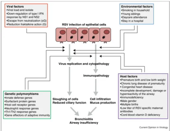
Figure 9: Factors influencing the pathogenesis and clinical disease caused by RSV infection in infants and children……….38
Figure 10: ROS-generating and ROS-involving reactions……….47
Figure 11: NOX2 structure and NADPH oxidase-dependent ROS production……….53
Figure 12: Structural organization of the DUOX2 NADPH oxidase……….56
Figure 13: Redox modification of cysteine residues………..59
Figure 14: NOX2 and DUOX2 in the antiviral airway defense………...208
Abbreviations
ADAM17 A Disintegrin and Metalloproteinase 17 AEC Airway epithelial cells
AIR Autoinhibitory region ALI Air-liquid interface
ARE Apical recycling endosome
ASGM1 Asialo-GM1
ASL Airway surface liquid ATP Adenosine triphosphate ATRA All-trans retinoic acid
BALF Bronchoalveolar lavage fluid
c-Flip Cellular FLICE-like inhibitory protein CARD Caspase recruitment domain
CBP Creb binding protein cDC Conventional dendritic cell
CF Cystic fibrosis
CFTR Cystic fibrosis transmembrane conductance regulator CH Congenic hypothyroidism
cIAP Cellular inhibitor of apoptosis
COPD Chronic obstructive pulmonary disease
COX Cyclooxygenase
DBD DNA binding domain
DC Dendritic cell
DCFDA Dichlorofluorescin diacetate
DD Death domain
DI Defective interfering
ds Double strand
DUOX Dual oxidase 1 DUOXA DUOX activator
EGFR Epidermal growth factor receptor EGFα Epidermal growth factor α EMCV Encephalomyocarditis virus
ENA-78 Epithelial neutrophil activating protein-78 ER Endoplasmatic reticulum
Erk Extracellular signal-regulated kinase ENaC Epithelial sodium channel
F Fusion protein
FAD Flavin adenine dinucleotide
FADD FAS-associated death domain-containing protein FI-RSV Formalin-inactivated RSV vaccine
G Glycoprotein
GAS γ-interferon activated sequence GCD Chronic granulomatous disease
GM-CSF Granulocyte-macrophage colony-stimulating factor Gro-a Growth-regulated oncogene α
HCV Hepatitis C virus
HIV Human immunodeficiency virus HN Hemagglutinin neuraminidase HNE Human neutrophil elastase IAD IRF association domain
ICAM Intercellular adhesion molecule
IFIT1 Interferon-induced protein with tetratricopeptide repeats 1
IFN Interferon
IFN-lR1 IFNλ receptor 1 IFNAR IFNα receptor
IKK IκB kinase
IL Interleukin
IP-10 IFNγ-induced protein IP3 Inositol triphosphate
IRF Interferon regulatory factor ISG Interferon stimulated gene
ISGF3 Interferon stimulated gene factor 3 ISRE Interferon stimulated response element JAK1 Janus kinase 1
JUNK C-Jun-NH2-terminal kinase
kb kilobases
leRNA Leader RNA
LGP2 Laboratory of genetics and physiology 2
LOX Lipoxygenase
LPO Lactoperoxidase LPS Lipopolysaccharide
LUBAC Linear ubiquitin chain assembly complex
M Matrix protein
MDA5 Melanoma differentiation-associated gene 5 MeV Measles virus
MIP-1α Macrophage inflammatory protein-1a MMP9 Matrix Metalloprotease 9
mRNA Messenger ribonucleic acid
MSK-1 Mitogen and stress related kinase 1
Muc Mucin
MuV Mumps virus
N Nucleocapsid protein
NADPH Nicotinamide Adenine Dinucleotide Phosphate Hydrogen NAP1 NAK-associated protein 1
NEBs Neuroepithelial bodies NEMO NF-κB modulator
NK Natural Killer
rich repeat containing X1
NOD Nucleotide-binding oligomerization domain
NOX NADPH oxidase
NS Non structural protein
OAS1 2’, 5’-oligoadenylate synthetase OSCN- Hypothiocyanate
P Phosphoprotein
PAMP Pathogen associated molecular pattern pDC Plasmacytoid dendritic cell
PI3K Phosphoinosite 3 kinase PIV Human parainfluenza virus PKA Protein kinase A
PKAc cAMP dependent protein kinase A PKC Protein kinase C
PKR Protein kinase R
PMA Phorbol-12-myristate-13-acetate PRR Pathogen recognition receptors PTP1B Protein tyrosine phosphatase 1B
RANTES Regulated and normal T cell expressed and secreted
RD Repressor domain
RHD Rel homology domain RNA Ribonucleic acid
RIG-I Retinoic acid inducible gene-I RIP1 Receptor interacting protein 1 RLR RIG-I-like receptor
RNAi RNA interference
RNS Reactive nitrogen species RSV Respiratory syncytial virus
RV Rhinovirus
SCN- Thiocyanite
SeV Sendai virus
SH Short hydrophobic protein SH3 C-terminal Src homology 3
SHP-2 Src-homology 2 containing protein phosphatase 2 SOCS Suppressors of cytokine signaling
SOD Superoxide dismutase SP Surfactant proteins
ss Single-strand
STAT Signal transducer and activator of transcription TAB TAK1-binding protein
TACE TNFα-converting enzyme TAD Transactivation domain
TAK1 Transforming growth factor β-activated kinase 1 TANK TRAF family member associated NF-kB activator TBK1 TANK-binding kinase 1
TCR T cell receptor TF Transcription factor TGFα Tumor growth factor α
Th T helper
TJ Tight junction
TLR Toll-like receptor
TM Transmembrane domain
TNFa Tumor necrosis factor α
TNFR1 Tumor necrosis factor receptor 1
TRADD Tumor necrosis factor receptor type 1-associated death domain protein
TRAF TNF receptor associated factor trRNA Trailer RNA
TSS Transcription star site Tyk2 Tyrosine kinase 2
VEGF Vascular endothelial growth factor VEGF Vasoactive Endothelial Growth Factor vRNAP Viral RNA-dependent RNA polymerase WHO World health organization
α Alpha β Beta γ Gamma ε Epsilon κ Kappa λ Lambda ω Omega
An meine zwei cocos, Mattias und Nathaniel
Man sieht nur mit dem Herzen gut,
das Wesentliche ist für die Augen unsichtbar
.
Acknowledgements
I am truly thankful to all the individuals that have contributed to and enriched my Ph.D. studies in so many different ways over the last years. Me sitting here and writing the following sentences would not be possible without you. Thank you! Merci! Danke!
D’abord j’aimerais remercier ma directrice de recherche Nathalie Grandvaux. Merci pour la confiance que tu m’as attribuée en étant la première étudiante de ton laboratoire. Merci pour les heures que tu as passées à m’enseigner tout le savoir nécessaire afin d’avancer dans ma recherche et la complétion de mon doctorat.
J’aimerais également remercier les membres du jury. Nikolaus Heveker, Guy Lemay et Albert van der Vliet, merci d’avoir accepté d’évaluer mes travaux de recherché de doctorat.
À tous les membres du laboratoire, merci de m’avoir soutenue pendant ces années, merci d’avoir partagé des heures et des heures au laboratoire avec moi. Si j’avais été toute seule dans ces moments, le travail n’aurait pas été le même! Merci Anton, Alexis et Annick d’avoir été présents pour moi, de m’avoir appris comment parlent les Québécois, de m’avoir fait apprécier plus le hockey. Merci Annick de penser encore à moi quand tu fais des petites activités avec ton trésor. Merci Esperance, pour ton soutien au laboratoire, ton esprit calme et tranquille, pour ton oreille ouverte à mes soucis lors des heures de manips dans la salle de culture. Fabrice, merci de ne m’avoir jamais jugée parce que j’ai plus que 30 ans, je n’ai pas 200 ami(e)s facebook et je ne sors pas à tous les jours de la semaine. Stéfany, ton arrivée au labo a donné beaucoup de plaisir à tout le monde, moi incluse. Merci ! Avec toi toutes les journées sont des bonnes journées, où le soleil brille et nos petits soucies deviennent insignifiants. Merci Julie, tu es sans comparaison la personne la plus agréable à travailler avec. Merci pour ton calme et tous les biscuits chinois bizarres que tu as partagés avec moi. Et il reste à remercier le triumvirat français, Lydie, Virginie et Nicolas. Merci de m’avoir réappris le français de la France. Merci Lydie pour ton élan contagieux au travail, pour toutes tes petites anecdotes sur la vie de la maman d’une
princesse. Virginie, tu es la plus efficace des efficaces au téléphone, j’aimerais t’engager pour régler mes comptes au téléphone. Merci pour tes efforts et conseils. Et Nicolas, qui est extrêmement bon avec les ordinateurs, mais un peu moins bon en pâtisserie, merci pour les bonbons, la crème glacée, les chocolats. Grace à toi je n’ai jamais eu des problèmes à garder mon poids.
Merci aux collègues des autres laboratoires. Il y a Nathalie, qui a été sympathique avec moi dès le début et qui n’a jamais cessé de l’être. Merci, ma meilleure amie de la Rive Sud. Merci aussi à toi, Sandy, co-habitante en diagonal du bureau d’étudiante. Merci pour tes petits 5-10 min que tu m’as consacrées à plusieurs reprises lorsque j’avais besoin de me changer les idées. Merci les filles pour des lunchs super agréables.
Merci à Malek, qui a partagé le même chemin que moi depuis le début, qui m’a encouragée à des diverses moments avec des délicieux Baklavas. Merci aussi à Amal, tant des conseils tu m’a donnés par rapport à tout. Merci Mohammed, avec toi on était jamais seule au labo, parce que tu travailles trop fort. Loubna, depuis que tu es partie ce n’est pas la même chose. Merci pour tout le temps que tu as pris pour moi sur le Rotorgene.
Merci aussi aux Manons (Bourcier et Livernois) pour toute votre aide organisationelle et vos histoires parfois hilarantes aux heures du lunch.
I also would like to thank my dearest and best friend Anna, whom I know since my very first days in Montreal. Thank you so much for having shared so many different steps during the last years with me: immunology benchmate, party friend, roommate, work colleague, friend, self-proclaimed godmother of my babies, park hang-out friend. Hopefully you have enjoyed all this as much as I did.
J’aimerais aussi remercier toutes les personnes qui se sont occupées de mon petit monstre Mattias. D’avoir la confiance de le laisser entre vos mains, de savoir qu’il est bien avec vous, a énormément aidé sur mon cheminement de travail. Merci Juliana! Merci aux éducatrices du CPE Viroulu!
Myriam et André, les beaux grands parents de mes enfants, merci également pour votre aide, votre soutien, votre écoute lors des dernières années de mes études. Merci pour toutes les journées où vous m’avez dépannée à garder Mattias s’il fallait aller au labo. Merci pour toutes les corrections que vous avez faites sur mes textes.
Mama und Papa, ohne eure unendliche Zuversicht in meine Ideen und Pläne, eure Unterstuetzung in all meine Projekte hätte ich es wohl nie soweit geschafft. Danke, dass ihr mich immer auf meinen Wegen, wenn sie auch recht weit von euch entfernt waren, begleitet habt.
Ian, merci pour tous tes encouragements, tes conseils, les discussions scientifiques et autres. Merci de toujours avoir compris ce que c’est faire un doctorat, faire de la recherche, avec ses hauts et ses bas. Ce travail n’aurait pas pu être fait sans toi. J’aimerais être un jour tellement importante pour toi comme tu l’étais pour moi lors des dernières années.
Mattias und Nathaniel, auch wenn es euch vielleicht nicht immer leicht gefallen ist mich bei diesem Studium zu unterstuetzen, mich zu vielen Stunden mit Zellen, Mikroskop, Viren und der Schreiberei zu teilen, möchte ich dass ihr wisst wie sehr ich euch für eure unendliche Geduld dankbar bin. Letztendlich habt ihr mir die Kraft gegeben in den allerschwierigsten Momenten nicht einfach alles aufzugeben. Ihr seid meine Welt, vielen vielen Dank.
1.1 The Paramyxoviridae – Respiratory viruses of etiologic
importance
The Paramyxoviridae family contains some of the most severe respiratory viruses, whose infection might lead to respiratory tract disease. One of these is Respiratory Syncytial Virus (RSV). The first chapter of this doctoral thesis aims to introduce notions of the Paramyxoviridae family of viruses in terms of their family organization, virion structure, genomic organization and replication cycle. The focus will be on characteristics related to the two relevant viruses used in this study, RSV and Sendai virus (SeV).
1.1.1 The Paramyxoviridae family
Paramyxoviridae viruses are enveloped, negative-sense, single-stranded RNA viruses
of the Mononegavirales order. The family is subdivided into two subfamilies, the Paramyxovirinae and the Pneumovirinae. The Paramyxovirinae subfamily divides itself into seven genera: Respirovirus, Rubulavirus, Morbillivirus, Avulavirus, Henipavirus, Acquaparamyxovirus and Ferlavirus (Knipe et al., 2001) (Table 1). Some important etiologic agents of this subfamily include different strains of Human Parainfluenza Virus (PIV; Respirovirus and Rubulavirus genera), Mumps virus (MuV; Rubulavirus genus), and Measles virus (MeV; Morbilivirus genus). Sendai virus (SeV), a member of the Respirovirus genus, is a murine parainfluenza virus type 1. It infects rodents and is believed to be the leading cause of pneumonia in mice (Faisca and Desmecht, 2007). In the laboratory, SeV is a prominent model in the study of the airway response to respiratory infections. The second subfamily, termed Pneumovirinae, is divided into two genera, Pneumovirus and Metapneumovirus, among which Respiratory Syncytial Virus (RSV; genus Pneumovirus) is an important etiologic agent (Table 1). RSV causes about 60% of all lower respiratory tract
infections in infants and is additionally a major cause for severe respiratory morbidity and mortality in elderly and immunocompromised individuals (Hall, 2001). It is also becoming increasingly acknowledged that RSV infection can lead to respiratory complications in healthy adults (Walsh, 2011). The World Health Organization (WHO) estimates that RSV is responsible for 64 million clinical infections and 160,000 deaths annually worldwide (Falsey et al., 2005). In the United States, RSV is the most frequent cause for hospitalization in infants (Leader and Kohlhase, 2002).
Table I: Examples of members of the Paramyxoviridae family Family Paramyxoviridae
Subfamily
Paramyxovirinae
Genus Rubulavirus Mumps virus (MuV)
Human parainfluenza virus type 2, type 4a and 4b Genus Avulavirus Newcastle disease virus (NDV)
Genus Respirovirus Sendai Virus (SeV)
Human parainfluenza virus type 1 and type 3 (hPIV1/3)
Genus Henipavirus Hendra virus (HeV) Nipah virus (NiV) Genus Morbillivirus Measles virus (MeV)
Subfamily Pneumovirinae
Genus Pneumovirus Human respiratory syncytial virus (hRSV) Genus Metapneumovirus Human metapneumovirus (hMPV)
1.1.2 RSV and SeV virion structure
Paramyxoviridae are pleiomorphic in structure – both spherical and filamentous
forms of the virus have been observed (Figure 1) (Bachi and Howe, 1973).
Paramyxoviridae contain a lipid bilayer envelope, which is derived from the plasma
membrane of the host cell during viral budding (Harrison et al., 2010). Inserted into this membrane envelope are two surface proteins: Glycoprotein (G) and Fusion protein (F) for RSV; and Hemagglutinin Neuraminidase (HN) and F protein for SeV (Figure 2). These membrane proteins serve two principal functions: first, viral attachment, and second, viral entry (Chang and Dutch, 2012). It is noteworthy to mention that RSV possesses a third membrane protein, the short hydrophobic protein (SH), which does not have a function in attachment or fusion. Inside the virion lies the nucleocapsid core, which contains the RNA genome surrounded by the Nucleocapsid protein (N), complexed to the Phosphoprotein (P) and the Large protein (L), the latter two forming the viral RNA-dependent RNA polymerase (vRNAP). Between the envelope and core, just below the inner membrane layer, lies the viral Matrix (M) protein, which is vital in determining virion architecture, and which is released from the core during virus entry. In addition, the viral M protein plays a central role in viral budding (Knipe et al., 2001).
Figure 1: RSV pleiomorphic forms.
1.1.3 RSV and SeV genome organization
The Paramyxoviridae genome is a negative-sense, single-strand genome of 15-19 kb that encodes 6 and 10 viral genes for SeV and RSV, respectively (Figure 3). The genome generally possesses a 3’-extracistronic region of approximately 50 bp, called the “Leader” region, and a 5’-“Trailer” region of 50-161 bp. Between genes, intergenic sequences are found that serve as termination and initiation sequences in mRNA synthesis (Knipe et al., 2001). The SeV genome encodes for the following proteins in a 3’-5’ order: 3’-N-P/C/V-M-F-HN-L-5’. P, C and V proteins are encoded from one P gene whose mRNA has several open reading frames. In contrast, the RSV genome encodes for 11 proteins in the following 3’-5’ order: 3’-NS1-NS2-N-P-M-SH-G-F-M2-L-5’. The M2 gene is transcribed into an mRNA with two overlapping reading frames
Figure 2: RSV virion structure.
Schematic representation of the RSV virion structure. The viral envelope is made up of the plasma membrane into which viral proteins F, G and SH are inserted. Beneath the membrane layer lies the M protein. The nucleocapsid is made up of the viral genome that is tightly encapsidated by N. N is complexed to P and L proteins that make up the vRNAP. Adapted from (Knipe et al., 2001).
resulting in the synthesis of two proteins, M2-1 and M2-2 (Knipe et al., 2001). RSV and SeV also code for accessory proteins that are not present in the infecting virion but have important roles as inhibitors of the host antiviral defense. Notably, NS1 and NS2 proteins of RSV, and V and C proteins of SeV can foster evasion of the antiviral response (Gotoh et al., 2001).
1.1.4 RSV and SeV replication cycle
1.1.4.1 Virus attachment and entry
Before infection of the host cell can occur, virus particles have to attach to their target cells, which brings the virion and host cell into close proximity. Then
Paramyxoviridae entry into the host cell is mediated by fusion of the virus with the
cellular membrane (Figure 4). These steps, attachment and entry, are assured by the viral attachment and fusion proteins G and F for RSV, and HN and F for SeV. In order for attachment to occur, certain viruses will attach to cell surface molecules found on the exterior of the host cell. This is the case for SeV, as SeV HN protein attaches to
Figure 3: SeV and RSV genome.
Schematic representation of the SeV and RSV genome. For explanation, see RSV and SeV genome organization. Adapted from (Knipe et al., 2001).
sialic acid containing cell surface glycoproteins and glycolipids (Villar and Barroso, 2006). RSV, on the other hand, attaches to glycosaminoglycans containing the disaccharide heparan sulfate and chondroitin sulfate via the G protein (Feldman et al., 1999; Krusat and Streckert, 1997). After attachment, the F protein initiates the fusion process, whereby the nucleocapsid is delivered into the cytoplasm. A recent publication has demonstrated that cholesterol-rich lipid raft domains are important for RSV entry into airway epithelial cells (AEC) (San-Juan-Vergara et al., 2012). Their viral entry can occur either through receptor-mediated endocytosis or direct penetration at the plasma membrane. Both processes have been described in the case of SeV and RSV infection (Kolokoltsov et al., 2007; Rasmusson et al., 1998; Srinivasakumar et al., 1991). In the context of RSV fusion with the plasma membrane, the protein nucleolin has recently been decribed as a novel fusion receptor as it interacts with RSV-F at the cell surface and is essential for efficient RSV infection (Tayyari et al., 2011).
1.1.4.2 Viral mRNA synthesis
Once the virion is released into the cytoplasm, the next strategic step in the virus cell cycle is the generation of viral proteins that will provide building material for new virions. Some viral proteins also serve to interfere with the host metabolism in order to increase the potential for efficient viral replication. The Paramyxoviridae life cycle is entirely cytoplasmic (Figure 4). The virus brings its own vRNAP, which is a complex consisting of a tetramer of P proteins and one L protein. P proteins serve to recruit L proteins to the viral genome, which is strongly encapsidated by the N protein (Cowton et al., 2006). This polymerase transcribes the incoming negative sense genome into 5’-capped, 2-O-methylated and 3’-polyadenylated mRNAs, an activity that is assured by the L protein (Liuzzi et al., 2005). All viral RNA synthesis begins at the 3’ end of the genome, where a cis-acting promoter sequence initiates the synthesis of short, non-coding leader RNAs. At the beginning of the viral cycle, vRNAP is restricted to the production of these leader RNAs and thereafter mRNAs (Cowton et al., 2006). Scrolling down the viral genome, vRNAP terminates and reinitiates at each gene junction, thereby generating an mRNA transcript for each encoded gene. However,
vRNAP does not always reinitiate mRNA synthesis at the next gene junction, as it has a tendency to dissociate from its template at these sites. This in turn leads to a decreasing gradient of mRNA abundance from the genome’s 3’ end onward. This gradient of mRNA results in defined quantities of the viral N protein, which determine the balance between mRNA synthesis and genome replication. Once a sufficient amount of N protein is present in the cell, viral mRNA synthesis stops and vRNAP engages into the viral replication process (Knipe et al., 2001). It is noteworthy to mention that the RSV accessory M2-1 protein interacts with P and viral RNA. M2-1 has been shown to be an essential transcription elongation factor, because in its absence RSV does not transcribe beyond the NS1 and NS2 genes (Fearns and Collins, 1999). M2-2 is non-essential for viral growth but may be involved in the regulation of balancing viral transcription and viral replication (Bermingham and Collins, 1999).
1.1.4.3 Genome replication
The Paramyxoviridae negative-sense genome replicates via an intermediary full-length complementary copy called the antigenome. For antigenome synthesis, vRNAP copies the negative sense genome but ignores all of the junctional signals and editing sites. It synthesizes an exact complementary positive-sense antigenome that becomes immediately bound by N proteins. This antigenome serves as a template for genome replication. The 3’ end of the antigenome contains a promoter that is recognized by vRNAP, which initiates synthesis of the genome in a manner similar to antigenome synthesis, i.e. ignoring all gene junctions (Cowton et al., 2006). Under conditions where sufficient unassembled N protein is present, encapsidation of the nascent genome prevents termination and leads to the synthesis of an encapsidated minus-strand genome.
1.1.4.4 Virion assembly and virion release
Once the viral genome has replicated, sufficient building blocks for new virion assembly have been synthesized and nucleocapsid assembly is terminated. Virion
assembly can therefore begin and will culminate in the release of new virions. Nucleocapsids are assembled in two steps: first, free N subunits associate with the genome to form the helical ribonucleoprotein structure of viral genomic RNA and N; second, the RNA:N complex associates with the P and L protein complex. Virion assembly then takes place at the cell surface (Harrison et al., 2010). The viral integral membrane proteins are synthesized in the ER and undergo maturation or glycosylation in the Golgi complex before being transported through the secretory pathway and integrated into the cellular membrane. The viral M protein associates with these transmembrane glycoproteins at locations where budding occurs. The clear mechanism of assembly at the membrane is still unknown but the viral M protein is thought to play a major role in bringing the nucleocapsid to the plasma membrane to finalize the formation of the budding virion (Ghildyal et al., 2006). In this process, viral proteins also interact with host cellular factors and these interactions will drive viral budding. For instance, M protein, as well as SeV C protein, interacts with cellular molecules of the vacuolar sorting pathway to organize the assembly of the nascent virion (Irie et al., 2007; Sakaguchi et al., 2005). For SeV and RSV it has been shown that viral budding preferentially takes place at apical side of AEC (Zhang et al., 2002). In this context, the cellular apical recycling endosome (ARE) plays an important role (Brock et al., 2003).
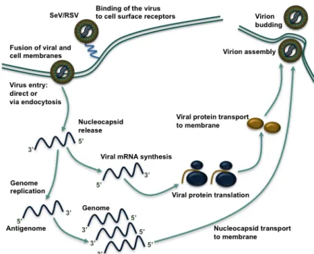
Figure 4: The Paramyxoviridae replication cycle.
Following viral entry, the negative-sense viral genome serves as a template for viral mRNA synthesis by the vRNAP. Viral protein synthesis then occurs. When enough building blocks have been generated, viral genome replication begins. The negative-sense viral genome is generated by the vRNAP via a positive-negative-sense antigenome intermediate. Viral genomes are rapidly encapsidated and transported to virion assembly sites at the membrane. Following assembly, virions are released by budding from the plasma membrane.
1.2 The airway response to respiratory virus infection
The airways are a complex barrier that separates the exterior environment with its hazardous components from our body. The following chapter will introduce airway physiology and airway defense strategies that serve to minimize or combat respiratory infections. These mechanisms range from general airway cell defense mechanisms to the specific detection of viral pathogens, leading to the initiation of a more global airway immune response. Lastly, Paramyxoviridae evasion of these mechanisms and resulting pathology will be discussed.
1.2.1 Airway physiology at the site of infection
The classical lung anatomy model consists of 24 generations of dichotomously branching airway tubes, where each tube divides into two smaller airways. At each division, the diameter of the airway decreases (Weibel, 1963). From the trachea to the bronchi (generation 0-10), the airways form a moderately tight pseudo-stratified ciliated columnar epithelium, exhibiting Transepithelial Electric Resistance (TEER) measurements of approximately 300 Ωcm2 (Wijkstrom-Frei et al., 2003). The main purpose of the epithelium in these proximal airways is to filter particles and kill invading microbes. The three main cell types present in this section of the airways are: ciliated cells, goblet cells, and basal cells. The main function of ciliated cells is to propel foreign particles and organisms out of this airway region via the mucociliary clearance mechanism. The goblet cell, which extends from the larger to the smaller bronchi, but is absent in the bronchioles, contains abundant granules for mucus secretion. Basal cells are the stem cell-like progenitor cells of the surface epithelium, and all ciliated and non-ciliated cells in the bronchial mucosa are derived from basal cells. Basal cells can survive mucosal injury, and consequently serve in the reconstruction of the bronchial mucosa (Fischer, 2009). The epithelium is covered with
a layer of airway surface liquid (ASL). The ASL is made up of two phases, the first of which is a periciliar layer of thin fluid film, into which the cilia beat at a rate of 12-16 beats/second (Ng et al., 2004; Rutland et al., 1982; Tomashefski et al., 2008). Glands which are present in the submucosa of the trachea and bronchi, and which contain mucus and serous cells, contribute to the secretion of this mucus layer. The second phase is a high-viscosity upper mucus layer, which originates from submucosal glands, but also from goblet cells. The ASL contains a variety of molecules that serve in airway antimicrobial defense (Ganz, 2002) (Vareille et al., 2011).
The membranous bronchioles are located in the descending airways. Here, the diameter of the airway decreases to about 1 mm (Tomashefski et al., 2008). Membranous bronchioles are lined by ciliated columnar epithelial cells and non-ciliated Clara cells. Clara cells replace the disappearing goblet cells. The Clara cell also contains an abundance of secretory granules that primarily secrete antimicrobial and immunomodulatory surfactant proteins (SP). Due to its function as a reserve and reparatory cell, the Clara cell replaces the basal cell in the bronchioles. The terminal bronchiole leads into the acinus, the functional unit of the lung, which consists of the respiratory bronchiole, the alveolar ducts, the alveolar sac and the alveoli. The alveolar sac and the alveoli are the site of gas exchange. The alveoli are made up of large, thin type I cells, which only account for 40% of the alveolar lining cells, but cover 90% of the alveolar surface; and small cuboidal type II cells, which constitute 60% of the surface cells, but cover only 5% of the alveolar surface (Tomashefski et al., 2008). Besides secreting surfactant, type II cells also function as a reserve cell, as they can mature into type I cells. In the alveolar sac, the epithelium is very tight and has TEER measurements of more than 2,000 Ωcm2 (Fischer et al., 2007). The alveoli are lined with a very thin fluid film and, as a result, antimicrobial factors secreted in this zone are highly concentrated.
For an efficient gas exchange of oxygen and carbon dioxide between air spaces and red blood cells, the alveolar arrangement is ideal. Alveolar interstitial cells are tightly surrounded by capillaries. The endothelial cell and epithelial alveolar type I cell cytoplasm is spread as thinly as possible, which leads to an air-blood distance of approximately 0.6 µm. At any given moment, about 200 mL of blood can be found
within the capillary network that spreads over 126 m2, which is equivalent to a 1.6 mL spread(Starosta et al., 2006) over 1 m2 (Tomashefski et al., 2008). The pulmonary endothelium of the alveolus, which occupies a surface area of more than 140 m2, is the largest and densest in the human body. Endothelial cells are connected to each other by loose junctions, which readily allow the passage of fluids, macromolecules and immune cells into the interstitial compartment (Tomashefski et al., 2008).
Leukocytes are continuously present in the airways, and in normal, non-inflamed lungs the majority of them are macrophages. More specifically, in Bronchoalveolar Lavage Fluid (BALF) of healthy individuals, 94% of all leukocytes were found to be macrophages, followed by lymphocytes (4%), neutrophils (1%), and eosinophils (0.7%). Plasma cells, basophils and mast cells can also be found in these compartments at very low numbers
1.2.2 Airway defense mechanisms
The air we breathe carries many potentially harmful microbes. To protect us against them, the lung harbors an array of defense mechanisms. On the one hand, the composition and tightness of the airways provide anatomic defense mechanisms, due to the presence of tight junction barriers that make it difficult for pathogens to cross into the interstitial space (Bergelson, 2009). The continuous beating of ciliated AECs also contributes to such anatomical defense mechanisms. On the other hand, factors secreted from AECs into the ASL are known to have potent antimicrobial or immunomodulatory functions. These factors include antimicrobial proteins and peptides, which serve to eliminate the invading pathogen (Figure 5) (Tomashefski et al., 2008). Factors like mucins, surfactant proteins, lactoferrin and human β-defensins have been shown to play roles in AEC infection by respiratory viruses, such as RSV (Grover et al., 1997; Kota et al., 2008; LeVine et al., 1999; Vareille et al., 2011). Cytokines and chemokines that in turn attract innate and adaptive immune cells in the fight against an infection are also secreted from AECs. More specifically, via the
secretion of antiviral cytokines, such as type I and III interferons (IFNs), AECs are able to alarm the surrounding cells and signal the presence of a viral pathogen. Via the secretion of proinflammatory cytokines, such as Tumor Necrosis Factor α (TNFα), and chemokines, such as Regulated And Normal T cell Expressed and Secreted (RANTES)/CCL5, into the submucosa, AECs are able to call for “back-up” and recruit and activate players of the innate and adaptive immune response axes, which work together to eliminate infection (Figure 6)(Vareille et al., 2011). The following paragraphs will discuss the nature and function of these cytokines and chemokines in more detail, with an emphasis on their role in respiratory virus infection.
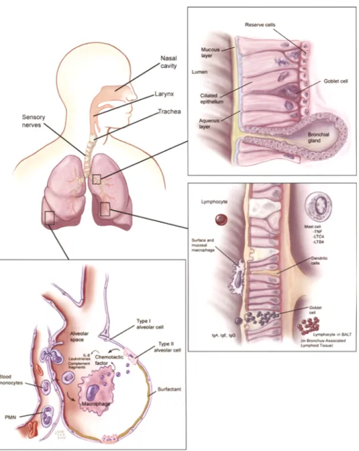
Figure 5: Summary of pulmonary physiology and defenses.
Airway defenses in the trachea and bronchi include cough reflexes and mucociliary clearance. Further, soluble antimicrobial mediators, antibodies as well as innate and adaptive immune players are present. Alveolar defenses rely on antimicrobial properties of surfactant and initial clearance by alveolar macrophages; if needed, additional inflammatory cells are recruited by chemoattractants produced by macrophages and epithelial cells. From (Tomashefski et al., 2008). With permission from Springer Verlag-Gmbh.
Type I and III IFNs
Following respiratory virus infection, AECs secrete considerable amounts of type I IFN, which is composed of 13 different IFNα species, IFNβ, IFNω, IFNκ, and IFNε (Ioannidis et al., 2012; Pestka et al., 2004). Additionally, AECs are considered to be a major source of the newly recognized type III IFNs, which include λ1/IL29, IFN-λ2/IL-28A and IFN-λ3/IL-28B (Kotenko et al., 2003; Okabayashi et al., 2011; Sheppard et al., 2003). Type I and type III IFNs trigger similar intracellular signaling pathways and biological activities (Zhou et al., 2007). As will be discussed below in more detail, IFNs induce the Janus Kinase (JAK)-Signal Transducer and Activator of Transcription (STAT) signaling cascade, which culminates in the transcription of a vast quantity of Interferon Stimulated Genes (ISGs) that are responsible for establishing an antiviral state (Stark and Darnell, 2012). Whereas type I IFNs execute their antiviral functions on diverse subsets of cells due to the ubiquitous presence of IFNα receptor (IFNAR), the primary targets of type III IFNs are epithelial cells of airway mucosal sites since IFNλ recetor 1 (IFN-λR1) chain is primarily expressed on epithelial cells (de Weerd and Nguyen, 2012; Mordstein et al., 2008; Mordstein et al., 2010).
.
Proinflammatory cytokines and chemokines
Aside from secreting IFNs, AECs respond to viral infection by releasing a vast number of different proinflammatory cytokines, which are crucial for the activation of the innate and adaptive immune responses.
Neutrophils are the first responders recruited into the infected airways following viral infection and their recruitment is mediated by the secretion of interleukin (IL)-8/CXCL8, Growth-regulated oncogene α (Gro-α)/CXCL11 and Epithelial Neutrophil Activating protein-78 (ENA-78)/CXCL5 from AECs (Message and Johnston, 2004; Vareille et al., 2011). The expression of these cytokines has been observed in RSV- and SeV-infected AECs (Oshansky et al., 2010; Villenave et al., 2010; Zhang et al., 2001a). Neutrophil survival and activation is assured by AEC-secreted granulocyte
colony-stimulating factor (G-CSF) and granulocyte-macrophage colony-stimulating factor (GM-CSF).
RANTES/CCL5, together with IL-5, GM-CSF, 1/CCL11, eotaxin-2/CCL24 and eotaxin-3/CCL26, recruits eosinophils to the infected site (Gleich, 2000). As with IL-8, RANTES chemokine levels following RSV infection correlate with disease severity (Hornsleth et al., 2001).
One further AEC-secreted cytokine that has been implicated in the recruitment of innate immune players to the lung is macrophage inflammatory protein-1α (MIP-1a)/CCL3, which can recruit macrophages and Natural Killer (NK) cells (Biron et al., 1999). Macrophages in turn find themselves activated by AEC-secreted IL-1β and TNFα (Message and Johnston, 2004). Conventional dendritic cells (cDCs) are recruited via MIP-3α/CCL20 secretion (Gill et al., 2005). Additionally, type I IFNs drive local DC differentiation and maturation. A second subset of DCs, plasmacytoid DCs (pDCs), is also recruited into the lung upon viral infection. The presence of pDCs is important for viral clearance as they are major IFNα producers (Smit et al., 2006).
T and B lymphocytes constitute the cellular and humoral axes of the adaptive immune response, respectively. Respiratory virus infection generally induces an important cellular immune response of CD4+, CD8+ and γδ-T cells (Braciale et al., 2012). CD8+ cytotoxic T cells play a crucial role in the clearance of viral infection. AECs can induce the migration of the CD4+ T helper 1 (Th1) subset of T cells to the mucosa via production of RANTES/CCL5 and IFNγ-induced protein (IP-10)/CXCL10, and that of CD4+ Th2 subset of T cells via production of IL-1β (Vareille et al., 2011). Th1 cells in turn produce IFNγ, IL-2, IL-12 and TNFα, which all contribute to efficient cellular virus clearance, whereas Th2 cells produce IL-4, IL-10 and IL-13, which play an important role in humoral immunity against viruses (Vareille et al., 2011).
1.2.3 The AEC response to viral infection
The above paragraphs discussed the secretion of cytokines from the airway epithelium. This cytokine secretion is specifically induced upon detection of the invading pathogen by the AEC. The following sections will elaborate the essential events that culminate in this cytokine secretion from virus-infected airway epithelial
Figure 6: AEC defense mechanisms against respiratory viruses.
AEC act as a barrier against respiratory viruses. The mucociliary apparatus and tight junctions (TJs) add mechanical, biological, and chemical protection. The airway epithelium also regulates both innate and adaptive immune responses, through production of antiviral substances such as IFNs, lactoferrin, and β-defensins in the mucus layer and production of cytokines and chemokines, which recruit and activate immune cells in the submucosa. From (Vareille et al., 2011), with permission from the American Society for Microbiology.
cells. These events constitute the core mechanism that drives innate and adaptive immunity – viral detection, which via the activation of specific signal transduction pathways leads to the activation of transcription factors (TFs), such as Interferon Regulatory Factor-3 (IRF-3) and Nuclear Factor-κB (NF-κB). These in turn regulate the gene expression of essential antiviral and proinflammatory cytokines and chemokines. The downstream molecular changes induced by these secreted factors on the airway epithelium and innate immune players will also be discussed. In order to convey only the most relevant knowledge to the understanding of this thesis work, the following section will focus mainly on the events of viral recognition and the antiviral and proinflammatory responses to RSV and SeV infection, since viruses of other families are recognized and signal in quite distinct patterns to induce antiviral and proinflammatory responses that are tailored to defending against them.
1.2.3.1 Viral recognition by Retinoic acid inducible gene-I (RIG-I)-like receptors (RLRs)
Recognition of a virus by infected cells requires detection of the viral genome or of its proteins. The intracellular RLR family, comprised of RIG-I, Melanoma differentiation-associated gene 5 (MDA5) and Laboratory of genetics and physiology 2 (LGP2) belongs to a series of intracellular Pathogen recognition receptors (PRRs) that detect viral genomes and initiate antiviral responses (Baum and Garcia-Sastre, 2010; Yoneyama et al., 2004). RIG-I has a central DExD/H-box RNA helicase domain, which serves in RNA binding and unwinding, and two N-terminal Caspase recruitment domains (CARDs), which are essential for the initiation of downstream signaling. Additionally, a C-terminal repressor domain (RD) can be found. The domain makeup of MDA5 is similar to that of RIG-I, with the exception that MDA5 does not contain a RD (Yoneyama et al., 2005). LGP2 also possesses helicase domains, but lacks the CARD domains present in RIG-I and MDA5. Several data have suggested that it is a regulator of RLR-signaling triggered by RIG-I or MDA5, although LGP2 has recently been implicated in the antiviral response following encephalomyocarditis virus
(EMCV) infection (Eisenacher and Krug, 2012; Venkataraman et al., 2007). RIG-I signaling activity, unlike that of MDA5, is autoregulated via intramolecular interactions between the CARDs and the RD (Saito et al., 2007). In the absence of an RNA ligand, RIG-I is inactive due to the interaction of these two domains, which free themselves following conformational changes only upon ligand binding. Once in an open confirmation, RIG-I becomes active and the CARDs are free to mediate the association with the downstream adaptor protein Mitochondrial antiviral signaling protein (MAVS) (also known as IFNβ promoter stimulator 1 (IPS-1), Cardif, or Virus-induced signaling adapter (VISA)) (Meylan et al., 2005; Saito et al., 2007; Seth et al., 2005; Xu et al., 2005). MAVS assembles into prion-like aggregates necessary for antiviral signaling (Hou et al., 2011). In this context, MDA5 has been demonstrated to form polar helical filaments on the bound RNA molecules, which nucleates the formation of these MAVS aggregates (Berke et al., 2012).
Generally, RIG-I and MDA5 recognize distinct RNA structures, which leads to the detection of specific viruses (Kato et al., 2006; Loo et al., 2008). However, some viruses can be detected by several different PRRs (Kato et al., 2006; Loo et al., 2008). Recognition by RIG-I is selectively activated by double-stranded (ds)RNA molecules with an uncapped 5’-triphosphate, whereas MDA5 preferentially recognizes longer dsRNA molecules (Hornung et al., 2006; Kato et al., 2008; Pichlmair et al., 2006). Branched higher order dsRNA structures and the absence of 2’O-methylation at the 5’-Cap are also recognized by MDA5 (Pichlmair et al., 2009; Zust et al., 2011). Moreover, sequence composition of the RNA molecule has been shown to be an important determinant in RNA recognition by these sensors. Indeed, PolyU/UC sites, which are polyuridine motifs that contain interspersed C nucleotides, are also important for the activation of RIG-I signaling (Hornung et al., 2006; Kato et al., 2008; Pichlmair et al., 2006). These overall sensing modalities are thought to restrict the detection of nucleid acids by these PRRs of foreign, non-self origin and prevent them from responding to host encoded genes (self). In terms of Paramyxoviridae infection, it is generally acknowledged that RIG-I and MDA5 play a role in detecting and initiating the AEC antiviral response to RSV and SeV infection (Gitlin et al., 2010; Kato et al.,
which exact RNA motifs are responsible for RIG-I and MDA5 activation in their case remains a matter of debate. Although the study of Rehwinkel et al. suggests that SeV genomic RNA could be the RIG-I ligand leading to IFNβ activation, this event is rather unlikely in vivo considering the tight encapsidation of the Paramyxoviridae genome by the N protein (Gerlier and Lyles, 2011; Rehwinkel et al., 2010). Baum et al. have suggested that Defective interfering (DI) genomes, which result during genome replication due to vRNAP jumping from one template to another and thereby generate short genomes with deletions, as well as antigenomes, could be the RIG-I ligand in SeV infection (Baum et al., 2010). Small leader RNAs (leRNA) and trailer RNAs (trRNA) synthetized during viral RNA transcription could also be RIG-I ligands, as these structures possess 5’-triphosphate motifs (Gerlier and Lyles, 2011). If any of these mentioned molecules fail to become encapsidated, they then form secondary structures that are readily recognizable by RIG-I or MDA5, which then activate antiviral signaling as discussed below.
1.2.3.2 IRF-3 and NF-κB activation in RLR signaling lead to antiviral and proinflammatory cytokines production
The production and secretion of type I IFNs, which drives the establishment of the antiviral state, is the culminating event of the RLR signaling cascade. NF-κB, in combination with IRF-3, ATF-2 and c-Jun, the latter two forming the AP-1 transcription factor, and the transcriptional enhancer Creb binding protein (CBP)/p300 form the enhanceasome that directs IFNβ transcription (Panne 2008). Less is known about the activation of AP-1 following viral infection, and this section will thus focus on the events that culminate in the activation of IRF-3 and NF-κB TFs. These TFs are also implicated in the regulation of type III IFN expression (Onoguchi et al., 2007; Osterlund et al., 2007; Thomson et al., 2009). It is generally thought that IFNβ and IFNλ1 are among the first IFN species to be induced upon viral infection, and that they are heavily dependent on IRF-3. The transcription of further IFNα and IFNλ2−3 genes requires the IFN-mediated induction of another member of the IRF family, IRF-7. The
IRF-3/IRF-7 heterodimers formed subsequently regulate the expression of further IFNα and IFNλ2−3 species, thereby amplifying the IFN-induced antiviral response (Figure 7).
Once RIG-I and MDA5 have bound viral RNA, these proteins interact with the downstream adaptor protein MAVS to form a MAVS-signalosome for further triggering of the antiviral signaling pathway (Belgnaoui et al., 2011). MAVS localizes to the mitochondria via a single-spanning Transmembrane domain (TM). Via its CARD domain, which protrudes into the cytoplasm, it interacts with RIG-I or MDA5 (Kawai et al., 2005; Meylan et al., 2005; Seth et al., 2005; Xu et al., 2005). RIG-I or MDA5 interaction with MAVS initiates the recruitment of the MAVS-signalosome and approximately 30 MAVS-interacting partners have been described to date (Belgnaoui et al., 2011). Interacting proteins are involved in antiviral and proinflammatory responses, but MAVS also interacts with mitochondrial proteins, as well as with proteins involved in cell death and autophagy. It is noteworthy to mention that, besides being located at the mitochondria, MAVS can also be found at peroxisomes, where it functions in the induction of ISGs independently of type I IFN (Dixit et al., 2010). These two distinct intracellular pools of MAVS have been shown to be necessary for efficient clearance of viral infection.
Once MAVS homo-oligomerizes, it recruits members of the Tumor necrosis factor receptor-associated factor (TRAF) family: TRAF3 and TRAF6. These are E3 ubiquitin ligases that assemble lysine 63-linked polyubiquitin chains, which constitute an important docking site for downstream signaling molecules (Belgnaoui et al., 2011). TRAF3, in complex with NF-κB modulator (NEMO), TRAF family member associated NF-κB activator (TANK) and NAK-associated protein 1 (NAP1), controls the activity of two non-canonical IKK-related kinases, TANK-binding kinase 1 (TBK1) and inducible Inhibitor of κB (IκB) kinase (IKKi/IKKε), which phosphorylate the transcription factor IRF-3 (as will be discussed below) (Guo and Cheng, 2007; Sasai et al., 2006; Zhao et al., 2007). MAVS interaction with TRAF6 forms a complex consisting of Transforming growth factor beta-activated kinase 1 (TAK1) and TAK1-binding protein 2 and 3 (TAB2 and TAB3), which dock onto the by TRAF6 generated
lysine K63-linked polyubiquitin chains. This leads to the activation of the IKK complex and consequent NF-κB activation. In addition, FAS-associated death domain-containing protein (FADD) has been identified in a complex with MAVS, and a FADD/caspase-8-dependent pathway has been proposed to be required for the activation of NF-κB downstream of MAVS (Kawai et al., 2005; Takahashi et al., 2006). Tumor necrosis factor receptor type 1-associated death domain protein (TRADD), an adaptor of the tumor necrosis factor receptor (TNFRI), is further recruited to MAVS and orchestrates the formation of a complex with TRAF3, TANK, FADD and Receptor interacting protein 1 (RIP1), which leads to the activation of IRF3 and NF-κB (Michallet et al., 2008).
IRF-3 activation. IRF-3 is a ubiquitously expressed TF. It is crucial in the early
antiviral response due to its function in the regulation of genes such as type I and type III IFNs, but also IFN-independent ISGs as well as proinflammatory cytokines such as RANTES and IL-6 (Grandvaux et al., 2002; Lin et al., 1999; Matsukura et al., 2006). IRF-3 possesses an N-terminal DNA binding domain (DBD) and a C-terminal domain, termed the IRF association domain (IAD), which serves in homo- or heterodimerization with members of the IRF family. Transcriptional activity of IRF-3 is controlled by C-terminal phosphorylation events in three clusters: Ser385 and Ser386 (cluster 1), Ser396 and Ser398 (cluster 2), and Ser402, Thr404, and Ser405 (cluster 3) (Lin et al., 1998). The IKK-related kinases TBK-1 and IKKε have been shown to be responsible for phosphorylation of residues Ser386, Ser396 and Ser402 of IRF-3 (Fitzgerald et al., 2003; Fujii et al., 2010; Mori et al., 2004; Sharma et al., 2003). C-terminal IRF-3 phosphorylation consequently induces a conformational change in IRF-3 that allows homo- or heterodimerization, nuclear localization, binding to IRF-3 or Interferon stimulated response element (ISRE) target sequences, and association with the co-activator CBP/p300 (Lin, Hiscott, MCB 1999) to regulate the expression of IRF-3 target genes. In the context of IRF-3 activation by phosphorylation, it is noteworthy to mention that other key residues have been identified recently. For instance, Ser173, a target for c-Jun-NH2-terminal kinase (JUNK), and Ser339 have
NF-κB activation. NF-κB is a homo- or heterodimeric transcription factor,
consisting of two of the following subunits: p65/RelA, RelB, cRel, p50 and p52. Of note, in airway epithelial cells, NF-κB is composed preferentially of p65/RelA and p50. Each NF-κB subunit contains an N-terminal Rel homology domain (RHD) that serves in the binding of κB sequences in target gene promoters and a C-terminal Transactivation domain (TAD) that is necessary for activating gene transcription (Hayden and Ghosh, 2008; Vallabhapurapu and Karin, 2009). In unstimulated conditions, NF-κB is retained in the cytoplasm via its association with the IκBα inhibitor of NF-κB. Upon phosphorylation of IκBα at Ser32 and Ser36 by the activated IKK complex, composed of NEMO, IKKα and IKKβ and the resulting proteasome-dependent degradation of IκBα, NF-κB translocates to the nucleus to activate transcription of target genes, such as TNFα, IL-8, and RANTES. It is noteworthy to mention that for full transactivation activity, NF-κB subunits have to undergo post-transcriptional modification, such as phosphorylation and acetylation. In this context, it has been shown that depending on the stimuli, phosphorylation of NF-κB at Ser276, Ser311, Ser529, Ser536 and Ser576 can be important for enhancing NF-κB-mediated transcription of target genes (Chen and Greene, 2004). For instance, in the case of RSV infection, Mitogen-and-stress-related kinase 1 (MSK-1)-induced phosphorylation of the p65 subunit at Ser276 has been shown to be essential for NF-κΒ dependent target gene transcription by ensuring Lys310 acetylation of p65 and efficient transcriptional elongation of mRNA transcripts (Brasier et al., 2011; Jamaluddin et al., 2009).
1.2.3.3 Viral detection by Toll-like receptors (TLRs)
TLRs are PRRs that reside either on the cell surface or in the endosomal compartments of cells. Out of the 13 mammalian TLRs identified to date, 10 are present in humans. Each TLR recognizes a unique Pathogen associated molecular pattern (PAMP), including peptidoglycans (TLR1/2 and 2/6), Lipopolysaccharide (LPS) (TLR4), lipoteichoic acid (TLR1/2 and 2/6), lipoproteins, lipopeptides (both TLR1/2 and 2/6), fungal zymosan (TLR2/6), bacterial flagellin (TLR5),
single-stranded (ss) (TLR7) or dsRNA (TLR3), and CpG DNA (TLR9) (Kawai and Akira, 2008). Considering their specific detection pattern for nucleic acids, it is evident that TLR3 and TLR7 are important candidates for the recognition of RNA viruses (Arpaia and Barton, 2011). With regard to TLR3, it is known that RSV infection upregulates TLR3 in AECs and sensitizes them to further stimulation with nucleic acids (Groskreutz et al., 2006). Further, TLR3 is important for RSV-induced chemokine expression in AEC (Rudd et al., 2005). Additionally, extracellular TLR2, 4 and 6 have been implicated in the immune response against RSV (Kurt-Jones et al., 2000; Murawski et al., 2009; Rudd et al., 2005). However, most of these data demonstrate the importance for TLRs in cells of the immune system, where TLRs are expressed in much higher levels than in AECs. Thus, in the airway epithelium, RIG-I and MDA5 are considered to be the main PRRs responsible for RSV and SeV detection.
1.2.3.4 The IFN-induced JAK-STAT pathway
When type I or III IFNs are secreted, they bind to their cognate receptors to trigger signaling cascades that mediate the induction of an antiviral state. Type I IFNs bind to the IFNα receptor (IFNAR), composed of a heterodimer of αR1 and IFN-αR2 chains, and induce a JAK-STAT signaling cascade that culminates in the activation of the Interferon-stimulated gene factor (ISGF3) transcription factor complex and transcription of a vast quantity of ISGs (Figure 7) (Stark and Darnell, 2012). Type III IFNs induce a JAK-STAT signaling cascade via their own specific receptor composed of a heterodimer of the IL-10 receptor 2 (IL-10R2) chain and IFN-λR1 chain, which also results in JAK-STAT signaling (Kotenko, 2011). The intracellular domains of the IFNAR1 subunits are associated with Janus protein tyrosine kinases, Tyrosine kinase 2 (Tyk2) and Janus kinase 1 (Jak1). The binding of type I IFNs to their receptor results in cross-activation of these kinases, which then phosphorylate their downstream substrates, STAT1 and STAT2 (Stark and Darnell, 2012). STAT1 and STAT2 heterodimerize and associate with IRF9 to form the ISGF3 transcription factor complex (Stark and Darnell, 2012). This complex then translocates to the nucleus, where it binds to ISRE sequences (Levy et al., 1989; Levy et al., 1988).
This results in the expression of hundreds of ISGs, whose united action defines the antiviral state (Schoggins and Rice, 2011).
In the context of type III IFN stimulation, the binding of IFNλ to its cognate surface receptors results in the activation of STAT1 and STAT2 by yet unknown kinases. These can, as is the case in type I IFN stimulation, participate in the formation of ISGF3 and its binding to target genes with ISRE sequences, or activate the expression of target genes that harbor γ-interferon activated sequences (GAS) in their promoters (Kotenko, 2011).
1.2.3.5 Establishment of the antiviral state by ISGs
The IFN-induced JAK-STAT pathway leads to the expression of hundreds of ISGs (Schoggins and Rice, 2011) that exert diverse functions to limit viral propagation. The list of ISGs is exhaustive and goes far beyond the scope of this thesis. Therefore, only a few classical ISGs, as well as new concepts in this field are discussed here.
As mentioned, ISGs target several steps of the viral replication cycle. Protein kinase R (PKR), for example, shuts down protein translation by phosphorylating the translation initiation factor eIF2α (Toth et al., 2006); interferon-induced protein with tetratricopeptide repeats 1 (IFIT1)/ Interferon stimulated gene 56 (ISG56) also targets translation initiation (Fensterl and Sen, 2011); MxA targets viral transcription by forming spherical structures around the viral nucleoprotein (Haller et al., 2007); and 2’, 5’-oligoadenylate synthetase 1 (OAS1), OAS2, OAS3 are capable of activating RNAseL, which functions in genome degradation (Kristiansen et al., 2011). Several ISGs have yet uncharacterized functions such as OASL and MxB to name but a few (Schoggins and Rice, 2011). It is noteworthy to mention that RIG-I and MDA5 are also ISGs. It is the combined action of these and the many other ISGs induced by a given viral infection that will determine the level of inhibition of viral infection. A novel concept in this field is that each virus has a unique, but partly overlapping “ISG profile” – a collection of genes that preferentially inhibits a given virus (Schoggins and Rice, 2011; Schoggins et al., 2011). A subset of ISGs, which includes RIG-I, MDA5,