A crystal of a typical EF-hand protein grown under microgravity diffracts
X-rays beyond 0.9 Å resolution
Jean-Paul Declercqa, Christine Evrarda, Daniel C. Carterb, Brenda S. Wrightb, Gérard Etiennec, Joseph Parelloc,d a
Université Catholique de Louυain, Unité CPMC, 1 place Louis Pasteur, B-1348 Louυain-la-Neuυe, Belgium b New Century Pharmaceuticals, Incorporation, 895 Martin Road, Huntsυille, AL 35824, USA
c UPRESA, 5074 CNRS, Faculté de Pharmacie, Montpellier, France
d The Burnham Institute, 10901 North Torrey Pines Road, La Jolla, CA 92037, USA
Abstract
We report on our recent observation that crystals of a typical EF-hand protein (parvalbumin or Pa; Ca-loaded component from pike muscle with isoelectric point 4.10) grown under microgravity conditions diffract X-rays to a resolution better than 0.9 Å. The crystals were grown in the US space shuttle and characterized at 100 K, using an X-ray synchrotron beam. An effective atomic resolution has been achieved and substates in the conformation of the protein are observed. Large crystals up to 3 mm were also obtained.
PACS : 81.10. - h; 81.10.Mx
Keywords : Protein crystallization ; Microgravity ; Atomic resolution ; Parvalbumin ; Calcium binding protein
1. INTRODUCTION
Improvements in protein crystal perfection, size and resolution as a result of growth under micro-gravity conditions have been documented in the past by several laboratories, most recently involving crystals of lysozyme, human antithrombin and thaumatin. In the case of tetragonal lysozyme, Snell et al. [1] have shown that the mosaicity of the crystals grown in such conditions was improved by a factor of three to four as compared with the mosaicity of earth-grown crystals, resulting in an improvement of the peak to background ratio in X-ray diffraction experiments. With antithrombin, Wardell et al. [2] demonstrated an increase of the resolution from 3 Ǻ with earth-grown crystals to 2.6 Å with microgravity crystals, which has allowed for the visualization of the flexible loops in the active site [3]. The same kinds of improvements were established with crystals of thaumatin [4] and the authors also noticed that the number of crystals grown in a microgravity environment was less and that their volume was usually larger.
The recently reported [5] crystal structure of the parvalbumin from pike muscle with isoelectric point 4.10 appeared to be an excellent candidate for microgravity experiments, with the aim to reach very high resolution. Indeed, when earth-grown crystals were allowed to obtain a resolution of 1.75 Å, there was only one molecule in the asymmetric unit, the packing was rather close, with a value of Vm = 1.99 Å
3
/Dalton [6], corresponding to a solvent content of 38.2%. Furthermore, the analysis of this structure from diffraction data at ambient temperature resulted in very low values of the temperature factors in all the regions of the molecule (B usually less than 10 Å2).
The importance of a very high resolution structure of a typical EF-hand protein is clearly established by our previous analyses of parvalbumins [7-10]. It has been proposed that under physiological conditions,
parvalbumins are likely to alternate between a Ca-loaded form and a Mg-loaded one following an early proposal by Pechere [11]. A previous crystal structure analysis of the same protein in four different environments [8] has shown that Ca/Mg exchange is accompanied by well-defined, although subtle changes in the conformation of the protein. Similar results were also observed in solution by two-dimensional (2D) NMR [12] with pike pI 5.0 isoform. NMR studies in solution have also shown that the hydrophobic core of parvalbumins should be highly mobile with its Phe side chains undergoing fast flipping motions on the ms time scale [13,14]. In the crystal, this could result in alternate conformations observable at high resolution. An accurate understanding of these phenomena requires a description of the structures at very high resolution.
2. EXPERIMENTAL PROCEDURE
As initially reported [7], the purity of the protein sample is likely to be a crucial parameter in establishing optimal crystallization conditions. Naturally occurring parvalbumins usually appear as a mixture of variants in a given species. This is the case of pike muscle with two parvalbumin components differing by their isoelectric points (4.10 and 5.0). Our initial X-ray crystallographic study with pike 4.10 PaCa2 clearly emphasized the question of the purity of the protein sample [7].
Crystallization experiments were performed in parallel on earth and under microgravity environment. In both cases, the experiments were realized with the same protein samples and in very similar conditions. For
microgravity, the crystals were grown in a protein crystallization apparatus for microgravity (PCAM) hardware facility [15] aboard the US Space Shuttle Columbia during the microgravity science laboratory missions (MSL) STS-83 and STS-94. The PCAM hardware provides a large number of chambers (630) for sitting drop
experiments which closely approximate common laboratory vapour-diffusion crystal growth methods. The sample receptacle is on a pedestal in the centre of a circular chamber, in an arrangement very similar to that of the CRYSCHEM MVD/24 plates. The reservoir holds a doughnut-shaped wicking insert containing a solution of the precipitating agent. In order to prevent nucleation or crystallization from commencing before reaching microgravity conditions, the protein and the reservoir compartments were hermetically sealed by an elastomer membrane and the vapour diffusion between the protein drop and the reservoir was initiated only after reaching microgravity conditions. For similar reasons, the communication between the two compartments was stopped before the landing. The earth-based experiments were conducted in CRYSCHEM MVD/24 plates with the same wicking inserts in the reservoirs.
The protein solutions were prepared by dissolving the lyophilized protein in deionized water containing 0.02% sodium azide, at a protein concentration ranging between 25 and 35 mg/ml. The precipitant solutions contained ammonium sulfate (2.25-2.40 M), EDTA (0.3 mg/ml) and Tris buffer for adjusting the pH between 7.75 and 8.25. The drops were formed by mixing between 5 and 15 µl of protein solution with the same volume of precipitant. There were 63 experiments conducted during each of the two flights and more than 200 on earth.
3. RESULTS
3.1. Crystals grown on earth
It was not possible to observe any crystals after 4 days as is the case under microgravity conditions (see below). After 16 days, bundles of needles were present, agglomerated together and glued to the bottom of the reservoir. The crystals were photographed 24 days after the beginning of the experiment. Typical results are shown in Fig. la. As can be seen, some needles seem to be well shaped, but are very difficult to harvest without mechanical damage.
3.2. Crystals grown during the NASA flight STS-83
For technical reasons, this flight (launch April 4, 1997) was interrupted after 4 days instead of the planned 16-days flight. In spite of this short duration, parvalbumin crystals were obtained. Although some drops contained agglomerated needles similar to those grown on earth after a longer experiment, many drops contained only a very small number of isolated crystals completely free from the walls or the bottom of the sample receptacle. An example of such a drop and a zoom on one selected crystal are presented in Fig. 1b and Fig. 1c, respectively. The largest dimension of these crystals was about 1 mm. The crystals were photographed within a day after the shuttle landed.
3.3. Crystals grown during the NASA flight STS-94
This flight (launch July 1, 1997) was a replay of STS-83 but this time, it lasted 16 days according to the initial plan. It allowed to grow many crystals similar in shape and dimensions to those originating from the previous flight. In addition, probably in relation with the longer duration of the mission, a number of larger crystals were also grown and one very large crystal with dimensions 3.1 × 0.5 × 0.5 mm3 (Fig. 1d) was thus obtained. It is by far the largest crystal of this protein grown to date.
Fig. 1. Morphology of crystals of the Ca-loaded parvalbumin pI 4.10. (a) Crystals grown on earth appear as an agglomerate of needles attached to the bottom of the reservoir, (b) Crystals obtained under microgravity conditions after the 4-days mission STS-83. (c) A zoom on a part of the previous image showing the crystal selected for characterization by synchrotron radiation X-ray diffraction, (d) During the 16-days flight STS-94, a crystal of dimensions up to 3 mm was thus obtained and is by far the largest crystal of this protein grown to date. The scale markers shown on each part of the figure represent 0.5 mm.
3.4. Characterization by X-ray diffraction
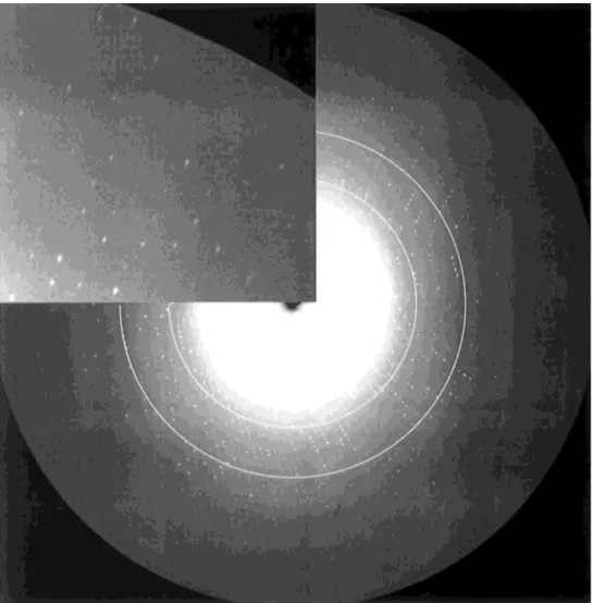
Crystals grown during the flight STS-83 were transported as sitting drops to the EMBL outstation in Hamburg (Germany). One selected crystal (Fig. 1c) was tranferred to a mother liquor containing 20% ethylene glycol as cryoprotectant and shock-cooled at 100 K. It was characterized by X-ray diffraction at the EMBL c/o DESY synchrotron facility at Hamburg, using the X-ray beam-line X11 operating at a wavelength of 0.9096 Å and equipped with a 30 cm Mar detector. The crystal gave a diffraction pattern to 0.91 Å resolution (Fig. 2). The details of the crystal data and of the data collection are summarized in Table 1. According to the diffraction pattern (Fig. 2) and to the intensity statistics (Table 1), it clearly appears that the crystal used was capable of diffracting X-rays to an even higher resolution. The resolution achieved was only limited by the instrument since it was impossible to shorten the crystal to detector distance due to a mechanical contact between the detector and the cryostat. The resolution could have been improved if we had used a beam-line delivering a shorter X-ray wavelength. The quality of a protein crystal is usually assessed by its mosaicity and diffraction resolution limit. An experimental mosaicity value of 0.235° was inferred with the crystal of parvalbumin diffracting to better than 0.9 Å resolution. According to the data available in the protein data bank [16], the resolution observed with the microgravity crystals of pike 4.10 PaCa2 corresponds to one of the highest resolutions achieved so far by X-ray diffraction with a protein with more than 100 amino-acid residues. However, a 125-residue protein has recently afforded crystals diffracting at 0.85 Å resolution using synchrotron radiation [17]. Up to now, 13 X-ray crystal structures of parvalbumins have been reported and deposited with the protein data bank [16]. The best resolution attained for these molecules was 1.50 Å.
A full data set was thus collected at 0.91 Å and is used to refine the structure at atomic resolution. Although this work is still in progress and will not be described here, it is worth noting that the quality of the electron density obtained so far clearly indicates that the atomic resolution has been reached (Fig. 3a). One of our objectives was
a possible observation of the alternate conformations which could occur in the hydrophobic core. As can be seen on Fig. 3b this objective will certainly be fulfilled.
Fig. 2. X-ray rotation image obtained from the crystal of pike parvalbumin shown in Fig. 1(c). It was recorded on a MAR Research imaging plate using the XI1 beam-line at EMBL Hamburg outstation. Rotation range 0.8°, crystal to dector distance 86.2 mm, exposure time 300 s. The inset (top left) gives an enlarged version of the high resolution region. The rings correspond to 1.5 and 1.2 Å resolution and the edge of the detector corresponds to 0.91 Å.
Table 1 Crystal data and data collection statistics
Crystal system Orthorhombic
Space group P212121
a, b, c (Å) 51.026, 49.807, 34.570
Mw (Dalton) 11390
Z 4
Vm (Å 3/Dalton) 1.93
Temperature of data collection (K) 100
Resolution range (Å) 25.0-0.91
Reflections collected 481 972
Unique reflections 63 763
Rmerge(I) Overall 3.6%
Highest resolution shell (0.92-0.91 Å) 12.4%
Highest resolution shell 97.3%
I/σ(I) Overall 14.2
Highest resolution shell 3.3
Fig. 3. (a) Example of electron density (Tyr 48) showing the atomic resolution (2Fo-Fc map contoured at 3.2 σ). (b) The electron density corresponding to two alternate conformations of the side-chain of Phe 30 (2Fo-Fc map contoured at 2.0 σ).
4. CONCLUSION
The effects of microgravity on the crystallization appear at different levels: 1. the highly improved quality of the crystals, as established by X-ray diffraction; 2. the improved growth rate (crystals of 1 mm in length after 4 days);
3. monocrystals with large dimensions (up to 3 mm).
The availability of crystals of a typical EF-hand calciprotein (parvalbumin), diffracting X-rays at 0.9 Å resolution or better corresponds to an unprecedented opportunity to understand cation-protein interactions with this class of proteins. The positions of hydrogen atoms are likely to be identified, as recently shown with an X-ray crystallographic study of a protein [18] at a resolution of 1 Å, thus adding accuracy to our description of hydrogen networks and water sites in parvalbumin [8,9]. Since our X-ray crystallographic analysis of pike 4.10PaCa2 at 0.91 Å resolution was carried out at low temperature (100 K), this analysis will provide us with information on parvalbumin sub-states which are rapidly interconverting at higher temperatures, as recently demonstrated in the case of an enzyme analysed at 120 K and 1.5 Å resolution [19]. The crystallographic analysis of a parvalbumin at atomic resolution appears to be highly relevant to our understanding of the mechanisms underlying divalent cation-binding to, as well as divalent cation-exchange in, the EF-hand proteins [20].
Acknowledgements
J.P.D. and C.E. are indebted to the "Fonds National de la Recherche Scientifique (Belgium)" and to the "Fonds de Développement Scientifique (Université Catholique de Louvain)" for financial support. G.E. and J.P. are thankful to "Centre National de la Recherche Scientifique" (Paris, France) for financial support. The use of the EMBL XII beamline at the DORIS storage ring, DESY, Hamburg is gratefully acknowledged. We thank the European Union for support of the work at EMBL Hamburg through the HCMP to Large Installations Project,
contract no CHGE-CT93-0040. PCAM experiments were supported by the National Aeronautics and Space Administration (NASA) Microgravity Science and Applications Division under contract NAS8-97247 to DC.
References
[1] E.H. Snell, S. Weisgerber, J.R. Helliwell, E. Weckert, K. Hölzer, K. Schroer, Acta Crystallogr. D 51 (1995) 1099. [2] M.R. Wardell, R. Skinner, D.C. Carter, P.D. Twigg, J.-P. Abrahams, Acta Crystallogr. D 53 (1997) 622.
[3] R. Skinner, J.-P. Abrahams, J. Whisstock, A.M. Lesk, R.W. Carrell, M.R. Wardell, J. Mol. Biol. 266 (1997) 601. [4] J.D. Ng, B. Lorber, R. Giegé, S. Koszelak, J. Day, A. Greenwood, A. McPherson, Acta Crystallogr. D 53 (1997) 724. [5] J.P. Declercq, B. Tinant, J. Parello, Acta Crystallogr. D 52 (1996) 165.
[6] B.W. Matthews, J. Mol. Biol. 33 (1968) 491.
[7] J.P. Declercq, B. Tinant, J. Parello, G. Etienne, R. Huber, J. Mol. Biol. 202 (1988) 349. [8] J.P. Declercq, B. Tinant, J. Parello, J. Rambaud, J. Mol. Biol. 220 (1991) 1017. [9] F. Roquet, J.P. Declercq, B. Tinant, J. Rambaud, J. Parello, J. Mol. Biol. 223 (1992) 705. [10] A. Padilla, A. Cavé, J. Parello, J. Mol. Biol. 204 (1988) 995.
[11] J.F. Pechère, in: R.H. Wasserman, R. Corradino, E. Carafoli, R.H Kretsinger, D.H. MacLennan, F.L. Siegel (Eds.), Calcium Binding Proteins and Calcium, Elsevier, North-Holland, New York, 1977, p. 213.
[12] Y. Blancuzzi, A. Padilla, J. Parello, A. Cavé, Biochemistry 32 (1993) 1302. [13] A. Cavé, CM. Dobson, J. Parello, R.J.P. Williams, FEBS Lett. 65 (1976) 190.
[14] A. Cavé, J. Parello, in: Les Houches, Session XXXIII, 1979, Membranes and Intercellular Communication, North-Holland, Amsterdam, 1981, p. 197.
[15] D.C. Carter, T.E. Dowling, Protein Crystallization Apparatus for Microgravity, U.S. Patent No. 5,643,540, 1997.
[16] F.C. Bernstein, T.F. Koetzle, G.J.B. Williams, E.F. Meyer, Jr., M.D. Brice, J.R. Rodgers, O. Kennard, T. Shimanouchi, M. Tasumi, J. Mol. Biol. 112 (1977) 535.
[17] U.K. Genick, S.M. Soltis, P. Kuhn, I.L. Canestrelli, E.D. Getzoff, Nature 392 (1998) 206. [18] S. Longhi, M. Czjzek, V. Lamzin, A. Nicolas, C. Cambillau, J. Mol. Biol. 268 (1997) 779. [19] S.D. Rader, D.A. Agard, Protein Science 6 (1997) 1375.