Université de Montréal
Calcium dynamics and related alterations in pulmonary
hypertension associated with heart failure
par Nour Dayeh
Département de Médecine Faculté de Médecine
Thèse présentée à la Faculté de Médecine
en vue de l’obtention du grade de Philosophiae Doctor (Ph.D) en Sciences biomédicales
option générale
Août, 2018
Cette thèse intitulée :
Calcium dynamics and related alterations in pulmonary hypertension associated with heart failure
Présentée par : Nour Dayeh
a été évaluée par un jury composé des personnes suivantes :
Dr Anique Ducharme, président-rapporteur
………. Dr Jocelyn Dupuis, directeur de recherche
………. Dr Jonathan Ledoux, co-directeur de recherche
………. Dr Guy Rousseau, membre du jury
………. Dr Sébastien Bonnet, examinateur externe
Résumé
L'insuffisance cardiaque (IC) représente un problème de santé important au Canada. La plupart des patients atteints d'IC développent une hypertension pulmonaire (HP), qui est un marqueur de la progression de la maladie et de son mauvais pronostic. Des progrès significatifs ont été réalisés pour le traitement de l'IC. Néanmoins, la morbidité et la mortalité chez les patients atteints d'IC avancée, qui ont développé l’HP reste élevée.
L'augmentation de la pression vasculaire pulmonaire (PVP) observée en HP entraîne une augmentation du tonus vasculaire et un remodelage vasculaire associés à des réponses vasodilatatrices altérées. En effet, une diminution des réponses vasodilatatrices a été observée dans l'HP. La dysfonction endothéliale est au cœur des altérations vasodilatatrices. Cette caractéristique de la plupart des maladies cardiovasculaires est associée à des altérations de l'homéostasie du calcium (Ca2+).
Bien que le Ca2+ global joue un rôle dans un grand nombre de fonctions cellulaires, la présente
thèse est concentrée sur l'impact de la signalisation calcique locale dans les cellules endothéliales (CE). Parmi les différents types de signaux calciques locaux, les pulsars ont été identifiés. Les pulsars calciques sont des évènements endothéliaux locaux dont l'activité est finement régulée par des agents physiologiques qui modulent les niveaux intracellulaires d'inositol 1,4,5-triphosphate (IP3) et de Ca2+. Les pulsars ont un effet sur plusieurs fonctions
cellulaires importantes. Dans les artères mésentériques, les pulsars induisent une relaxation des cellules musculaires lisses vasculaires. Jusqu'à présent, les mécanismes de régulation des pulsars Ca2+ restent à découvrir. Les caractéristiques spatio-temporelles des pulsars suggèrent qu'ils
pourraient jouer un rôle dans le contrôle du tonus vasculaire pulmonaire, impliquant potentiellement plus de canaux ioniques transmembranaires, ainsi que des protéines régulatrices. Les canaux TRP de la famille vanilloïde 4 (TRPV4) sont des canaux cationiques méchanosensitifs, non sélectifs, largement exprimés dans un nombre de tissus. L'activation des
canaux TRPV4 permet l'entrée de Ca2+ dans la cellule. Des études ont montré l'implication de
TRPV4 ainsi que d'autres canaux de la famille TRP dans l’HP.
Les mécanismes physiopathologiques liés au Ca2+ endothélial modulant le tonus vasculaire
rareté des études explorant la physiopathologie et les thérapies de l’HP du groupe II réside dans l'absence de modèles animaux validés pour l’étude de l’HP du groupe II, avec une détermination adéquate de la présence et de la sévérité de l'HP.
Les travaux issus de cette thèse ont identifié et caractérisé pour la première fois des pulsars Ca2+
intracellulaires dans l'endothélium pulmonaire et leurs altérations dans un modèle de souris cliniquement significatif de l’HP de groupe II qui a été développé. En outre, ce travail a révélé
l'implication des canaux TRPV4 endothéliaux dans la dérégulation des pulsars Ca2+ dans l’HP
du groupe II.
Mots-clés :
Abstract
Congestive heart failure (CHF) represents an important Canadian health problem. Most patients with CHF develop pulmonary hypertension (PH), which is an important marker that signals progression of the disease and its poor outcome. Significant advances have been made for the treatment of heart failure (HF). Nevertheless, the morbidity and mortality among patients with advanced heart HF, who have developed PH remains high.
Increased pulmonary vascular pressure (PVP) observed in PH leads to increased vascular tone and vascular remodelling associated with altered vasodilatory responses. It is noteworthy that a decrease in vasodilatory responses has been observed in PH. At the core of vasodilatory alterations lies endothelial dysfunction. This hallmark of most cardiovascular diseases is associated with alterations in calcium (Ca2+) homeostasis.
Although global Ca2+ plays a role in a wide range of cellular functions, this thesis work focused on the impact of local Ca2+ signalling in endothelial cells (ECs). Among the different types of local Ca2+ signals, Ca2+ pulsars were identified. Ca2+ pulsars are local endothelial Ca2+ signals whose activity is finely regulated by physiological agents that modulate intracellular levels of inositol 1,4,5-triphosphate (IP3) and Ca2+. Ca2+ pulsars have been shown to have an effect on several important cellular functions. In mesenteric arteries, Ca2+ pulsars induce endothelium-induced relaxation of vascular smooth muscle cells. Up until now, the regulatory mechanisms of Ca2+ pulsars remain to be uncovered. The spatio-temporal characteristics of Ca2+ pulsars suggest that they could play a role in the control of pulmonary vascular tone, potentially involving more transmembrane ion channels, as well as regulatory proteins. Transient receptor potential vanilloid 4 (TRPV4) channels are non-selective mechanosensitive osmo-regulated cation channels broadly expressed in a number of tissues. Activation of TRPV4 channels allows Ca2+ entry into the cell. A number of studies have shown the implication of TRPV4 as well as other channels from the TRP family in PH.
Endothelial Ca2+-related pathophysiological mechanisms modulating pulmonary vascular tone and leading to the development of group II PH are poorly defined. In addition, the scarcity of studies exploring the pathophysiology and therapies of group II PH resides in the lack of validated small animal models with an adequate determination of the presence and severity of PH.
The work in this thesis identified and characterized for the first time intracellular Ca2+ pulsars in pulmonary endothelium and their alterations in a clinically relevant mouse model of group II PH that was developed. In addition, this work revealed the implication of endothelial TRPV4 channels in Ca2+ pulsars dysregulation in group II-PH.
Keywords:
Table of contents
RÉSUMÉ ... I ABSTRACT ...III TABLE OF CONTENTS ... V LIST OF TABLES ...IX LIST OF FIGURES ...X LIST OF ABBREVIATIONS ...XI ACKNOWLEDGEMENTS ... XVI
I. CHAPTER I: INTRODUCTION ... 1
1. THE PULMONARY SYSTEM ... 2
1.1. ANATOMICAL AND FUNCTIONAL ORGANIZATION OF THE LUNGS ... 2
1.2 CELLULAR COMPOSITION OF THE LUNGS AND AIRWAYS ... 3
1.3 STRUCTURE OF PULMONARY ARTERIES, ARTERIOLES AND CAPILLARIES ... 4
1.3.1 The tunica intima... 4
1.3.2 The internal elastic lamina ... 5
1.3.3 The tunica media ... 5
1.3.4 The tunica adventitia ... 6
1.4 PULMONARY VEINS... 7
1.5 THE BRONCHIAL CIRCULATION ... 7
1.6 LYMPHATIC VESSELS... 7
1.7 REGULATION AND DISTRIBUTION OF BLOOD FLOW ... 8
1.8 PULMONARY VASCULAR TONE ... 8
1.9 PULMONARY VASCULAR RESISTANCE ... 9
1.10 ENDOTHELIAL CONTROL OF THE PULMONARY CIRCULATION ... 9
1.10.1 Endothelium-dependent vasodilation ... 9
1.11 VASOACTIVE PROPERTIES OF PULMONARY VEINS ... 12
2. INTRACELLULAR CALCIUM HOMEOSTASIS ... 14
2.1 INTRACELLULAR CALCIUM SOURCES AND TRANSPORTERS ... 14
2.1.1 Inositol 1,4,5-triphosphate receptors ... 14
2.1.2 Ryanodine receptors ... 15
2.1.3 SERCA pumps ... 16
2.1.4 Plasma membrane Ca2+-ATPase and Na+/Ca2+ exchanger ... 16
2.1 EXTRACELLULAR CALCIUM ... 17
2.1.1 Ca2+ influx from the extracellular space ... 17
2.2 CALCIUM SIGNALLING ... 17
2.2.1 Global calcium signalling ... 18
2.2.2 Local calcium signalling ... 18
3. THE TRANSIENT RECEPTOR POTENTIAL CHANNELS FAMILY ... 24
3.1 CLASSIFICATION OF TRP CHANNELS ... 24 3.2 STRUCTURE OF TRP CHANNELS ... 25 3.3 ACTIVATION OF TRP CHANNELS ... 26 3.4 THE TRPV FAMILY ... 26 3.4.1 Expression of TRPV channels ... 27 3.4.2 Activation of TRPV channels ... 27 3.5 TRPV4 CHANNELS... 28
3.5.1 Structure of TRPV4: from the gene to the protein ... 28
3.5.2 Permeability and selectivity of TRPV4 ... 29
3.5.3 Regulation of TRPV4 activity ... 30
3.5.4 Physiological role of TRPV4 channels... 32
3.5.5 TRPV4 regulation of vascular tone ... 32
3.5.6 Pathological role of TRPV4 ... 33
4 THE PULMONARY CIRCULATION IN DISEASE ... 35
4.1 PULMONARY HYPERTENSION ... 35
4.1.1 Historical background ... 35
4.1.2 Groups of pulmonary hypertension... 35
4.2 THERAPEUTIC STRATEGIES IN PULMONARY HYPERTENSION ... 38
4.2.1 ETR antagonists ... 38
4.2.2 PDE-5 inhibitors ... 39
4.2.3 PGI2 analogues ... 39
4.2.4 Calcium channel blockers ... 39
4.2.5 Soluble guanylate cyclase stimulators ... 40
4.2.6 Combination therapy ... 40
4.2.7 Other therapies in group I: pulmonary arterial hypertension ... 41
4.3 ADVANCEMENTS AND OBSTACLES IN GROUP II-PH TREATMENT ... 42
4.4 ANIMAL MODELS OF PULMONARY HYPERTENSION ... 44
4.4.1 Genetic animal models of PH ... 44
4.4.2 Single pathological insult models ... 45
4.4.3 Multiple pathological insult models (group I-PAH) ... 47
4.4.4 Models used for the study of group II- PH ... 48
5 LUNG CAPILLARY STRESS FAILURE AND ARTERIOLAR REMODELLING IN PULMONARY HYPERTENSION ASSOCIATED WITH LEFT HEART DISEASE (GROUP 2 PH) 51 II. CHAPTER II HYPOTHESIS AND OBJECTIVES OF THIS THESIS ... 63
III. CHAPTER III: RESULTS ... 66
Echocardiographic validation of pulmonary hypertension due to heart failure with reduced ejection fraction in mice ... 68
Localized endothelial Ca2+ signaling is strongly conserved amongst vascular beds ... 103
Involvement of endothelial TRPV4 in alterations of calcium pulsars in pulmonary hypertension associated with left heart failure ... 124
IV. CHAPTER IV: DISCUSSION ... 151
HEART FAILURE AND PULMONARY HYPERTENSION ... 152
Development of a model for PH-LHD ... 152
Setting specific parameters and cutoff values ... 153
THE PULMONARY ENDOTHELIUM IN HEART FAILURE ... 154
The integrity of pulsars ... 154
TRPV4 and pulmonary pulsars: location, location, location ... 156
Impact of PH-LHD on localized endothelial Ca2+ signalling. ... 157
LIMITATIONS AND FUTURE DIRECTIONS ... 160
CONCLUSION ... 164
ORIGINAL CONTRIBUTION TO THE LITERATURE ... 164
BIBLIOGRAPHY ... 165
ANNEXE: EXPRESSION OF PHOSPHOINOSITIDE SPECIFIC PHOSPHOLIPASE C ISOFORMS IN NATIVE ENDOTHELIAL CELLS ... 182
List of tables
Table 1 Clinical classification of pulmonary hypertension, adapted from Simmoneau et al., 2013, (5th WSPH Nice 2013. Main modifications to the previous Dana Point classification) ... 36 Table 2 Hemodynamic, functional, and structural changes observed during the progression of pulmonary hypertension ... Error! Bookmark not defined. Table 3 Basal pulsars’ kinetics and descriptive parameters in non-stimulated pulmonary arteries control mice and human pulmonary miscrovascular endothelial cells. ... 163
List of figures
Figure 1 The systemic and pulmonary circulations (http://academic.kellogg.cc.mi.us) ... 2
Figure 2 Structural differences between pulmonary arteries and mesenteric arteries of mice ... 6
Figure 3 Calcium blips and puffs (Charbel et al., 2013) ... 19
Figure 4 Calcium sparklets (Charbel et al., 2013) ... 21
Figure 5 Endothelial calcium pulsars, adapted from Ledoux et al., 2008 ... 22
Figure 6 Calcium wavelets (Charbel et al., 2013)... 23
Figure 7 TRP channels family ... 25
Figure 8 A: Structure of a TRP channel showing the 6 transmembrane domains and the pore region. B: Tetrameric structure of a TRP channel (Watanabe et al., 2009) ... 26
Figure 9 Schematic representation of TRPV4 protein... 29
Figure 10 Human endothelial pulsars ... 162
List of abbreviations
PH Pulmonary hypertension
HF Heart failure
mPAP Mean pulmonary arterial pressure PVR Pulmonary vascular resistance Group I-PAH Pulmonary arterial hypertension
PH-LHD Pulmonary hypertension associated to left heart disease
PA Pulmonary artery PV Pulmonary vein EC Endothelial cell NO Nitric oxide PGI2 Prostacyclin ET-1 Endothelin-1 Ang II Angiotensin II TxA2 Thromboxane A2
IEL Internal elastic lamina MEP Myoendothelial projection
SMC Smooth muscle cell
Ca2+ Calcium
CaM Calmodulin
MLCK Myosin light chain kinase
MLC20 Myosin light chain
ATP Adenosine triphosphate
MLCP Myosin light chain phosphatase
PVSMC Pulmonary vascular smooth muscle cell NANC Non-adrenergic non-cholinergic system PAP Pulmonary arterial pressure
NOS Nitric oxide synthase sGC Soluble guanylate cyclase
PKG cGMP-dependent protein kinase NCX Na+/Ca2+ exchanger
K+ Potassium
AA Arachidonic acid
Cox Cyclooxygenase
cAMP Cyclic adenosine monophosphate
PKA Cyclic adenosine monophosphate-dependent protein kinase A BKca Large conductance calcium-activated potassium channel (Kca 1.1) Kir Inward rectifying potassium channel
Kv Voltage-dependent potassium channel EDHF Endothelium-derived hyperpolarizing factors EETs Epoxyeicosatrienoic acids
H2O2 Hydrogen peroxide
IK Intermediate conductance Ca-activated K channel SK Small conductance Ca-activated K channel MEGJ Gap junctions at the MEP level
TGF-β Transforming growth factor β
ETA Endothelin receptor A
ETB Endothelin receptor B
COPD Chronic obstructive pulmonary disease
PLC Phospholipase C
IP3 Inositol triphosphate
DAG Diacylglycerol
ACE Angiotensin converting enzyme
5-HT Serotonin
PAH Pulmonary arterial hypertension
ER Endoplasmic reticulum
IP3R inositol 1,4,5-triphosphate receptor
RRy Ryanodine receptors
CaMKII Ca/CaM-dependent protein kinase II
SR Sarcoplasmic reticulum
NFAT Nuclear factor of activated T-cells HUVEC Human umbilical vein endothelial cells HPAEC Human pulmonary artery endothelial cells PIP2 Phosphatidylinositol 4,5-biphosphate eNOS Endothelial nitric oxide synthase STOC Spontaneous transient outward current VDCC Voltage-dependent Ca2+ channels TRPV4 Transient receptor potential vanilloid 4 TRP Transient receptor potential
TRPC Transient receptor potential canonic TRPM Transient receptor potential melastatin TRPP Transient receptor potential polycyctine TRPML Transient receptor potential mucopiline TRPA Transient receptor potential ankyrine TRPN Transient receptor potential nompC CNS Central nervous system
DRG Dorsal root ganglia
NGF Nerve growth factor
PI3K Phosphatidylinositol-3-kinase OTRPCA4 Osm-9-like TRP channel 4
VR-OAC Vanilloid receptor-related osmitically activated channel CaM-BD Calmodulin-binding domain
CYP450 Cytochrome P450 4α-PDD 4α-phorbol-12,13-didecanoate GSK1016790A N-((1S)-1-3-hydroxypropanoyl)-1-piperazinyl]carbonyl}-3-methylbutyl)-1-benzothiophene-2-carboxamide HC067047 2-Methyl-1-[3-(4-morpholinyl)propyl]-5-phenyl-N-[3-(trifluoromethyl)phenyl]-1H-pyrrole-3-carboxamide
WHO World health organization
Group III-PH PH associated with chronic disease and/or hypoxia Group IV-PH PH associated with chronic thromboembolic disease Group V-PH PH due to multifactorial mechanisms
BMPR2 Bone morphogenic protein receptor 2 ACVRL1 Activin kinase receptor type 1
HFrEF Heart failure with reduced ejection fraction HFpEF Heart failure with preserved ejection fraction
LV Left ventricle
PCWP Pulmonary capillary wedge pressure
TPG Transpulmonary gradient
IPF Idiopathic pulmonary fibrosis
CPFE Pulmonary fibrosis combine with emphysema PHCTE PH related to chronic thromboembolic disease
PDE Phosphodiesterase
VIP Vasoactive intestinal peptide
CO Cardiac output
MCT Monocrotaline
(HIF-1α)-k+ Mitochondrial reactive oxygen species inducible factor α
PAB Pulmonary artery banding
RV Right ventricle
SU-5416 Semaxinib
VEGF Vascular endothelial growth factor
RVH RV hypertrophy
RVSP RV systolic pressure
TAC Transverse aortic constriction
LVEDP Left ventricular end diastolic pressure RVEDP Right ventricular end diastolic pressure PPHN Persistent PH of the newborn
PAAT Pulmonary artery acceleration time
TAPSE Tricuspid annular plane systolic excursion TNF- α Tumor necrosis factor α
Acknowledgements
I would like to thank members of my jury, Dr. Anique Ducharme, Dr. Guy Rousseau, and Dr. Sébastien Bonnet for taking part in the completion of this thesis.
I would like to thank my supervisor Dr. Jocelyn Dupuis and my co-supervisor Dr. Jonathan Ledoux for their dedication to the realization of my thesis. I have experienced growth on a personal and intellectual level.
I would like to thank Dr. Dupuis for his guidance, mentorship, wise advice and his belief in my capacities at times when I did not believe in myself. I would like to thank Dr. Ledoux for his commitment, for constantly pushing me to think deeper and creatively, for his great ideas in my project, and for allowing me the chance to participate in different projects in his lab.
I would like to thank Emma Dedelis. I am very thankful for your sense of humour and sarcasm, which got me through some very rough times. And, of course thank you for your great technical assistance. Some experiments were humanly impossible for one person to do and you were always there. Thank you for the chocolate, and candy, and cookies etc…
Thank you to everyone who participated in my project, Yanfen Shi, Marc-Antoine Gillis, Louis Villeneuve, and to all members of both labs I was part of.
To Dr Yahye Merhi, thank you for not letting me give up, for being a listening ear and for your wise advice.
To my friends, Hneineh, Mira, Hiba, Hassan, and Omar. Thank you for being there for better or for worse. To friends I made at the Montreal Heart Institute, Sam, Celia, Sherine, Anh-tuan, and Nassiba, thank you for making MHI a pleasant and happy place to work at. I will cherish these times forever.
Mira, the bestie, the partner in crime, you give a completely new meaning to the word friendship!
To Peter, I am beyond words. Thank you for your patience, constant support and encouragement, and for being the most understanding at difficult times.
To my parents and two brothers, who have been my constant support system, thank you for your unconditional love and unlimited support. Joumana and Riad, everything I am and everything I will be is thanks to both of you. I love you and forever will be grateful.
To every single person who has brought happiness to my heart, thank you from the bottom of mine.
I am seeking. I am striving. I am in it with all my heart. Vincent Van Gogh
I. Chapter I: Introduction
Pulmonary hypertension (PH) is a common complication in heart failure (HF). It is considered as group II according to international clinical guidelines [1, 2]. It is characterized by a progressive increase in mean pulmonary arterial pressure (mPAP) and pulmonary vascular resistance (PVR) successively inducing right ventricular hypertrophy and death. The disease is under-diagnosed and clinically detectable in its later stage. Current clinically approved pharmacological treatments only slow the progression of the disease without treating it permanently and are primarily limited to pulmonary arterial hypertension (group I-PAH). Extensive ongoing clinical trials are yet to confirm their validity for the treatment of pulmonary hypertension associated to left heart disease (PH-LHD). PH-LHD therefore remains an important public health problem and a better understanding of the cellular and molecular mechanisms underlying this condition is necessary for the development of innovative and effective therapies.
1. The pulmonary system
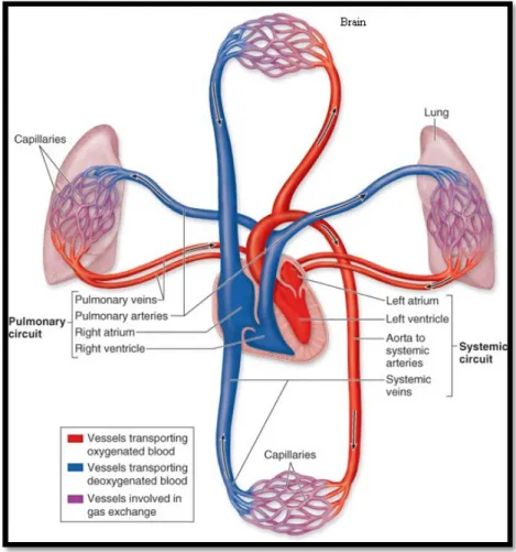
The main function of the cardio-pulmonary apparatus is to provide tissues with oxygen rich blood, thus allowing their proper functioning. Deoxygenated blood coming from the right ventricle reaches the lungs through the right and left pulmonary arteries (PAs). It circulates the arteries, arterioles, reaching the pulmonary capillaries. Gas exchange occurs within the alveolar-capillary surface, and oxygen rich blood returns through the pulmonary veins to the left heart.
Figure 1 The systemic and pulmonary circulations (http://academic.kellogg.cc.mi.us)
1.1.
Anatomical and functional organization of the lungs
In the thoracic cavity, the lungs are enclosed by the pleural membrane, which is composed of two layers: an outer membrane (the parietal pleura) that lines the chest wall and diaphragm, and an inner membrane (visceral pleura) separated by an inter-pleural space which contains fluid
secreted by both membranes. The right lung is divided into three lobes (superior, middle, and inferior), while the left lung is composed of two lobes (superior and inferior).
The lungs are connected to the trachea by the right and left bronchi that branch from the main bronchus. Each lobe in the lung is supplied by a bronchopulmonary segment. A lobe subdivides into smaller lobules separated by a septum with the branching of bronchi into bronchioles. Inside a lobule, a bronchiole subdivides into multiple branches.
Blood supply to the lungs begins with the PA that arises from the pulmonary trunk carrying deoxygenated arterial blood to the alveoli. The PA branches in parallel with the bronchi reaching the level of smaller arterioles. One arteriole and an accompanying vein supply and drain one pulmonary lobule. As they approach the alveoli, pulmonary arterioles become the pulmonary capillary network. At the level of the alveolar-capillary membrane, where the capillary wall meets the alveolar wall, gas exchange occurs.
1.2 Cellular composition of the lungs and airways
The trachea, bronchi, and bronchioles are lined with ciliated columnar epithelial cells, intercepted by serous cells, as well as club cells with a macrophage-like role. Starting at the level of terminal bronchioles, pulmonary epithelial cells lose their cilia and are intercepted by goblet cells capable of producing mucus.
The walls of alveoli are lined with two types of cells termed alveolar cells type I (squamous pneumocyte) and type II (granular pneumocyte), supported by a thin layer of connective tissue. Type I alveolar cells, which are the main cells paving the structure of alveoli, have extremely thin walls that allow easy gas exchange. Type II alveolar cells have a cuboidal shape, possess microvilli, and produce and secrete lung surfactant. Lung surfactant mainly composed of lipids and proteins, is secreted into the alveolar space. It has a number of functions that include prevention of lung collapse during expiration, supporting inspiratory opening of the lungs, and balancing hydrostatic filtration forces to prevent lung oedema formation. In addition to its biophysical functions, lung surfactant possesses immunological functions [3], and protects the lungs from micro-organisms and toxins. Macrophages are also found inside the alveoli and are responsible for keeping the lungs free of pathogens and other foreign matter that can enter
the alveoli with inhaled air. All the epithelial cells lining the respiratory tract except for type I alveolar cells secrete epithelial lining fluid, which covers the mucosa of the alveoli and airways. The pulmonary circulation comprises several cell types that compose the different layers of pulmonary vessels depending on vessel calibre and type.
1.3 Structure of pulmonary arteries, arterioles and capillaries
The pulmonary vasculature has a low pressure and low resistance under normal conditions. It accommodates 100% of the cardiac output [4]. Therefore, the structure of PAs and pulmonary veins (PVs) differ from the systemic one on a variety of levels. In general, pulmonary vessels are more compressible and distensible than systemic ones. Additionally, pulmonary vessels are thin-walled compared to their systemic counterparts, and have less vascular smooth muscle [5]. Similarly to systemic arteries, PAs are composed of three histologically distinct layers: the tunica intima, the tunica media, and the tunica adventitia.
1.3.1 The tunica intima
The tunica intima is the inner layer of PAs, as well as the only layer forming pulmonary capillaries [6]. It is a monolayer of endothelial cells (ECs) separated by intercellular tight junctions resting on a basal membrane that is secreted by ECs. It is responsible for gas exchange at the alveolar-capillary level, as well as endothelial control of the pulmonary circulation through the release of a number of molecules and factors (Nitric oxide (NO), prostacyclin (PGI2), endothelin 1 (ET-1), angiotensin II (Ang II), thromboxane A2 (TxA2). Pulmonary vascular ECs are aligned in the direction of blood flow, and exclusively express a number of proteins like lung endothelial cell adhesion molecule-1 and endothelial specific molecule-1 [7]. The pulmonary endothelium is a metabolically active surface that plays an important role in immunologic and inflammatory events [8]. Endothelial dysfunction is a major contributor to a wide range of diseases [9], notably PH . In fact, several studies have revealed that pulmonary ECs abnormalities (in morphology and function) were major contributors and sole initiators of the pulmonary remodelling process [10] [11] [12].
1.3.2 The internal elastic lamina
The internal elastic lamina (IEL) consists of elastic fibers and its thickness varies according to vessel diameter and vascular bed. The structure of the IEL can vary as well. Some IEL contain holes termed fenestrations. It general, the size and density of fenestrations are inversely proportional to vessel calibre and greatly dependent on vessel function [13, 14] [15] [16]. In the pulmonary circulation, the IEL is fenestrated and through its fenestrations, extensions from ECs pass and are in contact with smooth muscle cells (SMCs). These myoendothelial projections (MEPs) (around 0.5 µm width and 0.5 µm depth variable from one vascular bed to another) have been shown to play an important role in the control of vascular function through the release and diffusion of vasoactive substances [17] [18]. Additionally, it has been shown that modulation in the number of IEL holes occurs in pathological conditions [13], including PH [14]. Results regarding hole density in pathological states have varied. In some hypertensive rat models, hole density was found to be lower compared to normotensive rats in some stains, while it remained the same for both groups in other strains [13]. In PH, a study on patients with congenital cardiac shunts revealed that the number of IEL gaps in peripheral pulmonary arteries was lower in pre-acinar arteries compared to a control group[14].
1.3.3 The tunica media
The tunica media is the middle layer of PAs. It is mainly composed of longitudinally arranged SMCs, connective tissue, collagen and elastic fibers. It is separated from the tunica intima by the IEL. In PAs, the contribution of the media to the total wall thickness varies with vessel diameter. Additionally, the number and alignment of elastic lamina varies according to diameter and type. Pulmonary vascular SMCs are responsible for the maintenance of pulmonary vascular tone, vascular resistance, as well as contractile response upon stimulation. An increase in intracellular calcium (Ca2+) levels leads to its binding to the (Ca2+)-binding protein calmodulin (CaM). The then formed Ca2+-CaM complex binds to and activates myosin light chain kinase (MLCK) via the C-terminal domain of CaM. Activated MLCK phosphorylates myosin light chain (MLC20) resulting in the interaction of myosin and actin filaments in an ATP-dependent manner [19] [20]. De-phosphorylation of MLC20 by myosin light chain
phosphatase (MLCP) inhibits the interactions between myosine and actine filaments, and consequently ends vascular contraction.
1.3.4 The tunica adventitia
The tunica adventitia of PAs, the outermost layer, is separated from the media by the external elastic lamina. It is loosely organized, composed of fibroblasts and a collagen matrix that reinforces vessels and protects their integrity. The tunica adventitia of larger vessels contains vasa vasorum, a capillary network that nourishes the external tissues of the blood vessel wall [6]. In the pulmonary circulation, fibroblasts of the tunica adventitia have been shown to play an important role in the response to environmental stimuli [21].
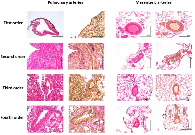
Figure 2 Structural differences between pulmonary arteries and mesenteric arteries of mice
Hematoxylin and eosin staining of cross-sections of mouse pulmonary and mesenteric arteries of different orders (scale bar: 10 mm).
1.4 Pulmonary veins
PVs run inferior to PAs throughout the lungs and are not accompanied by an airway. PVs arise from the pulmonary capillary network and carry oxygenated blood from the lungs to the heart. Unlike systemic vessels, PVs are not so physiologically and structurally distinct from PAs. Additionally, they possess a similar density of MEPs to that observed in PAs [22]. Small differences between PAs and PVs lie within the histological level, where PVs have thinner and less well organized walls than PAs of the same calibre [23]. Additionally, EC nuclei shapes are not similar between PAs and PVs: while ECs of PAs have an elliptical shape, ECs of PVs have a polygonal shape [24] [25].
Studies have confirmed the contribution of PVs to the total PVR [26] [27] [28].
1.5 The bronchial circulation
The bronchial circulation is smaller than the pulmonary circulation. Bronchial vessels usually originate from the aorta (90%) or intercostal arteries [29]. They enter the lungs at the hilum and supply the lower trachea, bronchi, bronchial branches, esophagus, and visceral pleura with oxygen and nutriments [30]. Additionally, they supply blood to the vasa vasorum of the thoracic aorta and pulmonary arteries and veins, as well as the nerves and lymph nodes in the thorax [30] that are not supplied by the pulmonary circulation. The bronchial circulation terminates at the level of terminal bronchioles where it joins pulmonary capillaries and venules. Bronchial arteries receive around 1% of the total cardiac output, and are high resistance, low capacitance vessels [30].
1.6 Lymphatic vessels
The lungs possess two sets of lymphatic vessels. Surface lymphatic vessels are located beneath the pleura, while deep lymphatic vessels parallel blood vessels in the lungs and extend along the bronchi and are not present in the walls of alveoli. They are responsible for clearing fluid and particulates.
1.7 Regulation and distribution of blood flow
A passive regulation of blood flow exists in the lung. The lung is divided into three zones determined by the relative values of the pulmonary arterial (Pa), pulmonary venous (Pv), and alveolar (PA) pressure. In an upright position, blood flow in the upper portion of the lung (zone 1) is extremely low due to the fact that alveolar pressure is greater than both Pa and Pv (PA> Pa > Pv). Zone 2 is the middle portion of the lung where Pa > PA> Pv, and blood flow remains impaired. Zone 3 is the lowest zone in the lung where the level of vessel recruitment is the greatest at rest because the arterial and venous pressures are both higher than the alveolar pressure allowing vessels to be maximally distended at all times. Following an increase in blood flow, there is blood vessel recruitment in zones 1 and 2, resulting in an even blood flow distribution throughout the lung.
In addition to passive flow regulation, an active regulatory system that includes sympathetic innervation, humoral mechanisms, and respiratory gases, is a key determinant of vascular pressure.
1.8 Pulmonary vascular tone
In contrast to the systemic circulation where a relatively elevated vascular tone participates in the regulation of arterial pressure, the basal pulmonary vascular tone is low under normal conditions. Pulmonary vascular ECs play an undisputed role in the maintenance of a low resting vascular tone [31]. Extensive work has shown that the low tone is attributed to a continuous circulating and local production of vasodilators like a tonic endothelial release of NO and PGI2 [32], as well as vasoconstrictors with the balance tipped in favour of vasodilators. Additionally, the control of pulmonary vascular tone happens via the autonomic nervous system where parasympathetic innervation (cholinergic) is responsible for the vasodilator effect, and sympathetic innervation (adrenergic) is responsible for the vasoconstrictor effect [33]. A non-adrenergic non-cholinergic (NANC) system also regulates pulmonary vascular tone [34]. Pulmonary vascular tone can be raised in response to a number of stimuli like changes in O2 pressures, which can result in vasoconstriction, as well as acidosis [35].
1.9 Pulmonary vascular resistance
The mean pressure in human PAs is around 15 mmHg. When reaching the pulmonary capillaries, the pressure drops to about 5 mmHg. Vascular resistance is defined as the pressure drop divided by the flow, which is identical in both systemic and pulmonary circulations. However, the pressure drop in the pulmonary circulation is much lower. Therefore, the pulmonary circulation has low resistance.
PVR is about 1/8 to 1/10 of systemic vascular resistance. According to Poseuille’s law (ΔP = 8hLQ / πr4), small variations in radius are to the fourth, and can therefore significantly affect flow (Q), and consequently PVR [36]. In PAs, variations of PAP have direct consequences on PVR. A sustained increase in PVR is a contributor to disease progression in a number of pathologies including PH [37] [38]. PVR can also increase in pulmonary veins following fluctuations in pressure.
1.10 Endothelial control of the pulmonary circulation
1.10.1 Endothelium-dependent vasodilation
Nitric oxide (NO)
NO is produced by a biochemical reaction triggered by the activity of the enzyme NO-synthase (NOS) on L-arginine [39]. Briefly, the completion of the reaction consumes oxygen and produces NO and H2O by conversion of L-arginine to L-citrulline. Three NOS isoforms have been described. Neuronal NOS (NOS-I), constitutively expressed in neuronal cells, inducible NOS (NOS-II), and endothelial NOS (NOS-III), found throughout the vascular endothelium, all three of which are expressed in the lung [40].
Newly synthesized NO diffuses through the cell membrane into the cytoplasm of adjacent SMCs, where it activates sGC and increases intracellular levels of cGMP [41]. The latter activates PKG, consequently causing a decrease in intracellular Ca2+ concentration due to inhibition of voltage and receptor-operated Ca2+ channels accompanied by increased cellular extrusion of Ca2+ through Ca2+- ATPase pumps and the Na+ / Ca2+ exchanger. PKG activity also
causes the activation of Ca2+-dependent K+ channels leading to cellular hyperpolarization. Additionally, PKG activates MLCP leading to SMC relaxation.
Prostacyclin (PGI2)
PGI2 is synthesized from arachidonic acid (AA) by the cyclooxygenase pathway (Cox). Once synthesized, PGI2 diffuses and binds to its receptors. These receptors activate the enzyme adenylyl cyclase located at the membrane level, resulting in an increase in the formation of cyclic AMP (cAMP). cAMP thus produced activates cAMP –dependent protein kinase A (PKA), capable of inducing relaxation of SMCs. On the other hand, PGI2 is capable of activating ATP-dependent K+ channels, Ca2+ dependent K+ channels, large conductance (BKca), inwardly rectifying K+ channels (Kir), and voltage-dependent K+ channels (Kv), all participating in relaxation of the vascular smooth muscle [42].
Endothelium-derived hyperpolarizing factors (EDHF)
While NO signalling appears to play a major role in the vasoreactivity of arteries, the dominant responses in small resistance arteries are related to the EDHF pathway [43]. Several factors are included in EDHF signalling, including electrical signalling through MEPs, an increase in intracellular Ca2+ levels that activates intermediate and small conductance K+ channels, IK and SK, and produces endothelial hyperpolarization, the extracellular accumulation of K+, Epoxyeicosatrienoic acids (EETs), hydrogen peroxide (H2O2), and type C natriuretic peptide. Endothelial hyperpolarization propagates along the blood vessels via homocellular gap junctions, and communicated to adjacent SMCs by communicating junctions at the level of the MEPs [44]. Gap junctions at the MEP level are composed of proteins called connexins that physically connect the cytoplasm of ECs and SMCs, which allows electrical and chemical coupling between the two cell types. Cyclic AMP facilitates the electrical transmission of endothelial hyperpolarization of smooth muscle layers by increasing the conductance of gap junctions of smooth muscle. In addition, activation of endothelial K+ channels also leads to accumulation of K+ ions in the intercellular space. The increase in extracellular K+ concentration can activate Na+/K+ ATPase and Kir which also produces hyperpolarization of the vascular SMC [45].
1.10.2 Endothelium-dependent vasoconstriction
Endothelin-1
Endothelin-1 is produced mainly by ECs [46] from a precursor, pre-endothelin, transformed into inactive big-ET and then into ET by the endothelin conversion enzyme. ET-1 production is increased by several stimuli, such as shear forces and hypoxia as well as by a number of neurohumoral factors and cytokines including AngII, vasopressin, catecholamines and transforming growth factor- β (TGF-β) [47] [48]. ETA and ETB are two G protein-coupled receptors (GPCR) that mediate the effects of ET-1. ETB is mainly found on ECs. Its activation leads to vasorelaxation. While ET-1 can be responsible for vasodilation when it binds to endothelial ETB receptors, its main role in vasoconstriction is mediated by its receptors on SMCs. Pulmonary vascular SMCs express both ETA and ETB receptors. Activation of these receptors induces contraction that contributes to the increase of vascular tone, and to the reduction of the flow. Briefly, activation of both receptors leads to vasoconstriction through the activation of phospholipase C (PLC), generation of inositol triphosphate (IP3) and diacylglycerol (DAG), and the consequent increase in intracellular Ca2+ followed by the activation of the vascular SMCs’ contractile system. Additionally, ET-1 can promote vascular remodelling by triggering SMC proliferation and collagen deposition.
In various pathological conditions affecting the pulmonary circulation, such as pulmonary arterial hypertension, chronic obstructive pulmonary disease (COPD), and Hypoxia, an increased synthesis of ET-1 is at the basis of the increase in tone [49] [50].
Thromboxane A2
TXA2 is synthetized through the activation of the COX1-2 pathway and has a half-life of a few seconds. Briefly, binding of TXA2 to its GPCRs, prostanoid receptor T1 and prostanoid receptor T2 (TP1 and TP2) leads to the activation of PLC and induces intracellular Ca2+ increase thus leading to vascular SMC contraction. Under physiological conditions, the production of TxA2 is countered by a production of PGI2. However, this equilibrium can be shifted in the presence of pathologies affecting endothelial function.
Angiotensin II
Ang II is synthetized in the lung as a result of the enzymatic conversion of angiotensin I by the endothelial angiotensin converting enzyme (ACE) [51]. It causes vasoconstriction through its binding to its receptors AT1 and AT2 on the surface of VSMCs. Briefly, binding of Ang II to its receptors results in the activation of PLC and the generation of IP3 and an increase in intracellular Ca2+ levels.
Serotonin
Serotonin (5-HT) is synthesized via the hydroxylation and subsequent decarboxylation of its precursor tryptophan. After synthesis, 5-HT is stocked in platelet dense granules and is released upon their activation. Serotonin induces vasoconstriction following its binding to its specific GPCR of several subtypes (14 distinct subtypes including: 1B/D, 2A and 2) abundantly expressed within the lung on pulmonary arterial SMCs. Serotonin has been implicated in a number of pathologies including PAH, where studies have clearly shown that drugs (ex: appetite suppressant fenfluramine and aminorex) that affected serotonin signalling had a marked effect on PAH progression [52, 53].
1.11 Vasoactive properties of pulmonary veins
In the pulmonary circulation, vasoactive mediators have effects on veins. In fact, PVs seem to have a stronger response than PAs to certain vasoactive factors [54] [55]. NO was shown to be produced by ECs of PVs of a number of animal models. Vascular response to NO revealed to be greater in PVs than PAs [56]. This effect was due to a higher amount and activity of NOS in PVs [56] [57]. Other animal studies have attributed this same effect to a more pronounced phosphodiesterase activity as well as a prominent role of PKG [58]. Additionally, PGI2 production was shown to be the same in adult ovine pulmonary arteries and veins [26]. A variety of studies showed that PVs possess greater sensitivities than arteries to a number of vasoconstrictors and vasoconstrictor stimuli like ET, thromboxane, leukotriennes, and hypoxia. In fact, in a number of animal models as well as in humans, ET-1 seems to be a more potent constrictor of pulmonary veins than arteries with a more notable effect in small veins [59] [60]
[61] [62]. Finally, TXA2 was found to be a constrictor of PVs in a number of animal models as well as humans [63] [64] [28]. It was found to induce vasoconstriction of human PVs through prostanoid receptor subtypes TP and EP1 [65].
Whether causing dilation or constriction, vasoactive molecules exert their effects on vascular tone through their control of Ca2+ levels. For example, vascular dilation through
EDHF can happen via IK and SK channels, activated following an increase in intracellular Ca2+. Additionally, can Ca2+ initiate the production of vasoactive molecules like NO for
example through the activation of eNOS. Ca2+ is required for the control and regulation
of a variety of cellular functions of the endothelium. It is at the base of the genesis of various endothelial factors that control resting pulmonary vascular tone and react to any variations caused by internal and/or external stimuli. Therefore, the regulatory mechanisms of endothelial intracellular Ca2+ levels have a significantimpact on vascular
2. Intracellular calcium homeostasis
Variations of intracellular Ca2+concentrationare at the core of a multitude of cellular
processes. Ca2+- dependent mechanisms include cellular differentiation, proliferation, contraction, secretion, gene expression and apoptosis [66]. Ca2+ homeostasis is the equilibrium between influx and efflux of Ca2+. In arteries (ECs and SMCs), Ca2+ plays an indispensable role; it is at the basis of the generation of molecules and factors responsible for vascular contraction and relaxation. These processes require a cross talk between ECs and SMCs. Bidirectional communication between both cell types is conferred by the presence of MEP [67].
2.1 Intracellular calcium sources and transporters
Ca2+ is present in the nucleus, mitochondria, endoplasmic reticulum (ER), and the Golgi apparatus. The ER is the most important intracellular Ca2+ reserve. In the ER, Ca2+ is present at a concentration of around 1 mM, while it is less concentrated in the cytoplasm (10-100 nM). In cellular compartments, Ca2+ can be present in a free form or bound to chelating proteins. In the ER/SR, Ca2+ exists under two forms: free Ca2+ or bound to a Ca2+ binding protein like calreticuline or calsequestrin. In the cytoplasm Ca2+ can also be bound to proteins like calmodulin and calcineurin.
2.1.1 Inositol 1,4,5-triphosphate receptors
Structure and expression of inositol 1,4,5-triphosphate receptors
The inositol 1,4,5-triphosphate receptor (IP3R) is a tetrameric protein of 250-300 KDa
that possesses six transmembrane segments and a Ca2+-passing pore. The protein consists of an NH2-terminal ligand and Ca2+- binding domain, a central modulatory domain, and a
COOH-terminal [68]. The N and C COOH-terminals are on the cytoplasmic side. In addition, the IP3R protein
possesses several phosphorylation sites [69]. Several homologues of IP3Rs exist (IP3R1, IP3R2,
and IP3R3). Four IP3Rs subunits assemble to form a functional channel. Subunits can assemble
to form homo- or hetero-tetrameric channels. Each receptor subtype has a different IP3-binding
calmodulin-binding) properties. IP3Rs were found to form clusters on the ER membrane when cytoplasmic
Ca2+ concentrations elevate [70] [71].
Properties of inositol 1,4,5-triphosphate receptors
Activation of IP3Rs requires both IP3 and Ca2+ [72] [73]. A functional channel is
activated following the binding of four IP3 molecules and Ca2+. In fact, the effect of Ca2+ on
IP3Rs is biphasic. Modest increases in cytosolic Ca2+ concentration (up to 300nM) enhance the
efficiency of IP3 binding to IP3R, whereas higher Ca2+ concentrations (µM) are inhibitory [74]
[72]. IP3R3 is Ca2+- independent. Additionally, IP3R activity can be regulated by variety of
proteins including PKA, Ca2+/CaM-dependent protein kinase II (CaMKII) and tyrosine kinases.
2.1.2 Ryanodine receptors
Structure of ryanodine receptorsRyanodine receptors (RyR) were initially identified in the sarcoplasmic reticulum (SR) of skeletal muscle [75]. They are tetrameric proteins with a molecular weight of around 550 kDa [76] [77]. Their C and N terminals are within the cytoplasm having the pore region contained within the C-terminal. The C-terminal is also responsible for channel selectivity while the N-terminal is responsible for the binding of Ca2+ and CaM [78]. Additionally, the N-terminal contains several phosphorylation sites. Three RyR isoforms exist: RyR1, RyR2, and RyR3. All three isoforms are expressed in pulmonary vascular SMCs [79] [80].
Activity of ryanodine receptors
RyR activity is modulated by intracellular cytoplasmic Ca2+ concentrations (1-10µM), as well as Ca2+ concentrations within the SR. High Ca2+ concentrations inhibit RyR activity (1-10mM) [81]. RyR are also inhibited by cytoplasmic Mg2+ through competitive binding against
Ca2+ [82]. Additionally, RyR can be activated by free cytosolic ATP [83]. Similarly, CaM has
a double effect on RyR [84]. Other molecules can affect the activity of RyR like PKA and CaMKII via phosphorylation [85] [86].
2.1.3 SERCA pumps
Ca2+ that has exited the sarco/endoplasmic reticulum is returned back via sarco-endoplasmic reticulum calcium ATPase (SERCA) pumps. SERCA pumps are located within the sarco/endoplasmic membrane, and exist in three identified isoforms: SERCA1, 2 and 3. They are 100 kDa proteins with a cytoplasmic loop and several transmembrane domains forming a pore. Ca2+ transfer into the sarco/endoplasmic reticulum happens through ATP breakdown allowing the translocation of two Ca2+ ions per one hydrolyzed ATP molecule [87] [88].
SERCA2 (SERCA2a and SERCA2b) isoform was found to be expressed in PVSMCs, with SERCA2a and SERCA2b being differentially distributed [89, 90]. SERCA2 and SERCA3 were found to be expressed in human pulmonary artery ECs [91].
2.1.4 Plasma membrane Ca
2+-ATPase and Na
+/Ca
2+exchanger
Plasma membrane Ca2+-ATPase are 120 to 140 kDa proteins with ten transmembrane domains and cytoplasmic N and C-terminal domains. Plasma membrane Ca2+-ATPase transporters are expressed on the plasma membrane and transport Ca2+ outside of the cell through ATP breakdown (1:1 ratio) and aid in maintaining the Ca2+ concentration gradient. Ca2+-ATPase transporters are the main factors responsible for maintaining intracellular Ca2+ concentrations at rest. They are activated by CaM [87].
The Na+/Ca2+ exchanger (NCX) is an 110kDa protein composed of nine transmembrane domains separated in two groups: five on the N-terminal side and four on the C-terminal side. In the middle, there is an intracellular loop [92]. NCX transports one Ca2+ ion into the extracellular space in exchange of three Na+ ions. NCX can also function in the opposite direction under certain conditions like membrane depolarization or an increase in intracellular Na+ during which the NCX exchanger lets one Ca2+ ion inside the cell in exchange for three Na+ ions [93].
2.1 Extracellular calcium
Under resting conditions, intracellular Ca2+ is kept at very low levels (» 100 nM) relative to an extracellular concentration of around 1-2 mM making the extracellular space a highly important Ca2+ source. At the level of the plasma membrane, different types of channels and pumps control
the exchange of Ca2+ions between the intra- and extracellular media.
2.1.1 Ca
2+influx from the extracellular space
A wide array of proteins controls the passage of Ca2+ into the intracellular space. The
list of channels includes several G-protein coupled receptors, typosine kinase recptors, and transient receptor potential channels (TRP), which will be discussed in part three of this thesis introduction. Additionally, Ca2+ entry into the cell is dependent upon the SOCE (store-operated Ca2+ entry) mechanism. In fact, a decrease in Ca2+ from the ER is sensed by a membrane protein called stromal interaction molecule 1 (STIM1) whose N-terminal region contains an EF-hand Ca2+-binding motif, while its C-terminal region contains a sequence involved in protein-protein interactions and activation of membrane channels leading to their opening and subsequent Ca2+ entry.
2.2 Calcium signalling
Intracellular Ca2+ signals are diverse and are defined and characterized based on several parameters represented by the localization, kinetics, and amplitude of Ca2+ increases. The nature and characteristics of an increase in Ca2+ can generate different physiologic responses. An intracellular Ca2+ increase can occur in a specific region within the cell or can extend throughout the entire cell generating a Ca2+ wave. The duration of an increase in intracellular Ca2+ has also consequences on the cellular response. For example, a transitory Ca2+ increase can lead to the translocation of certain transcription factors like nuclear factor of activated T-cells (NFAT) [94], while a sustained Ca2+ increase was shown to have variable effects like the proliferation of human umbilical vein endothelial cells (HUVEC) and human pulmonary artery endothelial cells (HPAEC) [95], and apoptosis of other cell types like proximal tubular renal cells [96].
2.2.1 Global calcium signalling
In ECs, a global cellular increase in Ca2+ occurs in two phases; one that is represented by a rapid and restricted in time Ca2+ increase, while the second phase is represented by a longer sustained increase in intracellular Ca2+. The first phase is the consequence of the activation of GPCRs or tyrosine kinase activated receptors, or mechanical forces like shear stress. Activation of those receptors leads to the production of IP3 and DAG from phosphatidylinositol
4,5-biphosphate (PIP2) via the activity of PLC. Activation of IP3Rs by IP3 and Ca2+ leads to the
release of Ca2+ from the ER [97]. Liberated Ca2+ will further activate more IP3Rs leading to Ca2+-induced Ca2+ release causing increases in Ca2+ throughout the cell (Ca2+ wave). The depletion of the ER from Ca2+ initiates the second phase termed store operated Ca2+ entry (SOCE), responsible for the sustenance of elevated Ca2+ levels. In fact, as mentioned in a previous section, ER depletion of Ca2+ causes a the activation of Ca2+ permeable channels within the cell membrane (TRPs, ORAI1), as well as the ER membrane (STIM1), leading to a global intracellular Ca2+ increase [98].
In SMCs, voltage-dependent Ca2+ channels mainly, mediate extracellular Ca2+ entry into the cell. However, other types of channels like TRP and ionotropic purinergic receptors (P2X) can also lead to Ca2+ entry into the cell. Ca2+ entry from the extracellular space is followed by Ca2+ release from the SR. Ca2+ store depletion is succeeded by the activation of STIM-ORAI causing a sustained Ca2+ increase [99].
2.2.2 Local calcium signalling
Although global Ca2+ plays a role in a wide range of cellular functions, in this thesis work we focus on the impact of local Ca2+ signalling. Several types of oscillatory localized Ca2+ patterns have been identified. Based on their spatial/temporal characteristics as well as their localization, Ca2+ transients are divided into several types: Ca2+ blips, Ca2+ puffs, Ca2+ sparklets, Ca2+ sparks, Ca2+ pulsars, and Ca2+ wavelets.
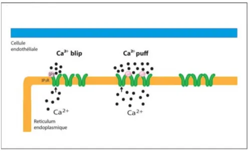
Ca2+ blips are spontaneous Ca2+ increases generated following the activation of one IP3R in the presence of a low concentration of IP3 [100] [101]. These events have a low duration of approximately 100 ms, and an amplitude measured at around 23 nM in cultured bovine pulmonary ECs [102]. Ca2+ blips have been observed in a number of vascular beds [103].
Calcium puffs
Ca2+ puffs are larger that blips and result from the concerted opening of a few IP3
channels in the same cluster. They were first observed in Xenopus oocytes [104] [105] and later on in HeLa cells [106]. They have been visualized in non-excitable cells [107]. Puffs have amplitudes ranging from ~ 50-500 nM, a spatial spread of ~ 2-4 µm2, and a total duration of ~ 1 second. A number of studies have concluded that the temporally and spatially coordinated recruitment of Ca2+ puffs is responsible for the generation of repetitive Ca2+ waves and
oscillations [101] [108].
Figure 3 Calcium blips and puffs (Charbel et al., 2013) Ca2+: Calcium
Calcium sparks
Ca2+ sparks are localized transient increases in Ca2+ that can occur after agonist stimulation as well as under basal conditions following its release through RyR. They have been
initially identified in cardiac cells [109]. Since then, Ca2+ sparks have been described in several
cell types like SMCs and skeletal muscle cells [110]. Sparks display variable spatial distribution with every occurrence. On average, Ca2+ sparks have a spatial spread ~ 12-14 µm2. The fusion of sparks generates propagating Ca2+ waves. Spontaneous spark activity results in a K+ current termed spontaneous transient outward current (STOC) [111]. Ca2+ sparks play a role in muscle
contraction, as well as a negative feedback to muscle contraction by inactivating voltage-dependent Ca2+ channels. Additionally, Ca2+ sparks activate SMCs large conductance Ca2+ -activated K+ channels affecting vascular tone [112] [113].
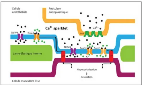
Calcium sparklets
Ca2+ sparklets are intiated following Ca2+ entry through membrane bound Ca2+-
permeable channels [114] [115]. Ca2+ sparklets were initially identified in vascular SMCs of
animals, and cardiac cells of animals, as well as ECs in mesenteric arteries of mice. Extensive investigations had previously concluded that sparklets generally depended on the opening of voltage-dependent Ca2+ channels (VDCC). However, endothelial sparklets are the result of the
opening of a specific type of Ca2+-permeable channels termed transient receptor potential (TRP)
channels. The first endothelial TRP-sparklets to be identified were transient receptor potential vanilloid 4-sparklets [116]. These sparklets occured repetitively at the same site (between IEL holes or at the end of the cells), with a spatial spread averaging around 11 µm2 [114]. Sparklets
were shown to have an effect on a number of proteins localized within MEPs like IK and SK leading to membrane hyperpolarization that is transmitted onto vascular SMCs through gap junctions [114]. Additionally, TRPA1-sparklets were identified in rat cerebral arteries [117]. They were found to induce dilation of cerebral arteries through the activation of IK channels [118].
Figure 4 Calcium sparklets (Charbel et al., 2013) Ca2+: Calcium
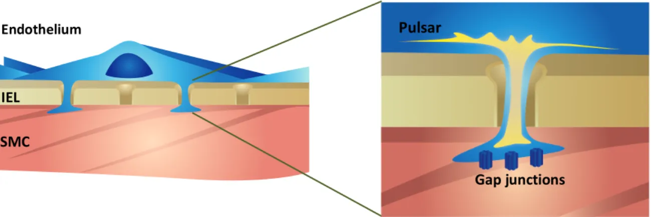
Calcium pulsars
Ca2+ pulsars were first characterized in mouse mesenteric arteries [16]. They are
generated following Ca2+ release from the ER after the activation of several clusters of IP3Rs.
Pulsars occur in ECs under resting conditions and were found to be blocked following the inhibition of PLC [16]. Ca2+ pulsars are a major player in endothelial Ca2+ signalling. This type
of signalling is initiated by the spontaneous activation of IP3Rs, and subsequent spatially restricted Ca2+ release from the ER, within MEPs. The regulatory mechanisms of Ca2+ pulsars
remain to be further explored. Ca2+ pulsars described in mesenteric arteries possess an average
Figure 5 Endothelial calcium pulsars, adapted from Ledoux et al., 2008
Illustration of a Ca2+ pulsar occurring in an IEL hole. IEL: internal elastic lamina. SMC: smooth muscle cell.
Calcium wavelets
Similarly to other aforementioned local Ca2+ transients, Ca2+ wavelets are also initiated
following the activation of IP3Rs [119]. Wavelets are localized in proximity to MEP and play a
role in myoendothelial feedback. Therefore, an initial vasoconstrictive stimulus that depolarizes vascular smooth muscle is followed by a Ca2+ response that includes the activation of endothelial
vasorelaxing actors that include NOS, SK, and IK [119].
Endothelium
IEL
SMC
Gap junctions
Figure 6 Calcium wavelets (Charbel et al., 2013) Ca2+: Calcium
The effect of localized Ca2+ signals on vascular tone often involves additional
transmembrane ion channels, as well as regulatory proteins. In addition to their abundant expression in ECs and SMCs, Transient receptor potential channels have been previously associated with the regulation of vascular reactivity.
3. The transient receptor potential channels family
The transient receptor potential (TRP) family of channels are membrane proteins that mediate the transmembrane flux of cations. TRP channels were first identified in the drosophila visual system where a characterized mutation resulted in an altered response to light [120] [121]. Since then, TRP channels have been described in a large number of organisms such as C. elegans as well as in mammals [122] [123]. TRP channels are expressed in many tissues and cell types. They are involved in a variety of primordial cellular functions including but not limited to cell survival and development, sensory transduction, endocytosis and exocytosis, membrane potential change, and enzymatic activity.
3.1 Classification of TRP channels
There are six subfamilies of TRP that have been identified: TRPC (Canonic), TRPV (Vanilloid), TRPM (Melastatin), TRPP (Polycystine), TRPML (Mucolipine), and TRPA (Ankyrine), and an additional subfamily that has also been described: TRPN (nompC) [124] [125]. In mammals, 28 channels are currently known to belong to six of the seven sub-families (TRPN is absent) [126] [127]. These six subfamilies are divided into two groups based on sequence and topological differences. Group 1 channels include TRPC, TRPA, TRPM and TRPV families; while group 2 includes members of the TRPML and TRPP subfamilies [128]. In humans, approximately 27 genes form the family of TRP channels are described [129].
Figure 7 TRP channels family, modified from Nilius et al., 2007
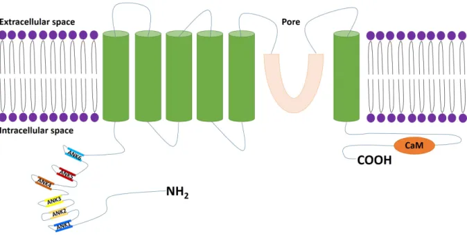
3.2 Structure of TRP channels
All TRP proteins are composed of six transmembrane domains separated by hydrophilic loops. There is a small hydrophobic segment that participates in the formation of the pore between the fifth and the sixth domains [130] [131], as well as cytosolic carboxy and amino termini with multiple protein-protein interaction sites. TRP channels owe their nomenclature to a small well-conserved segment known as TRP, consisting of 23-25 amino acids. This segment was shown to be involved in the regulation of channel activity by certain lipids such as PIP2
[132]. A functional TRP channel is formed by the association of four identical (homotetrameric channel) or different subunits (heterotetrameric channel) [133]. Finally, some protein domains, such as ankyrines, mainly involved in the anchoring of the channels to the cytoskeleton, are conserved in the N-terminal loop of the C and V families.
Figure 8 A: Structure of a TRP channel showing the 6 transmembrane domains and the pore region. B: Tetrameric structure of a TRP channel (Watanabe et al., 2009)
3.3 Activation of TRP channels
An important characteristic of TRP channels is their activation by a large number of stimuli allowing them to be involved in several cellular processes and pathological conditions [128]. TRP channels can be activated by exogenous stimuli such as mechanical forces, stretch, and thermal changes. Additionally, they can be activated by endogenous stimuli such as intracellular mediators, osmotic stress, Ca2+ concentration, and neurohormonal factors. They
can equally be activated by membrane depolarization. For example, activation of the TRPC subfamily members has been shown to be dependent on phospholipase C (PLC), store depletion, conformational coupling, and exocytosis [134]. TRPV channels are activated by high temperatures, chemical compounds [135], PH changes, proinflammatory cytokines, and mechanical stretch (the TRPV family of proteins will be detailed in a later section). TRPA proteins can be activated by chemicals and environmental irritants as well as bradykinin and poly-unsaturated fatty acids [136].
3.4 The TRPV family
TRPV channels were named after the first member TRPV1 was identified due to its reactivity to the inflammatory vanilloid compound capsaicin [137]. The TRPV subfamily is comprised of six members (TRPV1-6).
3.4.1 Expression of TRPV channels
TRPV channels are expressed in a multitude of tissues and cell types. TRPV1-3 are highly expressed in the central nervous system (CNS), particularly in the vagal ganglia and dorsal root ganglia (DRG). TRPV4 is expressed in the kidney, lung, heart, liver, ECs, SMCs, and DRG [138]. TRPV5-6 are found in the intestines, pancreas and placenta. TRPV5 is also expressed in the kidneys [128].
3.4.2 Activation of TRPV channels
A noticeable feature of TRPV channels is their activation by temperature [139]; for example, TRPV1 and TRPV2 are activated by relatively high temperatures (> 43° C and > 52°C respectively) [137]. TRPV3 and TRPV4 activation is increased at lower temperatures (33-39°C and 27-34°C respectively) [134]. TRPV5 and TRPV6 are not affected by thermal variations. TRPV proteins can equally be activated by PLC, arachidonic acid, changes in extracellular osmocity, and mechanical stretch [140]. TRPV channels activity can be affected by additional stimuli such as chemicals, low PH, proinflammatory cytokines, bradykinin, nerve growth factor (NGF), and phosphatidylinositol-3-kinase (PI3K). For example, a low PH contributes to the activation of TRPV1 channels [137] [141] due to the presence of two main PH-sensing sites [142], while TRPV1 and TRPV2 channels can increase in expression and activity following an elevation in pre-inflammatory cytokines [143]. Furthermore, studies have found that the excitability of TRPV1 was upregulated following bradykinin release during airway inflammatory reaction [144]. Moreover, nerve growth factor (NGF)and phosphatidylinositol 3-kinase(PI3K) were both found to cause the activation of TRPV channels; in fact, NGF sensitizes pain-receptor neurons through increased trafficking of TRPV1 channels. This mechanism was found to involve PI3K interaction with TRPV1 [145].
Amongst all TRPV family members, TRPV4 channels’ importance in the regulation of physiological processes has emerged due to their wide distribution, ability to be activated by a large range of stimuli, and their basal activity under normal conditions. Moreover, various pathologies have been attributed to the absence or abnormal functioning of these channels (the properties of TRPV4 channels as well as their role in physiological and pathological states will