Tendinopathy is a major problem in medicine and sports traumatology1. It is due, inter alia, to mechanical overload1. Painful and disabling, it frequently leads to athletes being unable to perform and may sometimes even impair their quality of life. It remains a challenge for the medical world, to the extent that its frequent resistance to conventional treatments rarely gives the patient a favourable outcome following therapeutic management1.
The development of platelet-rich plasma (PRP) offers new hope when therapeutic treatments such as non-steroidal anti-inflammatory drugs, corticosteroid injections, eccentric rehabilitation, shock waves etc. have been unsuccessful2. Current knowledge does not, at this time, consider PRP a primary treatment. This new treatment has drawn the attention of clinicians, especially those active in the
sporting environment. Furthermore, in January 2011, it was removed from the list of doping substances (www.wada-ama.org)2. PLATELET-RICH PLASMA
The platelets perform various functions during the process of haemostasis, they aggregate at the lesion edges to form a procoagulant surface area and are also involved in the inflammatory process through the release of various factors of vascular permeability, the increase in neutrophil chemotaxis and prostaglandin synthesis. Finally, they modulate the immune defence system. The ‘restorative’ properties of platelets are used because they support the healing process of various tissues (bone, skin, muscle and tendon)3.
Their cytoplasm contains granulations that are visible through an electron microscope, including alpha granules
containing various adhesion proteins (particularly the von Willebrand factor), coagulation factors, cytokines, metalloproteinases and various growth factors4. They exert a synergistic action with other signalling molecules and/or other growth factors and are involved in the healing process of tendons, notably4:
• Platelet-derived growth factor (PDGF): stimulates the production of other growth factors and proteins during the inflammatory phase and is involved in tissue remodelling.
• Transforming growth factor-β (TGF-β): stimulates migration and cell proliferation, collagen synthesis and regulates proteinases.
• Fibroblast growth factor (FGF): stimulates angiogenesis and during the proliferative phase, regulates migration and cell proliferation.
PLATELET-RICH
PLASMA FOR
TREATING CHRONIC
TENDINOPATHY
– Written by Jean-François Kaux, Belgium
• Vascular endothelial growth factor (VEGF): stimulates angiogenesis during the proliferative and remodelling phases.
• Hepatocyte growth factor (HGF): stimulates angiogenesis, cell proliferation and migration. It also has an anti-inflammatory and anti-fibrotic action.
• Insulin-like growth factor 1 (IGF-1): leads, during the inflammatory phase, to fibroblast proliferation and migration. During the remodelling phase it promotes the local production of collagen and various structures of the extracellular matrix.
The ratio between the various factors could influence the dynamic balance between the cells, as well as angiogenesis and the formation of the extracellular matrix.
PRP is obtained by centrifugation of autologous blood to obtain a platelet concentration greater than that of the autologous blood, which varies according to the production method5,6. PRP can be activated prior to its injection through the addition of thrombin or calcium chloride, the alternative being in-situ activation by collagen7. PRP also contains a variable amount of lymphocytes, which is suggested to have a detrimental effect on wound healing through the release of pro-inflammatory factors potentially responsible for degradation of the extracellular matrix7. The lack of red blood cells is also necessary because their lysis would result in a release of free radicals that can be damaging to tissue structures7. Currently, there is no formal consensus regarding the production method or the biological composition of PRP7. Based on pre-clinical studies, we can nonetheless
reasonably assume that the ideal PRP should not contain any white or red cells. However, this assertion is not clinically proven. PRECLINICAL STUDIES
Some laboratory studies (in-vitro and/or on animal) emphasise the acceleration of the tendon healing process after the injection of PRP, each growth factor exerting a specific action during healing8. PRP would cause the proliferation, migration and differentiation of cells derived from circulation, improving the initial phase of tendon healing8-13. Cellular activation can be explained by the local production of growth factors. This anabolic process initiates type I collagen synthesis9. PRP stimulates the proliferation of human tenocytes and collagen production but also slightly increases the expression of metalloproteinases involved in remodelling of the extracellular matrix14.
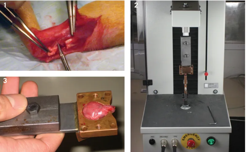
Figure 1: Animal experimental procedures. Surgical procedure: the Achilles and plantaris tendons were dissected. The plantaris tendon was
removed. The Achilles tendon was transected and a 5-mm defect was made. The fascia and the skin were each surgically closed before injection of PRP.
Figure 2: The traction-compression testing machine (106.2 kN, TesT GmbH, Dusseldorf, Germany) and an original clamping device type
'cryo-jaw' to perform the mechanical testing of the tendon.
Figure 3: Once the tendon had healed, the muscle-tendon-bone unit was fastened in the clamping device by freezing the muscular segment
(triceps surae) with liquid nitrogen and clamping it between the 'cryo-jaw'.
However, according to an animal model (progenitor cells of rabbit tendons), PRP was not sufficiently active to slow down the degenerative process of late stage tendinopathy, characterised by lipid deposits, calcification and an accumulation of proteoglycans15.
An injection of PRP improves tendon repair in rats, where the Achilles tendon had been previously sectioned. Within the PRP group, better maturation of the tendinous callus and an increase in its resistance to breakage were assessed9. PRP is suggested to improve the organisation of collagen fibres; stimulating neovascularisation and accelerating the healing process (which was also of better quality), confirming other results in the same experimental conditions9,16,17. After injecting PRP into
rabbits, the mechanical properties (tensile strength and response to mechanical stress) of the injured patellar tendon improves8. VEGF, a growth factor released by platelets, stimulates tendon healing and regulates the expression of other growth factors, including TGF-β18. Horses with injured tendons that are injected with PRP experience, after a few weeks, an increase in DNA, glycosaminoglycans and, in particular, collagen synthesis19. These tendons present better elasticity and a higher resistance to breakage19. Biochemical and histological analyses show a higher metabolic activity and more advanced tissue repair after injecting PRP.
Similar results have been reported in an animal model with patellar tendinitis and chemical calcaneal tendinitis (injection
of collagenase)20 – an ultrasound-guided intra-tendon injection of PRP promoted the healing of these chemical tendinopathies8. CLINICAL STUDIES
Although the effectiveness of PRP on tendon healing in-vitro or in animals seems to be confirmed, clinical studies are currently still controversial. In addition, there are a growing number of randomised and controlled studies reviewing PRP for treating tendinopathy.
Epicondylitis
The very first study specifically assessing the effect of PRP when treating tendinopathy was reported on epicondylitis21. The two small non-randomised groups (a single injection of PRP (n=15) vs. local anaesthetic
Images: Activated pure PRP (without
leukocytes) in the syringe and injections of PRP in various tendons (patellar and calcaneal) under aseptic condition and ultrasound guidance.
weeks, 6 months and 24 months. After 2 years, 94% of patients returned to sport and/ or previous professional activities; however, longitudinal observation of patient controls did not exceed 8 weeks and was not able to be compared to the PRP group further.
A first systematic review (literature until October 2013 included) concluded that there is evidence against the efficacy of PRP in the treatment of epicondylitis22. Of the six studies included, only four were considered high-level and among those, three showed that injections of PRP were not effective in treating epicondylitis, with one showing a real clinical efficacy. However, in a meta-analysis (articles until March 2014), seven randomised controlled trials (RCT) were selected (including 6 selected in the previous systematic review)23. When analysing the results of the different studies, a reduction in pain and algo-functional scores were observed within the patients in the PRP group compared to the control groups. It takes at least 3 to 6 months before this significant improvement could be observed. Another systematic review (articles until April 2014) also concluded that the use of PRP for treating epicondylitis is very promising in the short- to medium-term (up to 6 months)24. In conclusion, even though some evidence exists to support the use of PRP in chronic epicondylitis, the current literature does not provide strong evidence for its use in first-line treatment.
Tendinopathies of the rotator cuff
In the same meta-analysis (articles until March 2014), two RCTs compared the effect of PRP on tendinopathy of the rotator cuff with a control group23. Among them, one study (6 months) highlighted the effectiveness of two PRP injections compared to two injections with a simple dry needle under ultrasound guidance25. After the first injection, the two groups developed comparably, after the second injection of PRP, clinical improve-ment (pain and shoulder mobility) occurred, compared to the control group. The results of the meta-analysis (up to 1 year) showed favourable development compared with the control groups, but was less evident than for epicondylitis. This was confirmed in a final systematic review (articles up to April
2014), concluding that there was a lack of conclusive evidence to support the use of PRP in this indication24.
Patellar tendinopathy
A systematic review of 13 studies on PRP (articles up to July 2014) included two RCTs on the effect of PRP on patellar tendinopathy26. Two injections of PRP were compared with three shockwave sessions27. The PRP group showed better development than the shockwave group at 6 and 12 months for pain and algo-functional scores, showing that injections of PRP are more effective in the medium-term than the shockwaves in this indication. The authors of the systematic review therefore concluded that PRP would seem more useful for treating stubborn tendinopathy than other conservative treatments and can be regarded as a second-line therapy26. However, evidence from high-level studies is still needed. Three controlled studies (including 2 RCTs) analysed in another systematic review (articles up to February 2014) showed that for patellar tendinopathy, even if PRP is a safe and promising technique, its superiority over other conservative treatments is unproven28.
Calcaneal tendinopathy
Calcaneal or Achilles tendinopathy is very common among athletes, especially long distance runners1. A systematic review of 12 studies (articles up to July 2014), of which only one was an RCT (which was also included in a meta-analysis (articles up to March 2014) and in another systematic review (articles up to April 2014)) assessed PRP as a treatment for Achilles tendinopathy23,24,26. However, it did not show the efficacy of PRP (clinical and
ultrasound) compared to an injection of body fluid followed by a 3 months eccentric rehabilitation, with a 1 year follow-up29,30. This study has some limitations, including the lack of eccentric rehabilitation (which may be effective only before the injection) in all patients before inclusion31. Nevertheless, until now, there remains an absence of evidence for the use of PRP in calcaneal insertional tendinopathy.
Plantar fasciopathy
Also known by the name plantar fasciitis, it is an irritation of the fascia sheathing the tendons responsible for maintaining the plantar arch. It is not a tendon itself, but the symptoms and accompanying treatment are similar to those found in tendinopathy, which is why it has been included in this review.
A systematic review (articles up to June 2014) analysed eight studies including three RCTs32. Although current observations and conclusions are promising, they remain limited with regard to the change in pain and high-functional scores of the PRP group compared with the control group, justifying the continuation of high-level studies in this pathology.
DISCUSSION
Tendinopathy is a very common condition in sportspeople. Its treatment remains a challenge because many athletes disregard classical conservative treatments and the condition becomes chronic. For this reason, new treatments have been developed, including PRP injections. However, until now, despite the in-vitro efficacy of PRP on tissue regeneration8, there is an absence of strong evidence to support its use on tendinopathy22,24,26,28,32.
• PRP should be injected into the lesion, possibly under ultrasound
guidance. After the injection, submaximal and gradual eccentric
rehabilitation improves the quality of tendon healing.
• Based on the current literature, even though some evidence exists to
support the use of PRP injections to treat epicondylitis and patellar
tendinopathies, this treatment is not recommended as a first-line
treatment of tendinopathies.
PRP therapy has been used for several years in various surgical specialties, with favourable clinical results regularly suggested3. Through the release of platelet growth factors, PRP would likely be an attractive alternative therapy for chronic tendinopathy its easy preparation and low invasiveness are additional advantages3. Studies on the use of PRP have not reported any serious side effects33, nevertheless, it should be ensured that any side effects are actually published. Studies are difficult to compare because the methods of preparation, the qualitative characteristics of PRP (volume, platelet concentration, presence of leukocytes and erythrocytes), the injection technique and the post-injection rehabilitative protocol are not clearly defined.
After the injection, submaximal and gradual eccentric rehabilitation improves the healing quality of the tendon34. Indeed, PRP initiates a healing process secondarily developed by the vector of eccentric forces35.
PRP exhibits very variable platelet, erythrocyte and leukocyte concentrations depending on its preparation protocol5,6. The optimum concentration of platelets for therapeutic treatment of tendon injuries has not yet been validated. The pre-clinical literature suggests, however, the following7: • A platelet concentration of less than 106 platelets/μL (i.e. 3 to 4x blood platelet concentration) might be optimal, while a concentration higher than 1.2 x 10⁶ would have a paradoxical effect of inhibiting collagen synthesis.
• There is an ongoing debate on the effect of leukocytes. Some studies do not advocate the inclusion of white blood cells because they might slow the healing process due to the early
induction of a larger local inflammatory reaction and the presence of pro-inflammatory factors (cytokines and metalloproteinases) capable of degrading the extracellular matrix. PRP containing leukocytes would also reduce collagen synthesis. In addition to this it was shown that the anti-bacterial effect of PRP against Staphylococcus aureus, Staphylococcus epidermidis, Propionibacterium acnes and methicillin-resistant Staphylococcus aureus (MRSA) was not related to the presence of white blood cells.
• No red blood cells; once lysed they release various free radicals similar to those of leukocytes that could actually damage the surrounding tissue.
Ideally, one should be able to inject a reproducible PRP (same platelet concentration) into all patients. Currently, unlike all other PRP preparation techniques, only platelet collection via an apheresis machine can easily achieve this36,37.
Even if the product spreads from the injection site, it should be performed under ultrasound guidance while respecting aseptic precautions. Local anaesthesia is not recommended so as not to compromise the therapeutic potential of PRP, but this recommendation is not supported by strong evidence7. A preliminary activation (e.g. CaCl2) would allow for platelet degranulation and a more rapid release of growth factors, but its suggested effect is variable38. During the post-injection hyperalgic period, only painkillers and cryotherapy are allowed. Non-steroidal anti-inflammatory drugs must be avoided, at least from 10 days before to 21 days after the injection, as they may prevent PRP from working7.
Thereafter, sub-maximal eccentric rehabilitation of gradually increasing intensity combined with stretching will ‘guide’ tendon healing34. During the gradual return to sport any technopathy will be corrected and the tendon would be somewhat ‘protected’ using ‘functional’ orthotics (e.g. patellar band) or strapping7. The results at 3 months are used to evaluate the effect of the injection of PRP and discuss the indication of a second injection of PRP in case of a partial improvement of tendon symptoms. Indeed, two successive injections of PRP (15 days apart) do not seem to be justified37. Surgery should be discussed if there is no improvement.
CONCLUSION
Experimentally, through the local release of various growth factors, PRP stimulates tendon healing. This therapy could optimise the healing of pathological human tendons. However, based on the current literature, PRP injections are not recommended as first-line treatment for tendinopathies. PRP should be analysed to see whether it improves tendon healing in both humans and animals through further RCTs. Indeed, the effectiveness of PRP in clinical practice remains controversial despite interesting results for patellar tendinopathy and epicondylitis.
Jean- François Kaux M.D., Ph.D. Senior Consultant Department of Physical Medicine, Rehabilitation and Sports Traumatology FIFA Medical Centre of Excellence University Hospital and University of Liège Liège, Belgium Contact: [email protected]
Recommendations for future research
• A standardisation of the quality of the PRP is mandatory. The use of
an identical platelet concentration with the same technique among
all patients could help to improve this therapy. Moreover, a general
agreement on the presence or not of leukocytes is required, even if
pre-clinical studies advise against their presence.
• Randomised controlled studies are still needed to give strong evidence
for the use of PRP injections in chronic tendinopathies.
References
Available at www.aspetar.com/journal