HAL Id: tel-02871202
https://tel.archives-ouvertes.fr/tel-02871202
Submitted on 17 Jun 2020HAL is a multi-disciplinary open access archive for the deposit and dissemination of sci-entific research documents, whether they are pub-lished or not. The documents may come from teaching and research institutions in France or abroad, or from public or private research centers.
L’archive ouverte pluridisciplinaire HAL, est destinée au dépôt et à la diffusion de documents scientifiques de niveau recherche, publiés ou non, émanant des établissements d’enseignement et de recherche français ou étrangers, des laboratoires publics ou privés.
Triglyceride-sensing in the mesocorticolimbic system
and reward-driven behaviour control
Chloe Berland
To cite this version:
Chloe Berland. Triglyceride-sensing in the mesocorticolimbic system and reward-driven behaviour control. Tissues and Organs [q-bio.TO]. Université Sorbonne Paris Cité, 2018. English. �NNT : 2018USPCC297�. �tel-02871202�
Thèse de doctorat
de l’Université Sorbonne Paris Cité
Préparée à l’Université Paris Diderot
Ecole doctorale
Bio Sorbonne Paris CitéLaboratoire Unité Biologie Fonctionnelle et Adaptative, UMR CNRS 8251
Equipe Central COntrol oF Feeding behaviour and Energy Expenditure (C
2OFFEE)
Triglyceride-sensing in the
mesocorticolimbic system and
reward-driven behaviour control
Par Chloé Berland
Thèse de doctorat de Physiologie
Dirigée par le Dr Serge Luquet
Présentée et soutenue publiquement à l’Université Paris Diderot, le 11 Septembre 2018
Président du jury : Pr. Alain Zider, Université Paris Diderot Rapporteur : Pr. Stephanie Fulton, University of Montreal, Rapporteur : Dr Vincent Prévot, Université de Lille
Examinateur : Dr. Xavier Fioramonti, Université de Bordeaux, Examinateur : Dr. Patricia Parnet, Université de Nantes, Examinateur : Dr. Isabelle Niot, Université de Dijon
Contents
Contents ... 2
Acknowledgments ... 8
Foreword ... 10
Introduction ... 12
Chapter 1: The central control of feeding behaviour integrates multiple peripheral stimuli .. 13
A. Homeostatic regulation of food intake ... 13
1. Central regions involved in energy sensing ... 13
2. Peripheral molecules signalling energy status ... 15
3. Neurons regulating the energy balance: the melanocortin circuit ... 19
B. Reward-related networks controlling feeding behaviour ... 23
1. The dopaminergic pathway involved in hedonic aspect of feeding ... 24
2. The lateral hypothalamus, cross-talk between the homeostatic and non-homeostatic regulation of food-intake ... 28
3. Endocannabinoids control of food intake and energy balance ... 29
4. The opioid system encoding the ‘liking’ of food ... 31
C. Dysregulations of feeding behaviour: driven by food reward ... 32
1. Physiological role of dopamine ... 32
2. Compulsive feeding behaviours: can we talk about food addiction? ... 34
3. Overeating is often associated with dopamine signalling pathologies ... 35
D. Molecular basis underlying dopamine-driven reward ... 36
1. Dopaminergic firing and dopamine release in the synapse ... 37
2. Two classes of dopamine receptors ... 37
3. G protein induced signalling through PKA pathway ... 38
4. PKC/Calcium signalling pathway of dopamine receptors ... 40
5. Akt/GSK3 dopamine receptor signalling pathway ... 40
Chapter 2: Lipids as signalling molecules for the reward circuit ... 43
A. Nature and availability of dietary lipids in periphery ... 43
1. Lipid classification ... 43
2. Lipids availability and transport in periphery ... 45
B. Lipid access to the brain ... 48
1. Around the brain: the blood-brain-barrier, composed of endothelial cells, astrocytes, pericytes, neurons and microglia ... 48
C. Lipids as signalling molecules within the brain ... 54
1. Lipids as signalling molecules in charge of glucose homeostasis, food intake and energy expenditure ... 54
2. Impact of dietary lipids on reward and mood ... 55
3. Cognitive impairments ... 57
D. The lipoprotein lipase is expressed in the brain and converts TG into FFA ... 58
1. Lipoprotein lipase expression within the brain ... 58
2. LPL roles ... 58
3. LPL regulation ... 59
Aim of the thesis ... 60
Results ... 61
Chapter 1: Dopamine D2 receptor and dopamine signalling are direct target of nutritional triglycerides in the control of reward driven behaviours ... 62
A. Article information ... 62
B. Introduction ... 64
C. Materials and methods ... 66
D. Results... 70
1. The triglyceride-processing enzyme lipoprotein lipase is expressed in both mouse and human mesocorticolimbic structures. ... 70
2. Striatal TG metabolism decreases amphetamine-induced behavioral and molecular adaptations ... 70
3. Triglycerides exert modulatory actions onto dopamine signalling. ... 72
4. Central triglyceride delivery alters D2R-mediated functions while sparing D1R. ... 72
5. Centrally delivered triglycerides differently recruit post- and pre-synaptic components of the dopaminergic circuit regarding the time of exposure. ... 73
6. Central triglyceride delivery is reinforcing in lean but not obese animal. ... 73
7. Central triglyceride sensing differentially impacts food and non-food reward ... 74
8. Motivational and hedonic aspects of reward are differentially affected by central triglyceride delivery. ... 75
9. Lipoprotein lipase controls reward seeking behaviour through pre and post synaptic action 76 10. In humans, postprandial triglycerides control brain response to food-cues in dopamine receptor 2-depedent manner. ... 77
E. Discussion ... 79
1. Bridging dietary input to reward through triglycerides sensing in the reward system ... 79
2. Physiological implication of TG-mediated modulation of DRD2 signaling ... 80
F. Acknowledgments ... 84
G. Figure legends ... 85
H. Bibliography ... 89
I. Supplementary experimental procedures ... 112
Chapter 2: Palatable binge-drinking shapes dopamine signalling in the striatum through gut-striatal mechanisms ... 122
A. Article Information ... 122
B. Abstract ... 123
C. Material and methods ... 124
D. Introduction ... 127
E. Results... 128
1. Intermittent access to a caloric mixture quickly shifts eating pattern and triggers metabolic signatures ... 128
2. Gut peripheral signals, not circulating insulin and leptin, control bingeing ... 129
3. Intermittent access to a caloric drink triggers protein S6 kinase activation in the striatum and nucleus accumbens ... 130
4. Increased translational activity in the dorsal striatum and NAc is D1, not D2 dependent 131 5. Bingeing-like behaviour is associated with lipid signalling in reward related structures 132 F. Discussion ... 133
G. Bibliography ... 134
H. Figure Legends ... 146
Discussion ... 148
A. Results summary ... 149
B. Comments on NaCl 0.9% as a control for Intralipid in first study ... 149
C. Comments on D2R indirect pathway inhibition by central TG, and biphasic alteration of reward ... 151
1. Predilection site for TG sensing? ... 151
2. Putative mechanism for short-term effect of central TG delivery ... 152
3. Putative mechanism for long-term effect of central TG delivery ... 153
4. Long-term exposure to TG: a mechanism to explain reduced D2R signalling in obese? 154 D. Comments on different regulation of food reward and drug reward by central TG .. 154
E. TG metabolism and LPL role in lipid sensing ... 155
1. Putative role for LPL in D2R signalling ... 155
2. Other lipases involved in TG sensing? ... 156
3. LPL as the missing link to understand FFA and TG differences in lipid sensing? ... 157
G. Conclusion ... 158
Index ... 159
References ... 162
Figure 1 Prevalence of overweight (World Health Organisation database) ... 10
Figure 2 Central Regions Involved in the Energy Sensing ... 14
Figure 3 Peripheral signalling of energy status. ... 16
Figure 4 Some peripheral signals involved in feeding behaviour regulation. ... 17
Figure 5 Molecular integration of energy signalling: mTOR signalling. ... 19
Figure 6 The Melanocortin pathway composed of the antagonist NPY/AgRP and POMC neurons .... 20
Figure 7 Second order Neurons signalling and Food intake regulation ... 22
Figure 8 Interaction between metabolic signalling and mesocorticolimbic signalling. ... 23
Figure 9 The Basal Ganglia ... 25
Figure 10 The Mesocorticolimbic Pathway ... 25
Figure 11 Connectivity of the Nucleus Accumbens ... 26
Figure 12 Synaptic connections involving the Ventral Tegmental Area ... 26
Figure 13 Interactions between the mesocorticolimbic pathways and hunger related regions ... 27
Figure 14 Lateral hypothalamus interactions with the reward circuit ... 29
Figure 15 Endocannabinoids Synthesis. ... 30
Figure 16 Endocannabinoids and the control of energy balance ... 30
Figure 17 Endocannabinoids and the Dopaminergic Synapse ... 31
Figure 18 The nucleus accumbens µ-opioid hotspot involved in food ‘liking’ ... 31
Figure 19 Dopamine Synthesis and Degradation ... 32
Figure 20 Dopaminergic Firing in conditioned reward ... 33
Figure 21 Motivational and molecular adaptations in overeating. ... 35
Figure 22 Brain Dopamine and Obesity ... 36
Figure 23 Tonic and Phasic Firing of Dopaminergic Neurons ... 37
Figure 24 G-Protein coupled Receptors Signalling ... 37
Figure 25 DA Signalling in MSN induced by GPCR ... 39
Figure 26 Dopamine Receptors signalling through G-α-q subunits ... 40
Figure 27 Dopamine D2 Receptor signalling through beta-arrestin/Akt/GSK3 pathway ... 41
Figure 28 Dopamine Receptors Desensitisation and Internalisation through GRK/arrestins ... 42
Figure 29 Main Fatty Acids in the Brain. ... 44
Figure 30 Simplified Classification of lipid species in the brain ... 45
Figure 31 Triglycerides absorption in the enterocytes ... 46
Figure 32 Triglycerides transportation in the body ... 47
Figure 33 The blood brain barrier is formed by several layers protecting and isolating the brain from the periphery ... 48
Figure 34 A neurovascular unit ... 48
Figure 35 Molecular transports across the BBB ... 49
Figure 37 Fatty Acid Transporters in the Brain ... 51
Figure 38 Molecules involved in lipoproteins transport and hydrolysis are expressed within the brain. ... 53
Figure 39 Permeability changes at the blood-brain-barrier in obesity. ... 53
Figure 40 Central triglycerides sensing inhibits dopaminergic associated behaviours ... 56
Figure 41 Peripheral Triglycerides increase dopamine release in the NAc... 57
Figure 42 Lipoprotein lipase roles in the brain ... 59
Figure 43 Intralipid Composition ... 150
Figure 44 Mesocorticolimbic structures involved in TG sensing. ... 151
Figure 45 Putative effect of short term TG delivery in the MCL ... 152
Figure 46 Putative effect of long term central TG delivery ... 153
Figure 47 Schematic representation of altered neurotransmission in the dorsal striatum following H-FABP deletion. ... 156
Acknowledgments
This work would not have been possible without the tremendous amount of people who helped me unconditionally during these four years.
First, I would like to acknowledge the members of my jury for reading this manuscript and attending my PhD defence, I hope you will enjoy the reading as much as I enjoyed the writing.
Ensuite, je voudrais remercier Serge Luquet pour m’avoir recueillie dans son laboratoire plutôt que m’avoir laissée vagabonder en fac de lettres. Je ne te remercierai jamais assez pour avoir toujours été à l’écoute, et pour les innombrables discussions que nous avons eues, je ne me suis jamais sentie seule lors de cette thèse. Merci surtout d’avoir partagé mes éclats de joies pas du tout dissimulés lorsqu’un résultat positif sortait après des semaines de galères, je ne me serais probablement pas investie autant sans cet enthousiasme partagé que nous avons eu pour le projet.
Evidemment, je me dois de remercier ensuite Giuseppe Gangarossa, sans qui cette thèse ne serait que l’ombre d’elle-même. Merci de ta patience devant mes innombrables questions et mon manque d’organisation. Je ne mesure que trop la chance que j’ai eue d’avoir travaillé avec toi, et je voudrais ici souligner combien j’estime tout le temps que tu as passé auprès de moi, depuis ces weekends à pester en EPF, jusqu’à ces heures interminables à relire absolument tous mes écrits, y compris ce pavé interminable. Encore merci.
Je remercie nos collaborateurs au cours de ce projet, ils sont très nombreux. Merci à l’équipe de Martine Cador et Stéphanie Caillé-Garnier pour leur chaleureux accueil à Bordeaux, et leur aide dans l’établissement des protocoles de self-administration. Merci ensuite à l’équipe de Philippe Faure, et notamment Fabio Marti et Stefania Tolu, pour avoir réalisé tous les enregistrements d’électrophysiologie et avoir répondu à mes très nombreuses questions de novice. I would like to thank Tom Hnasko for all the precious advice regarding CPP analysis, reward and dopamine in general, and for the great exchanges we had in San Diego. Ali Shenasa, thank you for sharing so much with me, I’m not talking just about RNAscope data here, but also coffees in San Diego and French literature books, and mostly thank you for understanding the terrific burden that daily carotid infusions can bring in a student’s life, and still be happy to go for it. Finally, thank you to Dana Small and her team for sharing the human data with us.
Un immense MERCI à toute l’équipe COFFEE qui m’a épaulée tout au long de cette thèse. D’abord, merci pour leur aide en cette fin de thèse (un peu) chaotique à Julien Castel, Chloé Morel, Anne-Sophie Delbès, Enrica Montalban, Raphaël Denis, Claire Martin, Rim Hassouna, Daniela Herrera, Ewout Foppen, qui ont tous sans exception mis la main à la pâte dans ce travail, et qui en plus l’ont fait avec beaucoup d’humour et de bienveillance. J’apprends constamment auprès de vous, et travailler avec une telle équipe est un réel bonheur. Une petite mention supplémentaire pour mes collègues de bureau, vous qui étiez là depuis le tout début, merci de me faire autant sourire chaque jour, quelles que soient les circonstances !
Notre vie de laboratoire ne serait vraiment pas la même sans l’aide précieuse d’Olja Kacanski, qui arriver presque à nous faire oublier que l’administration universitaire est fort pénible parfois, et à Isabelle Le Parco, Daniel Quintas, Ludovic Maingault, Angélique Dauvin, Florianne Michel. Merci à chacun d’entre vous, pour votre gentillesse et votre aide de chaque jour en animalerie, pour votre bienveillance envers mes innombrables bestioles, et puis envers moi, aussi.
Je voudrais remercier tous ceux qui ont été près de moi ces quatre années, et qui m’ont toujours encouragé, sans toujours comprendre pourquoi je passais autant de temps au labo le weekend, mais qui somme toute, ont compris que cela me tenait à cœur, et même qui sont venus passer du temps à BNF malgré la froideur des rues pour me tenir compagnie pendant deux sessions de conditionnement. Je pense en particulier à un certain anniversaire qu’on a dû passer au Trac juste parce que j’avais manip, encore désolée pour ça les copains. A tous les copains de fac évidemment, les magistériens, les magistériens par procuration, ceux qui ne vivent pas en France aussi. Ça fait 4 ans qu’on est en thèse, 4 ans que je sais toujours pas sur quoi vous travaillez. Merci. Je remercie ici aussi l’équipe de Christophe, pour les midis passés ensemble, sans lesquels nos journées paraitraient bien plus longues, et le 5e
étage bien plus morose !
Enfin, merci à mes parents, à ma sœur, et à ma petite mamie, pour votre soutien dans ces interminables études. J’en ai fait du chemin, depuis mes balbutiements scientifiques devant la cheminée avec le boulier en bois, jusqu’aujourd’hui ! Merci d’avoir favorisé aux poupées les tours à bois et les cartes électroniques à souder quand on était petites, mine de rien, ça joue dans la décision de faire une thèse tout ça ! Et Thibeault, évidemment. Pour avoir été tellement drôle (Ha !) et parce que on était tellement persuadés qu’à l’issue de nos rédactions de thèses respectives, il y aurait au moins un mort, et que ce ne serait pas Mousse. Juste ça. Merci.
Foreword
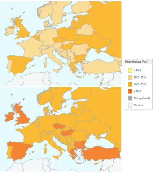
The World Health Organization (WHO) defines overweight and obesity as abnormal or excessive fat accumulation that may impair health. According to the WHO latest report, worldwide obesity (defined as a body mass index (BMI) greater than 30) has nearly tripled since 1975. In 2016, more than 1.9 billion adults (39%), 18 years and older, were overweight (BMI greater than 25). Among these, over 650 million were obese (13%). In France, 59,5% of the population is overweight. Because of the regular worldwide increase of overweight population across the years, obesity is now seen as a ‘major noncommunicable disease’, which kills more people than underweight because of associated diseases such as cardiovascular illnesses (mainly heart disease and stroke), diabetes, musculoskeletal disorders, some cancers (including endometrial, breast, ovarian, prostate, liver, gallbladder, kidney, and colon) (WHO, 2018).
Figure 1 Prevalence of overweight (World Health Organisation database)
Obesity and overweight are caused by an energy imbalance between calories consumed and calories expended. Physiologically, the brain regulates the body weight to a set point that will remain controlled during the life, by controlling the energy balance, that is to say the levels of input energy (food intake) versus the levels of output energy (metabolism, thermogenesis, activity). Evolutionary speaking, mammals developed a preference for energy-dense foods high in fat and sugar, to sustain an adequate level of energy no matter the circumstances. However, our lifestyles changed towards an increased intake of calorie-rich food, and a decrease in physical activity due to sedentary forms of work, or changing modes of transportation, that leads us to eat more for hedonic reasons than satisfaction of needs. Why are we so prone to eat more than we should? As we will see in the introduction of this thesis, the brain is in charge of energy regulation, thanks to the guidance of many molecular signals from the periphery. Among these signals, dietary lipids were found to signal within the brain, and participate in food intake regulation, and alter food reward and motivation. The aim of this thesis was to understand by which mechanisms the fat contained in a diet can potentially target the mesocorticolimbic system participating in food reward encoding.
Chapter 1: The central control of feeding
behaviour integrates multiple peripheral
stimuli
Living organisms require a fixed energy status with a balance between food intake and energy expenditure consisting in basal metabolism, thermogenesis, and physical activity. This regulation requires a communication between the brain, or central part, and the rest of the body, named periphery. In addition to nutrients requirements to sustain metabolic demands, several emotional factors, such as stress or boredom, can affect food intake and energy expenditure. For that reason, feeding can be qualified as either homeostatic, when it provides the necessary intake of calories to sustain life, or non-homeostatic, when it is driven by other processes, such as pleasure, appeal for food-reward, and often results in a higher intake than required. This chapter reviews the current knowledge regarding the central control of feeding behaviour.
A. Homeostatic regulation of food intake
The first cases of obesity involving central dysfunctions were described after noticing that tumours in the brain region of the hypothalamus were associated to massive gain weight and hyperphagic behaviours in patients (reviewed in Bray and York, 1979). Subsequent research in animals, involving hypothalamic lesions or electrical stimulations, pointed out the ventromedial region of the hypothalamus as inhibiting food intake (Hetherington and Ranson, 1939), and the lateral region of the hypothalamus as increasing food intake (Anand and Brobeck, 1951; Delgado and Anand, 1953). The major role of the hypothalamus in feeding behaviour control was then completed with the study of the two mice strains
ob/ob, homozygous mutant for the obese (ob) gene, and db/db, homozygous mutant for the diabetic
gene (db), displaying massive overeating and obesity, which showed the existence of a peripheral secreted peptide encoded by the ob gene, whereas the db gene would encode its corresponding central receptor (Coleman, 1973; Coleman and Hummel, 1969). More recent studies demonstrate that several central regions and not just the hypothalamus are actually involved in food intake regulation, thanks to neuronal communication between these different areas, and the periphery via different neurotransmitters and circulating signals.
1. Central regions involved in energy sensing
The brain is surrounded by an impermeable barrier, the blood-brain-barrier (BBB), which filters molecules accessing the brain, thus providing an efficient protection for the central nervous system. For
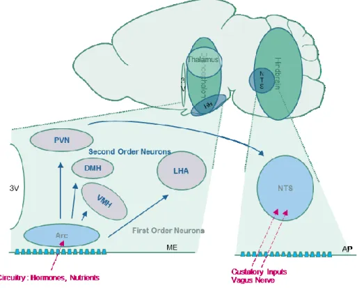
this reason, circulating molecules indicating the energy state from the periphery cannot easily reach the brain, unless some mechanisms allow the BBB crossing (for more details see II.B.1, page 48). Because they are located in brain regions with a thinner BBB, two main areas are able to sense the peripheral energy levels, thanks to a privileged communication with the blood circulation: the arcuate nucleus (Arc) of the hypothalamus, and the nucleus of the tractus solitary (NTS) (Morton et al., 2006, 2014; Schwartz et al., 2000) (Figure 2).
Figure 2 Central Regions Involved in the Energy Sensing
Blue nuclei contain first order neurons, grey nuclei contain second order neurons. Blue arrows represent the neuronal connections between the different areas. Pink arrows represent peripheral outputs signalling the energy state. 3V: third ventricle, HH: hypothalamus, NTS: Nucleus of the Solitary Tract, PVN: paraventricular nucleus, DMH: dorsomedial hypothalamus, VMH: ventromedial hypothalamus, LHA: lateral hypothalamus, Arc: arcuate nucleus, ME: median eminence, AP: area postrema.
i. Anatomy of the mediobasal hypothalamus
The hypothalamus, a brain part of the diencephalon, is located under the thalamus. This area is close to the median eminence (ME), a circumventricular organ with many permeable fenestrated capillaries, which facilitate the exchanges with the blood. Consequently, it collects and integrates information from the peripheral organs. The hypothalamus can be divided in different nuclei based on anatomical boundaries (Lechan and Toni, 2000), gene expression and function (Suzuki et al., 2012). Among these nuclei, the Arc is the closest to the ME, and is the first region to perceive peripheral signals. For that reason, the Arc neurons are called first-order neurons. These neurons work together with second-order neurons of the hypothalamus, located in several nuclei such as the paraventricular nucleus (PVN),
dorsomedial hypothalamus (DMH), ventromedial nucleus of the hypothalamus (VMH), and lateral hypothalamus (LH) (Figure 2).
ii. Anatomy of the Nucleus of the Tractus Solitary
The Nucleus of the Tractus Solitary (NTS) is a sensory nucleus located in the hindbrain. Because of its location near the area postrema (AP), a region composed of fenestrated capillaries, the NTS accesses nutrients circulating after a meal such as glucose (Ritter et al., 2000), or leucine, an essential branched-chain amino-acid (Blouet and Schwartz, 2012). It is also the first region of the central nervous system that receives neural inputs from the Vagus nerve (X), which brings information from the viscera such as the size of the stomach attesting the size of a meal (Phillips and Powley, 1996), and neurotransmitters from the gut. The NTS also receives gustatory signals from post-oral sites reflecting chemical, mechanical and nutrient properties of ingested foods (Van Buskirk and Erickson, 1977). Importantly, the NTS integrates signalling from the PVN of the hypothalamus (Blevins et al., 2004) (Figure 2), and these two structures, in addition to other structures, work together to sense the peripheral energy state (Morton et al., 2014).
2. Peripheral molecules signalling energy status
After a meal, the energy levels are high, the energy balance favours decreased food intake and increased total energy expenditure. Signals that will lead to this satiety state will be reported as ‘anorexigenic’ signals. On the other hand, during fasting, the energy levels are low, therefore the brain increases food intake and decrease energy expenditure. Signals that will lead to this hunger state will be reported as ‘orexigenic’ signals.
i. Nature of endogenous peripheral signals controlling feeding behaviour
The peripheral energy state signalling to the central nervous system are very diverse: gustatory inputs from the tongue, signals from the stomach and digestive system, post-prandial circulatory signals, energy storage signals (Figure 3, Figure 4).
Leptin is an anorexigenic peptidic hormone produced mainly by white adipose cells (Zhang et al., 1994), and its level of production is proportional to the level of adipocytes in periphery (Maffei et al., 1995a, 1995b). It is encoded by the obese gene ob, and administration of leptin to ob/ob mice with no functional
ob gene results in marked decreases in food intake and body weight (Ahima et al., 1996; Campfield et
al., 1995; Halaas et al., 1995; Pelleymounter et al., 1995). Leptin can reach the brain (Banks et al., 1996), where the leptin receptor encoded by the LepRb gene is mostly expressed in the Arc and parts of the VMH, DHM (Håkansson et al., 1996; Mercer et al., 1996).
Insulin is a peptidic hormone produced by the pancreatic beta cells depending on blood glucose levels. Insulin can reach the brain proportionally to its plasmatic levels (Baura et al., 1993), where insulin
1986). Intracerebroventricular1 (ICV) insulin injections decrease food intake and body weight (Ikeda et
al., 1986; Woods et al., 1979). Insulin receptor knock-out in the brain and more particularly in the Arc leads to hyperphagia and obesity (Brüning et al., 2000; Obici et al., 2002a).
élément sous droit, diffusion non autorisée
Figure 3 Peripheral signalling of energy status.
In purple: leptin signalling from the adipose tissue. In red: nutrients signalling. FFAs: Free Fatty Acids, aa: amino-acids. In yellow: ghrelin orexigenic signalling from the stomach. In blue: gastro-intestine satiety signals. CCK: cholecystokinin, GLP1: Glucagon-Like Peptide 1, PYY3-36: Peptide YY3-36. The green (+) represent the positive effect of signals on food intake, the red (-) represent the inhibitory effect of signals on food intake. Adapted from (Morton et al. 2014)
Several gastrointestinal peptidic hormones also act as satiety signals. Cholecystokinin (CCK) is secreted in the duodenum to release digestive enzymes and acts as an anorexigenic signal on the NTS (Gibbs et al., 1973). Recent studies showed that CCK is also produced in the NTS proportionally to the ingested nutrients (D’Agostino et al., 2016). Peptide YY3–36 (PYY3–36) is an enteric hormone released
from the gastrointestinal tract proportionally to the calorie-content of a meal (Adrian et al., 1985), that can reduce food intake in rodents and humans by targeting the Arc (Batterham et al., 2002, 2003). Glucagon-like peptide 1 (GLP-1) is a post-translational processing of the proglucagon gene, produced by intestinal cells after a meal, and by the NTS (Lockie, 2013) and ICV administration of GLP-1 reduces food intake in fasted rats (Turton et al., 1996).
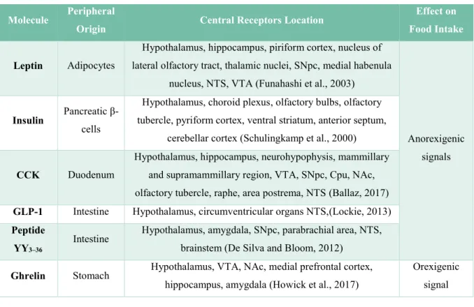
Molecule Peripheral
Origin Central Receptors Location
Effect on Food Intake
Leptin Adipocytes
Hypothalamus, hippocampus, piriform cortex, nucleus of lateral olfactory tract, thalamic nuclei, SNpc, medial habenula
nucleus, NTS, VTA (Funahashi et al., 2003)
Anorexigenic signals
Insulin Pancreatic
β-cells
Hypothalamus, choroid plexus, olfactory bulbs, olfactory tubercle, pyriform cortex, ventral striatum, anterior septum,
cerebellar cortex (Schulingkamp et al., 2000)
CCK Duodenum
Hypothalamus, hippocampus, neurohypophysis, mammillary and supramammillary region, VTA, SNpc, Cpu, NAc, olfactory tubercle, raphe, area postrema, NTS (Ballaz, 2017)
GLP-1 Intestine Hypothalamus, circumventricular organs NTS,(Lockie, 2013)
Peptide YY3–36
Intestine Hypothalamus, amygdala, SNpc, parabrachial area, NTS, brainstem (De Silva and Bloom, 2012)
Ghrelin Stomach Hypothalamus, VTA, NAc, medial prefrontal cortex,
hippocampus, amygdala (Howick et al., 2017)
Orexigenic signal
1 Intracerebroventricular: this injection technique delivers the molecule throughout the brain, with no location specificity, via
Figure 4 Some peripheral signals involved in feeding behaviour regulation.
CCK: cholecystokinin, GLP-1: glucagon-like peptide 1, SNpc: Substantia Nigra pars compacta, NTS: Nucleus of the Solitary Tract, VTA: Ventral Tegmental Area, Cpu: Caudate putamen, NAc: Nucleux Accumbens
On the contrary, when energy levels are low, ghrelin is a well-known orexigenic signal released from the stomach (Kojima et al., 1999). This peptide hormone is also expressed within the central nervous system, in a group of neurons adjacent to the third ventricle, in the mediobasal hypothalamus (Cowley et al., 2003). Circulating ghrelin levels change depending on the energy available, are high during a period of fasting, and decrease after eating (Ariyasu et al., 2001; Cummings et al., 2001). These ghrelin levels are regulated by calorie intake and circulating nutritional signals: the ingestion of food or glucose will induce a decrease of ghrelin, but not the ingestion of water, suggesting that the size of the stomach is not a regulator (Tschöp et al., 2000). Ghrelin has a central action and increases body weight by increasing fat storage, as shown with subcutaneous and ICV administrations in rodents (Tschöp et al., 2000).
Besides these classic signalling molecules (Figure 4), current research keeps identifying new molecules potentially involved in feeding behaviour regulation, like heparin for example, a mucopolysaccharide produced by basophils and mast cells, that could be a new orexigenic signal. Heparin levels are high during fasting and caloric restriction, and acute intraperitoneal heparin treatment promotes food intake and long term heparin treatment increases body weight (Zhu et al., 2017).
As we see in Figure 4, these signals can not only affect the hypothalamus, but also other limbic areas (amygdala, hippocampus, nucleus accumbens) and areas involved in reward and locomotion (ventral tegmental area, substantia nigra). Therefore, it is likely that those signals play several other central roles, yet not fully understood.
ii. Peripheral nutrients signalling for satiety
Carbohydrates, proteins, and lipids are nutrients absorbed in the intestine after a meal, and their metabolism will produce energy that will signal to the brain in order to maintain the energy balance. Most brain glucose is used primarily as a substrate for the energy needs of neurons and the surrounding glial cells. However, glucose was also shown to target neurons in the hypothalamus, and alter their firing rates, more particularly in the VMH and LH (Anand et al., 1964; Oomura et al., 1975). These glucose-sensitive neurons are able to adapt their electrical activity by directly detecting changes in extracellular glucose level (Fioramonti et al., 2004) through an adenosine tri-phosphate (ATP)-sensitive K+ channel, and
the mere deletion of those K+ neurons in the VMH leads to reduced food intake (Miki et al., 2001).
Proteins and amino-acids also contribute to the central homeostatic regulation of food intake and body weight. Moderately low-protein diets are associated with an increase in energy intake, while
high-protein diets reduce energy intake (Krauss and Mayer, 1965; Morrison et al., 2012). Several amino-acid carriers are expressed on the blood-brain-barrier, controlling the levels of each amino-acid reaching the brain (Hawkins et al., 2006). Interestingly, meal supplementation in the amino-acid leucine decreases food intake and growth (Rogers et al., 1967), and central leucine administration in the hypothalamus reduces food intake (Blouet et al., 2009).
Central lipid sensing was first evidenced with the work of Oomura, who showed that besides glucose-sensitive neurons, lipid-glucose-sensitive neurons could be excited with free-fatty acids (Oomura et al., 1975). In 1989, Golberg et al. noticed that one specific enzyme involved in triglycerides metabolism, the lipoprotein lipase, was present in the brain, and suggested that the presence of this enzyme may not be important to regulate circulating levels of triglycerides, but could be essential for cellular uptake, binding, and transfer of free fatty acids or other lipophilic substances (Goldberg et al., 1989). Later, ICV oleic acid injections blocked food intake and glucose production (Obici et al., 2002b), establishing a convincing signalling function for lipids in feeding regulation, that we will discuss further (see II.C.1, page 54).
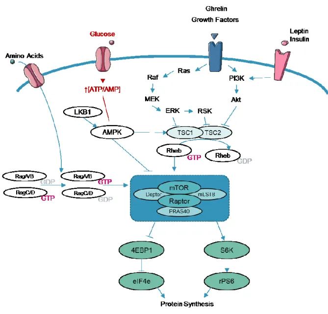
iii. Molecular integration of energy signalling: the mTOR pathway
Within animal cells, energy levels need to be balanced as well. The Mammalian Target Of Rapamycin (mTOR) is an evolutionarily conserved serine/threonine kinase that allows energy sensing and integrates many anabolic pathways involved in cell growth, cell cycle (Schmelzle and Hall, 2000). This phosphoinositide 3-kinase (PI3K)-related protein kinase (PIKK) family kinase is found in the cytoplasm as two different protein complexes named mTORC1 and mTORC2 (Giguère, 2018). mTORC1 links nutritional and energy status with the control of cell growth and regulates mRNA translation to allow protein synthesis, a cellular process that requires large amounts of ATP, as well as lipid synthesis (Lee et al., 2017a). This complex is activated by a GTPase enzyme called Ras homolog enriched in brain (Rheb), whose activity is controlled by the tuberous sclerosis complex (TSC) which converts active Rheb-GTP into inactive Rheb-GDP and supresses mTOR (Lipton and Sahin, 2014). Protein synthesis requires that mTORC1 complex phosphorylates two regulators, the p70 ribosomal S6 kinase (S6K) and the inhibitor of translation initiation eukaryotic translation initiation factor 4E (eIF-4E) binding protein (Burnett et al., 1998) (Figure 5).
In neurons, mTOR is important for synaptic plasticity, and it was shown that phosphorylation of S6K in the hypothalamus decreases with starvation, while its overactivation leads to overeating and obesity (Cota et al., 2006; Mori et al., 2009; Yang et al., 2012). Energy related hormones such as ghrelin, insulin and leptin modulate the mTOR pathway (Watterson et al., 2013). Central administration of leucine or leptin increases hypothalamic mTOR signalling and decreases food intake and body weight (Cota et al., 2006). Ghrelin activates mTOR in the hypothalamus in order to promote food intake (Martins et al., 2012). A disruption of the mTOR complex in the hypothalamus leads to hyperphagia
and obesity (Mori et al., 2009) and blunts leptin's anorectic effect (Cota et al., 2006). mTOR also senses nutrients like amino-acids within the cell and adapts the activity of S6K and eIF-4E subsequently (Hara et al., 1998), glucose through glycolysis and ATP synthesis (Dennis et al., 2001), and lipids (Foster, 2013; Menon et al., 2017).
Figure 5 Molecular integration of energy signalling: mTOR signalling.
Once in the cell, glucose is used to produce ATP, changing the [ATP/AMP] ratio. When AMP or ADP bind to AMPK, the kinase is activated and inhibits mTOR. The ERK and the Akt pathway are activated by growth factors, and inhibit the TSC1/TSC2 complex, a GTPase activating protein for Rheb. When the TSC complex is inhibited, Rheb bound to GTP accumulates, promoting mTOR activation. Amino-acids activate the Rag GTPases which bind to Raptor, and recruit the mTOR complex to the lysosomes, where it will meet Rheb-GTP to be active. Adapted from (Kim et al., 2013; Lipton and Sahin, 2014).
3. Neurons regulating the energy balance: the melanocortin circuit
Once the signals regarding the peripheral energy-state reach the brain, their information has to be mediated by specific populations of neurons in order to promote appropriate feeding behaviours. The
melanocortin circuit is one of the most studied pathways involved in feeding behaviour and glucose homeostasis maintenance. These neurones are mostly located in the hypothalamus and the NTS. Depending on the energy state, two mechanisms can be activated: the orexigenic pathway, or the anorexigenic pathway. The orexigenic pathway is stimulated by orexigenic signals when the energy levels are too low, promotes food intake, and decreases energy expenditure. The anorexigenic pathway is activated by anorectic signals when the energy state is too high, and promotes satiety by decreasing food intake and stimulates energy expenditure.
i. The NPY/AgRP and the POMC neurons: two antagonist first-order neuron populations integrating the energy levels information
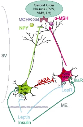
Figure 6 The Melanocortin pathway composed of the antagonist NPY/AgRP and POMC neurons
PVN: paraventricular nucleus, LH: lateral hypothalamus, VMH: ventromedial hypothalamus MCHR-4/3: Melanocortin Hormone Receptor 3 and 4, α-MSH: alpha melanocyte stimulating hormone, NPY: Neuropeptide Y, AgRP: Agouti-Related Protein, GABA: gamma-aminobutyric acid, POMC: pro-opiomelanocortin, CART: Cocaine and Amphetamine Related Transcript, LepR: leptin receptor, InsR: insulin Receptor, ME: median eminence. Adapted from (Cone et al 2006)
Once they cross the BBB, orexigenic and anorexigenic signals reach two distinct neuronal populations, called first order neurons because of their closeness to the median eminence: the Neuropeptide Y (NPY) Agouti-Related Protein (AgRP) neurons, and the Proopiomelanocortin (POMC) Cocaine and Amphetamine Related Transcript (CART) neurons (Figure 6). These two neuronal populations express the receptors for peripheral signalling molecules such as leptin, ghrelin or insulin receptor. They are antagonist, and work together to regulate food intake and energy expenditure.
The NPY neurons release three neuropeptides: NPY, AgRP, and γ-Aminobutyric acid (GABA), an inhibitory neurotransmitter. NPY has long been considered a major regulator of feeding (Clark et al., 1984), and injection of NPY in the PVN was shown to increase feeding and decrease energy expenditure (Billington et al., 1994). Following leptin discovery (Zhang et al., 1994), Stephens et al. (Stephens et al., 1995) showed that leptin inhibits the NPY/AgRP neurons just like insulin (Sipols et al., 1995) or PYY3–36 (Batterham et al., 2002), whereas they are stimulated by peripheral ghrelin (Kamegai et al., 2001), ghrelin released from the Arc (Cowley et al., 2003), and heparin (Zhu et al., 2017). Chemogenetic activation of these neurons by Designer Receptors Exclusively Activated by Designer Drugs (DREADDs)2 promotes food intake (Krashes et al., 2011). Because of these discoveries, the
NPY/AgRP neurons are therefore identified as orexigenic neurons responsible of food intake.
Adjacent to these cells, the POMC neurons co-express two neurotransmitter, POMC and CART. POMC is the polypeptide precursor of melanocortin, named α-melanocyte stimulating hormone (α-MSH). Once released from axon terminals, α-MSH binds to and activates neuronal α-melanocortin receptors 3 and 4 (MC3R and MC4R), located on second order neurons (Mountjoy et al., 1994; Ramachandrappa et al., 2013; Roselli-Rehfuss et al., 1993). The POMC neurons are activated by leptin, insulin, and inhibited by ghrelin (Pinto et al., 2004; Williams et al., 2010) (Figure 6). The POMC/CART are therefore identified as anorexigenic neurons, their activation prevents feeding behaviours.
As mentioned before, the NPY/AgRP and the POMC/CART neurons respond via an antagonist mechanism. There are several molecular reasons for this dichotomy. First of all, while α-MSH is an agonist of MC3R and MC4R receptors, AgRP is a strong antagonist. As such, a binding of α-MSH will activate the receptors on the second order neurons, while AgRP will inhibit them (Ollmann et al., 1997). Secondly, the NPY/AgRP also target the POMC/CART neurons, and release the inhibitory neurotransmitter GABA onto POMC neurons, which leads to their silencing. Finally, some anorexigenic signals like insulin and leptin bind transcription factors that will allow the transcription of AgRP but inhibit the transcription of POMC. That is for example the case of Forkhead 1 (FOXO1) that works oppositely to the transcription factor Signal-activated transcript-3 (STAT3) (Kim et al., 2006). These three mechanisms, antagonist neuropeptides for the MCR, GABA release, and opposite regulation for genes transcription, make these neuronal populations very distinct and allows opposite effects on food intake and energy expenditure.
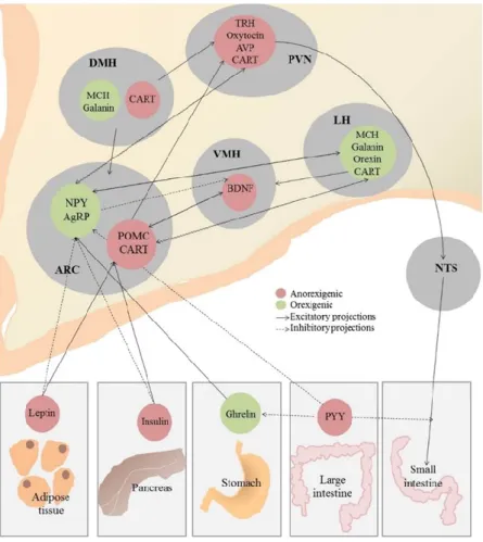
ii. The second order neurons release orexigenic and anorexigenic neurotransmitters and target higher regions of the brain
When activated, the NPY/AgRP and the POMC neurons target the second-order nuclei of the hypothalamus, the PVN, LH, or VMH, through the MCR3 and MCR4 receptors belonging to the
2 Designer Receptors Exclusively Activated by Designer Drugs (DREADD) are synthetic G protein-coupled receptors designed
coupled protein receptors family. Subsequent activation of the PVN triggers the release of anorexigenic neuropeptides such as the corticotropin-releasing hormone (CRH) and thyrotropin-releasing hormone (TRH) (Glowa and Gold, 1991; Vijayan and McCann, 1977). LH activation will release orexins A and B (Sakurai et al., 1998) and melanin-concentrating hormone (MCH) (Qu et al., 1996), which are well-studied orexigenic factors. In addition, the VMH activation releases brain-derived neurotrophic factor (BDNF), a neurotransmitter involved in synaptic plasticity having anorexigenic effects (Kernie, 2000) (Figure 7).
Figure 7 Second order Neurons signalling and Food intake regulation
ARC: Arcuate Nucleus of the hypothalamus, DMH: Dorsomedial Nucleus of the hypothalamus, PVN: paraventricular nucleus of the hypothalamus, LH: Lateral hypothalamus, VMH: Ventromedial hypothalamus, NTS: Nucleus of the solitary tract, MCH: melanin concentrating hormone, CART: cocain and amphetamine related transcript, TRH: thyrotropin releasing hormone, AVP: BDNF: brain derived neurotrophic factor, NPY: Neuropeptide Y, POMC: pro-opiomelanocortin, PYY: peptide Y3-36.
Finally, these orexigenic and anorexigenic circuits will connect to higher structures in the brain and motor control circuits, and they will regulate food intake. Recent investigation using optogenetics3
highlighted several communication between the hypothalamic melanocortin circuitry and brain regions
3 Optogenetics: this technique allows specific activation or inhibition of a specific subset of neurons genetically expressing
ionic channels coupled to light-sensitive opsins and activated by a specific wavelength. These channels will be subsequently targeted by the addition of an optic fibre, bringing light necessary for activation.
involved in reward and motivation (Aponte et al., 2011; Atasoy et al., 2012; Betley et al., 2013). For example, the ventral tegmental area (VTA), which will be described more precisely later (see section I.B.1, page 24), receives inputs from the lateral hypothalamus and the Arc, and contains ghrelin receptors (Abizaid et al., 2006; Zigman et al., 2006), and leptin receptors (Figlewicz et al., 2003) (Figure 8). Although this area was not initially thought to play a role in the regulation of food intake, it was shown that ghrelin injection in the VTA affects dopamine signalling and promotes food intake (Abizaid et al., 2006; Cone et al., 2015; Naleid et al., 2005), in addition to being an important factor that stimulates reward both in rodents, and in humans (Malik et al., 2008; Perelló and Zigman, 2012). For this reason, the semantic discrimination of homeostatic versus non-homeostatic feeding behaviour that we mentioned earlier seems difficult to untangle from an anatomical and neuronal perspective, and the hypothalamus and its related pathways alone are not sufficient to fully understand the drive associated to food intake mechanisms.
élément sous droit, diffusion non autorisée
Figure 8 Interaction between metabolic signalling and mesocorticolimbic signalling.
The reward circuit shown in light blue integrates peripheral and central mechanisms driven by homeostatic signals and environmental stimuli that result in goal-directed actions. The hypothalamus in light green is central to energy balance, integrates orexigenic and anorexigenic signals from the periphery and the brain and convey them to the reward system. Neurons in the nucleus of the tractus solitarius (NTS; dark blue) integrate peripheral satiety signals and convey the information forward to the locus coeruleus (LC), raphe, and beyond to modulate the sensitivity of upstream networks. Thus, orexigenic and anorexigenic peripheral signals directly influence not only hypothalamic nuclei but also mesocorticolimbic structures. PFC, prefrontal cortex; BLA; basolateral amygdala; Hipp, hippocampus; DS, dorsal striatum; NAc, nucleus accumbens; STN, subthalamic nucleus; VS, ventral striatum; SN, substantia nigra; VTA, ventral tegmental area; ARC, arcuate hypothalamus; LH,lateral hypothalamus;PVT, paraventricular thalamus; DA, dopamine; CCK, cholecystokinin; MCH, melanin-concentrating hormone; GLP1,glucagon-like peptide. From (Volkow et al., 2017)
B. Reward-related networks controlling feeding behaviour
Feeding, among other natural rewards such as breeding or drinking, is a crucial behaviour compulsory for survival, reproduction, and evolution of species. Living organisms evolved with a great preference for foods enriched with fat and sugar, which provide a lot of calories. These nutrients can be considered as potent rewards which promote eating even in absence of hunger, and trigger learned association between a stimulus and a reward. Indeed, this trait ensures that food is eaten when available and energy is stored in the body for future needs when resources are limited. The appealing taste of food, or palatability, is a crucial determinant of the decision to eat, and highly palatable foods can trigger eating at times when food would not otherwise be consumed. Brain circuits that process information related to food palatability are influenced by metabolic and hormonal signals that communicate information regarding the status of energy storage to the central nervous system. Satiety signals reduce perception of food tastiness (Figlewicz and Sipols, 2010; Fulton et al., 2000; Mietlicki-Baase et al., 2014; Nijs et al., 2010). Conversely, weight loss induced by fasting or caloric restriction stimulates hyperphagia partly by increasing the rewarding properties of food through leptin (Farooqi et al., 2007).
dopamine (DA) neurons, a subtype of midbrain cells in charge of encoding reward-related values, thus showing that a strict distinction between homeostatic and non-homeostatic signalling is not that simple or even possible.
1. The dopaminergic pathway involved in hedonic aspect of feeding
Historically, dopaminergic neurons were divided in two different pathways, the nigro-striatal, from the substantia nigra to the dorsal striatum, which help action selection (Gerfen and Surmeier, 2011), and the ventral mesocorticolimbic system, from the ventral tegmental area to the ventral striatum or nucleus accumbens, which promotes learning, reward, and motivated behaviours. Recent studies, using optogenetics and retrograde tracing strategies4, have shown that these two pathways are more mingled
than initially thought, and that some neurons of the substantia nigra project more ventrally, and can also innervate cortical and limbic areas, while some neurons of the ventral tegmental area project more dorsally (Morales and Margolis, 2017). These neurons are still being studied and their purpose is not known yet.
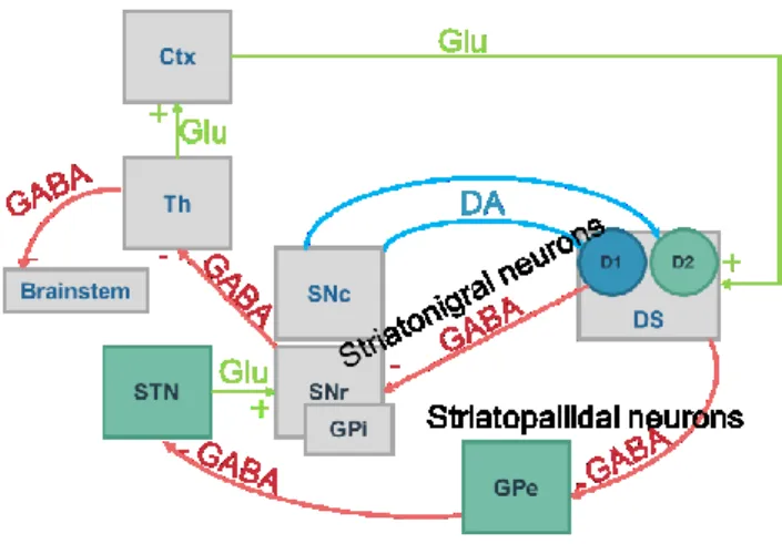
i. The Basal Ganglia in charge of movement
The basal ganglia is a set of highly interconnected nuclei, notably the striatum, the globus pallidus, the subthalamic nucleus, ventral pallidum and the substantia nigra (Ikemoto et al., 2015). DA is secreted from the substantia nigra pars compacta (SNpc) to the striatum. This projection of DA neurons is called nigrostriatal pathway, and its dysregulations result in motor-related diseases such as the Parkinson’s disease. The information then comes back to the substantia nigra pars reticulata (SNr), a region containing GABAergic neurons, and projecting to the thalamus. The most widely accepted model of the basal ganglia circuit is based on the segregation of information from the striatum to the SNr into a direct and indirect pathways. The direct pathway, or striatonigral pathway, contains medium spiny neurons (MSN) in the dorsal part of the striatum (DS) that express dopamine receptor D1 (D1R) and release GABA, and will project directly to the SNr, and to the internal globus pallidus (GPi). The indirect pathway contains GABAergic MSN expressing the dopamine receptor D2 (D2R), also called striatopallidal neurons, which will project to the external globus pallidus (GPe). The GPe also contains GABAergic neurons that will connect with the subthalamic nucleus. The subthalamic nucleus will finally relay the information to the SNr via glutamatergic inputs, where GABAergic neurons will convey information to the thalamus (Figure 9).
4 Retrograde markers: this technique allows to visualise neuronal connections from the synapse to the cell body by injecting a
Figure 9 The Basal Ganglia
In blue: neurons releasing dopamine (DA) from the Substantia nigra pars compacta (SNpc) to the Dorsal Striatum (DS). In red: GABAergic neurons of the direct striatonigral pathway from the DS to the Substantia Nigra pars reticulata (SNr), and indirect striatopallidal pathway from the DS to the globus pallidum external (GPe) to the Subthalamic nucleus (STN). In green: glutamatergic neurons conveying information from the thalamus (Th) to the cortex (Ctx).
ii. The mesocorticolimbic pathway
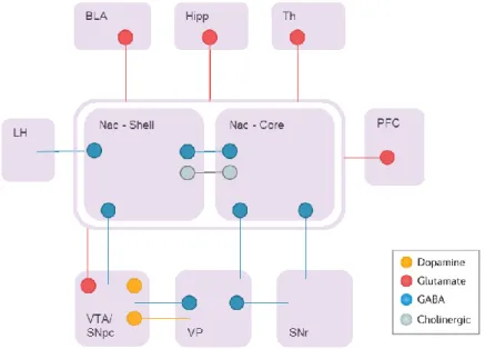
The mesocorticolimbic pathway (MCL) contains DA neurons from the VTA projecting to the ventral part of the striatum, called the Nucleus Accumbens (NAc), but also to the olfactory tubercle, the basolateral amygdala, the hippocampus and the prefrontal cortex (PFC) (Figure 10). The MCL is associated with goal-directed behaviours and motivation, as well as higher cognitive functions such as learning and memory. Dysfunctions of this circuitry can lead to different forms of maladaptive behaviours, such as addiction, mood disorders, compulsivity. Similarly to the basal ganglia system, the striatal MSN of the NAc also segregate in two pathways to return the information to the VTA. The D1R MSN will target the VTA via the direct pathway (Gerfen et al., 1990; Le Moine and Bloch, 1995), while MSN co-expressing the D1R, D2R and Adenosine A2a will target the ventral pallidum (VP) where neurons will relay the information to the VTA via the indirect pathway (Lu et al., 1998).
In blue: neurons releasing dopamine (DA) from the ventral tegmental area (VTA) to the nucleus accumbens (NAc). In red: GABAergic neurons NAc to the VTA, and indirect pathway from the NAc to the ventral pallidum (VP) to the Subthalamic nucleus (STN) and VTA. In green: glutamatergic neurons conveying information from the hippocampus (Hp), cortex (Ctx), basolateral amygdala (BLA).
The NAc is divided in two main regions, the core and the shell. The core is anatomically related to the DS in terms of connections and functions. The shell is highly heterogeneous, with a vast majority of GABAergic D1R MSN, D2R MSN, coexpressing D1R and D2R MSN, cholinergic and GABAergic interneurons (Gangarossa et al., 2013a). Whether D1R and D2R expressing MSNs are randomly distributed or exhibit inhomogeneous distribution patterns in the different subterritories of the NAc shell remains to be established. Due to the diversity of their output targets that include VTA, hypothalamus, VP, and brainstem, a simple division into direct and indirect pathways has been even more difficult to define for the NAc shell MSNs (van Dongen et al., 2008; Sesack and Grace, 2010; Zahm and Brog, 1992). Besides the dopaminergic inputs from the SNpc and the VTA, the NAc mostly receive glutamatergic inputs from the cortex, amygdala and the thalamus (Figure 11).
The ventral tegmental area contains the cell bodies of dopaminergic neurons, and sends its axons to the striatum. This area receives many glutamatergic and GABAergic inputs, including from the VP and lateral hypothalamus, and targets the NAc but also the Lateral Habenula, or the PFC (Figure 12).
Figure 11 Connectivity of the Nucleus Accumbens
The nucleus accumbens receives major glutamatergic inputs (in red) from the basolateral amygdala (BLA), hippocampus (Hipp), thalamus (Th), prefrontal cortex (PFC), dopamine (in yellow) from the ventral tegmental area (VTA) and substantia nigra pars compacta (SNpc). The main projections are GABAergic MSN reaching the lateral hypothalamus (LH), ventral pallidum (VP) and substantia nigra pars compacta (SNr). In grey : cholinergic interneurons.
élément sous droit, diffusion non autorisée
a. VTA DA neurons receive glutamatergic inputs from the medial prefrontal cortex (mPFC), pedunculopontine tegmentum (PPTg), laterodorsal tegmentum nucleus (LDTg), lateral habenula (LHb), periaqueductal grey (PAG), bed nucleus of the stria terminalis (BNST) and dorsal raphe nucleus (DRN). VTA dopamine neurons receive GABAergic inputs from the rostromedial mesopontine tegmental nucleus (RMTg), PAG, DRN, lateral hypothalamus (LHT) and ventral pallidum (VP). There are also local glutamate and GABA synapses onto VTA dopamine neurons arising from neurons within the VTA. b. VTA GABA neurons receive glutamatergic inputs from the LHb and mPFC and GABAergic inputs from the nucleus accumbens (nAcc) medium spiny neurons (MSNs) expressing the dopamine receptor D1. VTA GABA neurons receive both glutamatergic and GABAergic innervation from the PAG, DRN, LHT and BNST. Most BNST projections establishing synapses in the VTA are from GABA neurons that preferentially synapse on the VTA GABA neurons. c. VTA 'GABA-only' neurons target cholinergic interneurons of the nAcc and glutamatergic neurons of the LHb. d. VTA 'glutamate-only' neurons target glutamate neurons of the LHb and nAcc PV-expressing neurons, whereas VTA combinatorial glutamate–GABA neurons target glutamate neurons of the LHb. e.VTA 'dopamine-only' neurons establish symmetric synapses on MSNs in the nAcc. The combinatorial dopamine–glutamate neurons target nAcc MSNs, nAcc cholinergic interneurons, and mPFC parvalbumin (PV)-expressing GABA-releasing interneurons. The combinatorial dopamine–GABA neurons target nAcc MSN. From (Morales and Margolis, 2017).
iii. The dopaminergic pathway in feeding behaviour
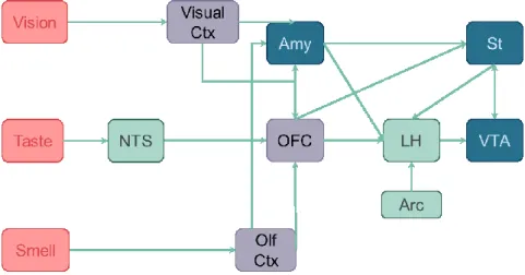
Feeding behaviour depends largely on the perception of food from the taste buds, colour, texture, smell, that will elicit rewarding properties of food thanks to the MCL. The rewarding effect of these cues depends on the satiety state (Morton et al., 2006; Rolls, 2005). As a consequence, the MCL needs to integrate not only cortical inputs related to food-cues, but also the inputs from the homeostatic pathway (Figure 13). For example, it was shown in primates that the NTS perceives taste information and connects to the VTA and NAc, that will assign reward to them.
Figure 13 Interactions between the mesocorticolimbic pathways and hunger related regions
The mesocorticolimbic system (MCL) is in blue. The striatum (St) receives inputs from the orbitofrontal cortex (OFC), amygdala (Amy), ventral tegmental area (VTA). In green, structures of the homeostatic system: the lateral hypothalamus (LH) receives input from the arcuate nucleus (Arc), nucleus of the solitary tract (NTS). In purple: visual cortex, olfactory cortex,orbitofrontal cortex. Adapted from (Morton et al., 2006; Rolls, 2005)
The NAc as well as the VTA contain receptors for signalling molecules involved in energy regulation such as leptin, insulin (Figlewicz et al., 2003), ghrelin (Naleid et al., 2005), CCK (Reum et al., 1997), or GLP-1 (Dossat et al., 2011; Mietlicki-Baase et al., 2014). For example, ICV delivery of leptin was shown to modulate reward (Fulton et al., 2000), and the absence of its receptor in ob/ob mice correlates
with a reduced DA signalling, demonstrating that leptin is not only a ‘homeostatic’ but also a ‘non-homeostatic’ signal (Fulton et al., 2006).
2. The lateral hypothalamus, cross-talk between the homeostatic and non-homeostatic
regulation of food-intake
The lateral hypothalamic area (LH) is a large and heterogeneous area with several distinct nuclei. It contains many populations of neurons, some expressing orexigenic neuropeptides such as orexin/hypocretin, MCH, galanin, others expressing anorexigenic neuropeptides such as neurotensin or CART. Most neurons in the lateral hypothalamic area express more than one peptide, and in addition may express either one of the classical neurotransmitters glutamate or GABA. It is one of the most extensively interconnected areas of the hypothalamus, allowing the reception of a vast range of information and the modulation of feeding functions accordingly. The LH joins rostrally the preoptic area and caudally the VTA, and is close to several hypothalamic nuclei such as the DMH, VMH, and Arc. As described earlier, the Arc sends NPY/AgRP and POMC/CART projections within the LH (for more details see I.A.3, page 19). The LH also receives many inputs from corticolimbic structures such as the PFC, olfactory cortex, hippocampus and amygdala, and from the shell of the NAc, and NTS (Kampe et al., 2009) (Figure 14). The LH has efferent projections to the hippocampus, amygdala, basal ganglia and thalamus, midbrain and pons, brainstem and spinal cord, and other nuclei of the hypothalamus (Figure 14). Because of this crossroads between the hypothalamus and the regions of the MCL, the LH was suggested to integrate reward-related input with information related to energy homeostasis (Mogenson et al., 1980; Stratford and Kelley, 1999), and to influence the MCL in turn (Leinninger et al., 2009).
The LH receives peripheral information via the Arc projections, and possesses glucose-sensitive neurons (Burdakov et al., 2005), as well as the ability to sense several signals such as ghrelin, GLP-1, leptin (Berthoud and Münzberg, 2011). Leptin stimulation in the LH inhibits food intake by signalling to the VTA (Leinninger et al., 2009) through orexin neurons (Peyron et al., 1998), and promotes locomotor activity (Nakamura et al., 2014). VTA orexin signalling is also involved in cocaine and morphine-induced hyperlocomotion and place preference through the MCL (Borgland et al., 2006; Narita et al., 2006), and orexin-deficient mice are less susceptible to develop drug dependence (Georgescu et al., 2003), while orexin injection into the VTA can reinstate an extinguished preference for drugs of abuse (Harris et al., 2005). These data support the large involvement of the LH and orexin neurons in reward encoding, and the superposition of pathways involved in reward and food intake. Using a model of reward-driven food intake in metabolically satiated rats (Zhang et al., 1998), (Kelley et al., 2005; Stratford and Kelley, 1999; Will et al., 2006) showed that food reward is associated with activation of orexin neurons in the LH, and that GABA agonist or glutamate
antagonist in the NAc can modulate LH activity to promote feeding. In other words, decreasing the GABAergic input of MSN from the NAc to the LH can promote food intake via orexins.
élément sous droit, diffusion non autorisée
Figure 14 Lateral hypothalamus interactions with the reward circuit
LepRb-expressing GABA neurons within the Lateral hypothalamus (LH) receives inhibitory inputs from D1-expressing medium spiny neurons (MSNs) of the nucleus accumbens (NAc). In turn, LH GABA neurons can inhibit ventral tegmental area (VTA) GABA-ergic neurons that disinhibit VTA DA neurons that may project onto D1-expressing MSNs of the NAc. LH OX neurons receives excitatory inputs from glutamatergic (Glu) innervation under the inhibitory control of presynaptic CB1 receptors, also located on presynaptic GABA-ergic terminals that synapse OX neurons within the LH. OX neurons are thought to form local microcircuits by synapting VTA DA neurons establishing contacts with D1-expressing MSNs of the NAc, which project back to LepRb-expressing GABA neurons. Glu-ergic innervation from LH to VTA DA neurons is also illustrated as relevant instance of insulin- and leptin-sensitive information processing within the LH-mesolimbic DA circuit. The Glu-ergic pathway, along with presynaptic CB1 receptors on Glu-ergic terminals contacting NAc MSNs illustrate an additional key component of the fine-tuned regulation of VTA DA neurons. Putative Glu-ergic neurons (yellow), GABA-ergic neurons (red), OX neurons (light blue) and DA-ergic neurons (blue). From (Coccurello and Maccarrone, 2018)
3. Endocannabinoids control of food intake and energy balance
Δ9-tetrahydrocannabinol (Δ9-THC) has long been used as an appetite-inducing drug; its consumption is often associated with increased wanting for food. This molecule targets the cannabinoid receptors 1 and 2 (CB1 and CB2), CB1 being expressed mostly in the brain while CB2 is mostly in periphery. The CB1 receptors are located in the basal ganglia, hippocampus, cerebellum, PFC (Herkenham et al., 1990) and activated by endogenous molecules named endocannabinoids. Cannabinoid receptors are G protein coupled receptors (GPCR) which modulate neurons activity via AC, and Ca2+ and K+ channels
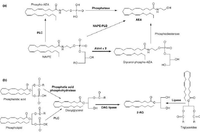
regulation (Matsuda et al., 1990). The most studied endocannabinoids are the N-arachydonoyl ethalonamine (anandamide, AEA) and the 2-arachidonoyl glycerol (2-AG), which are metabolised from arachidonic acid (Devane et al., 1992) (Figure 15). Unlike classical neurotransmitters, these molecules are not stored within vesicles but are produced on demand by two enzymes, the N-acylphosphatidylethanolamine-selective phospholipase D (NAPE-PLD) and the diacylglycerol lipase (DAG-lipase) (Di Marzo et al., 1994) (Figure 15). Their lifespan is controlled by a fast degradation by the fatty acid amide hydrolase (FAAH) and the monoacylglycerol lipase (MAG). Because of their hydrophobic nature, they can diffuse through plasmic membranes, and act as retrogrades messengers (Wilson and Nicoll, 2001).
Figure 15 Endocannabinoids Synthesis.
AEA: anandamide; NAPE-PLD: N-acylphosphatidylethanolamine-selective phospholipase D. PLC: phospholipase C; 2-AG: diacylglycerol; DAG: Diacylglycerol lipase. From (Matias and Di Marzo, 2007)
During fasting, endocannabinoids are more expressed in the limbic forebrain and hypothalamus, while repressed in a fed state (Kirkham et al., 2002). General activation of the CB1 increases food intake while its inhibition decrease food intake, and modulation of endocannabinoids signalling in the hypothalamus also affects food intake in similar ways (Cota, 2007).
Endocannabinoid modulate the MCL (French et al., 1997; Gessa et al., 1998) although very few CB1 are expressed in the VTA and NAc (Herkenham et al., 1990) (Figure 16). This regulation is due to neuronal populations expressing CB1 and projecting to midbrain DA neurons, like the glutamatergic neurons of the PFC and the subthalamic nucleus, or the GABAergic neurons of the striatum and those of the SNr (Marsicano and Lutz, 1999; Matsuda et al., 1993). Thus, CB1 can adjust the release of inhibitory and excitatory neurotransmitters and regulate DA neurons (Figure 17).
élément sous droit, diffusion non autorisée
Figure 16 Endocannabinoids and the control of energy balance
(a) In the NAc, endocannabinoids increase following food deprivation and are under the negative control of dopamine released from the ventral tegmental area (VTA). They presumably facilitate the disinhibition of hypothalamic nuclei conveying food intake-stimulatory signals (green dot), such as the MCH expressing neurons of the LHA. In the VTA, CB1 receptors might facilitate the activity of dopaminergic neurons through retrograde inhibition of other inhibitory GABAergic neurons. (b) In the hypothalamus, EC levels are under the positive control of glucocorticoids (Gluco) and, perhaps, NPY and ghrelin, and are under the negative control of leptin. CB1 receptors negatively control the expression of CRH and CART in the PVN and ARC,