D
DO
OC
C
TO
T
OR
RA
AT
T
D
D
Délivré parDiscipline ou spécialité :
Pr Doumit Monir Doyen UL Faculté Chirurgi Pr Sixou Michel Doyen
Pr Garcia Robert Doyen Paris VII Faculté Odontologie
Pr Boileau Marie-José Chef de Département ODF Univeristé Victor Segalen Bordeaux Pr Abdul Salam Hani Département Chiru
Pr Braga José Laboratoire Anthropobiologie UPS Faculté de Médecine Dr Faure Jacques Département ODF UPS Faculté Odontologie
Dr baron Pascal dépratement ODF UPS Faculté Odontologie Ecole doctorale :
Directeur(s) de Thèse : Rapporteu
Membres invités: Pr Palloudier Gerard Pr Ibrahim Nasseh Dr Treil Jacques .
Présentée et soutenue par
Titre : Les rapports tridimensionnels de la base du crâne intérêts en Orthodontie et A
T
T
H
H
È
È
S
S
E
E
En vue de l'obtention duD
DE
E
L
L’
’U
U
NI
N
IV
VE
E
RS
R
SI
IT
TÉ
É
D
DE
E
T
T
OU
O
U
Délivré par l'Université Toulouse III - Paul Sabatier Discipline ou spécialité : LABORATOIRE ANTHROPOBIOLOGIEJURY
Pr Doumit Monir Doyen UL Faculté Chirurgie Dentaire Liban Pr Sixou Michel Doyen UPS Faculté Odontologie Toulous
Pr Garcia Robert Doyen Paris VII Faculté Odontologie
Chef de Département ODF Univeristé Victor Segalen Bordeaux Département Chirurgie Maxillo-Faciale Mc Gill Univer
Braga José Laboratoire Anthropobiologie UPS Faculté de Médecine Dr Faure Jacques Département ODF UPS Faculté Odontologie
Dr baron Pascal dépratement ODF UPS Faculté Odontologie Ecole doctorale : Biologie Sante Biotechnologie
Unité de recherche : FR 2960
Directeur(s) de Thèse : Pr Braga J. , Pr Faure J. Rapporteurs : Pr Doumit M., Pr Boileau M.J.
Membres invités: Pr Palloudier Gerard Pr Ibrahim Nasseh Dr Treil Jacques .
Présentée et soutenue par OUEISS ARLETTE Le 8 Avril 2010
Les rapports tridimensionnels de la base du crâne et du massif maxillo intérêts en Orthodontie et Anthropobiologie
U
UL
L
OU
O
U
SE
S
E
LABORATOIRE ANTHROPOBIOLOGIE
e Dentaire Liban UPS Faculté Odontologie Toulouse Pr Garcia Robert Doyen Paris VII Faculté Odontologie
Chef de Département ODF Univeristé Victor Segalen Bordeaux University -Canada Braga José Laboratoire Anthropobiologie UPS Faculté de Médecine
Dr Faure Jacques Département ODF UPS Faculté Odontologie Dr baron Pascal dépratement ODF UPS Faculté Odontologie
Membres invités: Pr Palloudier Gerard Pr Ibrahim Nasseh Dr Treil Jacques .
fonctionne.
fonctionne.
fonctionne.
fonctionne.
La pratique, c’est quand tout fonctionne et que
La pratique, c’est quand tout fonctionne et que
La pratique, c’est quand tout fonctionne et que
La pratique, c’est quand tout fonctionne et que
personne ne sait pourquoi
personne ne sait pourquoi
personne ne sait pourquoi
personne ne sait pourquoi ».
».
».
».
Albert
Albert
Albert
Albert Einstein
Einstein
Einstein
Einstein, physicien américain (
, physicien américain (
, physicien américain (Nobel 1921).
, physicien américain (
Nobel 1921).
Nobel 1921).
Nobel 1921).
Remerciements à l'occasion de la défense publique
Remerciements à l'occasion de la défense publique
Remerciements à l'occasion de la défense publique
Remerciements à l'occasion de la défense publique
de
de
de
de ma Thèse de Doctorat.
ma Thèse de Doctorat.
ma Thèse de Doctorat.
ma Thèse de Doctorat.
Les travaux présentés dans cette thèse, subventionnée par
URCAM la sécurité sociale en France pour l’amélioration des
qualités de soins d’orthodontie en ville, ont été menés au
laboratoire d’Anthropologie de Toulouse. Je remercie le Professeur
Eric Crubezy pour m'avoir accueilli au sein de cet établissement
pour m'avoir accueilli au laboratoire, et m'avoir donné les moyens
de mener à bout cette étude.
La réalisation de cette thèse fut une occasion merveilleuse de rencontrer et
d’échanger avec de nombreuses personnes. Je ne saurais pas les citer toutes
sans dépasser le nombre de pages raisonnablement admis dans ce genre de
travail. Je reconnais que chacune a, à des degrés divers, mais avec une
égale bienveillance, apporté une contribution positive à sa finalisation.
Mes dettes de reconnaissance sont, à ce point de vue, énormes à leur égard.
Le sujet de thèse a été déposé fin novembre 2007. En Mai 2008, une
partie des chapitres 1 à 5 était déjà rédigée en anglais et déposée chez mon
directeur de thèse. Dans la même année, le collège des professeurs de la
Faculté a organisé sa commission annuelle d’évaluation de thèse et
l’appréciation était aussi positive que le jury me souhaite d’acquérir très
tôt la naturalisation française dans le but d’intégrer les Universités
Françaises et de gagner un élément important. Plus tard, ceci m'a incité à
donner une orientation plus pédagogique à ce travail et de mettre tous les
efforts en place pour soutenir le 8 Avril 2010 début de ma troisième
annéee.
Je tiens à remercier en tout premier lieu Jacques Faure qui a dirigé cette thèse.
Tout au long de mes années d’études en France, il a su orienter mes recherches aux
bons moments en me faisant découvrir le future de l’orthodontie au travers de ses
connaissances en recherche et en Orthopédie Dento-faciale tout en tirant partie de
ma formation en Orthodontie. Il a toujours été disponible pour d’intenses et
rationnelles discussions. Pour tout cela, sa confiance et son soutien, pour son
intuition et ses nombreux conseils pour l'aide compétente qu'il m'a apportée, pour
sa patience et son encouragement son oeil critique m'a été très précieux pour
structurer le travail et pour améliorer la qualité des différentes sections Je le
remercie vivement. De lui, j’ai toujours reçu non seulement les encouragements
dont le doctorant a tant besoin, mais aussi les précieux conseils pratiques que seul
un homme, ayant des qualités humaines comme lui, peut amener à prodiguer. C'est
grâce à lui que je me suis lancé dans une thèse de troisième cycle et je suis
heureuse qu'il ait accepté de diriger ma thèse et de faire partie de mon jury.
Je pense particulièrement au professeur Braga, mon directeur, mon promoteur,
pour la finesse de ses attitudes sur le plan aussi bien humain que scientifique. Ses
remarques successives ont permis d’améliorer les différentes versions de ce travail.
Il a toujours trouvé comme promoteur le juste équilibre entre la liberté qu’il m’a
laissée dans le choix des grandes orientations et dans la détermination des pistes à
suivre, d’une part, et un soutien total et sans faille dans les moments délicats,
d’autre part. Grâce à son approche respectueuse de la personne humaine, je me suis
continuellement senti à l’aise. Je lui en sais infiniment gré.
.
Mes remerciements vont aussi à Mr le Professeur Mounir Doumit pour m'avoir fait
l'honneur d'être président du jury et rapporteur de mon travail. J'exprime toute ma
reconnaissance à son aide tant sur le plan humain que professionnel. Quinze ans
passe il a pu être un vrai support un directeur un Doyen mais aussi un éclairant un
guide et un modèle exemplaire de réussite et de confiance.
Je suis reconnaissante à madame le Professeur Marie-José Boileau et au Professeur
Mounir Doumit d'avoir acceptés d'être rapporteurs de ma thèse. Leurs
commentaires et leurs questions m'ont permis de clarifier ma rédaction et m'ont
donné de nouvelles pistes de réflexion.
Docteur Baron Pascal
Docteur Baron Pascal
Docteur Baron Pascal
Docteur Baron Pascal
Je remercie Pascal Baron de ses conseils, de son soutien chaleureux, de sa foi dans
mon travail dans mes compétences, de m’avoir aidé et encouragé, à plusieurs
reprises, dans la réalisation de cette « thèse ». Il a su suivre et aimer, mon travail
dans sa durée.
Docteur Treil
Docteur Treil
Docteur Treil
Docteur Treil Jacques
Jacques
Jacques
Jacques
Dans le cadre de mes travaux, j’ai reçu du Docteur Treil Jacques des remarques
fines ainsi que de précieux avis et suggestions. On sait l’étendue de ses
connaissances en analyse tridimensionnelle et les remarques substantielles qu’il a
formulées pour la finalisation de ce travail. J’aurais souhaité qu’il lise encore et
toujours ces pages pour davantage les critiquer. Sans lui, ce travail n’aurait pas pu
être réalisé, le matériel de cette « thèse » provient de la Clinique Pasteur.
Nous vous remercions de la qualité de votre enseignement dont vous nous avez fait
bénéficier tout au long de nos études. Nous sommes très sensible a l honneur que
vous nous faites en acceptent de juger ce travail.
Professeur Robert Garcia
Professeur Robert Garcia
Professeur Robert Garcia
Professeur Robert Garcia....
Vous nous avez fait bénéficier de votre immense expérience. Nous apprécions la
qualité exemplaire de votre jugement nous vous en remercions d’avoir siéger à
notre jury .
Professeur
Professeur
Professeur
Professeur Abdel Salam Hani
Abdel Salam Hani
Abdel Salam Hani....
Abdel Salam Hani
Ce moment est pour nous, l’occasion de vous témoigner notre plus grande estime
et notre reconnaissance. Un grand merci pour votre attribution conseil et aide pour
accomplir et siéger à ce travail.
Docteur Njeim Ziad
Docteur Njeim Ziad
Docteur Njeim Ziad
Docteur Njeim Ziad
Vous avez toujours cru à mes compétences et capacités. Ce travail est le
témoignage de ma profonde amitié et respect.
Je remercie tous les membres du laboratoire d’Anthropobiologie avec qui j’ai
travaillé. Que l'ensemble du personnel et du secrétariat, soient aussi remerciés pour
l'atmosphère plus qu'agréable qu'ils contribuent à maintenir au sein du
département. Ma gratitude va particulièrement à Benoit et Adeline, au contact de
qui j’ai beaucoup appris, pour leur amitié, leurs conseils, et l'aide précieuse qu'ils
m'ont apporté tout au long de ce travail.
Pour votre amour, soutien et bienveillance, je vous offre mon travail. Au fil des
années vous m’avez supporté aidés et veillés sur ma réussite. Ce jour très
important de ma vie je vous l’offre comme témoignage de reconnaissance et
grande estime. Pour ma chère et adorable mère Je t’aime
A mes sœurs
A mes sœurs
A mes sœurs
A mes sœurs
Je suis très honorée et assez fière de vous offrir la primeur de mes efforts et de mes
sentiments pour exprimer respect et honneur envers vous .vous avez constitué
toujours un point de repère et un guide. Huguette pour ton cœur plein d’amour
Nancy Elsy et Dalia la vie sans vous n’aurez pas le même gout.
A m
A m
A m
A mes nièces
es nièces
es nièces Charel et Yvana
es nièces
Charel et Yvana
Charel et Yvana
Charel et Yvana et
et
et
et mon
mon
mon
mon neveu
neveu
neveu
neveu
Georges
Georges
Georges
Georges
mes anges adorés ce travail ne constitue qu’un cadeau pour vous
exprimer mon amour.
Ce travail te doit beaucoup… Qu’il soit pour toi le témoignage de mon infinie
reconnaissance pour ta compréhension, de privations et tes efforts communs.
J’ai une pensée très tendre à ton égard à ton soutien a ton apport quotidien à la
réalisation de ce travail. Tu m’as toujours donné l’espoir d’aller de l’avant. Tu
as été un bon conseiller. Ensemble sur ce long chemin on profitera de la vie
ensemble.
Live laugh and Smile Remember this always ARO thank you a lot……..
Live laugh and Smile Remember this always ARO thank you a lot……..
Live laugh and Smile Remember this always ARO thank you a lot……..
Live laugh and Smile Remember this always ARO thank you a lot……..
Love for ever
Love for ever
Love for ever
Love for ever
Belbacha
Belbacha
Belbacha
Belbacha
Je tiens à te remercier Gizlaine pour les discussions intéressantes et fructueuses,
pour la grande collaboration, et pour le temps que nous avions passé à Toulouse.
La ville en rose n’aurait pas le même gout sans toi, pour toutes les nuis blanches
qu’on a passe ; pour les bons moments de ma vie pour toutes ses belles choses je
te remercie et surtout le cadeau que tu m’as honoré par ta présence le jour de ma
soutenance.
Merci à mes collègues et amis pour les bons moments j'ai eu dans notre groupe.
J'ai bien aimé l'atmosphère, leur amitié et leur soutien. Merci à Gizlaine
Belbacha une sœur et une amie, Ramzi, Toni, Ahmed, Hicham, Lina, Fidele,
Wassim, Hani, Nadine et Mazen, Hicham Fekri, Hajj Kamel et les bon moments
a la faculté merci a vous tous, pour la grande amitié au fil des ans. J’ai une
infinie liste d’amis et je ne ferai pas le pari de les énumérer sans risque d’en
omettre certains. Je m’astreins à un devoir de reconnaissance à l’égard de tous.
Ce fut un plaisir de vous connaitre tous et bénéficier de votre support.
Et un grand merci a la diaspora Libanaise sur Toulouse, Charbel Zamlouty,
Charbel Chamo, Pierre Rahmé, Touffic, Elias, Dany, Jospeh Assad et Joelle le
couple né à Toulouse … puis Dany et Julie un plaisir de vous connaitre et de
partager de bon moments avec vous et mon petit Nano.. Kaddoum comment
t’oublier et Zahi Hallit le père des FL… Enfin mais pas la fin une pensée à
Nelly, 3ammo Fouad et Christian Merci encore….vous êtes tous une deuxième
famille la liste est surement longue mes pensées vont vers vous tous !!!!!!!!!!!!
qui véhiculent des apports et du progrès dans la science.
1
ABSTRACTS
2
ABSTRACT
There is a great interest in the recent literature about relationship between cranial base shape and facial disharmonies or malocclusions. But the conclusions are contradictory, due to insufficient samples and very basic method to appreciate cranial base shape.
The aims of this work are double: First we wanted to analyze the basi-cranial shape and its relationships with maxilla-facial schemes or malocclusion. Second we wanted to analyze the maxillo-facial shape and its relationships with malocclusions.
We used 3D data of 312 patients selected with great pathologies “border line surgery”, and we analyze these data by two methods: morphometric geometry processes and a specific 3D maxillo-facial analysis elaborated in Toulouse.
We can conclude about our main questions:
• Basicranial configuration is not significantly correlated with types of malocclusion, it is remarkably stable, and it does not play any etiologic role in malocclusion appearance.
• On the contrary, maxillofacial specific configurations, corresponding to different types of malocclusion, can be described precisely.
Some secondary results can be deduced from this work:
• We could perfect and test methods of 3D analysis applied to complex anatomies
• We could precise the pathologic schemes (antero-posterior, vertical, transversal, asymmetry) and their interconnections.
• We selected the 3D most determinant parameters.
• We made a proposal of threshold for surgical orientation concerning these parameters.
4
Résumé
La littérature accorde un intérêt soutenu à l’étude des relations entre la morphologie basi-crânienne et les dysmorphies maxillo-faciales ou les malocclusions. Mais les conclusions sont contradictoires à cause de l’insuffisance des échantillons et du caractère schématique des méthodes de description de la forme basi-crânienne.
Le but de ce travail est double : d’abord nous voulions analyser la forme basi-crânienne et ses rapports avec les schémas maxillo-faciaux ou les malocclusions. Ensuite nous voulions analyser la morphologie maxillo-faciale et ses relations avec les malocclusions. Nous avons utilisé les données scanner de 312 patients sélectionnés comme ayant des pathologies importantes « limite chirurgicale » et analysé ces donnée par deux méthodes : les procédés de la morphométrie géométrique et une analyse maxillo-faciale spécifique élaborée à Toulouse.
Nous pouvons conclure à propos des deux principales questions :
• La configuration basi-crânienne n’est pas significativement corrélée avec les différents types de malocclusion, elle reste très stable et elle ne joue aucun rôle étiologique dans le développement des malocclusions.
• Au contraire, des schémas maxillo-faciaux spécifiques correspondant aux différentes malocclusions peuvent être décrits avec précision.
Des conclusions secondaires peuvent encore être déduites de ce travail :
• Nous avons mis au point et testé les méthodes d’analyse tridimensionnelles des structures anatomiques complexes
• Nous avons précisé les schémas pathologiques (antéropostérieur, vertical, transversal, asymétrie) et leur interdépendance
• Nous avons sélectionné les paramètres 3D les plus déterminants
• Nous avons proposé des valeurs seuil pour une orientation chirurgicale.
5
TABLE OF CONTENTS
7
TABLE OF CONTENTS
1. Chapter I: Introduction………19
2. Chapter II: Cranial Base Configuration...……….27
2.1General introduction to the cranial base..……….30 2.2Overview of the cranial base growth……..………...31 2.3Growth changes in the cranial base flexion………...34 2.4Evolution of cranial base flexion………..36 2.5Description of measurements techniques of the cranial base in the literature…...40 2.6Relationship between the cranial base angle and maxillo-facial disharmonies...43
3. Chapter III: Assessment of facial dysmorphy and asymmetry………46
3.1 Assessment of the facial asymmetries………...50 3.2 Overview on the facial asymmetries………...52 3.3Three dimensional method for evaluating asymmetries ………...54 3.3.1 Review of the method for evaluating asymmetry ………..56 3.3.2 Three dimensional Methods………57 3.4Assessment of maxillo-facial disharmonies………..57 3.4.1 Appreciation of malocclusion……….57 3.4.2 Appreciation of maxillo-facial disharmonies………...58 3.5Facial asymmetries and maxillofacial disharmonies………...58
4. Chapter IV: Aims of the study…………..………...61
4.1 Basicranial shape and maxillo-facial shape: Morphometric Geometric Techniques………64
4.2. Maxillo-facial disharmonies and malocclusion: 3D analysis Faure & Treil ……68
5. Chapter V: Materials and Methods………..69
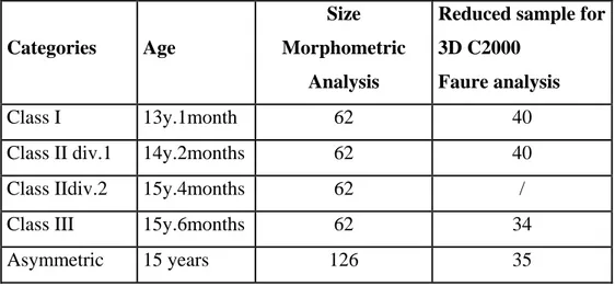
5.1 Materials: Samples and sub-samples ………..75 5.2 Methods ……….83 5.2.1 Landmarks description and identification………85
8 5.2.2 Reproducibility and validity of the landmarks……….90 5.2.2.1 Technique Error Measurement TEM……….91 5.2.2.2 Procrustes superimposition of the two configurations.………..92 5.2.2.3 Calculation of coefficient of correlation ρ……….92 5.2.3 Geometric morphometric technique……….92 5.2.3.1. Procruste superimposition ………...93 5.2.3.2 Principal compoment analysis ………..94 5.2.3.3. Thin plate analysis ………...95 5.2.3.4 Goodall test ………..95 5.2.3.4 Wilk’s lambda test……….96 5.2.3.6 Measure of B angle for basicranial shape configuration ……..97 5.2.4. Euclidean distance matrix analysis EDMA……….97 5.2.5. Superimposition of the biological shape after 3D reconstruction……...98 5.2.6 Three dimensional study of asymmetry by using C2000 and Treil & Faure 3D analysis………..99 5.2.6.1 Selection of landmarks and teeth……….100 5.2.6.2 3D imagery………..100 5.2.6.3 Cephalometric reconstruction………..101 5.2.6.4. Cephalometry………..102
6. Chapter VI: Results………113
Introduction to the results section………...116 6.1 Internal consistency and validity of the data……….117 6.1.1. Technical Error Measurement TEM………..117 6.1.2. Procrustes superimposition of the two configurations and then statistically tested by Wilks lambda value. ………...119 6.1.3. Calculation of coefficient of correlation ρ (MEDCAL; Lin 1989……121 6.1.4. Critic of the sample ………..121 6.2. Result section of basicranial configuration………..124 6.2.1. Study of basicranial configuration by use of Geometric morphometric technique ………125
6.2.1.1. Basicranial shape variation……….126 6.2.1.2. Thin plate analysis ……….127
9 6.2.2. Statistical test… ………130 6.2.2.1. Goodall test……….130 6.2.2.2. Wilk’s lambda test………..133 6.2.3. Measure of angle В………133 6.2.4. Euclidean distance matrix analysis (EDMA): A test for shape comparisons………...134 6.2.4.1. A test for shape comparisons Class I vs Asymmetric group………135 6.2.4.2. A test for shape comparisons Class I vs Cl III group……….135 6.2.4.3. A test for shape comparisons Class I vs Cl II division 2 group………135 6.2.4.4. A test for shape comparisons Class I vs Cl II division 1 group………136
6.2.5. Superimposition of the biological shape after 3D reconstruction ……136 6.2.5.1. Superimposition of the asymmetric subjects vs Cl I ……….138 6.2.5.2. Superimposition Class III maxillo-facial disharmonies vs Cl I ……….141 6.2.5.3. Superimposition Class II Division 1 maxillo-facial disharmonies vs Class I………...143 6.2.5.4. Superimposition Class II Division 2 maxillo-facial disharmonies vs Class I………..146
6.3 Results of maxillo-facial disharmonies……….151 6.3.1 Assessment of the asymmetry by use of Morphometric geometric…...152 6.3.1.1. Facial architecture shape variation……….152 6.3.1.2. Thin plate analysis ……….155 6.3.1.3. Statistical test……….159
6.3.1.3.1. Goodall Test………159 6.3.1.3.2. Wilk’s lambda test ………..160 6.3.2. Euclidean distance matrix analysis (EDMA): A test for shape comparisons……….161
10 6.3.2.1. A test for shape comparisons Class I and Asymmetric group………161 6.3.2.2. A test for shape comparisons Class I and Class III group…..162 6.3.2.3. A test for shape comparisons Class I and Class II division 2………...163 6.3.2.4. A test for shape comparisons Class I and Class II division 1………...164 6.3.3. Superimposition of the biological shape after 3D reconstruction…….165 6.3.4. Assessment of the asymmetry and study of pathologies by the use of C2000, cépha3DT and Treil & Faure analysis………171 6.3.4.1. Description of the samples regardless of the asymmetry…...171 6.3.4.1.1. Reference Sample………172 6.3.4.1.2. Class III sample………172 6.3.4.1.3. Class II sample……….175 6.3.4.1.4. Asymmetric sample………..178 6.3.4.2. Study of asymmetry of the samples……….179
6.3.4.2.1. Reference sample……….181 6.3.4.2.2. Class III sample………182 6.3.4.2.3. Class II sample……….184 6.3.4.2.4. Asymmetric sample………..186 6.3.4.2.5. Tridimensionnal asymmetry: correlation between transversal, vertical and antero-posterior asymmetry parameters………..188
7 Chapter VII: General Discussion……….192
7.1 Discussion Basicranial configuration and relation with maxillo-facial disharmonies and malocclusions………..195
7.1.1. Discussion about research methodology……….197 7.1.1.1. Methods used to explore the relation of basicranial flexion and types of maxillo-facial disharmonies and our proposal……….197 7.1.1.2. Sample……….200 7.1.1.3. Landmarks and Angles………201
11 7.1.2. Discussion of the results ……….203 7.1.2.1. Cranial base flexion and growth………..203 7.1.2.2. Cranial base configuration and maxilla-facial disharmonies……….206 7.2 Discussion: assessment of the Asymmetry and other pathologies…………...212
7.2.1 Assessment of the Asymmetry and other pathologies: discussion about methods ………212
7.2.1.1 The samples……….213 7.2.1.2 Advantage of three dimensional techniques…………213 7.2.2 Assessment of the Asymmetry and other pathologies: discussion about results ………217 7.2.2.1. Facial asymmetry was seen frequently with a high prevalence in the maxillo-facial deformities……….217 7.2.2.2. The mandible showed the most important asymmetry……….218 7.2.2.3. Left-sided facial laterality had more chances to occur than right-sided deviation………..220 7.2.2.4. Definition of a reference three D plan for asymmetry diagnosis and classification. ……….223 7.2.3. Assessment of the Asymmetry and study of other pathologies by the use of 3D analysis Treil and Faure: discussion of results……….225 7.2.3.1 Determinants parameters and limit values…………...226 7.2.3.1.1 Class II sample……….226 7.2.3.1.1.1 Correlation with ANB and AoBo...226
7.2.3.1.1.2 Test t strongest difference……….227 7.2.3.1.2 Class III sample……….227
7.2.3.1.2.1. Correlation with ANB and AoBo..227 7.2.3.1.2.2 Test t strongest difference………..228 7.2.3.1.3. Synthesis………..229 7.2.3.1.4. Limit surgical values………230 7.2.3.2 Precision of 3D description compared with 2D description………...230
7.2.3.2.1. Class II sample……… 230
12 7.2.3.3. Relations with other pathologies……….234 7.2.3.3.1. Vertical pathology………235 7.2.3.3.1.1. Comparison of medium profiles…235 7.2.3.3.1.2. Correlations matrix analyze ……236 7.2.3.3.1.3. Interpretation……….236 7.2.3.3.2. Transversal pathology………..238 7.2.3.3.2.1. Class III and class II samples……240 7.2.3.3.2.2. Asymmetric sample………...240 7.2.3.3.2.3. Interpretation……….241 7.2.3.3.3. Asymmetry………...241
8 Chapter VIII: Conclusion……….243 9 Annexes………..249 9 Bibliography ……….279
13
LIST OF TABLES
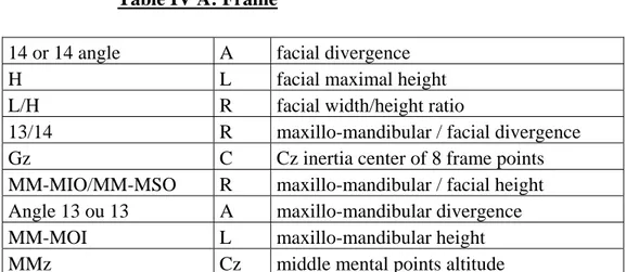
Table I: Description of the sample of the study………..76 Table II: Basicranial and facial architecture Landmarks definition ………...87 Table III: Antero-posterior Parameters of the study of the Asymmetry in the 3D analysis of
Faure and Treil. (Frame, Base, Teeth and arches)………..103
Table IV: Vertical Parameters of the study of the Asymmetry in the 3D analysis of Faure and
Treil. (Frame, Base, Teeth and arches)………...104
Table V: Transversal Parameters of the study of the Asymmetry in the 3D analysis of Faure
and Treil. (Frame, Base, Teeth and arches)………105
Table VI: Asymmetry: Transversal Parameters of the study of the Asymmetry in the 3D
analysis of Faure and Treil. (Teeth and arches, bases and envelop)………...106
Table VII: Asymmetry: Vertical Parameters of the study of the Asymmetry in the 3D
analysis of Faure and Treil. (Teeth and arches, bases and envelop)………...107
Table VIII: Asymmetry: Anteroposterior Parameters of the study of the Asymmetry in the
3D analysis of Faure and Treil. (Teeth and arches, bases and envelop)……….108
Table IX Technical Error measurement (TEM) for basicranial configuration………..118 Table X Goodall’s F-Test with a 1% confidence level of the four groups of malocclusion and
Class I reference group for basicranial configuration……….131
Table XI Goodall’s F-Test with a 1% confidence level between the maxillo-facial
disharmonies for basicranial configuration ………132
Table XII β measured between the two plans Po and P1………..133
Table XIII Goodall’s F-Test with a 1% confidence level of the four groups of malocclusion and Class I reference group for facial architecture………...159
Table XIV Goodall’s F-Test with a 1% confidence level between the maxillo-facial
disharmonies for facial architecture………159
Table XV 2D Parameters for the description of the sub-samples………..170 Table XVI Correlation matrix of transversal, vertical and anteroposterior asymmetry…...186
14
LIST OF FIGURES
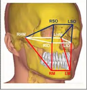
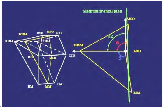
Figure 1 Cranial base angle measured in the literature………39 Figure 2 3D Treil & Faure analysis to describe malocclusion……….77 Figure 3 Illustration of the different sub-samples………....78 Figure 4 The landmarks for the basicranial configuration and facial architecture are related
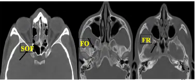
with trigeminal design………...85
Figure 5: The Landmarks identification on the CT Scan……….86 Figure 6 Definition of basicranial shape configuration………88 Figure 7: The basicranial landmarks configuration identified on the axial CT Scan………...88 Figure 8: Definition of facial architecture ………...89 Figure 9: The facial landmarks identified on the CT Scan………..89 Figure 10: The direct orthogonal landmark………...109 Figure 11: Main anteroposterior envelop parameters………...109 Figure 12: Main anteroposterior basis parameters………109 Figure 13: Procruste superimposition of the mean shape basicranial configuration for the
validity of the landmarks………120
Figure 14: Procruste superimposition of the mean facial architecture for Asymmetric group to
test the reliability and validity of identification of landmarks………120
Figure 15 Mean basicranial configuration of the Class I morphology derived from 62 Class I
individuals in untransformed space………..………...125
Figure 16 PCA Analysis representing mean shapes that correspond to the five samples of the
study Class I, Class II div 1, Class II div 2, Class III and Asymmetric………..126
Figure 17: Superimposition of the five sample of the study. This qualitative analysis shows
the high stability between the five groups of malocclusion………127
Figure 18 PCA Analysis representing mean shapes that correspond to the five samples of the
study Class I, Class II div 1, Class II div 2, Class III and Asymmetric………..128
Figure 19 Total splin for overall pattern cranial base configuration for the five sub samples of
the study………..129
Figure 20 Shape changes derived from TPS analysis of cranial base configuration between
normal and the pathologic maxilla-facial disharmonies. ………...130
Figure 21 3D reconstruction of the normal subject ………...137 Figure 22 The shell-to-shell BC differences of Class I / asymmetric subject 1 ………139 Figure 23 The shell-to-shell BC differences of Class I / asymmetric subject 2……….140
15
Figure 24 The shell-to-shell BC differences of Class I / Class III subject 1………..141 Figure 25 The shell-to-shell BC differences of Class I / Class III subject 2………..142 Figure 26 The shell-to-shell BC differences of Class I / Class II division 1 subject 1……..144 Figure 27 The shell-to-shell BC differences of Class I / Class II division 1 subject 1……..145 Figure 28 The shell-to-shell BC differences of Class I / Class II division 2 subject 1……..147 Figure 29 The shell-to-shell BC differences of Class I / Class II division 2 subject 2……..148 Figure 30: Mean facial architecture configuration of the Class I morphology derived from 62
Class I individuals………...153
Figure 31: Superimposition of the five sample of the study. This qualitative analysis
describes the location and prevalence of asymmetry in relation to different types of maxillo-facial disharmonies……….153
Figure 32: PCA Analysis representing mean shapes of facial architecture that correspond to
the 4 samples of the study superimposed to the mean shape of Cl I reference group to study the asymmetry……….155
Figure 33: PCA Analysis representing mean shapes that correspond to the five samples of the
study Class I, Class II div 1, Class II div 2, Class III and Asymmetric………..156
Figure 34: Shape changes derived from TPS analysis of facial architecture configuration
between the mean configurations of facial architecture shape in all the sample of the study CL I reference group, CL III, CL. II div.1, CL. II div.2, Asymmetric………157
Figure 35: Total splin for overall pattern facial architecture configuration for the five sub
samples of the study………158
Figure 36 3D reconstruction of Class I subject……….164 Figure 37 The shell-to-shell FA differences of Class I / asymmetric subject 1……….166 Figure 38 The shell-to-shell FA differences of Class I / Class III……….167 Figure 39 Superimposition of the mean shape of facial architecture of the five samples…..168 Figure 40 Superimposition of the mean shape of orbital level………..168 Figure 41 Characteristics of the mean values of the Class III sample in 2D and 3D
analysis...174
Figure 48 Characteristics of the mean values of the Class II sample in 2D and 3D
analysis………177
Figure 43: Asymmetry of the Class II sample ………..241 Figure 50: Asymmetry of the Class III sample ……….241 Figure 51: Asymmetry of the Class II sample Frontal view………..242 Figure 52: Asymmetry of the Class III sample Frontal view……….242
17
ANNEXES
Figure 1 3D reconstruction of the normal subject ………...251 Figure 2 3D reconstruction of subject of Class II division 1 ………252 Figure 3 3D reconstruction of subject of Class II division 2 ………253 Figure 4 3D reconstruction of subject of Class III ………...254 Figure 5 3D reconstruction of Asymmetric subject ………..255 Table I Sorted elements of the shape difference Class I and Asymmetric group for basicranial
configuration………...257
Table II Sorted elements of the shape difference Class I and Class III group for basicranial
configuration………...258
Table III Sorted elements of the shape difference Class I and Class II division 2 group for
basicranial configuration……….259
Table IV Sorted elements of the shape difference Class I and Class II division 1 group for
basicranial configuration……….260
Table V Sorted elements of the shape difference Class I and Asymmetric group for Facial
architecture ……….261
Table VI Sorted elements of the shape difference Class I and Class III group for Facial
architecture………..262
Table VII Sorted elements of the shape difference Class I and Class II division 2 group for
Facial architecture………...263
Table VIII Sorted elements of the shape difference Class I and Class II division 1 group for
Facial architecture………...264
Table IX: 3D anteroposterior parameters………..265 Table X: 3D vertical parameters………266 Table XII: 3D transversal parameters………267 Table XIIa: 3D transversal parameters………..268 Table XIIb: 3D vertical parameters………...269 Table XIIc: 3D anteroposterior parameters………...270 Table XIII: R coefficient correlation ANB/AoBo………271 Table XIV: Surgical Diagnosis ………...272 Table XV: Correlation matrix vertical/anteroposterior (Cl II) ……….273 Table XVI: Correlation matrix vertical/anteroposterior (Cl III) ………..274 Table XVII: Correlation matrix vertical/anteroposterior (Asymmetric) ………..275 Table XVIII: Correlation matrix transversal/anteroposterior (Cl II) ………...276
18
Table XIX: Correlation matrix transversal/anteroposterior (Cl III) ………..277 Table XVIII: Correlation matrix transversal/anteroposterior (Asymmetric ……….278
19
INTRODUCTION
21
CHAPTER I: INTRODUCTION
La base du crâne est en relation avec l’encéphale, l’oropharynx, les voies aériennes supérieures, l’articulation temporo-mandibulaire, les organes sensoriels. La flexion marquée de la base du crâne spécifique de l’espèce humaine est donc naturellement supposée être reliée au développement encéphalique, aux fonctions de déglutition, de mastication, de respiration et de phonation ainsi qu’à la bipédie.
Pour de nombreux auteurs il existe des relations entre forme basi-crânienne et pathologie maxillo-faciale ou pathologie occlusale. La plupart de ces travaux sont basés sur des téléradiographies profil crâne et des analyses bidimensionnelles tant pour l’appréciation de la forme basi-crânienne (schématisée simplement par l’angle basi-crânien) que pour l’architecture maxillo-faciale.
La prise en compte des asymétries s’appuie, elle, le plus souvent, simplement sur des données cliniques occlusales.
Le but de ce travail est d’abord de tester les méthodes d’analyse tridimensionnelles des structures anatomiques complexes, ensuite nous analyserons les formes basi-crâniennes, maxillo-faciales et leurs relations ; les questions essentielles sont :
• Existe-il un lien entre la forme basi-crânienne et les pathologies maxillo-faciales et / ou occlusales ?
• Quels sont les liens entre les différentes pathologies maxillo-faciales et / ou occlusales entre elles (antéropostérieures, verticales, transversales, asymétries)?
• Quels sont les limites du pathologique et du chirurgical?
Nous emploierons deux méthodes : la morphométrie géométrique et les outils connexes, et une analyse céphalométrique spécifique mise au point à Toulouse.
Nous utiliserons un échantillon étendu comportant, outre les cas de référence, essentiellement des cas de pathologies extrêmes.
22 CHAPTER I: INTRODUCTION
One structure where ontogeny and phylogeny have been extensively investigated is the human cranial base, which is unique among primates in being highly flexed (Gould, 1977; Dean and Wood, 1984; Lieberman and al., 2000; Jeffery, 2003). Phylogenetically, the cranial base is the oldest part of the cranial skeleton. It forms a support for the brain and has, suspended from it, the structures involved in respiration, swallowing, and vocalization. It has a major connection with the sensory organs (visual, auditory), and is involved in the movement of the pharynx inferiorly during early childhood that is thought to be essential for production of complex vowel sounds. Information on the cranial base has been applied to fields of comparative anatomy, primatology, human evolution, craniofacial growth and development. It has been established that modern humans have more flexion in the cranial base when compared to other primates (Lieberman and McCarthy, 1999; Ross and Ravosa, 1993). There are number of theories that have been proposed to explain the acute basicranial flexion seen in humans, these hypotheses are not necessarily mutually exclusive. They can be divided into neural hypotheses (Gould, 1977; Ross, 1993; Ross and Henneberg, 1995; Ross and Ravosa, 1993; Strait, 1998; Strait and Ross, 1999), speech and language hypotheses (Laitman, 1985; Laitman and Heimbuch, 1982; Laitman et al., 1978; Lieberman et al., 1972) and postural/bipedalism hypotheses (Ashton, 1957; Weidenreich, 1924). The increase in relative brain size and development of bipedal posture both seem to be influence in the increased flexion of the cranial base in humans compared to other primates (Strait, 1999; Strait and Ross, 1999).
The cranial base provides support for the brain and adaptation during growth between the developing neurocranium and viscerocranium. Because of this junctional location between cranium, midface, and glenoid fossa, the cranial base has the potential to influence growth of both cranium and face. Prenatal development during embryogenesis and the fetal period indicate the chondrocranium and basicranial region are mainly replaced by bone, starting in the second month of embryonic development. The human basicranium is evident by week 4 prenatally between the cranial part of the neural tube and the foregut. Previous reports in the literature describe general growth of the cranial base in terms of age changes from birth to early adulthood. The ontogenic and phylogenic role of basicranium as a functional piece
23
between neuro-cranium and vicerocranium was seen in terms of functional matrix (Moss and al., 1955; Moss and al., 1984) and recently in terms of morphological integration and modularity (Bastir et al., 2003).
The role of the basicranium and its potential interaction and contribution to normal and pathological variation of the face is a frequently addressed and clinically relevant issue in craniofacial biology. Also in anthropology there is an increasing interest in the developmental relations between facial and basicranial structures.
If the basicranium is involved in maxillo-facial evolution (ontogenic and phylogenic) it is surely involved in pathologies. Despite the studies on basicranial flexion among different species, the relationship between cranial base configuration and facial prognathism has been of interest to anthropologists, particularly in relation to racial differences. Huxley (1924) used the basicranial axis on sagittal sections of dried skulls to elucidate racial variation, and to suggest the possibility of an association between this variable and malocclusion. Björk (1955), using cephalometric radiographs, demonstrated the existence of a relationship between cranial base morphology and jaw relationship. The hypothesis of Björk was based on the following explanation, the maxilla and mandible articulate with different limbs of the cranial base, and therefore it is possible that variations in growth and orientation of the cranial base region could lead to a differential movement of the mandible in relation to the maxilla. The two limbs of the cranial base form a flexion of 130°– 135° at sella. The maxilla appears attached to the anterior segment and the mandible to the posterior segment. It would be reasonable to assume, just from this geometric relationship, that any change in flexion would alter maxillary and mandibular positions relative to the cranial base as well as to each other. This in turn may influence the skeletal pattern and type of malocclusion.
A number of studies have attempted to identify craniofacial differences between the classes of malocclusion. Hopkin et al., (1968), using Articulare to represent the posterior limit of the cranial base, described a linear relationship between the cranial base angle and prognathism with the angle systematically reducing from Class II, via Class I, to Class III individuals. Scott (1958) suggested that a number of factors determine or influence static jaw position and, consequently, the degree of prognathism in individual cases. These factors included the cranial base angle, the extent to which the mandible and maxilla moved forward in relation to the cranium and the amount of surface bone deposition along the facial profile from nasion to menton. The association between cranial base morphology and different types
24
of malocclusion is not fully understood. Contradictory results were noted in previous studies described in the literature. These studies measured a cranial base angle based and limited just on three landmarks, these studies missed the morphologic characteristics of cranial base configuration and in that way were not able to characterize global craniofacial morphology or regional changes in the cranial base itself.
It is possible to criticize greater part of these works; due to their measurement methods. The basicranial morphology till the latest research in the literature is mainly simplified and described as “the basicranial angle flexion”. Traditionally, the basicranial flexion is investigated as a degree of angulations defined by three points or two plans (Zuckerman, 1955; Levihn, 1967; Birch, 1968; Jeffery, 1999; Jeffery and Spoor, 2002; Lieberman et al., 2000). The maxillofacial morphology is commonly appreciated in orthodontics by the use of 2D cephalometric analysis. Unfortunately very few 3D biometric approaches are studied in the literature. Most recent studies take account of general shape of cranial base (Lieberman, 2007), by using 27 landmarks qualified as 3D landmarks , chosen to represent the cranial base, but the landmarks are unilateral selected on CT scan (Sella, Foramen caecum, Sphenoidale, Posterior cribiform plate and Bregma).
This research work will focus on the maxillofacial morphology, using 3D tools and defining a “basicranial configuration” representing the realistic configuration of the cranial base.
Once the “basicranial configuration” is established, it seems interesting to appreciate the relationship between cranial base shape and disharmonies or malocclusions from one hand, and to specify the malocclusions and disharmonies tied together and their interconnections in the other hand.
For this purpose we need:
• A wide sample of patients with pathologies. • Three dimensional data of these patients.
25
This review will first present a summary of the research that has gone into cranial base flexion and craniofacial morphology as it relates to the aims of the present work. This includes an overview of cranial base growth, and a description of studies on cranial base flexion regarding the maxillo-facial disharmonies. With the background to the study established, the aims and hypothesis of the current research will be explained in more detail.
The purpose of this study is to investigate morphology of the cranial base configuration in different types of malocclusion, to determine relationship between cranial base and maxillofacial disharmonies and/or malocclusions.
The data provides a baseline material for subsequent studies of the maxillo-facial specificity in the different types of malocclusion.
For this work a tridimensional method will be applied, and conjointly the use of the morphometric geometry methods and a 3D cephalometric program based on a reference tied to the cranial base.
The first part of the research work used the morphometric geometry methods to:
• Test them as a way to describe the cranial base or the maxillo-facial morphology.
• Test the relationship between cranial base morphology and malocclusion. • Test the relationship of dysmorphies between themselves (overall asymmetry/
other pathologies).
The second part of this research work used the 3D cephalometric analysis C2000 and Cépha 3DT elaborated by J.Treil and J. Faure with CIRAD (Montpellier), and it used the conventional statistical methods to compare pathological samples. In this second part we try to precise the definition of main pathologies, anteroposterior, vertical, transversal, asymmetrical ones, and overall to precise the definition of surgical common threshold.
After that we study the relationship of pathologies between themselves, overall asymmetry/other pathology.
26
We can dispose for our work of a wide sample (374) of patients with maxilla-facial disharmonies. These patients were collected in a study program evaluating detection, diagnosis and therapeutic follow up of surgical patients (URCAM French Social National Insurance, FAQSV funds). That was an excellent study sample to understand the relationship of maxilla-facial and occlusal pathologies.
The sample will be presented first, the justification of the research approaches, the methods, the results and the discussion will be presented successively.
27
CRANIAL BASE CONFIGURATION
29
CHAPTER II: CRANIAL BASE CONFIGURATION
Ce chapitre bibliographique présente d’abord les principaux résultats concernant la forme de la base du crâne en général caractérisée par l’angle crânien ou la flexion basi-crânienne. La flexion basi-crânienne chez homo sapiens est spécifique parmi les primates.
L’embryologie et la croissance ont été étudiées et on retiendra surtout le rôle des sutures et les variations de l’angle basi-crânien augmentant pendant la vie fœtale puis diminuant après la naissance dans les premières années.
La diversité et le caractère très schématique des méthodes d’appréciation de la forme basi-crânienne peuvent expliquer pour beaucoup le peu d’homogénéité des résultats.
Les conclusions concernant les relations entre forme basi-crânienne et pathologie maxillo-faciales et/ou occlusales sont souvent contradictoires.
L’idée « logique » d’une évolution occlusale de la Casse II à la Class I puis à la Class III à mesure de la fermeture de l’angle crânien et de la diminution des segments basi-crâniens est loin de faire l’unanimité.
30 CHAPTER II: CRANIAL BASE CONFIGURATION
In this section we will present a summary of the research that has gone into cranial base flexion and maxillofacial disharmonies. This includes an overview of the cranial base growth, a description of studies on the cranial base flexion and the various hypotheses regarding the evolution of cranial base flexion in humans.
With the background to the study established, the aims and hypotheses of the current research will be explained in more detail in the chapter four.
2.1 General introduction to the cranial base
In studies of craniofacial growths, early researchers assumed that the bones of the cranial base grew at similar rate to brain, and that little growth occurred after about seven years (George, 1978). As a result, most studies of the growth of the skull use the stability of the cranial base as a reference to assess growth in other bones of the cranial skeleton.
Variation in craniofacial morphology is evident during development and growth. It occurs as variation between modern humans, fossil hominins, and primates. Much of this variation arises during growth, with some influence of mechanical factors. This variation is the result of the same growth regulating hormones acting in various ways on body tissues so that they grow at different rates (Bijlmsa, 1983; Dixon and Sarnat, 1982; Goss, 1972; Raisz, 1988). The resulting differential growth arises from variation in the rate and duration of growth of the craniofacial bones, and occurs from early stages of embryonic development until after puberty, and possibly well into adulthood (Dixon and al., 1997; Enlow, 1990; Lewis and Roche, 1988). It produces differences in size and in shape (Huxley, 1924; Moss and al., 1984; Thomposon, 1942). In some individuals a disturbed pattern of differential growth can lead to malocclusion of the teeth or disproportion in facial morphology (Cantu, 1997; Enlow and Azuma, 1975; Nanda, 1955; Pirinen, 1994; Richtsmeier, 1985; Ricketts, 1960; Simmons, 1999; Trenouth, 1981, 1984, 1985; Wilhelm, 2001).
31 2.2 Overview of the cranial base growth
Ford (1958) analyzed the growth of the cranial base. After measuring differences in a cross-sectional collection of skulls, grouped into dental ages, it was found that growth at the sphenoid-occipital synchodrosis ceases after seven years. He found that the presphenoid and basisphenoid fuse shortly before birth but cartilage remains here for some time. At the time of birth, growth centers in the cranial base are between basisphenoid and basioccipital (spheno occipital synchodrosis), and between presphenoid and frontal bones. After birth, during the first year, the mesethmoid center appears in the area of the cribriform plate. This permits growth anteriorly and posteriorly between frontal and sphenoid bones while cartilage persists. The cribriform plate ceases to growth by about two years, while the thickness of the frontal bone continues to increase through adolescence. Between 30% and 60% of the growth of the craniofacial is complete by birth (Thilander, 1995), while over 80% of the growth of the cranial base is complete by six years (Myers, 1995). However, Ford’s study (1958) shows that the individual bones can follow either a neural or a general growth pattern. Increases in distances between nasion and foramen caecum and between sella and basion follow the general, somatic growth pattern. Foramen caecum to sella and the sagittal length of the foramen magnum show a neural pattern, which means that growth areas around the foramen magnum are silent by the time of eruption of the first molar ( Ford, 1958; Zuckerman, 1955). Once the brain has ceased growing, the anterior cranial base still needs to grow to allow for facial development. This occurs through the development of the frontal and ethmoidal air sinus, with development of the supraorbital region and the interorbital septum. In a comparison of cranial base growth in primates, examining samples of infants, juveniles and adults, it was found that the anterior cranial base growth seems to follow the pattern of facial skeleton, rather than the brain or endocranial cavity. In contrast, the posterior regions of the cranial base follow the growth pattern of the endocranial cavity (Michejda, 1975).
Studies on the growth of the head have attempted to document the normal growth of the cranial base at different ages. Many of these have taken the form of longitudinal, serial measurements of cranial growth (Bhatia and Leighton, 1993; George, 1978). An equal number have investigated growth cross-sectionally (Burdi, 1969; Diewert 1983; Zuckerman, 1955).
32
Enlow et al., 1975 investigated craniofacial development and variation; they note that all components of the facial skeleton are closely related during growth. For example, the anterior cranial base is equivalent to the upper naso-maxillary complex, which is closely related to the inferior part of the maxilla. Any changes in size or displacement of any of these regions will influence the others. In a description of the growth of the craniofacial skeleton through remodeling and displacement through serial tracings registered on vertical and horizontal reference lines, it is noted that most of the significant changes in growth of the cranial base and movement of the maxilla are not apparent if sella-nasion is used as a reference plane (Enlow et al., 1971). Enlow et al., 1975 explained the increases in length of the cranial base and demonstrate that it occur posterior to sella, which means that sella is moved relatively forward. The endocranial surface of the clivus, and the middle cranial fossa are resorptive, while deposition occurs ectocranially. This causes anterior relocation of the clivus and anterior wall of the middle cranial fossa. Simultaneously, superior displacement of the entire skull occurs by growth at the occipital condyles. Growth of the cranial base causes inferior and anterior displacement of the maxilla and mandible, but the effects are less evident in the mandible because of the angle of the inclination of the cranial base relative to the mandible. The authors also note that the sella-nasion plane is not an anatomically effective dimension to represent the upper face and/or cranial base. It passes across different bones that have different patterns and sites of growth, it is not related to an architecturally important landmark in the skull, and it does not include the posterior regions of the anterior cranial base.
In a landmark study about age changes in the cranial base, Zuckerman (1955) studied, dry modern human skulls, not separated into sexes or racial groups. Results show that the cranial base is more than 50% of its adult size by eight years. Basioccipital, basisphenoid, presphenoid and ethmoid all continue to grow until adulthood. The posterior part of the cranial base, measured from basion to the pituitary point, is longer in skulls with permanent dentition compared to those with deciduous dentition, and is longer in adults than in those with only the second molar erupted. Similar results are seen for the anterior cranial base. The sagittal diameter of the foramen magnum shows significant differences between juveniles with deciduous dentition and those with the permanent teeth erupting (six to eight years), but no differences are present between the latter group and adult skulls. These data suggest that the posterior parts of the cranial base cease growing sooner than the anterior part, which continue growing up to and beyond puberty, and contribute to the adolescent growth spurt.
33
Zuckerman (1955) noted that there is no reason to say that the growth patterns in the cranial base will correspond to dental ages in individuals, especially with adolescence where averages will underestimate growth changes.
Melsen (1982) was the first researcher to examine histologicaly the cranial base. Samples of the middle cranial base were taken from autopsy materiel, age range 0 to 20 years, compromising 76 males and 50 females. She used tetracycline staining techniques to identify surfaces where bone apposition was occurring. It was found that the endocranial surface of the anterior cranial base, consisting of the frontal bone and cribriform plate, ceases remodeling activity in most cases by four years of age. Apposition occurs on the anterior sphenoid surface (jugum sphenoidale) which has the effect of raising the level of the bone, as mentioned by Björk (1955). Growth in length of this part of the cranial base occurs by growth at fronto-ethmoidal, frontal and ethmoidal sutures. In the frontal and spheno-ethmoidal sutures, no growth was seen after seven years.
In a study of boys between 12 and 20 years, Björk (1955) found that the cranial base increases in length by sutural growth. The anterior cranial fossa stops growing around ten years, and the growth of the upper face after this point is mostly by apposition on the frontal bone. The posterior cranial base increases in length through growth at the sphenoid-occipital synchodrosis, causing an endocranial displacement of basion. Lateral growth of the posterior cranial base also occurs, through growth at the sutures, and continues as long as there is growth in the upper face. Changes in the cranial base do not appear to be reflected in movement of the sella, representing the sphenoid bone in this instance. The nasion-sella line is stable in relation to the floor of the anterior cranial fossa during adolescence. As demonstrated by a case study of anchondroplasia, the normal development of the cranial base is largely dependent on the normal growth of the spheno-occipital synchodrosis. Changes in the cranial base with age continue as long as the head and face continue to grow. Angles of the cranial base and face show a lot of individual variation in both directions with age.
2.3 Growth changes in the cranial base flexion
All prenatal studies have been based on cross-sectional data and the points used to measure the “saddle angle” differ slightly among the investigations. In a very early study in 1857, Virchow findings have been interpreted often as showing an increase in the saddle
34
angle. He measured the angle between the planum ethmoidal and the clivus. Pankow (1950) also used the same landmarks used by Virchow and reach the same conclusion.
Studies of cranial base flexion in prenatal individuals are necessarily cross-sectional, and it has been found by some researchers that the angle increases during prenatal development (Ford, 1956; Lieberman et al., 1972).
Cranial base flexion measured in longitudinal studies appears to show little change from 2 years after birth (Lieberman and McCarthy, 1999). According to Diewert (1983), the average cranial base angle between basion – sella - nasion in a sample of fetuses increases from 117 degrees to 127 degrees. George (1978), states that the angle at birth is 142 degrees, and has stabilized to 130 degrees by the age of five years. In a sample of children from the Belfast Growth Study the basion – sella – nasion angle was measured at 129 degrees in the oldest age group (15 years), with a standard deviation of about two degrees (Kerr, 1978). In additional work, examining the same children over a ten year period, it was found that the average angle of cranial base flexion remained constant.
Different cranial base angles measured nasion-sella-basion is related to different craniofacial forms. Rotation of the cranial base is related to rotation of the brain case and rotation of the facial skeleton. This is apparently related to interactions between different growth processes during development, producing a wide range of individual variation. A decreased cranial base angle will produce a more prognathic face, as measured by the protrusion of the upper and lower jaws (maxilla and mandible). Changes in cranial base flexure or shape can produce a relocation of the glenoid fossa in relation to the anterior areas of the cranial base (Bjork 1955; Kieser et al, 1999). However, protrusion of the mandible is also dependent on the growth processes occurring in the mandible, such as growth at the condylar processes in different directions, apposition or resorption at gnathion, and remodeling/growth at the gonial angle.
In a longitudinal and cross-sectional study of the cranial base angle it was found that the cranial base angle decreases during the first two years of life (George, 1978). These decreases were recorded in three different angles of cranial base flexion: nasion-sella-basion, internal frontal bone point-sella-basion, and the clival inclination relative to sphenoidale-frontal line. Following this, flexion shows individual patterns, with flexion increasing in some individuals and decreasing in others. It is suggested that in early childhood there are two patterns of growth, one operating until two years, and one after two years. The later pattern of
35
growth may also be interpreted as growth adjustments occurring in other areas. Considering the similar results of Björk (1955), it seems that the growth during the early post natal period follow a fixed pattern of growth as shown by high correlation between means and standard deviations, with individual variation (George, 1978).
Kvinsland (1971) found high correlations between different angles in the cranial base, these angles were between the anterior and posterior cranial base, intersecting at sella, which decrease in flexion with increasing developmental size. The angle between prosphenion-sella-basion measures the angle of the sphenoidal and occipital parts of the cranial base, and shows considerable individual variation, but no real relationship to developmental stage. The angle of the anterior base is measured by the anterior cranial base point-prosphenion-sella. The intersection between the most anterior part of the cribriform plate and the more vertical uppermost part of the nasal septum, in the midsagittal plane can be measured as a further angle in the cranial base, representing the spheno-ethmoidal part of the anterior cranial base. It shows an increase in the sample measured. All these angles were positively correlated. It was also found that individuals with a large cranial base angle had large anterior face height, measured from nasion-gnathion and relatively more posterior rotation of the mandible. Increased growth was observed in the anterior cranial base compared to the posterior cranial base, with contributions from sphenoid and ethmoid parts of the anterior cranial base being fairly equal. It is suggested that most of the angular changes in the anterior cranial base occur around the spheno-ethmoid junction.
Lieberman (2007) measure the growth changes in two estimates of craniofacial flexion between different groups of hominoids. The angles were internal flexion angle, measured between the anterior end of cribriform palte-tuberculum sella-nasion, of the cranial base, and a craniofacial angle staphylion-hormion-basion. In this investigation, sagittal radiographs of juvenile crania of gorillas, chimpanzees and modern humans were studied. Comparisons were also made to juvenile fossil crania. It was found that the internal flexion angle increases during growth in gorillas. The same angle in chimpanzees and modern Homo sapiens remained relatively stable during development, with a slight decrease in the angle in Homo (more flexed, average decrease of less than 5°). The craniofacial angle increased in both chimpanzees and gorillas but decreased in humans.