People
'
s
Democratic
Republic
of
Algeria
Higher
Education Ministry
And Scientific
Research
University
of El
-
Chahid Hamma
Lakhdar
El
-
Oued
Faculty of Natural
Sciences
and Life
Department
of
Cellular
and
Molecular
Biology
Master's Memory
In order to
obtain
a
diploma
of
an
Academic
Master
In
Biological
Sciences
Specialty
:
Applied
Biochemistry
Theme
Presented
by:
Legmairi Maroua
Salem Saadia
June 14
th, 2020
President : Mme. Mahboub Nasma (MCB) EL-Oued University Examiner : Mme. Bouras Beya (MAB) EL-Oued University Supervisor : Dr. Derouiche Samir (MCA) EL-Oued University
Epidemiological and risk factor study, evaluation of
biochemical and hematological parameters and Receiver
Operating Characteristic Curves analysis of some oxidative
stress markers in man and women stroke Patients of
Touggourt region
ءادهلإا
دغسب هنح مؼننن ةاُلحا ةىسق هادَ جهمتح هم لىإ
شُؼنا
,
كسمأ اوأ و ٍحاىطخ لوأ جُشم هم لىإ
هدُب
,
نذ لىإ
مجسنا ك
. "ٍبأ " مُظؼنا
سهطب ٍبوزد ثسسُح هم لىإ
اتهاىػد
,
جؼجش و ءاطؼنا في جوافح و جوأمط و ثىخحا هم لىإ
,
ٍحاحانج مك قحخسح هم لىإ
,
. "ٍمأ " ٍحاُح في ةأسما مظػأ لىإ
تؼبزلأا ٍحوز نازدج لىإ
,
اج ام مك هَرنا لىإ
ز
اوداج نامصنا اُهػ
,
زامػ ٍحىخإ لىإ
,
هَدنا زىو
,
ٍبسؼناو مُهس
.
جنهو امهك ٌزشأ نددش هم لىإ
,
له هم لىإ
ه
تهحسلما هره لىإ ليىصو في مُظػ مضف
,
اهسماب ةدحاو مك ٍحاىخأ لىإ
. تمطافو ةدزو سكرناب صخأو
. اهيربكن لاىصو اهيرغص هم تيهئاػ داسفأ مك لىإ
في مهاس هم لىإ
ٍمهؼم هم دحاو فسح ىن و ٍمُهؼح
ٍئادخبلاا
لأا لىإ
نُؼماجاا ةسحاكدنا و ةرحاس
.
داهخجا و دبج انُؼس اذإ مُحخسم ءٍش لا نأب نايملاا و علاطلاا بح انُف عزش هم لىإ
,
شَوزد زىخكدنا " ةودقنا لىإ
. " يرسم
سكرناب صخأ و ٍئاقدصأ غُجم لىإ
تهَصو
,
تلحاصنا
,
ةسهصنا
,
يدلها زىو
,
ءاسمأ
,
تشئاػ و ءامُش
.
ك لىإ
. تؼضاىخلما انحسكرم يىخمح ًهػ علاطلاا بحأ و انػىضىم هاىهخسا هم م
تَدؼس
ءادهلإا
فياقثنا ًِهؼنا فيسؼلما هدْصز دًّصحً تفسؼلما بسكن َؼسّ بناط مك لىا ثحبنا ارى ُدىا
.
. ايئاػدً اتهلاص في َِدَاس ٍي لىا
ِبزد يرنح لياْهنا ثسيس ٍي لىا...
دٌجٌنا في ةأسيا عًزأ لىا
:
تْناغنا ِيا
.
افك اَْدنا ٌا نيًهػ ٍي لىا
ح
...
...
.
...
تفسؼلماً ىهؼنا ايحلاسً
صّصؼنا ِبا :ٌٌكنا في مجز صػاً ىظػا لىا
.
ىػاسب لىا صاخ ءادىا
تهئاؼنا
:
دبػ
ٍّدنا ءاْض.زٌننا
تّادى.ٌايما.ضدنس.
.
ثاقّدص لىا
ثاصّصؼنا
:
زٌَ .تناى.ءاسما.ٍّسسَ.ةيرىش
ٍدلها
ً
تشئاػ
.
غْجم لىا
َفشخسي لاًػ
ٌاًْهس
ثايرًػ
ثسقخب
.
لىا قًْؼنا سّدقخناً ىْظؼنا ٌانخيلااً سكشنا مّصبج ودقحأ
لياغنا ُذاخسأ
ٍي لي وحني الم يرسم شًّزد زٌخكدنا فسشلما
زاً وْجٌحً ديجً جقً
غْجشحً داش
.
ةًسي
Acknowledgment
Firstly we thank God the exalted the majestic who helped us in completing this scientific research and who secured health wellness and determination praise be to god very much .
we extend our sincere thanks and appreciation to supervisor Dr . DEROUICHE Samir for all his information and guidance provided , which contributed to enriching the subject of our study in its various aspects .
We thanks Mme. MAHBOUB Nasma , who has given us the honor to accept the presidency of this thesis, respectful tribute .
We thank Mme. BOURAS Beya, for having agreed to examine our thesis and the honor for her presence, permit us to express to her our deep respect.
We also thank the members of the Sliman Amirat Hospital Touggourt : Dr. BAHRI Mouna , Dr. KOUL Abd Elhak , head of the internal medicine department for men Mr . BOUSOUFA Abd Ellatif , and for women Ms. Abla , head of the rehabilitation authority Mr. KDIDI Abd Elrazak , head of laboratory Mr . Fateh .
We would like to thank Miss GOUBI Sana responsible of laboratory , our friend BEDOUI Samah , Mr BEN NADJI Belgacem from Hospital of 8 MAY for helping us and facilities our research.
Finally, we not forget to thank our family , friends and everyone who helps us to complete this thesis .
Stroke is one of the important cerebrovascular diseases and it represent leading cause of mortality and disability in the world . The aim of our work is carried out an epidemiological study, evaluation of some risk factors and biological and oxidative stress markers in patients with stroke in Touggourt region .
Our epidemiological study was carried on 605 stroke patients admitted to hospital Sliman Amirat in the period (January 2014 to August 2018) in Touggourt region .Our socioeconomic risk factors study was conducted on 130 voluntary (65 healthy voluntary and 65 stroke patients), with 40 control and stroke persons were used for biological markers study (Hematological , Biochemical, Electrolyte, and oxidative stress markers ) for screening and follow-up stroke . The results of epidemiological study, show that 605 cases of stroke recorded 84% of patients were aged more than 50years. On the other hand, 47% of stroke patients are men versus 53% are women. In this study, 60% of the patients had a ischemic stroke whereas 15% of the people were affected by hemorrhagic stroke. Concerning length of stay, the majority cases 80% stay in hospital less to 10 days of a total cases study.
Concerning the risk factors study , our results demonstrated that a strong association between some socioeconomic behaviors and clinical history with stroke . But the high levels of family stress and excess in treatment with anticoagulants are the most dangerous risk factors with ( OR = 12,687 , OR=12,310 ) respectively , in contrast the brush of teeth daily and anemia safety are important protective factors against this disease . For the biological study , our results revealed significant change ( p< 0,05 ) of some hematological , biochemical , electrolytes and oxidative stress markers in stroke patients as compared to controls with a simple differences between women and men .
From this study we found that some of oxidative stress markers have a high sensitivity , specificity and AUC values which qualify them to be important marker for diagnosing and predicting stroke in women and men . On one hand this study showed a significant (p<0,05) relationship between the changes of some specifics oxidative stress markers with one of the hematological , electrolyte or biochemical markers . on the other hand we found a significant relationship (p<0,05 ) between some oxidative stress markers and risk factors .
In conclusion several socioeconomic and clinical factors contributed to the dispersion and development of the stroke in Touggourt region . in addition A change in hematological , biochemical , electrolyte markers and their relationship with oxidative stress contributes to the development or complication of the disease after stroke. Suggesting that MDA and SOD testing used as systemic marker
لاا ٔ ثاٛفٕهن ٙسٛئشنا ببسنا مثًحٔ تٛغايذنا تٛئاعٕنا عاشيلأا ىْأ ٍي ةذحأ ْٙ تٛغايذنا تخكسنا . ىناعنا ٙف تقاع اُهًع ٍي فذٓنا تٛئابٔ تساسد ءاشخا ْٕ , دآخلاا شٛٚاعي ٔ تٛخٕنٕٛبنا شٛٚاعًنا غعب ىٛٛقح ٔ شطخنا ميإع غعب ىٛٛقح غايذنا تخكسنا ٗػشي ٖذن ٘ذسكأخنا . ثشقح تقطُي ٙف تٛ ٗهع تٛئابٕنا اُخساسد جٚشخأ 506 ( ةشخفنا ٙف ثاشًٛع ٌاًٛهس ٗفشخسي ٗنا ىٓناخدا ىح تٛغايذنا تخكسناب اؼٚشي ٙفَاخ ٍي 4002 ثٔا ٗنا 4002 . ثشقح تقطًُب ) ٗهع تٚداظخقلاا ٔ تٛعاًخخلاا شطخنا ميإعن اُخساسد جٚشخأ 030 ( اعٕطخي 56 ٔ ءاحطأ 56 ناب اؼٚشي عي ) تٛغايذنا تخكس 20 ىح عشًناب ٍٛباظي ٔ ) ذْإشك ( ءاحطأ ٍٛب ضخش وذنا ثإَكي ( تٛخٕنٕٛبنا ثاششؤًنا تساسذن ىٓياذخخسا , تٛئاًٛٛك ٕٛبنا , كنر ٔ ) ٘ذسكأخنا دآخلاا ميإع ٔ دسإشنا ٌأ تٛئابٕنا تساسذنا حئاخَ ثشٓظأ . تٛغايذنا تخكسنا تعباخي ٔ ضحفن 605 د تخكس تناح . آهٛدسح ىح تٛغاي 84 ٍي % ٗػشًنا ذٚزح ٍع ىْساًعأ 60 ٖشخأ تٛحاَ ٍي .تُس , ٌاف 24 مباقي لاخشنا ٍي ىْ تٛغايذنا تخكسنا ٗػشي ٍي % 63 % بٛطأ تساسذنا ِزْ ٙف . ءاسُنا ٍي 50 بٛطأ اًُٛب تٚساقفإ تطهدب ٗػشًنا ٍي % 06 . تٛفزُنا تخكسناب صاخشلأا ٍي % تياقلإا ةذًب قهعخٚ اًٛف , ثلااحنا تٛبناغ ٗقبح 00 ٍي مقأ ٗفشخسًنا ٙف ٪ 00 تسٔسذًنا ثلااحنا ٙناًخإ ٍي واٚأ . شطخنا ميإع تساسذب قهعخٚ اًٛف , شٓظأ ٍٛب إٚق اؽابحسا دٕخٔ اُدئاخَ ث ثاٛكٕهسنا غعب ٔ تٛغايذنا تخكسنا حٕخنا ٍي تٛناعنا ثإٚخسًنا ٍكن . ٘شٚشسنا خٚساخنا ٔ تٚداظخقلاا ٔ تٛعاًخخلاا ثاداؼًب جلاعنا ٙف ؽاشفلاا ٔ ٘شسلأا ش ( ةسٕطخ شثكلأا ٌلاياعنا اًْ شثخخنا OR = 12,687,OR=12,310 ٔ اٛيٕٚ ٌاُسلأا فٛظُح مباقًناب . ٙنإخنا ٗهع ) تٛخٕنٕٛبنا تساسذهن تبسُناب . عشًنا ازْ ذػ تًٓي تٛئاقٔ ميإع ْٙ وذنا شقف ٍي تيلاسنا , شٛبك شٛغح ٍع اُدئاخَ جفشك ( 06 , (p <0 , إَكي غعب ٙف ٕٛبنا شٛٚاعًنا ٔ وذنا ث تٛئاًٛٛك تخكسنا ٗػشي ٙف ٘ذسكأخنا دآخلإا ثاششؤئ دسإشنأ لاخشنأ ءاسُنا ٍٛب تطٛسب ثافلاخخا دٕخٔ عي ذْإشناب تَساقي تٛغايذنا . أخنا دآخلاا ثاششؤي غعب ٌا اَذخٔ تساسذنا ِزْ ٙف ىٛق ٔ تٛعَٕ ٔ تٛساسح ىٓٚذن ٘ذسك AUC تٛناع ىٓهْؤح تٓخ ٍي . لاخشنا ٔ ءاسُنا ٖذن تٛغايذنا تخكسناب ؤبُخنا ٔ ضٛخشخهن تًٓي ثاششؤي إحبظٛن , دٕخٔ تساسذنا ِزْ ثشٓظأ ( ُٕ٘عي ؽابحسا (p<0,05 وذنا ثإَكي ٖذحا عي دذحًنا ٘ذسكأخنا دآخلاا ثاششؤي غعب شٛغح ٍٛب , شٛٚاعًنا ٔأ دسإشنا خٔ ٖشخأ تٛحاَ ٍي . تٛئاًٛٛك ٕٛبنا ( إُٚعي اؽابحسا اَذ (p<0,05 .شطخنا ميإع ٔ ٘ذسكأخنا دآخلاا ثاششؤي غعب ٍٛب واخخنا ٙف , تٛعاًخخا ميإع ةذع جًْاس , تقطُي ٙف تٛغايذنا تخكسنا سٕطح ٔ ساشخَا ٙف تٚشٚشس ٔ تٚداظخقا وذنا ثإَكي ٙف شٛٛغح ٗنا تفاػلإاب . ثشقح , لاع ٔ دسإشنا ٔ تٛئاًٛٛك ٕٛبنا شٛٚاعًنا ٙف ىْاسٚ ٘ذسكأخنا دآخلإاب آخق ساٛعي لاًعخسا ذشخقَ . تٛغايذنا تخكسنا ذعب ّحافعاؼي ٔا عشًنا سٕطح MDA ٔ SOD ٍي تًٓي تٛخٕنٕٛب ثاششؤًك فشكنا ثٛح , . تٛغايذنا تخكسنا تعباخي ٔ ضحف : ةيحاتفم تاملك تٛغايذنا تخكسنا , ميإع شطخ , ٘ذسكأح دآخا , ّٚساقفإ , . تٛفزَ
ACA : Anterior cerebral artery. ACha : Anterior choroidal artery. AComn :Anterior communicating artery. AF :Atrial fibrillation.
AICA :Anterior inferior cerebellar artery. AIS :Acute Ischemic stroke.
ALP : Alkaline phosphatase. ATP: Adenosine triphosphate. AUC: Area under curve.
BHT :Butylated hydroxytoluene . CNII: Optic nerve.
CN III: Coulometer nerve. CAT: Catalase .
CBC: Complete blood count.
CI95%:Asymptotic 95% Confidence Interval . CMP :Comprehensive metabolic panel . CNS: Central nervous system.
CRP :C-reactive protein . CT: Computed tomography . Cu SO 4 : Copper sulfate . DNA: Deoxyribonucleic acid .
FDA: Food and Drug Administration FNS : Hematological analysis. GPX :Glutathione peroxidase . GSH: Reduced Glutathion . HCl : Hydrochloric acid. HDL: High density lipoprotein HGB: Hemoglobin.
HgbA1c: Hemoglobin A1c . H2O2:Hydrogen Peroxyde .
ICA: Internal carotid. ICH :Intra cerebral hemorrhage .
IL-6: Interleukin 6 . IS. Ischemic stroke.
IV t-PA : Intravenous alteplase .
KH₂PO 4,K₂HPO 4 :Phosphate-buffered . LDL : Low density lipoprotein .
LTA4: Leukotriene A4 . LTC4: Leukotriene C4. LYM: Lymphocytes.
M1 MCA: : Middle cerebral artery. MDA: Malondialdehyde .
Met : Methionine . NaCl :Sodium Chloride.
NBT :Nitroblue tetrazolium. NEUT: Neutrophil .
OR :Odds Ratio.
OphA : Ophthalmic artery. ORAC: Oxygen Radical Absorbance Capacity.
OS :Oxidative stress. P : P values .
PCA : Posterior cerebral artery.
PComm : Posterior communicating artery.
PICA: Posterior inferior cerebellar artery . PLT: Platets .
R2: Person correlation . RBC: Red blood cell.
RNAs : Ribonucleic Acid Synthesis . RNS: Reactive Nitrogen Specie . ROS :Reactive Oxygen Specie . SCA: Superior cerebellar artery.
SHA : Subarachnoid Hemorrhage. SOD: Superoxide Dismutase. TBA: Thiobarbituric acid . Tc: Total cholesterol .
TOAST : Trial of ORG10172 in acute stroke . TSH: Thyroid-stimulating hormone .
US :United States . Utox: Urine toxicology. Vit c : Vitamin c .
WBC: White Blood Cell .
Number Title Page Figure 1 Schematic of the circle of Willis showing anatomical relations . 04 Figure 2 Distribution of perforating branches of the middle cerebral, anterior
choroid , and posterior cerebral arteries . 06 Figure 3 Schematic of Ischemic stroke . 09
Figure 4 Hemorrhagic stroke. 10
Figure 5 Global variation in stroke burden and mortality . 11
Figure 6 Risk factors of stroke . 14
Figure 7 Endogenous sources of free radicals/ROS . 19 Figure 8 Enzymatic and non-enzymatic classification of antioxidants. 21 Figure 9
Pathological signaling pathways involved in mitochondrial function and reactive oxygen species)ROS) generation in the cerebral
ischemic cascade .
22
Figure 10 Distribution of the hospital stroke patients among women's and
men's . 33
Figure 11 Distribution of the hospital stroke patients according to the age
group. 33
Figure 12 Distribution of the hospital stroke patients according to the stroke
type. 34
Figure 13 Distribution of the hospital stroke patients according to the region . 34 Figure 14 Distribution of the hospital stroke patients according to the
hospitalization during . 35
Figure 15 Blood pressure in control and stroke patient group . 39 Figure 16 Biochemical parameters in control and stroke patient group . 41 Figure 17 Alkaline phosphatase activity in control and stroke patient group . 42 Figure 18 Electrolytes concentration in serum of control and stroke patient . 43 Figure 19 ROC Curve for oxidative stress markers in women (A) and men (B). 44
Number Title Page
Table 1 Reactive oxygen species. 18
Table 2 Reactive nitrogen species . 18
Table 3 Description of study population . 36
Table 4 Comparison of the clinic factors of stroke patients and control
(N=130) . 37
Table 5 Comparison socioeconomic of the factors of stroke patients
andcontrols ( N=130 ) . 38
Table 6 Hematological parameters levels in control and stroke patient groups . 39 Table 7 Oxidative stress parameters in the blood of control and stroke patient . 44 Table 8 Sensitivity, specificity and AUC values of some oxidative stress
markers in women group . 45
Table 9 Sensitivity, specificity and AUC values of some oxidative stress
markers in men group . 45
Table 10 Correlation between biological markers for patients women's . 46 Table 11 Correlation between biological markers for patients men's . 25 Table 12 Multivariate analysis to multiple predictors (risk factors) and oxidant
status in the population study (women) . 24 Table 13 Multivariate analysis to multiple predictors (risk factors) and oxidant
Dedications Acknowledgment Abstract Abbreviation list Figures list Tables list Introduction
First part : Bibliographic part
I. Anatomical and physiological aspect of brain 04
I.1.Vascular supply of the brain 04
I.1.1 Middle Cerebral Artery 05
I.1.2 Anterior Cerebral Artery 05
I.1.3. Posterior Cerebral Artery 05
I.1.4. Vertebra basilar 05
I.2 Normal cerebral blood flow 06
I.3. Brain diseases 06
I.3.1.Alzheimer‗s disease 06
I.3.2.Parkinson‘s disease 07
I.3.3.Huntington‘s disease 07
I.3.4.Brain tumor 07
II. Cerebrovascular accident ( stroke ) 08
II.1. Definition and classification of stroke 08
II.1 .1.Ischemic stroke 08
II.1.1.1. Definition 08
II.1.1.2. Pathophysiology 08
II. 1.2.Hemorragic stroke 09
II.1.2.1. Definition 09
II.1.2.2. Pathophysiology 09
II.2. Burden of stroke 10
II.2.1. Epidemiology and mortality 10
II.2.2. Economic impact of stroke 11
II.3. Risk factors of stroke 11
II.3.1. Non Modifiable risk factors 11
II.3.1.1 .Age 11
II.3.1.2.Gender 12
II.3.1.3.Race 12
II.3.1.4.Heredity 12
II.3.2. Modifiable risk factors 12
II.3.2.1.Hypertension 12
II.3.2.2.Heart diseases 12
II.3.2.3.Diabetes 13
II.3.2.4.Dyslipidemia 13
II.4.3.Electromagnetic Tomography 15
II.4.4. Examples for biomarker 15
II.5. Consequences of stroke 15
II.6. Treatment of stroke 16
II.6.1. Treatment of ischemic stroke 16
II.6.1.1.Intravenous thrombolysis 16
II.6.1.2.Intra-arterial thrombolysis 16
II.6.1.3.Endovascular therapy 16
II.6.1.4.Neural stem cells therapy 16
II.6.1.5.Neuroprotection therapy 17
II.6.1.6.Physical therapy 17
II.6.2. Treatment of hemorrhagic stroke 17
III. Oxidative stress 17
III.1.Definition 17
III.2.Free radicals 18
III.2.1.Definition 18
III.2.2.Sources of free radicals 19
III.3.Antioxidants 20
III.3.1.Endogenous antioxidants 20
III.3.2.Exogenous antioxidants 21
III.4.Oxidative stress and stroke 21
Second part : Experimental part I. Materials & Methods
I.1. Patients and reagents 25
I.1.1. Study period 25
I.1.2. Epidemiological and Risk factors study 25
I.1.3. Biological study 25
I.1.4.Reagents 26
I.2. Methods 26
I.2.1. Collection of data 26
I.2.1.1. Sample collection 26
I.2.2. Method of Hematological analysis 26
I.2.3. Biochemical parameters assay 26
I.2.4. Method of electrolytes analysis 27
I.2.5. Method of estimating oxidative stress parameters 27 I.2.5.1. Preparation of erythrocyte homogenate 27 I.2.5.2. Separation of leukocyte homogenate 27
I.2.5.3. Malondialdehyde assay 27
I.2.5.4. Determination of Reduced glutathione level 27 I.2.5.5. Determination of superoxide dismutase activity assay 28 I.2.5.6.Determination of antioxidant power 29 I.2.5.7.Determenation of vitamin C concentration 30 I.2.5.8.Determenation of total thiol concentration 30
I.3. Statistical analysis 31
II. Results
II.1.Epidemiological study of stroke patients 33
II.1.1.Distribution of the hospital stroke patients by sex 33 II.1 .2. Distribution of the hospital stroke patients according to the age group 33 II.1.3. Distribution of the hospital stroke patients according to according to the 34
II.1.4.Distribution of the hospital stroke patients according to according to the region
34 II.1.5.Distribution of the hospital stroke patients according to the
hospitalization during
35
II.2. Risk factors Study of stroke 35
II.2.1. Description of study population 35
II.2.2. Study of socioeconomic and clinic factors 36 II.3.Study of biological markers and predictive factors 38
II.3.1.Blood pressure 38
II.3.2.Hematological markers 39
II.3.3.Biochemical markers 40
II.3.4.Enzymatic marker 41
II.3.5.Electrolytes level 42
II.3.6.Oxidative stress markers 43
II.3.7.Predictive factors study 44
II.3.8.Correlation between biological markers 46 II.3.9.Correlation between biological markers and risk factors 47
III. Discussion
III.1.Epidemiological study of stroke in the Touggourt region 50
III.2.Study of risk factors for Stroke 50
III.3.Biological marker study 52
III.4.Study of oxidative stress markers 54
Conclusion 60
Bibliographical references 63
Introduction
Cerebrovascular accident is a disease that affects the system of brain vasculature (Susan et al., 2012). Stroke is one of the most devastating worldwide neurological disorders (Mahmoud et al., 2019), because a gradual increase in the number of people have a stroke worldwide which threatens the general health of society on the economic and medical side (Hui-Chen et al., 2019), in addition stroke causes several neuronal problems such as neurological disability in the long term which causes several deaths worldwide (Rothwell et al., 2004).
The number of stroke patients is gradually increasing worldwide, with an estimated 5.71 million people with stroke in 2004 According to the World Health Organization (WHO), this number is expected to reach 7.8 million in 2030 (Serge et al., 2001). The Magrebia region has reported varied epidemiological transition among the all Arab and North Afreca from 1990 to 2017 , there are 13.008, 01cases of ischemic stroke and 5.184. 36 cases of hemorrhagic stroke in Algeria (Zaidi et al.,2019), according to the last WHO data published in 2017 stroke deaths in Algeria reached 22.917 or 13.19% of total death .
Stroke is diagnostic by radiological examination like nuclear magnetic resonance imaging (Antonio et al.,2002); Stroke (cerebrovascular accident) is classified into two major types; ischemic which presents (85%) and hemorrhagic (15%) (Parisa & Melissa,2019 ; Abdullah et al., 2018).
In addition to gender, age and genetic factors, the rest of stroke risk factors are closely related to people‘s daily habits and behaviors (Zheng et al., 2020). Lifestyle plays an important role in the etiology of this disease, including diet (Derouiche et al., 2018). Several known risk factors for stroke include cardiovascular disease, obesity, diabetes mellitus, smoking, and dyslipidemia (Daniela et al., 2019).
Identification of inflammatory biomarkers such as TNF-alpha, IL -6 as well as lipid markers including LDL-c and HDL-c, combined with imaging features may identify asymptomatic patients at high risk for stroke (Martinez et al., 2019). Tissue plasminogen activator (t-PA) is the only FDA approved drug for ischemic stroke but has a limited therapeutic time window of 4.5 hours (Chen et al., 2020).
cause of several pathologies (Derouiche et al., 2018), including acute ischemic stroke (Žitňanová et al., 2016). The main reason for the aggravation of cerebral ischemia-induced brain injury is the abnormal increase of oxygen free radicals, and the enhancement of oxygen free radical reactions is an important reason for cerebral edema secondary to CH (Zheng et al., 2019).
In light of these data, the aim of our work is based on the realization of two following complementary aspects:
The first part: is an epidemiological study and evaluation of some risk factors associated with cerebrovascular accident in Touggourt region
The second part: is an biological study concerns the determination of the variation and signification of some biochemical, hematological and oxidative stress markers in the prognostic and following up of stroke in women and men of Touggourt region.
First part
I. Anatomical and physiological aspect of brain
The brain is Despite representing only an extremely energy demanding organ. 2 of body mass, it consumes 20% of the total energy in a resting state (Jackman et al., 2015). An adequate blood supply is instrumental to normal brain functioning. The brain‘s vasculature is composed of a complex network of arterioles, capillaries, venules, and veins that regulate cerebral blood flow (CBF) and maintain the integrity of the blood brain barrier (BBB) (Kane et al., 2019).
I.1.vascular supply of the brain
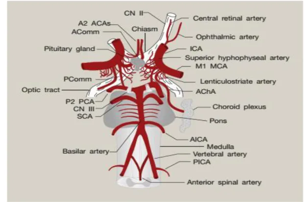
The brain is vascularized by 4 main arteries; internal carotid arteries and arteries right and left vertebral (figure 01). The two carotid arteries form the anterior circulation, the vertebral arteries when they unite in a basilar trunk thus forming the circulation posterior (Scremin et al., 2015 ; Chandra et al., 2017). The internal carotid arteries originating from the common carotid arteries, and the vertebral arteries. They form the Circle of Willis at the base of the brain, from which the main cerebral arteries are advancing and branching at the surface of the brain (Lindauer, 2017).
I.1.1. Middle Cerebral Artery
The middle cerebral artery (MCA) supplies a large area of the lateral surface of the brain and part of the basal ganglia and the internal capsule via four segments (M1, M2, M3, and M4). The M1 (horizontal) segment supplies the basal ganglia, which is involved in motor control, motor learning, executive function, and emotions. The M2 (Sylvain) segment supplies the insula, superior temporal lobe, parietal lobe, and the infer lateral frontal lobe (Kumral et al., 2002).
I.1.2. Anterior Cerebral Artery
The anterior cerebral artery (ACA) provides blood supply to the frontal, prefrontal, primary motor, primary sensory, and supplemental motor cortices. The sensory and motor cortices receive sensory information and control movement of the contralateral lower extremity. The supplemental motor area contains the Broca area, which is involved in the initiation of speech. The prefrontal cortex is used to organize and plan complex behavior and is thought to influence the personality (Brandt et al., 2000).
I.1.3. Posterior Cerebral Artery
The superficial posterior cerebral artery (PCA) supplies the occipital lobe and the inferior portion of the temporal lobe, while the deep PCA supplies the thalamus and the posterior limb of the internal capsule, as well as other deep structures of the brain. The occipital lobe is the location of the primary and secondary visual areas, where sensory input from the eyes is interpreted. The thalamus relays information between the ascending and descending neurons, while the internal capsule contains the descending fibers of the lateral and ventral cortical spinal tracts (Brandt et al., 2000).
I.1.4. Vertebra basilar
The vertebra basilar region of the brain is supplied by the vertebral arteries and the basilar arteries that originate within the spinal column and terminate at the Circle of Willis. (figure 02). These areas supply the cerebellum and brainstem (Jensen et al., 2005).
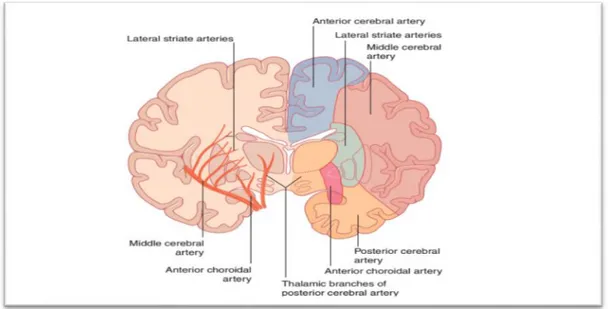
Figure 02: Distribution of perforating branches of the middle cerebral, anterior choroid , and posterior cerebral arteries (Kenna,2019).
I.2. Normal cerebral blood flow
CBF is defined as the blood volume that flows per unit mass per unit time in brain tissue and is typically expressed in units of ml blood ∕ (100 g tissue min) . The normal average cerebral
blood flow (CBF) in adult humans is about 50 ml ∕ (100 g min) (Fantini et al., 2016). I.3. Brain diseases
Brain diseases including Alzheimer‘s, Parkinson‘s, Huntington‘s and brain tumors and cerebrovascular accident are globally challenging issues (Jin et al., 2020).
I.3.1.Alzheimer‘s disease
Is a neurodegenerative disorder characterized by significant cognitive deficits behavioral changes, sleep disorders, and loss of functional autonomy (Lehmann et al., 2016). This neurodegenerative disease process is characterized classically by tow hallmark pathologies: β amyloid plaque deposition and neurofibrillary tangles of hyper phosphorylated tau (Weller et al., 2018). The number of patients suffering from AD is growing rapidly as the population ages worldwide (Lehmann et al., 2016). APOE4 is the human isoform strongest and most highly replicated genetic risk factor for AD (Halliday et al., 2015).
I.3.2.Parkinson’s disease
Parkinson‘s disease (PD) was first described by Dr.James Parkinson in 1817 as a «shaking palsy» (DeManged et al., 2015). It is the most common movement disorder and repress-ents the second most common degenerative disease of the central nervous system (Tysnes & Storstein., 2017), Affecting people mainly in later years of life (Sveinbjornsdottir,2016). Neuropath logically, PD is defined by loss of dopaminergic neurons in the substantial nigra pars compacta of the midbrain (Lill, 2016) which characterized neuropath logically by the presence of α-synuclein-containing Lewy bodies in the substantial nigra of the brain (Tysnes & storstein, 2017).
I.3.3.Huntington’s disease
Huntington‘s disease (HD) is a fully penetrant neurodegenerative disease caused by dominantly inherited CAG tri-nucleotide repeat expansion in the hunting in gene on chromosome 4 (Colgan & Tabrizi, 2017). This progressive neurodegenerative disorder accompanied by symptoms, such as dementia, chorea and depression (Choi et al., 2018). The people affected cannot think, talk, and move properly. Basal ganglia cells get damaged, which controls these capacities (Ghosh et al., 2019).
I.3.4.Brain tumor
Brain tumor is any mass that outcomes from unusual developments of cells in the brain. It might influence any individual at any age (Aswathy et al., 2017). Brain tumors classified to benign or low-grade (grade I and II) and malignant tumors or high-grade (grade III and IV). Benign tumors are non-progressive (non-cancerous) so considered to be less aggressive (Mohsen et al., 2018). Tumors can directly destroy all healthy brain cells. It can also indirectly damage healthy cells by crowding other parts of the brain and causing inflammation, brain swelling and pressure within the skull (Logeswari & Karnan, 2010). Primary tumors start in the brain and do not usually spread to other parts of the body and secondary tumors are formed by cancer cells from a primary tumor originated elsewhere in the body and spread to the brain (Mohsen et al., 2017).
II. Cerebrovascular accident ( stroke ) II.1. Definition and classification of stroke
Stroke is a type of cerebrovascular disease that involves the vessels of the central nervous system (Vida, 2004), defined as a clinical picture of vascular origin and a focal neurological deficit (Martin et al., 2011 ; Mevlut et al., 2020) (burst of cerebral arteries , hemorrhage , or occlusion by o thrombus or other particles ) leading to cerebral dysfunction (Vida, 2004).This deficit characterized by the rapidity of symptom resolution (within 24 h) , with or without demonstrable new hemorrhage or infraction in the brain (Martin et al., 2011 ; Prakash et al., 2018).
Strokes are sub classified into ischemic and hemorrhagic types, based on the underlying pathogenesis (Fatahzadeh & Glick, 2006).
II.1.1.Ischemic stroke II.1.1.1.Definition
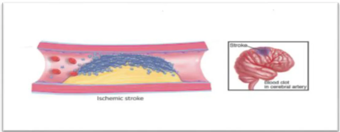
Ischemic stroke is the most prevalent type of stroke ( Pradeep et al., 2012). Defined as neurological event , charactezed by a sudden lack or complete interruption of blood flow in a brain supplying artery (figure 03) by a thrombus or embolus (Ning-Ning et al.,2018 ; Mani et al., 2019 ; Fei et al., 2020) , and reduces oxygen and energy supply to the critical tissues of the brain resulting in rapid cell death and cones ponding loss of neurological function (ahmed et al., 2014). The TOAST classification1 defines 5 IS subtypes based on etiology: 1) large-artery atherosclerosis, 2) cardio embolism, 3) small-large-artery occlusion, 4) stroke of other determined etiology, and 5) stroke of undetermined etiology (Jackova et al., 2019).
II.1.1.2. Pathophysiology
The event resulting from any subtypes of ischemic stroke result in the loss of blood supply, oxygen , nutrients and elimination of metabolic wastes (French et al., 2016). Neuronal death in ischemia occurs through apoptosis, necrosis, and autophagy after changes obstruct normal neuronal functioning such as ATP depletion and loss of ion exchange function of membrane pumps, terminal depolarization and glutamate mediated calcium excite toxicity, calcium dependent enzyme activation, generation of free radicals, and ultimately degradation of cellular molecules (Gupta et al., 2017).
Figure 03: Ischemic stroke (Bonaca M P et al., 2014). II. 1.2.Hemorragic stroke
II.1.2.1. Definition
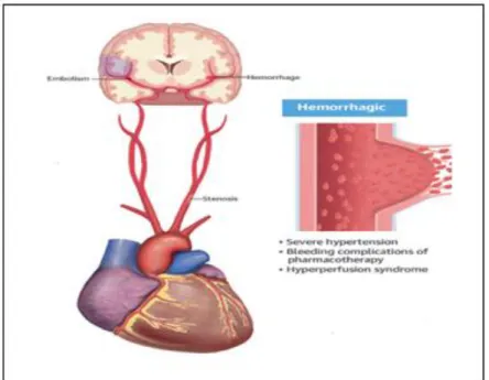
Hemorrhagic strokes due to an abnormal vascular structure or the rupture of a blood vessel (figure 04), including two main types intra cerebral hemorrhage (ICH) and subarachnoid hemorrhage (SAH) (Li H et al., 2018 ; Shao et al., 2019). ICH refers to bleeding within the brain itself, while SAH refers to bleeding that occurs outside of the brain tissue but still within the skull, precisely between the arachnoid mater and pia mater (Qu et al., 2019). II.1.2.2. Pathophysiology
Intra hemorrhage (ICH) is usually caused by rupture of small penetrating arteries secondary to hypertensive change or other vascular abnormalities (Qureshi et al., 2001). Its pathophysiology consists of three distinct phases : (1) initial hemorrhage , (2)hematoma expansion and (3) peri hematoma edema (Magistris et al., 2013).The initial injury mechanism in ICH is compressing brain parenchyma by hematoma 's mass effect , resulting in physical disruption of parenchymal architecture (Qureshi et al., 2003) in contrast a secondary mechanism of brain injury is related to clotting cascade after endothelial damage and hemoglobin breakdown (An et al., 2017).
The pathophysiology of SAH is complicated and includes vasospasm (VS), micro-circulatory dysfunction, neuronal apoptosis (Qu et al., 2019).
Figure 04: Hemorrhagic stroke (Devgun et al., 2018). II.2. Burden of stroke
Globally , stroke is a leading cause of mortality and disability and there are substantial economic costs for post stroke (Johnson, 2019).
II.2.1. Epidemiology and mortality
In 2017, stroke was the second leading cause of death worldwide (Jiang et al., 2020) .with 15 million strokes and 5.8 million stroke-related deaths per year (Al-Senani et al., 2019). The absolute number of people with incident strokes has significantly increased by 81% from 1990 to 2017 (Martins et al., 2019). Women are at higher risk of stroke than men, with a lifetime likelihood of 20% versus 17% (Robine & Bernstein, 2015). Ischemic stroke is by far the most common type of stroke, accounting for approximately 70%-90% of all stroke cases (Wang et al., 2017 ; Xu et al., 2013). Ischemic stroke mortality after one year is still at a high concentration (Slot et al., 2008 ; Xu et al., 2020). In 2016, there were 116,4 million of disability-adjusted life-years DALYs du to stroke (Feigin et al., 2016), in other words From the 795,000 new sufferers of stroke (figure 05) , 26% remain disabled in basic activities of daily living (Framingham cohort) and 50% have reduced mobility due to hemiparesis (Katan & Luft, 2018).
Figure 05:Global variation in stroke burden and mortality (Norrving & Kissela, 2013). II.2.2. Economic impact of stroke
For the years 2001–2005, the average cost for medication and for outpatient stroke rehabilitation services in the first year after discharge were $11,145 per patient with $7318 spent for rehabilitation services and $3376 for medication (Rajsic et al., 2018).
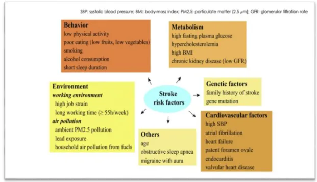
II.3. Risk factors of stroke
Risk factors are certain characteristic that give an increase likelihood of having stroke (Choudhury et al., 2015). Risk factors for hemorrhagic and ischemic stroke are similar, but there are some notable differences ( Boehme et al., 2017) . Risk factors for stroke are usually divided into non modifiable including age , Gender , Race and Heredity and modifiable such as hypertension, Heart disease, Diabetes mellitus, Dyslipidemia, Obesity, Smoking, Excess alcohol consumption (figure 06) (Arboix et al., 2015 ; Choudhury et al., 2015).
II.3.1. Non Modifiable risk factors II.3.1.1 .Age
For each consecutive decade after 55 years of age, the risk for stroke approximately doubles . The prevalence of stroke for individuals older than 80 years of age is approximately 27%, compared with 13% for individuals 60 to 79 years of age (Grysiewicz et al.,2008).
II.3.1.2.Gender
In general, stroke is more prevalent in men than in women . The incidence of stroke in the young (aged 35–44) is highest in women, however . The increased risk associated with pregnancy is most significant postpartum (Grysiewicz R A et al., 2008).
II.3.1.3.Race
In relation to race/ethnicity, African-Americans and some Hispanic/Latino American groups have a higher incidence and mortality of stroke .Prevalence and severity of risk factors and markers such as hypertension, diabetes, and obesity have been used to explain, at least in part, these disparities in stroke rates. Also, social determinants may play an important role (Farooq & Gorelick, 2017).
II.3.1.4.Heredity
Genetic factors that may be associated with stroke include family history of stroke, a number of inherited autosomal-dominant trait coagulopathies (eg, protein C and S deficiencies) There are many other genetic conditions (eg, inherited metabolic disorders, certain intracranial aneurysm alleles and mutations in the amyloid precursor protein genes causing inherited cerebral amyloid angiopathy syndromes) (Farooq & Gorelick, 2017).
II.3.2. Modifiable risk factors II.3.2.1.Hypertension
Hypertension is the most important modifiable risk factor for stroke, with a strong, direct, linear, and continuous relationship between blood pressure and stroke risk (Boehme et al., 2017).
II.3.2.2.Heart diseases
Heart diseases such as ; Atrial fibrillation (AF) has long been recognized to be a major risk factor for stroke, and this has only increased with the aging of the US population (Boehme et al., 2017). The Utilization of long-term anticoagulation therapy in the treatment of AF patients significantly increases the risk of future intra cerebral hemorrhage (ICH) (Elkhatib et al., 2020).
II.3.2.3.Diabetes
Diabetes is an independent risk factor for stroke disease Compared with patients without, patients with diabetes have at least twice the risk for stroke, and approximately 20% of patients with diabetes will die from stroke (Assy et al., 2019).
II.3.2.4.Dyslipidemia
The relationship between dyslipidemia and stroke risk is complex, with an increased risk for ischemic stroke with increased total cholesterol, and a decreased risk for ischemic stroke with elevated HDL cholesterol . Total cholesterol, meanwhile, is inversely associated with hemorrhagic stroke, with hemorrhagic stroke risk increasing as total cholesterol decreases (Boehme et al., 2017).
II.3.2.5.Obesity
Obesity is an established risk factor for the development of vascular diseases such as stroke (Oesch et al., 2017). It has classically been associated with disruption of pathways controlling lipid and glucose metabolism, however recent evidence has shown that obesity also has an inflammatory component , which is well established factor in stroke risk and outcome (Haley & Lawrence, 2016).
II.3.2.6.Smoking
Cigarette smoking is a causal risk factor for stroke that is dose- and duration- dependent, biologically plausible ,and interacts synergistically with other factors such as BP and risk of stroke (Towfighi & Hill, 2017).
II.3.2.7.Alcohol Consumption
Heavy alcohol use likely increases the risk of stroke by increasing the risk of hypertension, atrial fibrillation, cardiomyopathy, and diabetes ( Towfighi & Hill, 2017).
Figure 06 : Risk factors of stroke(Zhang et al., 2019 ). II.4. Diagnostic
The initial step in the evaluation of the acute stroke patient is accurate diagnosis (Zweifler, 2017). Acute stroke management in a patient presenting with focal neurologic deficits includes obtaining neuroimaging, most commonly a non-contrast head computed tomography (CT). On the other hand, magnetic resonance imaging (MRI) (Manwani et al., 2018). In addition, the use of a biomarker test (Monbailliu et al., 2017).
II.4.1.Magnetic Resonance Imaging
is an excellent modality for diagnosis of acute stroke with sensitivity of 94% -100% (Manwani et al ., 2018). Multimodal MRI can delineate the presence, size, location, extent and effects of acute brain ischemia, identify the hypo perfused tissue that is at risk of infarction, and show additional features of the cerebrovascular pathology. Magnetic Resonance Imaging (MRI) can also detect or exclude ICH with an accuracy comparable to CT (Merino & Warach, 2010).
II.4.2.Computed Tomography
Multimodal Computed Tomography (CT) , including CT angiography (CTA)and CT perfusion (CTP) (Merino & Warach, 2010 ).With the limited availability of MRI diagnostics for acute ischemic stroke in routine stroke care, attempts have been made to use CT for determining the time from onset (Audebert & Fiebach, 2015). The major value of acute CT
scanning is in differentiating intra cerebral hemorrhage from ischemic stroke and other pathologic processes such as subdural hematoma, tumor, and abscess (Zweifler, 2017).
II.4.3.Electromagnetic Tomography
Is an emerging imaging modality with significant clinically viable potentials , Electromagnetic Tomography (EMT) may present an effective supplement to current imaging modalities for acute and chronic assessment of perfusion related brain injuries (Semenov S et al., 2015).
II.4.4 .Examples for biomarker
The use of a biomarker test could add important adjunct information to make the diagnosis of AIS and thereby increase the likelihood of administering thrombolytic treatment within the therapeutic window (Monbailliu et al., 2017). Such as :
MicroRNAs (miRNAs) are a class of small non-coding RNAs .these biomarkers can be used as powerful tools in monitoring different stages of stroke and stroke outcomes (Mirzaei et al., 2017).
the levels of several inflammatory mediators correlate with stroke severity and outcome. Inflammatory biomarkers such as CRP or several pro inflammatory cytokines, specially IL-6, TNF-α and neutrophil-to-lymphocyte ratio (NLR) among other molecules have been widely associated with stroke diagnostic (Simats et al., 2016; Szegedi et al., 2017).
Plasma concentrations of the astroglial protein S-100b, a cytosolic calcium-binding protein, have been found to correlate with the extent of tissue damage (infarct volume) and neurologic outcome (Reynolds, 2003).
Other markers include a lipid panel, HgbA1c, Utox, and TSH (if there is aclinical suspicion for atrial fibrillation) along with the standard CBC and CMP (Stack & Cole, 2017).
II.5. Consequences of stroke
Motor impairment is the most common deficit after stroke, which either happens as a direct consequence of the lack of signal transmission from cerebral cortex or as a slowly
II.6. Treatment of stroke
II.6.1. Treatment of ischemic stroke
At present, there is no reparative treatment, and thus, this injury leads to permanent brain dysfunction in the patient. Therefore, we need to reduce the burden of disease in stroke patients and seek to reverse neurological damage by reducing CNS injury and promoting neural regeneration (Xie et al., 2020).
II.6.1.1.Intravenous thrombolysis
Intravenous thrombolysis with Al-teplase, a recombinant tissue plasminogen activator, is the standard medical treatment for acute ischemic stroke within 4.5 hours after the onset of symptoms (Thomalla et al., 2018 ; Mistry et al., 2017).
II.6.1.2.Intra-arterial thrombolysis
Intra-arterial thrombolysis in patients with middle cerebral artery territory strokes and M1 or M2 occlusion within 6 h of symptom onset. The IMS (Interventional Management of Stroke) study explored this latter approach because investigators noted the relative lack of efficacy of IV t-PA on large strokes and patients with major arterial occlusions; there are several ongoing investigations (Marsh & Keyrouz, 2010).
II.6.1.3.Endovascular therapy
Reperfusion therapy remains the only proven treatment for acute ischemic stroke (Rebello et al., 2016) . endovascular thrombectomy ; represented one of the only two Food and Drug Administration (FDA)-approved therapies are currently available to ischemic stroke patients after the tissue plasminogen activator (tPA) (Kaisera, 2020) .
II.6.1.4.Neural stem cells therapy
Neural stem cells are the precursor cells present in the neuro-epithelium along the neur-axis during mammalian fetal development (Zhang et al., 2019).there are many sources of sum an neural stem cells (Kokaia et al., 2018). the transplantation of stem cells induce to an extension of the time window for drug intervention improvement of neurological deficits, reduction of infarct volume, pro-regenerative cerebral reorganization, mitigation of post-stroke neuro-inflammation, and tissue restoration (Sinden et al., 2017).
II.6.1.5.Neuroprotection therapy
The term neuroprotection has also been used to describe the theoretical concept of decreasing additional neuronal injury that occurs upon reperfusion of the ischemic brain(Patel et al., 2017). One of neuroprotection method is the administration of near-infrared laser therapy in which light energy at a wavelength of 808 nm is applied directly to the shaved skull of the patient in an effort to enhance brain recovery through a process called photo bio stimulation (Yip et al., 2008) .
II.6.1.6.Physical therapy
Physical therapy remains the only approved intervention aimed at improving behavioral impairments (Ghuman, 2016).Rehabilitation involves repetitive motor practice to promote use-dependent neuroplasticity and functional recovery, and primarily occurs in the first 6 months after stroke (Ackerley et al., 2016).
II.6.2. Treatment of hemorrhagic stroke
For hemorrhagic stroke, limited treatments are available, which focus on controlling the bleeding and reducing the pressure caused by the bleeding immediately after stroke onset (Corey et al., 2020).
III. Oxidative stress III.1.Definition
Oxidative stress is broadly referred to as an imbalance between the generation of the Free radicals which are inherently unstable molecules because of balance between the production of reactive species (reactive oxygen specie ROS and reactive nitrogen specie RNS) and their clearance by the anti-oxidant defense system in favor of the former ( Stefanovic et al., 2019 ; Betteridge, 2000). Moreover, oxidative stress has been proven to be associated with many diseases, including cancer, diabetes, chronic kidney disease (CKD), cardiovascular diseases (CVDs), neurodegenerative diseases , inflammation ,atherosclerosis and ageing (El Assar et al., 2019 ; Shahidi & Zhong, 2015).
III.2.Free radicals III.2.1.Definition
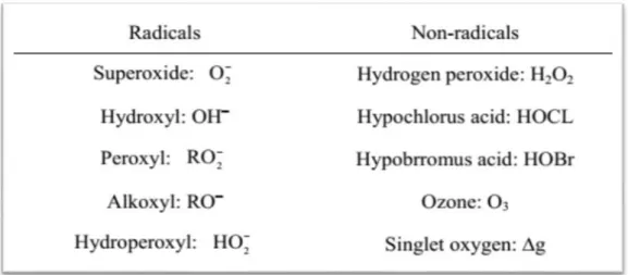
A free radical can be defined as an atom or molecule containing one or more unpaired electrons in valency shell or outer orbit and is capable of independent existence. Both ROS and RNS collectively constitute the free radicals and other non-radical reactive species (Phaniendra et al., 2015 ; Kehrer et al., 1993) (table 01 and table 02). These radicals can be produced in cells by losing or accepting a single electron, therefore, behaving as oxidants or reductants (Lobo et al., 2010). The main process of ROS generation in mitochondria could be schematically presented as O2 → O2- → H2O2 → OH (Luo et al., 2019).
Table 01 : Reactive oxygen species (Rahman et al., 2012).
III.2.2.Sources of free radicals
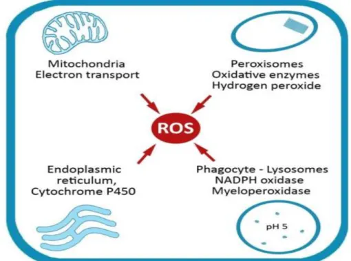
Living aerobic organisms stay under the influence of free radicals due to the constant formation of reactive oxygen species (ROS) and reactive nitrogen species (RNS) by endogenous and exogenous sources (Kruka et al., 2019).
Endogenous sources of ROS
The endogenous sources of ROS (figure 07) include mitochondria, cytochrome P-450 metabolism, peroxisomes, and inflammatory cell activation (Macrophages and neutrophils contain a group of enzymes called the NADPH oxidase complex, which, upon activation generates superoxide radicals and hydrogen peroxide ) (Bhattacharya, 2014).
Figure 07 : Endogenous sources of free radicals/ROS (Santo A et al., 2016).
Exogenous sources of ROS
ROS and RNS can also be generated through exposure to external factors such as oral bacteria, ionizing and ultraviolet radiation, food, and air pollution, alcohol, and cigarette smoking, heavy metals and certain chemotherapeutic drugs ( Żukowski et al., 2018 ; Santo et al., 2016 ).
III.3.Antioxidants
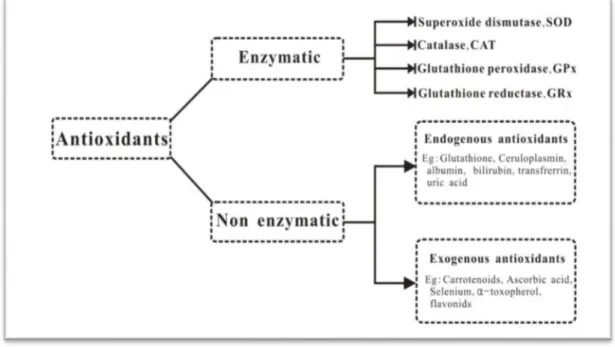
Antioxidant state necessary for Natural physiological function. An imbalance in the case of oxidation / antioxidants can cause injury to the tissues of the organism (Vidhya et al., 2018). Antioxidant defense protects biological systems from free radical toxicity and includes both endogenous and exogenous molecules , that function interactively and synergistically to neutralize free radicals,( Krishnamurthy & Wadhwani, 2012 ; Birben et al., 2012). The term ‗antioxidant‘ refers to any molecule stable enough to donate an electron to a rampaging free radical and neutralize it, thus reducing its capacity to damage a target molecule (Li et al., 2016). The antioxidants are present in significant amounts in commonly consumed fruits, vegetables, beverages (juices, tea, coffee), nuts and cereal products (Mirończuk-Chodakowska et al., 2018). In a biological system, antioxidants can be categorized as enzymatic or non-enzymatic (figure 08) (Zhao et al .,2016) .
III.3.1.Endogenous antioxidants
Enzymatic antioxidant
Enzymatic Activities of superoxide dismutase (SOD), catalase (CAT) and glutathione peroxidase (GPX) constitute a first line antioxidant defense system which plays a key and fundamental role in the total defense mechanisms and strategies in biological systems (ghodaro & Akinloye, 2017). SODs are metal-containing proteins that catalyze the removal of superoxide , O2 is converted by SOD to H2 O2 , which is decomposed to water and oxygen by
CAT, preventing hydroxyl radicals production (Krishnamurthy & Wadhwani, 2012 ; Liguori et al., 2018). However, catalase is absent in the mitochondria, hence the reduction of H2O2 to
water and lipid peroxides to their corresponding alcohols is carried out by Glutathione Peroxidase(GPx) (ghodaro & Akinloye, 2017). Additionally, GSH-Px converts peroxides and hydroxyl radicals into nontoxic forms by the oxidation of reduced glutathione (GSH) into glutathione disulfide and then reduced to GSH by glutathione reductase (Birben et al., 2012).
Non enzymatic antioxidant
Non-enzymatic antioxidants include endogenously produced GSH or dietary compounds (Zhao et al., 2016). The endogenous non-enzymatic antioxidants are molecules that inter act with RONS and terminate the free radical chain reac tions: bilirubin, α-tocopherol (vitamin E), and β-carotene are present in blood while albumin and uric acid account for 85% of antioxidant capacity in plasma (Wu JQ, Kosten & Zhang, 2013).
Figure 08 : Enzymatic and non-enzymatic classification of antioxidants (Li et al., 2016).
III.3.2.Exogenous antioxidants
Dietary antioxidants such as vitamin E, vitamin C, carotenoids, some minerals (e.g. ZnMn, Cu, Se) and polyphenols (flavonoids, phenolic acids, stilbenes, lignans) can affect the activity of endogenous antioxidants. Endo- and exogenous antioxidants may act synergistically to maintain or reestablish redox homeostasis ( Mirończuk-Chodakowska et al., 2018).
III.4.Oxidative stress and stroke
Oxidative stress (OS) plays an important role in the pathogenesis of nervous tissue damage in the presence of stroke (Maksimova et al., 2019). Ischaemia would be followed by reperfusion leads to increased production of free radicals (figure 09) (Setyopranoto, 2016).(Chehaibi et al., 2016). Superoxide is one of the most important ROS in the central nervous system [CNS] (Rodrigo et al., 2013). Nitric oxide (NO) plays an important role in vessel dilatation and inflammation (Cheng et al., 2017). The brain is especially prone to free radical damage for several reasons. It is very rich in polyunsaturated fatty acids, which are particularly vulnerable to free radical induced peroxidation, but also has a low content of
Histologically, stroke is characterized by an ischemic core [infarct] surrounded by a ―penumbra‖ [peri-infarct] region (Rodrigo et al., 2013), Increased production of superoxide anions, hydroxyl radicals, and peroxy nitrite or nitrogen dioxide has been shown in infiltrating phagocytes, vascular and glial cells in the penumbra (Sidorov et al., 2019). As a result Neurons are killed rapidly within minutes and the tissue in the ischemic core is irreversibly damaged even if blood flow is reestablished (Li et al., 2018). The immune-induced inflammation. Brain ischemia and reperfusion is known to activate the complement system and form complexes such as C5b-9. Circulating dendritic cells, lymphocytes, monocytes, monocyte derived macrophages, and natural killer cells modulate inflammatory responses and thereby facilitate the thrombo-inflammatory response (Yang et al., 2019).
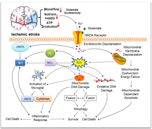
Figure 09: Pathological signaling pathways involved in mitochondrial function and reactive oxygen species)ROS) generation in the cerebral ischemic cascade (Yang, 2018).
Second part
Materials
&
I. Materials and Methods I.1. Patients and reagents I .1.1. Study period
The period of our study is 6 Months , (from September 2019 to February 2020) at the medicine service and medical analysis laboratory of Sliman Amirat hospital (Touggourt) and at the Faculty of Natural Sciences and Life at the University of Echahid Hamma Lakhdar El-Oued.
I.1.2. Epidemiological and Risk factors study
The stroke reports of 605 patients from hospital of Sliman Amirat in Touggourt were collected and information such as the patient‘s name, age, sex, type of stroke, region of patients and during of hospitalization was noted. The data was compiled to get the percentage distribution and graphs were drawn for the individual data.
A standard questionnaire was used to obtain the baseline information which Contribute in our study,with face-to-face interviews by us . this questionnaire asked for 130 volunteers divided into 65 healthy group as a control and 65 stroke patients and used for evaluation of some Risk factors of the disease.
I.1.3. Biological study
For biological study, this study is carried out on 40 volunteers ; 10 volunteers women aged between 36-77years, were divided into two groups ; a group of 05 healthy control women aged 54.4±2.11 year, and 05 women has stroke aged 56.20±6.80 years. 30 men volunteers of age between 35-79years , were divided into two groups ; a group of 15 healthy control men aged 56±3.32 year, and 15 men has stroke aged 57.75±4.01 years .
Inclusion criteria
Voluntary live in TOUGGOURT region .
Control group in good health, does not have any pathology. Voluntary was surfed from stroke .
Exclusion criteria
I.1.4.Reagents
Ethylene diamine tetraacetic acid (EDTA), Hydrogen Peroxyde (H2O2) ,Hydrochloricacid (HCl) , Thiobarbituric acid (TBA) , Salicylic acid , Methanol, Tris ,Trichloroacetic acid (TCA) , Copper sulfate (CuSO 4) , Ascorbic acid, DTNB (5,5′-Dithiobis(2-nitrobenzoic acid )), Sodium chloride (NaCl) , Butylated hydroxytoluene (BHT) ,Phosphate-buffered (KH₂PO 4,K₂HPO 4), folin , NBT , Methionin , riboflavin .
I.2. Methods
I.2.1. Collection of data
We used a questionnaire including social and clinical data for each volunteer , after that collecting the risk and protective factors associated with the accident vascular cerebral . I.2.1.1. Sample collection
About blood sampling for both groups is done morning fasting . It is performed in the vein of the end of the elbow. Blood samples is collected in three tubes . Dry tubes are centrifuged at 3000 rpm for 10 minutes, then obtained the serum to achieve the dosage of oxidative stress ( MDA, GSH , vitamin C , SOD ,ORAC and total thiol ) parameters .
The anticoagulant tube (EDTA) is mixed well and then assays the hematological and oxidative stress ( MDA , GSH ) parameters .
The anticoagulant tube (Heparin) are centrifuged at 2000 rpm for 5 minutes , then recover the plasma to realize the dosage of biochemistry parameter: Glucose, triglyceride , HDL , LDL , cholesterol, total bilirubin , alkaline phosphatase activity (ALP) , total protein , albumin , uric acid and electrolyte markers (potassium , sodium , chlorine ) .
I.2.2. Method of Hematological analysis
Hematological analysis (FNS) is performed by the hematology Auto analyzer. I.2.3. Biochemical parameters assay
Glucose, triglyceride, HDL, LDL, cholesterol, total bilirubin , total protein , albumin and uric acid Were determined by Auto analysis (BIOLIS24j) use commercial kits from Spinreact, (Spainref:glucose-20121, triglyceride-20131, HDL-20113, cholesterol- 20111, total bilirubin-20131, total protein-1001291, albumin-1001020,uric acid- 1002011. and enzyme
marker were also measured using commercial kits (Spinreact, ref: alkaline phosphatase-20015).
I.2.4. Method of electrolytes analysis
Determination of the ion gram parameter (potassium , sodium , chlorine ) by automatic electrolyte analyzer (Easylute) .
I.2.5. Method of estimating oxidative stress parameters I.2.5.1. Preparation of erythrocyte homogenate
Blood EDTA tubes contents are centrifuged at 2000 rpm for 10 min and removed the plasma. The cap of EDTA tube was lysis with 50 ml of TBS buffer (EDTA4.24 M;tris 1.21M; pH=7) and incubated 30 min in freezer. After incubation centrifuged at 2500 rpm for 10 min and the obtained supernatant (erythrocyte homogenate) was used for the determination of antioxidant activity (Miller et al., 1988).
I.2.5.2. Separation of leukocyte homogenate
After removing the plasma and separation of erythrocyte, the rest of EDTA tube contents centrifuge at 2000 rpm for 10 min. Wash pellet with lysis buffer and shake incubate in freezer for 30 min. After incubation centrifuged at 2500 rpm for 10 min followed this step by washing with lysis buffer until the Leukocyte pairing and then recovered to make the dosage of stress tests (Miller et al., 1988).
I.2.5.3. Malondialdehyde assay
MDA was measured according to the method described by (Yagi, 1976). Thiobarbituric acid 0.67% (w/v) was added to aliquots of the sample previously precipitated with 10% trichloroacetic acid (w/v). Then the mixture was centrifuged, and the supernatant was heated (100°C) for 15 min in a boiling water bath. Then cool in a cold water bath for 30 minutes, leaving the tubes open to allow evacuation of the gases formed during the reaction and the absorbance was measured at 532 nm using a spectrophotometer. The concentration of TBARS was determined using the molecular extinction coefficient of MDA (a=1.53 105 M-1.Cm-1). I.2.5.4. Determination of Reduced glutathione level
dithio-bis-2-nitrobenzoic acid, which is called reagent of Ellman with SH groupings exist in GSH briefly, 800 μL of homogenate are added to 200 μL of salicylic acid (0.25 ) and centrifuge at 1000 rpm for 5 minutes. 500 ml of supernatant are then mixed with 1000 μl of tris buffer (tris 0.4 mol, 0.02 molNaCl pH = 8.9) and 25 µL of DTNB (0.01 mol.L-1). After 5 minutes of incubation, the absorbance is read at 412 nm (Weakbeker& Cory, 1988).
13133:Absorption constant of SH groups at 412 nm.
OD :The absorbance reader by the spectrophotometer.
521.1ml :Total volume of blend.
521ml: Volume of solution float.
1:Volume of protein mixture.
0.8 ml :Volume of homogeneous solution without protein exists in 1ml.
GSH: Concentration of glutathione.
I.2.5.5. Determination of superoxide dismutase activity assay
The assay method of SOD activity using the NBT by the superoxide anion (O2 .), is used as a basis for detecting of presence of SOD by measuring the spectrophotometrically absorbance at 560 nm (Beauchamp & Fridovich,1971).
Collect in tubes Blank Sample EDTA-Met 0.1mM EDTA-13mM Met 1000μL 1000μL Phosphate buffer (50Mm) 892.2μL 892.2μL Sample 0 50 Phosphate buffer 1000μL 950μL NBT (75μM) 85.2μL 85.2μL Riboflavin (2μM) 22.6μL 22.6μL
Expression of results
I.2.5.6.Determination of antioxidant power a. Principle
The total antioxidant power of the serum,its capacity to absorb free oxygen radicals (ORAC: Oxygen Radical Absorbance Capacity), is estimated by the ability of red blood cells to resist free radical-induced hemolysis in vitro in the presence of plasma according to the method of (Oyaizu, 1986). This method is based on the time-dependent monitoring of red blood cell hemolysis induced by a free radical generator.
b. Treatment of red blood cells
Centrifuge donor blood at 2000 rpm for 10 min and remove plasma.
Wash gently 1 volume of the pellet with 2 volumes of physiological saline (without lysing the RBCs), then centrifuge again at 2000 rpm for 5 min.
c. Operating mode Control tube
Add 1 ml of RC: 20 μl of CuSO4 (2 mM), 20 μl of H2O2 (30 ) and 2 ml of physiological saline, then stir gently. Incubate for 5 min at room temperature, centrifuge for 5 min at 2000 rpm.
Read the OD at 450 nm from the supernatant and put it back into the tube and stir gently.
Repeat this operation every 10 minutes for 1 hour. Standard tube
To 1 ml of GR are added: 20 μl of CuSO4 (2 mM), 20 μl of H2O2 (30 ) and 2 ml of physiological saline, and 20 μl of vitamin C (400 μM) and then stir gently. Incubate for 5 min at room temperature, centrifuge for 5 min at 2000 rpm.
Test tube
To 1 ml of RC are added: 20 μl of CuSO4 (2 mM), 20 μl of H2O2 (30 ) and 2 ml of physiological saline, and 20 μl of serum (400 μM) and then stir gently. Incubate for 5 min at room temperature, centrifuge for 5 min at 2000 rpm.
Read the OD at 450 nm from the supernatant and put it back into the tube and stir gently.
Repeat this operation every 10 min for 1 hour (t0, t10, t20, t30, t40, t50, t60, and average the latter:
ΣDO = Σ (t0, t10, t20, t30, t40, t50, t60) / 7
To calculate the total antioxidant power using two methods. Calculate method
I.2.5.7.Determenation of vitamin C concentration
The plasma vitamin c is measured according to the method of Jacota and Dani (1982) using the Folin reagent and a range of ascorbic acid . Brieflt , add ml of plasma to 0.5 mol of the TCA solution ( 10٪ ) . Vortex then place the tube in an ice bath for 30 min . centrifuge at 3000rpm for 10 min . take 0.75 ml of the supernatant to which 0.75ml of distilled water and 150µl of Folin (1\10) are added vortex and incubate for 15 min at room temperature . Read the OD using a blank spectrophotometer at 769 nm and determine the vitamin C concentration (µg \ml) (Jago & Dani ,1982 ).
I.2.5.8.Determenation of total thiol concentration
The serum total thiol was determined according to the method described by Ellman9. Aliquot of the supernatant (50µL) was mixed with 1ml of tris base (0.25M)-EDTA(20mM) buffer and absorbance was taken at 412 nm . this was followed by the addition of 5 ml of 10 mM DTNB , after which another absorbance was taken after 15 min . The absorbance of DTNB blank and sample blank also taken and the total thiol were calculated using the formula
Where :
A = Absorbance of supernatant B=DTNB blank absorbance C = Sample blank absorbance
Extinction coefficient ( 13.600 L mol -1 cm -1 ) I.3. Statistical analysis
Statistical analysis is performed by the SPSSV20.0 software results comparisons were carried out by using the Student T test to compare means among the groups, Correlation analysis was carried out using Pearson Correlation test and regression analysis was used for other analysis and statistical data. Differences were considered statically significant at p <0.05.