« La Faculté n'entend donner aucune approbation aux opinions émises dans les thèses : celles-ci sont propres à leurs auteurs. »
LERISSON Erol TABLE DES MATIÈRES
4
LISTE DES ABRÉVIATIONS : INTRODUCTION GÉNÉRALE
3DVR : Three dimension volume rendering AAA : anévrysme de l’aorte abdominale
AOMI : Artériopathie oblitérante des membres inférieurs
ATA : Anévrysme thoraco-abdominal
CBCT : Cone beam computed tomography
ceCBCT : Contrast enhanced cone beam computed tomography CTA : Computed angiographic tomography
DALYs : Disability-adjusted life-years EVAR : Endovascular aneurysm repair
ESVS : European society for vascular surgery HU : Hountsfield unit
MIP : maximum intensity projection minIP : Minimum intensity projection MPR : multiplanar reconstruction SVS : Society for vascular surgery TDM : Tomodensitométrie
LISTE DES ABRÉVIATIONS : ARTICLE
3DVR : Three dimension volume rendering
ALARA : As low as reasonably achievable
AP : Uniplanar angiography
BEVAR : Branched endovascular aneurysm repair
CBCT : Cone beam computed tomography
ceCBCT : contrast enhanced cone beam computed tomography
CEUS : Contrast enhanced ultrasonography
CI : Confidence interval
CT : Computed tomography
CTA : Computed tomography angiography
DUS : Dupplex ultrasonography
EVAR : Endovascular aneurysm repair
FEVAR : Fenestrated endovacular aneurysm repair
FU : Follow up
HR : Hazard ratio
IQR : Interquartile range
MIP : maximum intensity projection
MPR : multiplanar reconstruction
TABLE DES MATIÈRES
TABLE DES MATIERES ... 7 RESUME ... 9 INTRODUCTION GENERALE ... 11 GÉNÉRALITÉS ... 11 ÉPIDÉMIOLOGIE ... 12 DIAGNOSTIC ... 13PRISE EN CHARGE DES AAA ... 14
RÔLE DE LA TOMOGRAPHIE VOLUMETRIQUE À FAISCEAU CONIQUE (CBCT) DANS LA PRISE EN CHARGE DES AAA. ... 21
POURQUOI REALISER UNE TOMOGRAPHIE VOLUMETRIQUE A FAISCEAU CONIQUE APRES EVAR ? ... 25 OBJECTIF ... 28 REFERENCES BIBLIOGRAPHIQUES ... 29 ARTICLE ... 35 INTRODUCTION ... 39 MATERIAL AND METHODS ... 41 RESULTS ... 48 DISCUSSION ... 63 CONCLUSION ... 69 CONCLUSION GENERALE ... 71 REFERENCES BIBLIOGRAPHIQUES ... 72 ANNEXES ... 79
RÉSUMÉ
Impact de la tomographie volumétrique par faisceau conique
(CBCT) sur les résultats à moyen terme après exclusion
endovasculaire des anévrysmes de l’aorte abdominale
Contexte :
Lors d’une précédente étude, il était constaté que le CBCT permettait de diagnostiquer plus d’anomalies techniques sur les endoprothèses aortiques (plicature, endofuite distale) en fin de procédure que l’angiographie 2D conventionnelle. Ceci entrainait plus de gestes complémentaires non planifiés pour corriger ces anomalies, mais les bénéfices à long terme de cette prise en charge plus « agressive » restaient débattus. Le but de cette étude est d’évaluer si le traitement per-opératoire des anomalies détectées par le CBCT améliore les résultats à long terme après EVAR.
Méthode:
Il s’agit d’une étude monocentrique rétrospective, incluant tous les patients traités par endoprothèses bifurquées, fenêtrées et branchées en salle hybride sur deux périodes. De décembre 2012 à juillet 2013, le succès technique était contrôlé par une angiographie 2D en fin d'intervention, puis par un angioTDM (CTA) avant la sortie (groupe 1). D'octobre 2013 à avril 2014, un CBCT peropératoire (40°/sec - 35cc d'iode dilué dans 35 cc de sérum physiologique) était réalisé, suivi d'une échographie doppler de contraste (CEUS) avant la sortie (groupe 2). Si une anomalie technique était détectée sur l’angiographie ou le CBCT elle était traitée dans le
LERISSON Erol RÉSUMÉ
10
même temps opératoire. Le taux de réinterventions liées à l’endoprothèse (EDP) et la survenue de complications tardives liées à l’EDP (définies par la mortalité liée à l'aorte, endofuites de type 1 ou 3 secondaires, les plicatures ou thromboses de vaisseaux cibles ou de jambage et l’accroissement >5mm du sac anévrysmal) étaient étudiés.
Résultats :
133 patients étaient inclus dans l’étude (79 dans le groupe 1 et 54 dans le groupe 2) avec un suivi médian de 48 [36-59] mois. Les diamètres aortiques initiaux étaient respectivement de 55 (51-60) mm et 53 (51-58) mm. 40 complications tardives liées à l’EDP survenaient chez 25 patients (31.7%) dans le groupe 1 et 12 évènements chez 9 patients (16.7%) dans le groupe 2 (p=0.05). Les taux de réinterventions liées à l’EDP étaient respectivement de 18.9% (n=15) et de 14.8% (n=8) (p=0.46). En analyse multivariée, le CBCT ne réduisait pas significativement le taux de réintervention ou de complications tardives liés à l’EDP, mais prévenait la majoration du diamètre au cours du suivi (OR=0.25, IC95% 0.07-0.91, p=0.035).
Conclusion :
Cette étude est la première à étudier l’impact à moyen terme d’un CBCT réalisé en routine après EVAR. Nos résultats montrent que la correction per-opératoire d’anomalies constatées sur le CBCT réalisé en fin de procédure protège d’une expansion du sac anévrysmal, et suggèrent une tendance à la réduction du taux de complications tardives liées à l’endoprothèse et de réinterventions liées à l’endoprothèse.
INTRODUCTION GÉNÉRALE
GÉNÉRALITÉSUn anévrysme artériel est défini par une dilation focale et permanente d’une artère avec perte de parallélisme, et dont le diamètre est supérieur de 50% au diamètre sain d’amont (1). Les anévrysmes artériels peuvent se manifester sur l’ensemble de l’arbre artériel, la localisation extracrânienne la plus fréquente étant la localisation aortique.
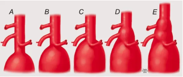
Bien que les anévrysmes aortiques puissent survenir sur n’importe quel segment de l’aorte ; de son origine, la racine aortique, jusqu'à sa terminaison, la bifurcation aortique, la localisation la plus fréquemment observée est sous rénale (30% de cas). (2) Un anévrysme de l’aorte abdominale sous rénale (AAA) est défini par un diamètre supérieur à 30 mm. A l’étage abdominal, lorsque le sac anévrysmal englobe l’origine d’une ou plusieurs artères à destinée rénale ou viscérale, on parle d’anévrysme aortique complexe. Cette définition est anatomique et englobe les entités suivantes (Figure 1) :
Figure 1 A-E: Classification anatomique des anévrysmes aortiques complexes (© Mayo Clinic)
LERISSON Erol INTRODUCTION GÉNÉRALE
12
• A : Les AAA sous rénaux à collet court, dont le collet proximal ; défini par la longueur d’aorte saine entre l’artère rénale la pus basse et l’origine du sac anévrysmal ; est compris entre 4 et 10 mm,
• B : les AAA juxta-rénaux, dont le collet proximal est compris entre 0 et 4 mm, • C : les AAA para-rénaux, englobant l’origine des artères rénales,
• D : les AAA para-viscéraux, englobant l’origine de l’artère mésentérique supérieure et du tronc cœliaque,
• E : et les Anévrysmes thoraco-abdominaux de type 4 (ATA 4) selon la classification de Crawford, défini par une extension de l’anévrysme jusqu’au hiatus diaphragmatique (classification des ATA selon Crawford en annexe 1).
La pathologie anévrysmale aortique est le plus souvent d’origine dégénérative, athérosclérotique (3) (4). Dans 5 à 10 % des cas, l'étiologie est plus rare : anévrysme septique, dystrophies héréditaires du tissu conjonctif (maladie de Marfan, maladie d'Ehlers Danlos), lésions inflammatoires (maladie de Takayasu, maladie de Behçet) et traumatique. L’origine syphilitique est désormais exceptionnelle dans les pays développés.
ÉPIDÉMIOLOGIE
L’incidence de la maladie anévrysmale augmente avec l’âge et est plus importante dans les pays développés, ainsi en 2010 l’incidence des anévrysmes de l’aorte abdominale (AAA) chez les patients âgés de 40 à 44 ans était de 0.96 nouveau cas pour 100.000 habitants dans les pays développés (IC 95 % : 0.65 à 1.41) contre 175.12 (IC 95 % : 159.43 à 191.64) chez les patients âgés de 75 et 79
ans (5). La prévalence des AAA de plus de 4 cm chez les hommes entre 55 et 64 ans est estimé à 1 % dans les pays développés, puis augmente de 2 à 4% par décennie. (6)
Les facteurs de risque fortement associés au développement d’une maladie anévrysmale sont le sexe masculin OR = 5.93 (4.26–8.25), le tabagisme actif OR = 2.97 (1.20–7.30), une histoire familiale d’anévrysme aortique OR = 9.64 (1.72–53.98) (6)(7)(8), une artériopathie oblitérante des membres inférieurs (AOMI) OR=2.50 (2.12–2.95), une histoire de cardiopathie ischémique préexistante OR=2.28 (1.90– 2.74) et une hypertension artérielle essentielle OR=1.33 (1.14–1.55). (9).
L’histoire naturelle est l’augmentation progressive du diamètre du sac anévrysmal, puis la rupture de l’anévrysme conduisant à une hémorragie massive et au décès du patient avant la possibilité d’une prise en charge chirurgicale dans près de 80 % des cas (10)(11). Le risque de rupture est proportionnel à la taille de l’anévrysme, le taux de rupture annuel pour les petits anévrysmes (entre 30 et 39 mm) étant inférieur à 0.3%, il s’élève à 2.3% pour les AAA mesurant entre 50 et 54 mm qui représentent la grande majorité des AAA au moment du diagnostic (12). Le risque de rupture augmente ensuite de manière exponentielle pour atteindre 32.5% lorsque l’AAA dépasse les 70 mm (13)(12)(14).
DIAGNOSTIC
Les AAA sont le plus souvent asymptomatiques et découverts fortuitement par un examen d’imagerie (radiographie de l’abdomen, scanner, IRM, ou échographie) réalisé dans le contexte d’une autre pathologie. La découverte d’une masse battante pulsatile abdominale à l’examen clinique est devenue rare et doit faire suspecter un AAA. En cas de rupture, la clinique est bruyante et associe douleur abdominale
LERISSON Erol INTRODUCTION GÉNÉRALE
14
d’apparition brutale et choc hémorragique. Il s’agit d’une urgence thérapeutique qui nécessite le transfert immédiat du patient au bloc opératoire en cas d’instabilité hémodynamique. Si l’état hémodynamique du patient le permet, un angioscanner est réalisé en urgence afin de confirmer le diagnostic et de préciser la localisation de l’anévrysme par rapport aux artères rénales et digestives afin d’orienter le nature du geste chirurgicale.
PRISE EN CHARGE DES AAA
Afin de prévenir le risque de rupture, un traitement chirurgical préventif électif est retenu selon une balance bénéfice (prévention de la rupture) risque (morbi/mortalité liée à l’intervention). Le seuil thérapeutique retenu par les sociétés savantes européennes (ESVS) et américaine (SVS) pour un traitement électif d’un AAA sous rénal est de 55 mm chez l’homme et 50 mm chez la femme, ou un anévrysme évolutif (croissance annuelle > 10 mm). (15) (16) (Annexes 2-3).
Un anévrysme devenu symptomatique est une indication au transfert du patient en urgence dans une structure adaptée comprenant une équipe de chirurgie vasculaire (12).
TRAITEMENT CHIRURGICAL OUVERT DES AAA
Technique
Les principes de la chirurgie moderne des AAA par mise à plat et reconstruction in situ on été posés par A. Carrel (première mise à plat et reconstruction in situ par tissu autologue en 1948). La technique a été modifiée durant la seconde moitié du XXème siècle avec l’introduction des prothèses en
LERISSON Erol INTRODUCTION GÉNÉRALE
16
Figure 2A-1C Technique de mise à plat d’un AAA sous rénal. D Reconstruction in situ par tube aorto-aortique simple. E Reconstruction in situ par tube bifurqué aorto bi-iliaque. D’après Rutherford et al.
Résultats de la chirurgie conventionnelle
La mortalité post opératoire précoce associée au traitement chirurgical ouvert des AAA sous rénaux (s’étendant de l’intervention chirurgicale jusqu’à J30) est de 1% à 4.6% lorsque l’on observe le bras chirurgical des quatre grands essais randomisés comparant le traitement chirurgical conventionnel au traitement endovasculaire (EVAR 1, DREAM, OVER, ACE) (20)(21)(22)(23). Le clampage aortique est responsable de modifications importantes de l’hémodynamique qui entrainent une surcharge pour le système cardio-pulmonaire. La morbidité périopératoire est ainsi dominée par les complications respiratoires qui touchent jusqu’à 17% des patients, les complications cardiaques chez 10 à 15 % des malades, puis les complications rénales chez 10% des malades (24). Au cours du suivi, près de 20% de ces patients seront également exposés à des complications liées à la voie d’abord chirurgicale nécessitant une réintervention (occlusion, éventration) (25).
Les grandes séries traitant des AAA complexes (AAA juxta-rénaux, AAA supra-rénaux et ATA IV) objectivent une mortalité post-opératoire précoce allant de 0.8% à 6.8 % en cas de clampage supra-rénal (26)(27)(28), et de 2.8 à 11% en cas de clampage supra-cœliaque (29)(30). La nécessité d’un clampage supra-rénal, ou supra-cœliaque s’accompagne d’un risque d’altération de la fonction rénale, de l’ordre de 12 à 20%, avec un risque de dialyse permanente de 1 à 3 % (31).
TRAITEMENT ENDOVASCULAIRE DES AAA
Technique
En juin 1990, Juan Parodi réalisa l’implantation de la première endoprothèse aortique à Buenos Aires (32). Depuis, le design des endoprothèse a largement progressé, et l’usage des endoprothèses aortiques dans le traitement des AAA simples s’est démocratisé. Ainsi le traitement endovasculaire est devenu le traitement de première intention des AAA sous rénaux simples et en cas d’anatomie favorable pour sociétés savantes européennes (ESVS) et américaines (SVS) (15) (16) (Annexes 2-3). Le principe général du traitement endovasculaire repose sur l’exclusion de l’anévrysme de la circulation générale par la mise en place d’une endoprothèse au sein du sac anévrysmal. L’anévrysme n’est donc pas retiré comme lors d’une chirurgie ouverte conventionnelle.
L’endoprothèse est constituée d’une armature en alliage métallique auto-expansible (stent) à l’intérieur de laquelle est cousue une membrane étanche en tissu synthétique (polyester ou polytétrafluoroéthylène microporeux). Précontrainte dans une gaine de faible diamètre, elle est déployée dans l’anévrysme après abord d’une artère fémorale commune (par ponction ou abord mini-invasif), insertion du dispositif puis navigation dans le réseau iliaque (Figure 3). L’étanchéité du montage repose sur la parfaite coaptation entre l’endoprothèse et la paroi artérielle sur des segments de vaisseau sain en amont et en aval de l’anévrysme appelés zone d’étanchéité. Différentes plateformes commerciales et designs existent, la plateforme utilisée dans notre institution est la Zenith © (Cook, Bloomington, UK). Elle repose sur endoprothèse modulaire en trois parties constituées d’un corps principal bifurqué, et de deux jambages, qui viennent assurer l’étanchéité au sein des artères iliaques.
Figure 4 A-D: Plateforme Cook Zenith fenestrated d’endoprothèse fenêtrée sur mesure © Cook Medical. A Segment proximal fenêtré. B Corps bifurqué. C Jambage controlatéral. D Illustration d’une endoprothèse fenêtrée (4 fenêtres) après mise en place au sein d’un ATA IV © Mayo Clinic.
Résultats
Quatre grands essais randomisés ont comparé les résultats du traitement chirurgical conventionnel au traitement endovasculaire des anévrysmes sous rénaux (EVAR 1, DREAM, OVER, ACE) (22)(23)(24)(25). Ces essais ont montré que le traitement endovasculaire conférait un avantage durant la période post-opératoire précoce avec des taux de mortalité allant de 0.5% a 1.7%. Cependant, ce bénéfice est perdu au cours du suivi avec une mortalité similaire au traitement conventionnel à deux à trois ans de l’intervention initiale. Le traitement endovasculaire des anévrysmes complexe donne des bons résultats également, le taux de mortalité post-opératoire précoce étant de 4% (31).
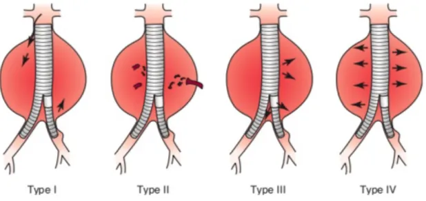
L’inconvénient majeur du traitement endovasculaire est la survenue de complications spécifiques, liées à l’endoprothèse au cours du suivi. Les premières générations d’EDP étaient responsables d’un taux important de réinterventions secondaires, jusqu’à 15 % des patients nécessitaient au moins une réintervention au cours du suivi (33). Ces réinterventions sont la conséquence de complications mécaniques survenant sur la structure de l’endoprothèse. Les complications les plus fréquemment observées après EVAR sont les endofuites qui correspondent à un flux sanguin artériel persistant au sein de l’anévrysme (figure 5), les migrations d’endoprothèse, les plicatures et thromboses de jambage et la couverture accidentelle d’une artère rénale (34)(35).
LERISSON Erol INTRODUCTION GÉNÉRALE
20
Figure 5 : Illustration des types d’endofuites (EF) rencontrés après EVAR. Les EF de type 1 sont des fuites proximales ou distales au niveau des zones d’étanchéité de l’endoprothèse. Avec les EF de type 3, qui correspondent à une mauvaise étanchéité entre les composant de l’EDP, elles constituent des endofuites à haut débit. Les EF de type 2, proviennent du reflux d’artères collatérales au sac anévrysmal. Les EF de type 4, plus rares, proviennent d’une porosité de l’EDP. D’après White GH, May J, Waugh RC, Chaufour X, Yu W. Type III and type IV endoleak: toward a complete definition of blood flow in the sac after endoluminal AAA repair. J Endovasc Surg. nov 1998;5(4):305‑9.
L’utilisation des endoprothèses fenêtrées et branchées s’est accompagnée de nouvelles sources potentielles de complications liées à l’EDP (au niveau des fenêtres ou des branches). Dans une série de 166 patients ayant bénéficiés de la mise en place d’une endoprothèse fenêtrée (FEVAR) ou branchée (BEVAR), Verhoeven et al. ont objectivé un taux de réinterventions liées à l’EDP de 17% à 3 ans de l’intervention initiale, une majorité de ces interventions étant due à des endofuites à haut débit.(36)
RÔLE DE LA TOMOGRAPHIE VOLUMETRIQUE À FAISCEAU CONIQUE (CBCT) DANS LA PRISE EN CHARGE DES AAA.
Le développement rapide des techniques endovasculaires et l’apparition des endoprothèses fenêtrées et multibranches se sont accompagnés de l’avancée des technologies d’imagerie ; de guidage pendant l’intervention et, de contrôle post-opératoire. La prise en charge des AAA est réalisée dans des salles de chirurgie dites « hybrides » qui combinent un équipement d’imagerie, et d’angiographie très performants dans un environnement stérile et contrôlé. Ces salles hybrides permettent de réaliser des procédures très complexes avec une dose de rayonnement ainsi qu’une quantité de produit de contraste nettement plus faibles qu’auparavant (37)(38)(39)(40).
Généralités
En Imagerie médicale nous désignons par le terme tomographie l’ensemble des techniques d’imagerie qui permettent de reconstruire le volume d’un objet d’intérêt.
Lors d’une acquisition scannographique classique (TDM), un générateur de rayons X émet un faisceau qui est projeté est continu, en éventail (fan beam), puis traverse le patient placé sur une table mobile qui avance dans le plan de coupe. En face, se trouve une couronne de détecteurs, dont le nombre peut atteindre 128 sur les systèmes les plus récents (on parle alors de scanner multibarettes). Le faisceau de rayon X est analysé après atténuation par le volume anatomique d’intérêt. La rotation du couple émetteur-détecteur ainsi que l’avancée de la table d’imagerie dans le plan de coupe permettent d’acquérir un grand nombre d’images qui sont ensuite reconstruites afin de représenter le volume anatomique d’intérêt en trois dimensions (figure 6).
plans de l’espace. Les voxels produits par un scanner hélicoïdal classique sont de forme parallélépipédique rectangle, le volume est dit anisotrope avec une résolution qui diffère dans les trois plans de l’espace.
La limite principale du CBCT est un niveau de contraste plus faible que le TDM, et ce surtout pour les tissus mous. La plage dynamique permise par un scanner conventionnel est de 4096 niveaux de gris (soit 12 bits), tandis que le CBCT permet de mettre en évidence des différences d’atténuation de l’ordre de 10 UH(42)(43). Sa seconde limite et la taille du volume acquis, limité par la surface du capteur plan.
En salle hybride, le capteur plan associé à l’arceau mobile et la suite logicielle intégrée permettent de réaliser des CBCT. L’injection de produit de contraste iodé via un injecteur automatisé couplé au système d’imagerie permet de réaliser une angiographie 3D rotationnelle (ceCBCT). Les différents paramètres que l’on peut modifier et qui vont impacter la qualité d’image sont :
- Le nombre de projections (200 à 400 images planes) - Le degré de rotation (180 à 250 degrés)
- La vitesse de rotation, le temps de rotation
- Les paramètres de l’injecteur (volume, débit et pression d’injection) - La collimation du capteur plan
Dans notre institution, le système implanté en salle hybride est le système Innova CT (GE Healthcare, Waukesha, Wisconsin, USA).
LERISSON Erol INTRODUCTION GÉNÉRALE
24
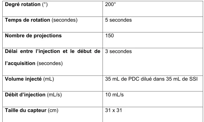
Le contrôle final après l’exclusion endovasculaire d’un AAA en salle de chirurgie hybride repose sur CBCT avec injection de produit de contraste.
Degré rotation (°) 200°
Temps de rotation (secondes) 5 secondes
Nombre de projections 150
Délai entre l’injection et le début de l’acquisition (secondes)
3 secondes
Volume injecté (mL) 35 mL de PDC dilué dans 35 mL de SSI
Débit d’injection (mL/s) 10 mL/s
Taille du capteur (cm) 31 x 31
Table 1 : Protocole de CBCT injecté. Innova CT (GE Healthcare, Waukesha, Wisconsin, USA).
POURQUOI REALISER UNE TOMOGRAPHIE VOLUMETRIQUE A FAISCEAU CONIQUE APRES EVAR ?
L’inconvénient majeur du traitement endovasculaire des AAA est la survenue de complications secondaires liée à l’endoprothèse et la nécessité de réinterventions au cours du suivi. Au total, jusqu’à 15 % des patients nécessitent une reprise chirurgicale au cours de leur suivi après EVAR (33). La plupart des complications surviennent dans la période post-opératoire précoce (jusqu’à 10% des malades à J30 (20). Les anomalies précoces les plus fréquentes après EVAR sont des endofuites à haut débit (type 1 et 3), des thromboses et plicatures de jambage, ou la couverture accidentelle d’une artère rénale (34)(35). Avec l’introduction des endoprothèses fenêtrées et multibranches, les sources d’anomalies techniques sont encore plus nombreuses, ainsi jusqu’à un tiers des patients nécessitent une reintervention au cours de leur suivi, les endofuites à haut débit et les compressions de stent en représentant la moitié (44)(45).
Ces anomalies nécessitent le plus souvent une reprise chirurgicale rapide afin de prémunir les patients de complications plus graves comme la rupture aortique en cas d’endofuites à haut débit, l’ischémie aigue de membre en cas de plicature de jambage et la défaillance rénale en cas de thrombose ou de plicature de stent à destinée rénale.
L’angiographie 2D est l’examen de référence pour évaluer le succès technique après EVAR. Cependant, cet examen présente une limite majeure puisqu’il s’agit d’un examen en deux dimensions. Il est ainsi souvent difficile d’évaluer l’origine d’une endofuite ou la présence d’une plicature dans la structure métallique de l’endoprothèse et il est fréquemment nécessaire de réaliser des acquisitions répétées sous plusieurs incidences(46), ce qui augmente la dose de produit de contraste
LERISSON Erol INTRODUCTION GÉNÉRALE
26
potentiellement néphrotoxique injecté au patient, ainsi que l’exposition aux radiations ionisantes. La sensibilité et la spécificité de l’angiographie 2D pour détecter les endofuites ont été estimées à respectivement 63 % IC95 [60-70] et 77% IC95 [58-100]. La plupart des équipes réalisent donc un angioscanner avant la sortie du patient ou lors du premier mois après l’intervention ; pour pallier aux limitations de l’angiographie 2D et pour évaluer convenablement l’intégrité de l’endoprothèse et l’absence de complications mécaniques. La sensibilité et la spécificité de l’angioscanner à détecter les endofuites à haut débit sont nettement meilleures que l’angiographie 2D et ont été estimées à respectivement 92 % IC95[80-100] et 90% IC95[85-92]. (47)
Cependant il s’agit d’un compromis non satisfaisant. En effet les complications détectées relèvent d’une reprise chirurgicale afin d’être corrigées ce qui représente une morbidité supplémentaire pour les patients. De plus, certaines anomalies comme les endofuites à haut débit nécessitent une prise en charge rapide pour éviter la rupture. La détection de ces anomalies trop tardivement au cours du suivi pourrait mettre les patients en danger.
L’angiographie 3D rotationnelle (ceCBCT), permet de répondre à plusieurs de ces problématiques. il s’agit d’un examen volumétrique, réalisable en salle hybride et permettant de réaliser des reconstructions de qualité proche d’un angioscanner(48).
Le CBCT a montré dans plusieurs études qu'il réduisait le taux de réintervention précoce après EVAR, en permettant une meilleure évaluation de l'intégrité de l'endoprothèse et des anomalies techniques par rapport à l'angiographie 2D.
Biasi et al. (49)ont démontré que le ceCBCT était plus performant que l’angiographie 2D pour évaluer le succès technique après EVAR. Chez les 80 patients qui ont
bénéficié du ceCBCT, L’angioscanner réalisé avant la sortie n'avait diagnostiqué aucune complication liée à l'endoprothèse qui n'avait pas été décrite sur le ceCBCT. Schultz et ses collaborateurs (50)ont montré que le CBCT pouvait détecter de façon fiable les complications liées à l'endoprothèse et qu'il était meilleur que l’angiographie 2 pour détecter les kinks et thromboses de jambage (figure 7) , ce qui permettait d'améliorer les résultats opératoires et de réduire les taux de réintervention précoce. Törnqvist et al (51)ont démontré que le CBCT était supérieur dans la détection des problèmes structurels.
Figure 7 A-C: Comparaison d’un contrôle final par angiographie uniplanaire et CBCT. A, Plicature en sortie de jambage iliaque droit non vu par l’angiographie 2D B-C, Plicature de jambage visible sur le CBCT après reconstruction. (Intra-operative
Cone Beam Computed Tomography can Help Avoid Reinterventions and Reduce CT Follow up after Infrarenal EVAR P. Törnqvist *, N. Dias, B. Sonesson, T.Kristmundsson, T. Resch°
Dans une étude récente, Tenorio et al. ont évalué les bienfaits du CBCT dans une large cohorte de 386 F/BEVAR. Ils ont démontré que le taux d'interventions secondaires précoces (à 30 jours) était significativement plus faible chez les patients
LERISSON Erol INTRODUCTION GÉNÉRALE
28
ayant bénéficié d’un CBCT que chez ceux qui avaient subi des interventions sans TCCB (4 % vs 10 %, p=0,022)(39).
Dans une étude précédente (52), nous avions montré que le ceCBCT permettait de diagnostiquer un plus grand nombre d’anomalies techniques sur les endoprothèses aortiques en fin de procédure que l’angiographie 2D conventionnelle. Ceci entrainait plus de gestes complémentaires (nouvelle dilatation, extension de jambage, etc…) non planifiés pour corriger ces anomalies, mais les bénéfices à long terme de cette prise en charge plus « agressive » restaient débattus.
OBJECTIF
L’objectif de ce travail est d’évaluer si la réalisation d’un ceCBCT et la correction peropératoire des anomalies détectées par le ceCBCT améliorent les résultats à moyen terme après EVAR.
RÉFÉRENCES BIBLIOGRAPHIQUES
1. Johnston KW, Rutherford RB, Tilson MD, Shah DM, Hollier L, Stanley JC. Suggested standards for reporting on arterial aneurysms. Subcommittee on Reporting Standards for Arterial Aneurysms, Ad Hoc Committee on Reporting Standards, Society for Vascular Surgery and North American Chapter, International Society for Cardiovascular Surgery. J Vasc Surg. mars 1991;13(3):452‑8.
2. Gloviczki P, Pairolero P, Welch T, Cherry K, Hallett J, Toomey B, et al. Multiple aortic aneurysms: the results of surgical management. J Vasc Surg. janv 1990;11(1):19‑27; discussion 27-28.
3. Sierevogel MJ, Pasterkamp G, Strauss DPV de K and BH. Matrix Metalloproteinases: A Therapeutic Target in Cardiovascular Disease [Internet]. Current Pharmaceutical Design. 2003 [cité 23 oct 2019]. Disponible sur: http://www.eurekaselect.com/63986/article
4. Brophy CM, Marks WH, Reilly JM, Tilson MD. Decreased tissue inhibitor of metalloproteinases (TIMP) in abdominal aortic aneurysm tissue: a preliminary report. J Surg Res. juin 1991;50(6):653‑7.
5. Sampson UKA, Norman PE, Fowkes FGR, Aboyans V, Song Y, Harrell FE, et al. Estimation of Global and Regional Incidence and Prevalence of Abdominal Aortic Aneurysms 1990 to 2010. Global Heart. 1 mars 2014;9(1):159‑70.
6. Singh K, Bønaa KH, Jacobsen BK, Bjørk L, Solberg S. Prevalence of and risk factors for abdominal aortic aneurysms in a population-based study : The Tromsø Study. Am J Epidemiol. 1 août 2001;154(3):236‑44.
7. Lederle FA, Johnson GR, Wilson SE, Aneurysm Detection and Management Veterans Affairs Cooperative Study. Abdominal aortic aneurysm in women. J Vasc Surg. juill 2001;34(1):122‑6.
8. Lederle FA, Johnson GR, Wilson SE, Chute EP, Littooy FN, Bandyk D, et al. Prevalence and associations of abdominal aortic aneurysm detected through
LERISSON Erol RÉFÉRENCES BIBLIOGRAPHIQUES
30
screening. Aneurysm Detection and Management (ADAM) Veterans Affairs Cooperative Study Group. Ann Intern Med. 15 mars 1997;126(6):441‑9.
9. Cornuz J, Sidoti Pinto C, Tevaearai H, Egger M. Risk factors for asymptomatic abdominal aortic aneurysm: systematic review and meta-analysis of population-based screening studies. Eur J Public Health. déc 2004;14(4):343‑9.
10. Adam DJ, Mohan IV, Stuart WP, Bain M, Bradbury AW. Community and hospital outcome from ruptured abdominal aortic aneurysm within the catchment area of a regional vascular surgical service. J Vasc Surg. nov 1999;30(5):922‑8.
11. Huber TS, Harward TR, Flynn TC, Albright JL, Seeger JM. Operative mortality rates after elective infrarenal aortic reconstructions. J Vasc Surg. sept 1995;22(3):287‑93; discussion 293-294.
12. Greenhalgh RM, Forbes JF, Fowkes FG, Powel JT, Ruckley CV, Brady AR, et al. Early elective open surgical repair of small abdominal aortic aneurysms is not recommended: results of the UK Small Aneurysm Trial. Steering Committee. Eur J Vasc Endovasc Surg. déc 1998;16(6):462‑4.
13. Gorham TJ, Taylor J, Raptis S. Endovascular treatment of abdominal aortic aneurysm. Br J Surg. juill 2004;91(7):815‑27.
14. Badger SA, Harkin DW, Blair PH, Ellis PK, Kee F, Forster R. Endovascular repair or open repair for ruptured abdominal aortic aneurysm: a Cochrane systematic review. BMJ Open. 1 févr 2016;6(2):e008391.
15. Wanhainen A, Verzini F, Van Herzeele I, Allaire E, Bown M, Cohnert T, et al. Editor’s Choice - European Society for Vascular Surgery (ESVS) 2019 Clinical Practice Guidelines on the Management of Abdominal Aorto-iliac Artery Aneurysms. Eur J Vasc Endovasc Surg. janv 2019;57(1):8‑93.
16. Chaikof EL, Dalman RL, Eskandari MK, Jackson BM, Lee WA, Mansour MA, et al. The Society for Vascular Surgery practice guidelines on the care of patients with an abdominal aortic aneurysm. Journal of Vascular Surgery. 1 janv 2018;67(1):2-77.e2.
17. Dubost C, Allary M, Oeconomos N. Resection of an aneurysm of the abdominal aorta: reestablishment of the continuity by a preserved human arterial
graft, with result after five months. AMA Arch Surg. mars 1952;64(3):405‑8.
18. Matas R. Surgery of the vascular system. AMA Arch Surg. 1 janv 1956;72(1):1‑19.
19. Wiersema AM, Jongkind V, Bruijninckx CMA, Reijnen MMPJ, Vos JA, van Delden OM, et al. Prophylactic perioperative anti-thrombotics in open and endovascular abdominal aortic aneurysm (AAA) surgery: a systematic review. Eur J Vasc Endovasc Surg. oct 2012;44(4):359‑67.
20. Greenhalgh RM, Brown LC, Kwong GPS, Powell JT, Thompson SG, EVAR trial participants. Comparison of endovascular aneurysm repair with open repair in patients with abdominal aortic aneurysm (EVAR trial 1), 30-day operative mortality results: randomised controlled trial. Lancet. 4 sept 2004;364(9437):843‑8.
21. Blankensteijn JD, de Jong SECA, Prinssen M, van der Ham AC, Buth J, van Sterkenburg SMM, et al. Two-year outcomes after conventional or endovascular repair of abdominal aortic aneurysms. N Engl J Med. 9 juin 2005;352(23):2398‑405. 22. Lederle FA, Freischlag JA, Kyriakides TC, Padberg FT, Matsumura JS, Kohler TR, et al. Outcomes following endovascular vs open repair of abdominal aortic aneurysm: a randomized trial. JAMA. 14 oct 2009;302(14):1535‑42.
23. Becquemin J-P, Pillet J-C, Lescalie F, Sapoval M, Goueffic Y, Lermusiaux P, et al. A randomized controlled trial of endovascular aneurysm repair versus open surgery for abdominal aortic aneurysms in low- to moderate-risk patients. J Vasc Surg. mai 2011;53(5):1167-1173.e1.
24. Schermerhorn ML, O’Malley AJ, Jhaveri A, Cotterill P, Pomposelli F, Landon BE. Endovascular vs. open repair of abdominal aortic aneurysms in the Medicare population. N Engl J Med. 31 janv 2008;358(5):464‑74.
25. De Bruin JL, Baas AF, Buth J, Prinssen M, Verhoeven ELG, Cuypers PWM, et al. Long-term outcome of open or endovascular repair of abdominal aortic aneurysm. N Engl J Med. 20 mai 2010;362(20):1881‑9.
26. Knott AW, Kalra M, Duncan AA, Reed NR, Bower TC, Hoskin TL, et al. Open repair of juxtarenal aortic aneurysms (JAA) remains a safe option in the era of fenestrated endografts. J Vasc Surg. avr 2008;47(4):695‑701.
LERISSON Erol RÉFÉRENCES BIBLIOGRAPHIQUES
32
27. Chong T, Nguyen L, Owens CD, Conte MS, Belkin M. Suprarenal aortic cross-clamp position: a reappraisal of its effects on outcomes for open abdominal aortic aneurysm repair. J Vasc Surg. avr 2009;49(4):873‑80.
28. Landry G, Lau I, Liem T, Mitchell E, Moneta G. Open abdominal aortic aneurysm repair in the endovascular era: effect of clamp site on outcomes. Arch Surg. sept 2009;144(9):811‑6.
29. Patel VI, Ergul E, Conrad MF, Cambria M, LaMuraglia GM, Kwolek CJ, et al. Continued favorable results with open surgical repair of type IV thoracoabdominal aortic aneurysms. J Vasc Surg. juin 2011;53(6):1492‑8.
30. Martin GH, O’Hara PJ, Hertzer NR, Mascha EJ, Krajewski LP, Beven EG, et al. Surgical repair of aneurysms involving the suprarenal, visceral, and lower thoracic aortic segments: early results and late outcome. J Vasc Surg. mai 2000;31(5):851‑62.
31. Rao R, Lane TRA, Franklin IJ, Davies AH. Open repair versus fenestrated endovascular aneurysm repair of juxtarenal aneurysms. J Vasc Surg. janv 2015;61(1):242‑55.
32. Parodi JC, Palmaz JC, Barone HD. Transfemoral intraluminal graft implantation for abdominal aortic aneurysms. Ann Vasc Surg. nov 1991;5(6):491‑9. 33. Sampram ESK, Karafa MT, Mascha EJ, Clair DG, Greenberg RK, Lyden SP, et al. Nature, frequency, and predictors of secondary procedures after endovascular repair of abdominal aortic aneurysm. J Vasc Surg. mai 2003;37(5):930‑7.
34. Hobo R, Buth J, EUROSTAR collaborators. Secondary interventions following endovascular abdominal aortic aneurysm repair using current endografts. A EUROSTAR report. J Vasc Surg. mai 2006;43(5):896‑902.
35. Stather PW, Sidloff D, Dattani N, Choke E, Bown MJ, Sayers RD. Systematic review and meta-analysis of the early and late outcomes of open and endovascular repair of abdominal aortic aneurysm. Br J Surg. juin 2013;100(7):863‑72.
36. Verhoeven ELG, Katsargyris A, Bekkema F, Oikonomou K, Zeebregts CJ a. M, Ritter W, et al. Editor’s Choice - Ten-year Experience with Endovascular Repair of Thoracoabdominal Aortic Aneurysms: Results from 166 Consecutive Patients. Eur J
Vasc Endovasc Surg. mai 2015;49(5):524‑31.
37. Hertault A, Maurel B, Midulla M, Bordier C, Desponds L, Kilani MS, et al. Editor’s Choice – Minimizing Radiation Exposure During Endovascular Procedures: Basic Knowledge, Literature Review, and Reporting Standards. European Journal of Vascular and Endovascular Surgery. 1 juill 2015;50(1):21‑36.
38. Steuwe A, Geisbüsch P, Schulz CJ, Böckler D, Kauczor H-U, Stiller W. Comparison of Radiation Exposure Associated With Intraoperative Cone-Beam Computed Tomography and Follow-up Multidetector Computed Tomography Angiography for Evaluating Endovascular Aneurysm Repairs. J Endovasc Ther. août 2016;23(4):583‑92.
39. Tenorio ER, Oderich GS, Sandri GA, Ozbek P, Kärkkäinen JM, Macedo TA, et al. Impact of onlay fusion and cone beam computed tomography on radiation exposure and technical assessment of fenestrated-branched endovascular aortic repair. J Vasc Surg. 4 déc 2018;
40. Rolls AE, Rosen S, Constantinou J, Davis M, Cole J, Desai M, et al. Introduction of a Team Based Approach to Radiation Dose Reduction in the Enhancement of the Overall Radiation Safety Profile of FEVAR. European Journal of Vascular and Endovascular Surgery. 1 oct 2016;52(4):451‑7.
41. Scarfe WC, Farman AG. What is cone-beam CT and how does it work? Dent Clin North Am. oct 2008;52(4):707‑30, v.
42. Gupta R, Cheung AC, Bartling SH, Lisauskas J, Grasruck M, Leidecker C, et al. Flat-panel volume CT: fundamental principles, technology, and applications. Radiographics. déc 2008;28(7):2009‑22.
43. Gupta R, Grasruck M, Suess C, Bartling SH, Schmidt B, Stierstorfer K, et al. Ultra-high resolution flat-panel volume CT: fundamental principles, design architecture, and system characterization. Eur Radiol. juin 2006;16(6):1191‑205. 44. Mastracci TM, Eagleton MJ, Kuramochi Y, Bathurst S, Wolski K. Twelve-year results of fenestrated endografts for juxtarenal and group IV thoracoabdominal aneurysms. J Vasc Surg. févr 2015;61(2):355‑64.
LERISSON Erol
34
et al. Fenestrated endovascular grafting: the French multicentre experience. Eur J Vasc Endovasc Surg. mai 2010;39(5):537‑44.
46. Bianchini Massoni C, Gargiulo M, Giovanetti F, Freyrie A, Faggioli G, Gallitto E, et al. Adjunctive stenting of endograft limbs during endovascular treatment of infrarenal aortic and iliac aneurysms according to 3-projection completion angiography. J Endovasc Ther. août 2011;18(4):585‑90.
47. Armerding MD, Rubin GD, Beaulieu CF, Slonim SM, Olcott EW, Samuels SL, et al. Aortic aneurysmal disease: assessment of stent-graft treatment-CT versus conventional angiography. Radiology. avr 2000;215(1):138‑46.
48. al TP et. Optimizing imaging for aortic repair. - PubMed - NCBI [Internet]. [cité 5 mars 2019]. Disponible sur: https://www.ncbi.nlm.nih.gov/pubmed/25644824
49. Biasi L, Ali T, Ratnam LA, Morgan R, Loftus I, Thompson M. Intra-operative DynaCT improves technical success of endovascular repair of abdominal aortic aneurysms. J Vasc Surg. févr 2009;49(2):288‑95.
50. Schulz CJ, Schmitt M, Böckler D, Geisbüsch P. Intraoperative contrast-enhanced cone beam computed tomography to assess technical success during endovascular aneurysm repair. J Vasc Surg. sept 2016;64(3):577‑84.
51. Törnqvist P, Dias N, Sonesson B, Kristmundsson T, Resch T. Intra-operative cone beam computed tomography can help avoid reinterventions and reduce CT follow up after infrarenal EVAR. Eur J Vasc Endovasc Surg. avr 2015;49(4):390‑5. 52. Hertault H, Maurel B, Pontana F, Martin-Gonzalez T, Spear R, Sobocinski J, et al. Benefits of Completion 3D Angiography Associated with Contrast Enhanced Ultrasound to Assess Technical Success after EVAR. Eur J Vasc Endovasc Surg. mai 2015;49(5):541‑8.
ARTICLE
Intraoperative cone beam computed tomography to improve
outcomes after endovascular aortic repairs.
ABSTRACT
Background:
This study aims at evaluating if combining intraoperative contrast-enhanced cone beam computed tomography (ceCBCT) and postoperative contrast enhanced ultrasound (CEUS) to assess technical success after standard and complex endovascular aortic repair (EVAR & F/B-EVAR) could reduce late stentgraft-related complications, and/or stentgraft-related reinterventions. In a previous study, higher rates of early high-flow endoleak and limb kink were detected with ceCBCT, and conducted to immediate correction during the initial procedure. However, the long-term benefits of a better detection resulting in a more "aggressive" intra-operative attitude would remain debatable. We here report long-term outcomes of the same cohort of patients.
Methods:
All consecutive patients treated with infrarenal bifurcated or custom-made fenestrated/branched stentgrafts in our hybrid room (IGS 730, GE Healthcare) over the study period were included. From December 2012 to July 2013, final 2D completion angiogram (25cc of iodine at 15cc/sec) was performed at the end of the
LERISSON Erol
36
procedure for each patient, and then a computed tomography angiography (CTA) before discharge (group 1). From October 2013 to April 2014, an intraoperative ceCBCT was performed, followed by CEUS during the 30-day post-operative period (group 2). Comparative analyses of late outcomes were undertaken.
The primary endpoint included late stentgraft-related complications, a composite factor defined by aneurysm-related death, type 1 or 3 endoleaks, kink or occlusion of target vessels, kink or occlusion of iliac limb and aortic sac enlargement. The secondary endpoint was stentgraft-related reinterventions.
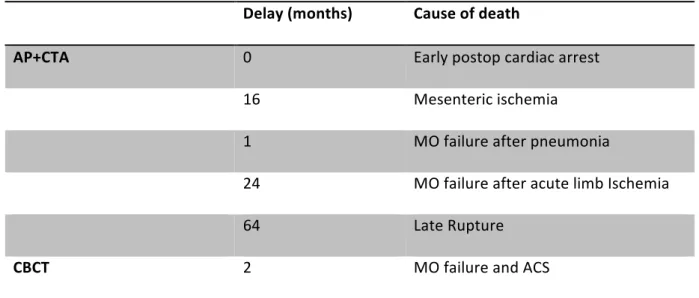
Death from any cause and related to stentgraft were also depicted.
Results:
A total of 134 patients (79 in group 1 and 55 in group 2) were enrolled with a median follow up of 48 months [IQR 36-59]. 40 late stentgraft-related complications occurred in 25 patients in group 1 (31.7%), 12 events occurred in 9 patients in group 2 (16.7%) p=0.05, with a median time from index procedure at 24 [IQR 12-38] months and 24 [IQR 12-38] months respectively (p=0.80).
Reintervention rates were respectively of 18.9% (n=15) in group 1 and 14.8% (n=8) in group 2 (p=0.5), with a median time from the index procedure at 14 months [IQR 4.0-28.0] and 26 [IQR 4,3-45.8] months respectively (p=0.58) in group 1 and in group 2. In multivariate analysis, CBCT did not significantly reduce the rate of late reintervention or stent-graft related adverse events but appears to protect from aortic sac enlargement OR=0.25, IC95% 0.07-0.91, p=0.035.
At the end of follow-up, there was no significant difference between the two groups in terms of death from any cause and aortic-related mortality.
Conclusion:
This study is the first to report the influence of routine ceCBCT on mid-term outcomes after standard and complex EVAR. Our results show that ceCBCT would protect from aortic sac enlargement and suggest a trend to reduce late stentgraft related reinterventions and late stentgraft related events. A randomized prospective study is
INTRODUCTION
Endovascular aortic repair (EVAR) has become the 1st treatment option in aortic aneurysms following specific anatomical conditions. The first generations of infrarenal stentgrafts were associated with high rates of complications and secondary procedures. It has been reported that 15 % of patients underwent ≥1 secondary procedure during follow-up (128/10/y 13:18), with up to 1/3 of them occurring within 30 postop days (2). Type 1 and 3 endoleaks, stent migration, graft and limb thrombosis, accidental renal artery coverage are the most common reasons for secondary procedure (3)(4) (5)(6).
Most of stentgraft-related complications might be explained by technical issues during the index procedure not identified on the final 2D-angiogram. Completion uniplanar angiography (usually anteroposterior) is recommended for evaluating technical success after EVAR, although it has been shown as insufficient to detect all technical procedural issues especially high-flow endoleaks. Both sensitivity and specificity of uniplanar angiography to detect endoleaks have been estimated respectively at 63% [IC95=60-70%] and 77% [IC95=58-100%]; while for CTA (Computed Tomography Angiogram) it is 92% [IC95=80-100%] and 90% [IC95=85-92%] (7)(8)(9).
As a consequence, potential technical failures are missed with 2D angiogram and secondary detected with early postoperative CTA; those latter would conduct to early reinterventions which represent additional cost and additional morbidity.
The introduction of complex endovascular aneurysm repairs including branched and/or fenestrated stentgrafts with additional bridging stent(s) increases the potential sources of technical failure and requires final 3D reconstructions (10)(11).
LERISSON Erol INTRODUCTION
40
Additionally, the cost-effectiveness of EVAR remains debatable mostly due to the rate of secondary interventions. This emphasizes the need for completion imaging strategies with improved sensitivity for endoleaks or kinks. (12)(13)(14)(15).
Final contrast-enhanced CBCT (ceCBCT) has the potential to improve outcome after EVAR with better depicting of technical failure. CBCT is an intraoperative 3D-imaging modality, offering high-spatial resolution and produce volumetric data able to be reconstructed and processed in a workstation during procedure. Several studies have established the short-term benefits of ceCBCT (16)(17)(18)(19).
In a previous study, ceCBCT has shown reduction in both radiation exposure and contrast media volume injection after infrarenal endovascular aortic repair (EVAR) and complex endovascular aortic repair (F/BEVAR) compared to a strategy including 2D angiogram and postoperative CTA (20). Moreover, higher rates of high flow endoleaks and limb kinks were detected with ceCBCT enabling immediate correction during the initial procedure.
The present study reports late outcomes of this initial cohort where ceCBCT has been completed during the index procedure (EVAR, F/B-EVAR).
MATERIAL AND METHODS
Patient selectionPatient selection and groups constitution have been described previously (20), and are briefly reviewed here. All consecutive patients treated with infrarenal bifurcated stentgraft (EVAR) or fenestrated (FEVAR) and branched (BEVAR) stentgrafts in our hybrid room (IGS 730, GE Healthcare, France) over the study period were included. Patients with renal insufficiency (DFG< 60 mL/min) or requiring emergency treatment were not included. Arch and isolated thoracic endovascular procedures were excluded.
Population was divided in 2 groups:
Group 1: From December 2012 to July 2013, a 2D completion angiogram (25cc of iodine, at 15cc/sec, (Omnipaque 350, GE Healthcare)) was performed at the end of the procedure for each patient to assess technical success; a computed tomography angiography (CTA) was then performed before discharge (group 1).
Group 2: From October 2013 to April 2014, an intraoperative ceCBCT (40°/sec – 70cc of iodine (Omnipaque 350, GE Healthcare) diluted by half with saline at 10cc/sec with 3s of delay between the iodine injection and CBCT acquisition) was performed to assess technical success, followed by CEUS within the 30-day post-operative period (group 2).
Procedures
All procedures were performed under fusion imaging guidance, according to the ALARA principles(21) (22). All patients underwent repair with Zenith plateform devices (Cook Medical, IN, USA).
LERISSON Erol MATERIAL AND METHODS
42
Primary technical success of the procedure was defined according to the reporting standards and including: the successful introduction and deployment of the stentgraft in the aneurysm with secure proximal and distal fixation without open surgical conversion and/or death, the absence of stentgraft-related complication (including high-flow endoleaks (type 1 and 3)), patent stentgraft without significant kink (> 30% of luminal stenosis) of iliac limb extensions and/or bridging stents, and no access complications (23).
Technical details
ceCBCT
ceCBCT acquisition (40°/sec) was performed at the end of the procedure. A spin test was run before the acquisition to ensure the absence of collision between the patient, the table and the gantry, and to check whether the acquisition volume is properly centered on the stentgraft.
A power injector was used for contrast media injection with the following parameters: injection of 35 mL of iodine (Omnipaque 350, GE Healthcare), diluted with 35 mL of saline at a flow rate of 10 mL/second, the delay between the contrast injection and the acquisition was set at 3 seconds.
The 31x31 cm flat panel robotic gantry was rotated over 200 degrees, 150 angiographic projections were acquired during 5 seconds with 1.33 degrees increment. The projections images were automatically transferred to a dedicated workstation and automatically reconstructed (Advantage Window; GE Healthcare, Milwaukee, USA). The resulting volume was an 18.3 cm diameter cylinder, reconstructed data were displayed using cross-sectional axial reconstruction,
multiplanar (MPR), maximum intensity projection (MIP) and 3D volume rendering (3DVR) views.
2D completion Angiography
Completion angiography was performed using a power injector with the following parameters: injection of 25 mL of contrast medium (Omnipaque 350, GE Healthcare), diluted by half with saline with a flow rate at 10 mL/second.
Two experienced endovascular therapists reviewed final control imaging, aiming at depicting high flow endoleaks (type I or III), limb kinks and evaluating the integrity and good positioning of bridging stents through fenestrations.
When above-mentioned issues were detected, immediate correction was attempted. It is referred later in the manuscript as additional unplanned procedures at the index surgery.
Contrast-Enhanced Ultra-Sound (CEUS)
CEUS is an imaging modality used routinely in our center after endovascular aortic repairs. It is performed by three experienced vascular therapists. (24)(25)
US examinations include a standard morphological investigation in B-mode followed by a blood flow analysis in pulse wave modality. CEUS was performed after the administration of an intravenous bolus of 2.5 mL of SonoVue (Bracco, Milan, Italy), flushed with a 5 mL bolus of isotonic saline solution. Endoleak detection was performed at a low mechanical index (0.2e0.3) and with the focus positioned behind the aorta to delay bubble destruction.
LERISSON Erol MATERIAL AND METHODS
44 CTA
In group 1, the postoperative three-phase CTA was performed within 7 days of the procedure on a 64-slice multi-detector CT system (Brillance 64, Philips Healthcare, Best, The Netherlands). The injection protocol includes the administration of 120 mL of a contrast agent with 350 mg of iodine per milliliter (Ioversol, Optiject; Guerbet, Roissy, France) at a flow rate of 4 mL/second. The acquisition was initiated by bolus tracking within the aorta at the level of the thoraco-abdominal junction. The triphasic acquisitions (unenhanced, arterial, and delayed phases) were systematically obtained with a maximum tube current of 150-250 mAs, dependent on automatic selection, and a longitudinal dose modulation (ACS & Z-DOM; Philips Healthcare)
Data collection
Data were collected in a prospective database with retrospective analyses. Demographic characteristics, medical history, anatomical measurements and surgical data of the population were collected.
Follow up
Follow-up visits were scheduled at 6, 12, 18 months after the procedure and yearly thereafter. All patients underwent a physical examination at each follow-up visit, and abdominal color duplex ultrasonography. A reference three-phase CTA was performed during the first 12 months period after the procedure and patient’s risk was stratified accordingly.
Thereafter, follow up CTA were realized yearly. All available CTA completed over the FU period were analyzed on a dedicated 3D-workstation (Aquarius, Terarecon). Aortic sac diameter was measured and collected after centerline reconstruction, endoleaks, stengraft/bridging stents integrity and/or patency were reported.
All post-operative complications and reinterventions were collected, and the delays between the index procedure and the post-operative complications were noted.
Objectives
The primary endpoint was late stentgraft-related complications, a composite criterion defined as aneurysm-related deaths, secondary high flow endoleaks including type 1 and 3 endoleaks, secondary kink or occlusion of bridging stent, secondary kink or occlusion of iliac limb, and aortic sac enlargement.
The secondary endpoint was stentgraft-related reinterventions defined as all device related procedures occurring after the index procedure. Additional outcome included death from any cause.
Outcomes were assessed in accordance with SVS reporting standards for EVAR (23). An endoleak observed after the perioperative period (>30 days) was considered as a secondary endoleak (26). In the same way, any bridging stent or iliac limb kink or thrombosis depicted after the perioperative period was considered as secondary. Aortic sac enlargement was considered as significant if ≥5mm. Aneurysm-related mortality was defined as all deaths within 30 days of the primary procedure, within 30 days of any re-intervention attributable to the aneurysm, from other aneurysm-related causes (including graft infection), or from secondary aneurysm rupture after repair (23). Stentgraft-related reinterventions occurring within the 30-day postoperative
LERISSON Erol MATERIAL AND METHODS
46
period or during the initial hospitalization (if>30 days) were deemed as early-reinterventions.
Statistical Analysis
All statistical analysis were performed using SAS advanced statistics 2.0 (IBM,New York, USA). Descriptive study of the cohort was performed. Continuous variables were expressed as median with inter-quartile range (IQR=[Q1-Q3]), or mean with standard deviation. Categorical variables are presented as a percentage and 95% confidence interval.
Comparisons between categorical variables were performed with the chi-square test (or Fisher exact test) and between continuous variables with the Student t test, or with the Mann-Whitney test.
A composite criterion defined as late stentgraft-related complications and including aortic-related deaths, secondary high flow endoleaks including type 1 and 3 endoleaks, secondary kink or occlusion of bridging stent, secondary kink or occlusion of iliac limb, and aortic sac enlargement (defined by an enlargement >5mm) was created. Survival analyses were performed according to the Kaplan- Meier model. Curves were compared with a log-rank test, and then results were adjusted with a hazard ratio model according to non-comparable preoperative variables.
Relationships between ceCBCT, reinterventions, late stentgraft-related complications and sac enlargement were analyzed in binary logistic regression or hazard ratio models (when duration between the index procedure and the event was available).
When relationship between the composite criterion and a quantitative covariate was not linear, the covariate was recoded into a binary variable. Recoding threshold was determined by minimizing the Bayesian information criterion of the model. Multivariate models were constructed by including all variables with p-value <0.10 in univariate analyses or considered clinically relevant. Selection of covariates was done manually until a relevant and meaningful model was obtained and then refined according to different automatic selection procedures (ascending, descending or stepwise).
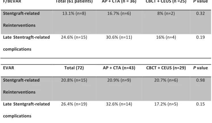
Sub-analyses were performed according to the type of endograft (standard EVAR versus F/BEVAR). A p value <.05 was considered significant.
LERISSON Erol RESULTS
48
RESULTS
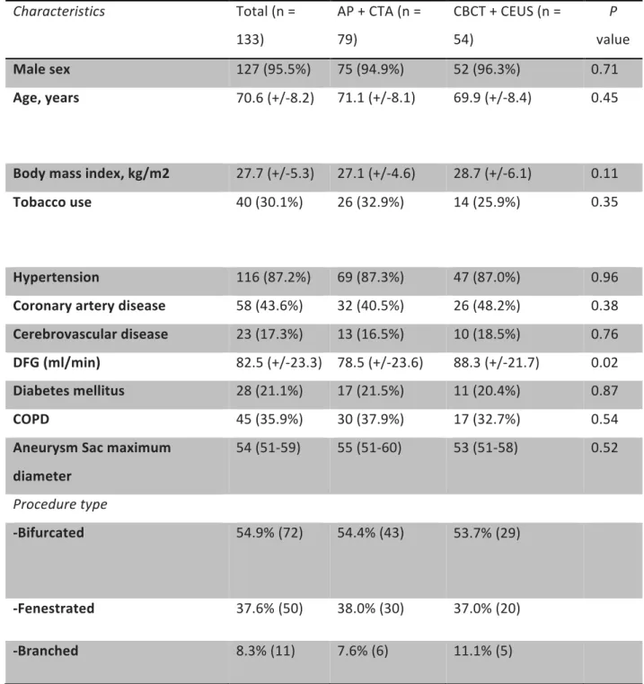
79 consecutive patients were included in group 1 and 54 patients in group 2. Baseline characteristics were previously described(20) and presented in table 1. Briefly, mean age of our cohort was 70.6 years and 95.5% were male. The mean maximum diameter of the aneurysm sac at initial procedure was 54 mm. 72 patients were treated with a bifurcated stentgraft, and 61 with F/BEVAR. In the F/BEVAR subgroup, devices with fenestrations only, branches only, and a combination of both were used. The average number of target vessel included per patient was 3.5+/-0.2 and 34 patients (25.6%) had four targeted vessels or more.
The two groups were similar in terms of comorbidities. The mean time of follow-up was higher in group 1, 54 months [IC95%: 37-61] and 47.5 months [IC95%: 35-50] respectively (p=0.005).
Early outcomes were also previously reported (20). Peri-operative event detection rates were 8.9%(n=7) and 33.3% (n=18) in group 1 and 2 (p<.001). Additional procedures at the index surgery were performed in 7 patients (8.9%) in group 1 versus 17 (31.5%) in group 2 (p=0.001).
Table I. Baseline characteristics of the patients Characteristics Total (n = 133) AP + CTA (n = 79) CBCT + CEUS (n = 54) P value Male sex 127 (95.5%) 75 (94.9%) 52 (96.3%) 0.71 Age, years 70.6 (+/-8.2) 71.1 (+/-8.1) 69.9 (+/-8.4) 0.45 Body mass index, kg/m2 27.7 (+/-5.3) 27.1 (+/-4.6) 28.7 (+/-6.1) 0.11 Tobacco use 40 (30.1%) 26 (32.9%) 14 (25.9%) 0.35 Hypertension 116 (87.2%) 69 (87.3%) 47 (87.0%) 0.96 Coronary artery disease 58 (43.6%) 32 (40.5%) 26 (48.2%) 0.38 Cerebrovascular disease 23 (17.3%) 13 (16.5%) 10 (18.5%) 0.76 DFG (ml/min) 82.5 (+/-23.3) 78.5 (+/-23.6) 88.3 (+/-21.7) 0.02 Diabetes mellitus 28 (21.1%) 17 (21.5%) 11 (20.4%) 0.87 COPD 45 (35.9%) 30 (37.9%) 17 (32.7%) 0.54 Aneurysm Sac maximum diameter 54 (51-59) 55 (51-60) 53 (51-58) 0.52 Procedure type -Bifurcated 54.9% (72) 54.4% (43) 53.7% (29) -Fenestrated 37.6% (50) 38.0% (30) 37.0% (20) -Branched 8.3% (11) 7.6% (6) 11.1% (5)
LERISSON Erol RESULTS
50
Late stent-graft related complications
40 late complications occurred in 25 patients in group 1 (31.7%), 12 complications occurred in 9 patients in group 2 (16.7%), with a median time from index procedure at 24 [IQR 12-38] months and 24 [IQR 12-38] months in both groups Late complications are reported in table II.
At 48 months from the index procedure, freedom from late stent-graft related complications probabilities were 72.4% (95 % CI [62.7-83.6]) for the AP+CTA group and 82% (95 % CI [71.2-94.4]) for the CBCT+CEUS group (logrank P=0.36). Freedom from stent-graft related late complications estimates by Kaplan Meier method are reported in Figure 1.
Table II. Follow up
Follow Up Total (n = 133) AP + CTA (n = 79) CBCT + CEUS (n = 54)
Mean follow up, months 48 (36-59) 54 (37-61) 47.5 (35-50) High Flow Endoleaks Ia Ib III 12 (9 %) 6 (5.5%) 5 (3.8%) 1 (0.8%) 10 (12.7%) 5 (6.3%) 5 (6.3%) - 2 (3.7%) 1 (1.9%) - 1 (1.9%) Target vessel/Bridging stent kink 1 (0.8%) 1 (1.3%) - Bridging stent fracture - - - Bridging stent thrombosis 4 (3%) 2 (2.5%) 2 (3.7%) Limb kink 2 (1.6%) 1 (1.3%) 1 (1.9%) Limb thrombosis 7 (5.3%) 5 (6.3%) 2 (3.7%) Aortic sac enlargement >5mm 18 (13.5%) 15 (18.9%) 3 (5.6%) Accidental renal artery coverage 1 (0.8%) - 1 (1.9%) Mean Maximum sac diameter at end of FU (mm) 45 (38.3-55) 45 (38-55) 46 (40-54) Aortic sac evolution (mm) -6 (-14;-0) -7.5 (-15;0) -5 (-13;-1)
LERISSON Erol RESULTS
52
High flow endoleaks rates were respectively 12.7% in group 1 (n=10/79) and 3.7% in the group 2 (n=2/54). Five type 1A endoleaks where detected in group 1 (6.3%), whereas only 1 type 1A endoleak was detected in group 2 (1.9%). Five Type 1B endoleaks where detected in group 1 whereas none occurred in group 2 . One Type III endoleaks was detected in group 2.
At some point of the follow up, 15 (18.9%) patients experienced an aortic sac enlargement in the group 1 and 3 patients (5.6%) in the group 2. Freedom from aortic sac enlargement is reported in Figure 2b. In multivariate analysis, ceCBCT appears to independently protect from aortic sac enlargement (OR=0.25, IC95% [0.07-0.91], p=0.035]). In our serie, age >70 was also associated with a increased risk of aortic sac enlargement OR=4.06, IC95%[1.24-13.32], p=0.021.
Stentgraft type was not predictive of late stentgraft related complication OR=1.02, (95% CI, [0.51-2.01]), p=0.966
In multivariate analysis, CBCT was not protective from the onset of late stentgraft related event HR=1.733, 95% CI [0.758-3.959], p=0.192. The diagnosis of a high flow endoleak during initial procedure in both groups was predictive of the onset of late stent-graft event during follow up HR=2.844, 95% CI[1.028-7.864], p=0.044.
LERISSON Erol RESULTS
54
Stentgraft related reinterventions
A total of 19 reinterventions were performed in 15 patients in group 1 (18.9 %), and 9 reinterventions were performed in 8 patients in group 2 (14.8%). with a median time from index procedure at 14 months (IQR [4.0-28.0]) months and 26 months (IQR [4.3-45.8]) months respectively, p=0.58.
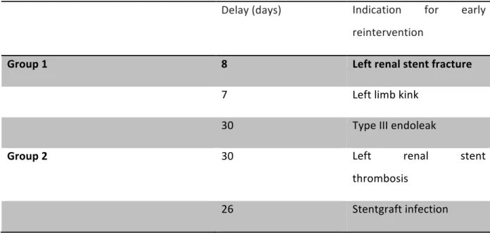
Three early reinterventions (within 30 days of initial EVAR or within first hospitalization) were performed in group 1 (3.8%), and 2 (3.7%) in group 2 (p=.978). Early reinterventions are reported in Table IIIA.
Table IIIA. Indications for early stentgraft-related reinterventions (≤30 day)
Delay (days) Indication for early reintervention
Group 1 8 Left renal stent fracture
7 Left limb kink
30 Type III endoleak
Group 2 30 Left renal stent
thrombosis
26 Stentgraft infection
Forty-eight months after the initial procedure, the freedom from reintervention rates were 80.9% (95% CI, 72.2-90.5) in group 1 (AP+CTA) and 82.5% (95 % CI, 70.9-96.1) in group 2 (CBCT+CEUS) (p=0.86). Freedom from reintervention estimates by Kaplan Meier method are reported in Figure 2.
The main indication for reintervention in group 1 was high flow endoleaks (n=10/19, 52.6%).
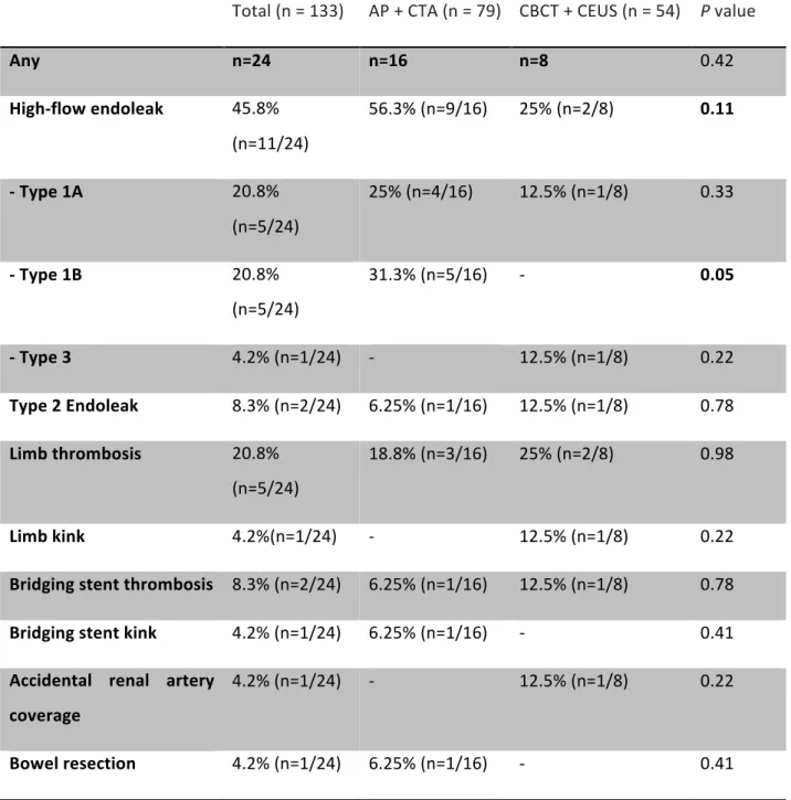
In group 2 (CBCT + CEUS), main causes for reintervention were high-flow endoleaks (n=2/9, 22.2%), limb graft thrombosis (n=2/9, 22.2%) and limb graft kink (n=2/9, 22.2%). No patient required a secondary procedure for type 1B endoleaks, and one patient required a proximal fenestrated cuff to correct a type 1A endoleak. ndications for stentgraft-related reinterventions occurring after early follow-up (>30 days) are listed in table IIIb.
LERISSON Erol RESULTS
56
Table IIIb: Indications for stentgraft-related reinterventions (>30 days)
Total (n = 133) AP + CTA (n = 79) CBCT + CEUS (n = 54) P value
Any n=24 n=16 n=8 0.42 High-flow endoleak 45.8% (n=11/24) 56.3% (n=9/16) 25% (n=2/8) 0.11 - Type 1A 20.8% (n=5/24) 25% (n=4/16) 12.5% (n=1/8) 0.33 - Type 1B 20.8% (n=5/24) 31.3% (n=5/16) - 0.05 - Type 3 4.2% (n=1/24) - 12.5% (n=1/8) 0.22 Type 2 Endoleak 8.3% (n=2/24) 6.25% (n=1/16) 12.5% (n=1/8) 0.78 Limb thrombosis 20.8% (n=5/24) 18.8% (n=3/16) 25% (n=2/8) 0.98 Limb kink 4.2%(n=1/24) - 12.5% (n=1/8) 0.22 Bridging stent thrombosis 8.3% (n=2/24) 6.25% (n=1/16) 12.5% (n=1/8) 0.78 Bridging stent kink 4.2% (n=1/24) 6.25% (n=1/16) - 0.41
Accidental renal artery coverage
4.2% (n=1/24) - 12.5% (n=1/8) 0.22