HAL Id: dumas-01674722
https://dumas.ccsd.cnrs.fr/dumas-01674722
Submitted on 3 Jan 2018HAL is a multi-disciplinary open access archive for the deposit and dissemination of sci-entific research documents, whether they are pub-lished or not. The documents may come from teaching and research institutions in France or
L’archive ouverte pluridisciplinaire HAL, est destinée au dépôt et à la diffusion de documents scientifiques de niveau recherche, publiés ou non, émanant des établissements d’enseignement et de recherche français ou étrangers, des laboratoires
Comparison of induced gamma band responses to
electrocortical stimulation and functional MRI during a
language task: are they redundant or complementary to
assess cognitive postoperative outcome in epilepsy
surgery?
Pauline Cuisenier
To cite this version:
Pauline Cuisenier. Comparison of induced gamma band responses to electrocortical stimulation and functional MRI during a language task: are they redundant or complementary to assess cognitive postoperative outcome in epilepsy surgery?. Human health and pathology. 2017. �dumas-01674722�
AVERTISSEMENT
Ce document est le fruit d'un long travail approuvé par le
jury de soutenance et mis à disposition de l'ensemble de la
communauté universitaire élargie.
Il n’a pas été réévalué depuis la date de soutenance.
Il est soumis à la propriété intellectuelle de l'auteur. Ceci
implique une obligation de citation et de référencement
lors de l’utilisation de ce document.
D’autre part, toute contrefaçon, plagiat, reproduction illicite
encourt une poursuite pénale.
Contact au SID de Grenoble :
bump-theses@univ-grenoble-alpes.fr
UNIVERSITE GRENOBLE ALPES
FACULTE DE MEDECINE DE GRENOBLE
Année : 2017 N°
COMPARISON OF INDUCED GAMMA BAND RESPONSES TO
ELECTRO-CORTICAL STIMULATION AND FUNCTIONAL MRI
DURING A LANGUAGE TASK:
ARE THEY REDUNDANT OR COMPLEMENTARY TO ASSESS COGNITIVE
POST-OPERATIVE OUTCOME IN EPILEPSY SURGERY ?
Apport des activités cérébrales gamma comme méthode de cartographie fonctionnelle et marqueur du devenir cognitif dans le bilan pré-chirurgie de l’épilepsie.
Comparaison avec les stimulations électriques cérébrales et l’IRM fonctionnelle lors d’une tâche de langage
.
THESE PRESENTEE POUR L’OBTENTION DU DOCTORAT EN MEDECINE DIPLOME D’ETAT
CUISENIER Pauline
THESE SOUTENUE PUBLIQUEMENT A LA FACULTE DE MEDECINE DE GRENOBLE* Le 24 mars 2017
DEVANT LE JURY COMPOSE DE
Président du jury : Monsieur le Professeur Philippe KAHANE Membres
Madame le Professeur Monica BACIU Madame le Professeur Elena MORO
Madame le Professeur Agnès TREBUCHON
Madame le Docteur Marcela PERRONE-BERTOLOTTI directrice de thèse
*La Faculté de Médecine de Grenoble n’entend donner aucune approbation ni improbation aux opinions émises dans les thèses ; [Données à caractère personnel]
Remerciements
A Monsieur le Professeur Philippe Kahane,
Merci de me faire l'honneur de présider cette thèse. Votre disponibilité et votre expérience m’ont été particulièrement précieuses tout au long de mon internat et pour la réalisation et enfin la relecture rigoureuse de ce travail. Vous savez éveiller en nous la joie de travailler et de connaître. Merci de votre chaleureux soutien.
A Madame le Professeur Monica Baciu,
Je vous remercie de me faire l’honneur de juger ce travail. Veuillez trouver ici l’expression de mes sincères remerciements.
A Madame le Professeur Agnès Trébuchon.
Je suis très honorée que vous ayez accepté d’expertiser ce travail et de participer à mon jury de thèse. Veuillez recevoir l’expression de toute ma gratitude.
A Madame le Professeur Elena Moro.
Votre énergie et votre détermination font de vous un exemple. Vous avez fait de la formation des internes de neurologie l’une de vos priorités, nous vous en remercions. Merci d’avoir accepté de faire partie de mon jury et de me pousser vers le meilleur de moi-même.
A Madame le docteur Marcela Perrone-Bertolotti.
Je suis honorée que tu aies accepté de diriger ce travail ainsi que mon année de Master 2. Merci de ton aide et de ta disponibilité, jusqu’en Uruguay, la semaine comme le week-end depuis l’élaboration du projet jusqu’aux dernières et nombreuses relectures. Ces 18 mois à tes côtés à découvrir le monde de la recherche ont été très enrichissants.
A celles et ceux qui ont permis la réalisation de ce travail :
A Pierre Deman, un immense merci pour ton temps et ton aide sans lesquels ce travail n’aurait pas abouti. Merci également de ta patience face à mes ignorances informatiques. A IntrAnat Electrodes qui aura grandi en parallèle de ce travail.
A Bénédicte Testud, qui a rejoint ce projet en route. Merci de ton aide précieuse pour le traitement des données IRMf.
A Alexis Robin. Merci d’avoir lu avec moi 48956 cartes gamma, plutôt 5 fois qu’une, (soit 244 780 cartes…) Continue comme ça car on te veut dans l’équipe neurogrenoble ;-)
A Lorella Minotti, pour son enseignement rigoureux de la neurologie et son hospitalité à l’italienne ainsi qu’à toute l’équipe du laboratoire d’Exploration et de Neurophysiopathologie de l’Epilepsie pour son accueil et son aide tout au long de ce projet mais également de mon internat : Marie-Pierre Noto et Patricia
Boschetti pour leur soutien quasi maternel et Valérie Damon pour son organisation impeccable et
bienveillante.
Au Professeur Alexandre Krainik. Merci de m’avoir donné accès aux données IMR. Merci de votre enseignement durant mon semestre à vos côtés en neuroradiologie.
Enfin, à tous les patients qui ont accepté de participer à ce projet.
A celles et ceux m’ont encadrée et entourée au cours de mon internat :
Aux équipes médicales :
- De neurologie du CHU de Grenoble ; merci de m’avoir aidé au cours de ces quatre années d’internat, tout en partageant votre savoir et votre expérience clinique. Je suis heureuse de rejoindre vos équipes et de pouvoir continuer à apprendre à vos côtés.
- De neuroradiologie du CHU de Grenoble : les Drs Grand, Tahon, Kastler et Heck ; vous m’avez beaucoup appris durant mon semestre à vos côtés. Au Dr Arnaud Attye, qui aura réussi à me faire aimer le scanner ORL (ok je n’ai pas tout compris). J’espère que notre projet commun aboutira.
A Anne Sophie Job-Chapron. Merci de ton soutient bienveillant et de tes conseils avisés, cette année, et tout au long de mon internat. Tu es notre exemple à tous !
A Mathieu Vaillant. Mon grand frère neurologue qui a guidé mon choix post ECN. Merci de m’avoir vendu du rêve, tu ne m’avais pas menti ! A ta petite famille en croissance que j’aime tant.
A Isabelle Favre, ma première assistante. Au cours de mon semestre à tes côtés, tu m’as appris à être une interne rigoureuse. Merci de m’avoir donné l’envie de continuer.
A Olivier Casez dit papa Casez. Tu es celui que l’on consulte pour ses connaissances neurologiques mais également œnologiques, pour ses bonnes adresses ou sa grande expérience culinaire. Merci d’avoir facilité mon quotidien à de nombreuses reprises ; du vélo de la grand-mère jusqu’à la recette de la tarte aux fraises-mascarpone en passant par les doublures de mes gardes !
Travailler avec vous est une chance, j’espère en profiter au maximum.
Aux équipes paramédicales, AS, IDE, MKDE, avec qui j’ai beaucoup partagé : tout particulièrement Sabrina, Lydie, Juliette et Emilie qui m’ont appris à gérer un patient à mes débuts, A maman Maryse et notre amour commun pour gwada. A l’équipe d’HDJ, meilleur service du monde pour petit-déjeuner, déjeuner, goûter, prendre le thé, diner (ah non c’est fermé à cette heure-là) mais aussi pour travailler ! A toute l’équipe de Neuro A, qui tremble, freeze, chante, et crise. Au manips de neuroradio, pour leur bonne
A Mes Co-Internes :
A Sébastien pour tes talents de craqueur de logiciels et tes cours improvisés de Word et Zotero. Ton oreille attentive et neutre a su guider mon chemin. Merci de ton soutient.
A Clémentine pour avoir STABILOté le terrain et les joysticks mais surtout pour ton amitié. Les rôles de Marco et Luigi, déboucheurs de tuyau nous ont liées à jamais, tellement que nous en inventions des thrombolyses. Merci d’avoir été là dans les moments difficiles.
A Jérémie, allias Anselme papapoussin. Nous avons formé le binôme qu’il ne fallait pas croiser en avis «
elle a fait l’attaque ! », le binôme de choc en HDJ « nous voulons du fromage, et un peu de sucre en poudre », et formerons un binôme de choc en tant que CCA, je l’espère.
A Nastasia, qui m’a supportée et soutenue en période de thèse. Tu es l’amie sincère qu’il faut avoir, parfois brutale mais mais qui fait avancer. T’avoir à mes côtés ce semestre m’a été précieux.
A Marie qui me fait tant rire. Merci de m’apporter ton petit brin de légèreté et ton ouverture au monde extra-médicale. Prochaines étapes ensemble : Saint-Egrève et le Mont Aiguille.
A Fanny, j’ai eu la chance de réviser le DIU de Neurophysiologie à tes côtés, avec succès. Merci d’avoir accepté mon cadeau empoisonné ! A Anne la voyageuse à bicyclette, spécialiste des bonnes attentions. A
Giovanni merci pour la découverte de petit biscuit longuement écouté durant la rédaction de ce travail. A Sarah la souriante, Guillaume le voyageur, la belle Hélène, Thomas le taquin. Aux plus jeunes Lucie, Loïc et Hugo qui écriront la suite.
A Francesco Cavallieri, il mio professore d'italiano. Grazie per il suo regalo de un pocco della Dolce Vita. A mes co-interne neuroradiologues, « la dream team » : Yann, avec qui j’ai découvert le TDM des rochers et surtout décrit le phénomène Pink Floyd en scanner. Corentin, le plus drôle, ne change rien ! Pierre le plus gentil et attentionné des co-interne ever.
A ceux qui ont facilité mon travail au quotidien :
Les ophtalmologues : Fréderic et Georges toujours partant pour une OCT rapide. Les cardiologues :
Aure-Elise et Adrien qui m’ont dépanné plus d’une fois face à un ECG ou pour une ETT. Les psychiatres,
membres du feu cercle des neuro-psy : Benoit Rumbach et Fabien Droux pour leur sens de l’humour et leurs connaissances toujours à la pointe. Anaïs Dartevel : une belle rencontre tardive de mon internat ;-). Aux heures passée ensemble à discuter jupon, étoilés, mais également anticorps anti PDC. A Sébastien
Baillieul : ton optimisme et ta solidarité font de toi une personne à rencontrer absolument !
Et surtout les radio-imageurs : Amis qu’il fait bon retrouver en garde mais surtout en terrasse ou autour d’un d’une tarte citron-meringuée ; la mafia dijonnaise (Mathew et René dit Rooney), Perol, et enfin
Moumoune, last but not least, à condition que tu ne me piques pas mon mec.
A mes maitres d’externat
Au Professeur Chavanet, qui m’a montré le bon chemin, et a marqué mes études de médecine. Au
Professeur Giroud, qui a créé des vocations chez plusieurs générations d’externe dont la mienne. A JFM
grâce à qui nous avions un mental de guerrier. A Pifu, qui nous a confié les clefs de son bureau pendant 2 ans, ce qui nous a permis d’atteindre nos objectifs ;
A Mes Amis
De Carnot, nos si belles années :
Ben et sa tchach, Garzai qui disparaît dès qu’on le retrouve, Nayef le bon conseiller qui vous veut du bien, Romblax le joyeux voyageur, la belle Morgane, Eric Hockety pockety wockety wock. Charlène, alias Charlou, du collège à la fac on en a partagé des moments ensemble ! Paul, avec qui la P1 fut adoucie par Plus belle la Vie. Vincent, notre pharmacien préféré au regard ravageur, membre de la bienveillante famille Vaillant.
A Pierre Emmanuel, Pilou, mon jumeau de P1, membre du cercle « auto-proclamé » du Jurançon. A nos samedi soir à Gallien à réviser la biocellulaire. Tu étais déjà le meilleur ! Alexia et toi êtes un couple que j’admire pour son courage. Qu’Alaska vous apporte beaucoup de bonheur…Et des nuits calmes.
A Louis, qui a été là de Carnot à la fac, en passant par l’Irlande, Paris et j’en passe. Merci pour ces séances intenses de révisions, tous les dimanches soir, avec ou sans macaron. Nabiha et toi m’avez beaucoup apportée.
A Jean Baptiste, « Rien ne vaut le trésor de tant de souvenirs communs, de tant de brouilles, de réconciliations, de mouvements du cœur. On ne reconstruit pas ces amitiés- là. » A. de St Ex.
A Victor, mon binôme de choc de toujours. Merci de ton amitié sans faille qui m’est si précieuse. A ta Mathilde. Tu as trouvé la meilleure ! Un couple imbattable à la coinche, avec qui il ne faut pas oublier de compter les atouts, biathlètes sans limite à la martin F. avec qu‘il fait si bon partir. Ne quittez pas Grenoble, sinon nous serions obligés de vous suivre !
De l’Externat :
A Christophe B., allias Toff, le meilleur des co-externes, qui gère dans TOUTES les situations ! Les patients ont de la chance de t’avoir ! A Kevin C., ton mental de tueur a été précieux pour moi en D4. A Louise M. merci pour ta joie et ton accueil nantais et à Pierre F. pour ta bonne humeur, j’espère que nous ferons enfin cette traversée de la Chartreuse ensemble. A la douce Sarah S. A Noé L., retrouvée dans les méandres de la recherche clinique, pour son cache-cache C1. A Eloïse F., la plus jolie, ma bonne copine que je ne vois pas assez.
De Grenoble
A Bleuenn. Ta bonne humeur permanente fait de toi une amie très précieuse. Ces quelques mois de colocation furent heureux ! A Thomas, le capitaine exemplaire et surtout à ma belle filleule Alice.
A Hélène qui a réussi à me transformer en CAFiste et me faire aimer le ski de rando.A Aurélien. Les Feuillu-Flesch, à qui rien ne fait peur pas même les kms en voiture, (ou pour des chaussures de ski). Vous êtes des amis en or. A Marie et Max, rois des pentes et de la tarte flambée. Votre calme cache une énergie débordante dont j’espère profiter plus.
A Irina, Ira. Merci pour la poésie et les rêveries que tu as apporté à mon internat et à notre année recherche. Ce travail est aussi dédié aux lanceuses de patates… A Mathieu et Anna. Les kitesurfeurs chamoniards qui maitrisent la bacalhau mieux que personne, où que vous alliez nous serons là.
A Alexandre qui nous a montré de chemin de l’exode dijonnais avec talent, Et Julie mon élégante motivatrice pumpeuse. Merci de vos bienveillantes attentions notamment durant ces derniers mois de labeur.
Aux anneciens, Marion Thomas Charlotte et Romain qu’il est toujours agréable de retrouver. A Sam, rideur sur toute surface, à l’oreille attentive et attentionnée. A Marjo U. toujours souriante, partie à la découverte de la Suisse. D’ici et d’ailleurs
Anaïs, Charles, Philippe et Hortense #miglietouzetoujoursautop #bitofaction, je vous adore. Merci également à T O U C A N T O C O d’avoir participé à ce projet
.
A Ma Famille
A mes parents,
Je vous remercie pour votre soutien permanent, et votre amour et pour tout ce que vous m’avez transmis ; la curiosité, l’amour des voyages, la culture des belles choses, l’envie d’apprendre. Papa, puis-je espérer un jour être aussi admirée et respectée de mes patients que toi. Maman, merci de m’avoir transmis ta sensibilité et ton sourire.
A ma grand-mère, pour son amour
Au Pad. Tu souhaitais que tes petits enfants sachent encore utiliser un BIC, rassures toi ! J’espère que tu es fier de nous. Et à ceux parti trop tôt sans qui je n’aurais pas été là.
A ma famille, aux Lugnier et Curie ainsi qu’aux Elnécavé et Hubert qui dans mon cœur en font partie. A ma filleule Lucie. Vous êtes venu de loin pour me soutenir. Merci de toutes vos attentions.
To my Australian family, Kathy and Eric. I am very grateful for your help on this work and for your love. Aux Maillot, ma deuxième famille.
A Anne et Bernard, je vous remercie de votre accueil si chaleureux que ce soit à Dijon, à la mer ou à la montagne. A Clémence, Maud et Nicolas, avec qui je vais apprendre ce que c’est que d’avoir des frères et sœurs. J’espère que je serai à la hauteur.
A Olivier
A nous deux toujours plus, toujours plus vite, les pieds sur terre, la tête dans les nuages, cela promet…Les clefs n’ont qu’à bien se tenir. Merci d’être aussi joyeux, de déborder de projets et d’idées en tout genre, et surtout de me faire autant rire. A tes cotés tout est possible. Merci de t’être battu.
Table of contents
Remerciements ... 6
Table of contents ... 11
Abstract ... 12
1. Introduction ... 12
2. Materials and Methods ... 17
2.1 Patients selection and characteristics ... 17
2.2 Electrode implantation and localization ... 18
2.3 Stereotactic intracerebral EEG (SEEG) recordings ... 19
2.4 Electrical cortical stimulations (ECS) ... 19
2.5 Functional mapping using task-related gamma-band responses (GBR) ... 20
2.6 Functional mapping using functional MRI (fMRI) for language ... 21
2.7 Surgery and post-operative follow-up ... 22
2.8 Data analysis ... 23
2.8.1 ECS analysis ... 23
2.8.2 Language-related GBR ... 24
2.8.3 Language-fMRI BOLD responses ... 24
2.8.4 Functional data fusion ... 25
2.8.5 Comparison between GBR, ECS and fMRI maps ... 26
2.8.6 Effect of surgery ... 27
3. Results ... 28
3.1. Functional Maps ... 28
3.2. Comparison between GBR and ECS ... 29
3.3. Comparison of GBR maps with fMRI activations ... 30
3.4. Correlation between functional mapping methods ... 31
3.5. Effects of surgery ... 32
3.5.1. Effects of surgery on behavioural performances ... 32
3.5.2. Effect of GBR resection on behavioural performances ... 33
4. Discussion ... 35 Conclusion ... 40 Appendix ... 41 References ... 44 Serment d’hippocrate ... 44
Abstract
Rationale: Like any brain surgery, epilepsy surgery can have various negative effects on cognition and
behaviour. The best way to anticipate post-operative deficits is to test, before surgery, the ‘functional integrity’ of the brain. Different methods can be used for that purpose, including neuropsychological testing, functional neuroimaging (fMRI), and Electro-Cortical Stimulation (ECS). None of these approaches, however, has been proved accurate enough to reliably anticipate the risks of postoperative cognitive deficits.
Study aims: This last decade, intracranially recorded task-related Gamma Band Responses (GBR) has
emerged as a putative biomarker of various cognitive processes. The present study aimed at comparing the GBR method with the classical fMRI and ECS procedures for localizing language functions, and at evaluating whether the removal of brain tissue exhibiting GBR might have significant consequences on language processing.
Methods: Fourteen epileptic patients who underwent stereotactic intracerebral EEG (SEEG) recordings and
who were subsequently operated on were included in the study. SEEG signals were recorded while the patients performed a language task involving two different language processes: semantic and phonologic. Language-related GBR (50–150 Hz) were analysed at every recording site, and compared with the results of ECS-elicited clinical responses. These findings were also compared with the results of fMRI language assessment in a sub-group of 6 patients. Post-operative language outcome was evaluated in patients in whom at least one GBR+ site was resected versus those in whom no GBR+ site was resected.
Results: GBR predicted ECS language interference with a high specificity (92.4%) and negative predictive
value (NPV, 95.5%), but with a low sensitivity (11.7%) and positive predictive value (PPV, 7.4%). Similar findings, although a little bit weaker, were found when comparing GBR and fMRI (specificity: 89.2%, NPV: 84.7%, sensitivity: 8.4%, PPV: 12.2%). Resections that included GBR+ sites, as compared with resections that spared GBR+ sites, did not show any significant language decline both in semantic and phonological conditions. Only a few patients, however, had a resection that included GBR+ sites (n=2 for semantic condition, n=2 for phonological condition).
Conclusion: GBR mapping appears as a useful tool for pre-surgical evaluation of language
functions, complimentary to ECS, that might be used to build a preliminary functional map to reduce the duration of ECS sessions. Although preliminary, these findings emphasise the benefit of a multimodal screening for language features before epilepsy surgery.
Keywords:
epilepsy surgery; stereo-electroencephalography; gamma band response; brain functional1. Introduction
Resective surgery is the only treatment able to suppress drug-resistant seizures in epileptic patients. This therapeutic surgery is possible only if the Epileptogenic Zone (EZ) is focal, clearly identified, and can be safely removed (Kahane & Landré, 2008 for a review of EZ definition). The identification of the EZ, and the evaluation of its functional integrity is a difficult process which requires intracranial EEG (iEEG) recordings in 25-50% of the cases. However, even when using such iEEG information, epilepsy surgery still fails in a substantial ratio of patient (Téllez-Zenteno, 2010), and post-operative cognitive/psychiatric deficits still exist in a significant number of the cases (Baxendale et al., 2006 ; Helmstaedter, 2004), especially for language processes (Sherman et al., 2011).
Language is a high-level and non-monolithic cognitive function supported by an extended network of brain regions and by several complex dynamic processes (Price, 2012) such as lexico-semantic, phonological and motor commands. Brain structures which support such processes are relatively well described in literature. For instance, the semantic network involves mainly the pars orbitalis and triangularis of the inferior frontal gyrus (Bookheimer, 2002) and the left mid and inferior temporal gyri (Binder et al., 2009), while the phonological network is mostly supported by the pars opercularis of the left inferior frontal gyrus (Vigneau et al., 2006) and the left posterior and superior temporal regions (Démonet et al., 1992). Nevertheless, an important inter-subjects variability exists on brain language representation and this variability is more important in epileptic patients (Baciu & Perrone-Bertolotti, 2015 for a review). Hence, pre-operative language mapping should delineate eloquent areas at the individual level.
Currently, many tools and methods are available for pre-surgical evaluation of cognitive anf language processes, of which none has been proved accurate enough to reliably anticipate the risks of postoperative deficits. These exams include neuropsychological testing, functional neuroimaging, chemical inactivation (Wada test), and Electro-Cortical Stimulation (ECS).
ECS, as performed during iEEG, is classically considered as the ‘gold standard’ method for the identification of the cortical regions that have to be spared during surgery (David et al., 2010; Penfield & Jasper, 1954 ; Lesser et al., 1994 ;Ojemann et al., 1989). It is an invasive method, the clinical validity of which to assess post-operative outcome has been reported (Haglund et al., 1994; Hermann & Wyler, 1988). However, ECS mapping has several limitations: (i) ECS has a limited spatial sampling, since it can be applied only at the sites where the intracranial electrodes are implanted; (ii) ECS is a time-consuming method, that may occasionally lead to seizures (Lesser et al., 1984) and therefore limitates the repetition of ECS and subsequently reduces inter-subject reproducibility; (iii) ECS does not always trigger objective clinical signs, especially in associative cortices such as language areas; (iv) elicited signs can be subtle, especially in the language domain (e.g., a specific lexico-semantic deficit), and they may be therefore difficult to detect or interpret; (v) patient's subjective experience can be misleading, or may add difficulties for ECS interpretation; (v) it is currently not clear whether ECS trigger inhibition or activation of neuronal networks (David et al., 2010). For all these reasons, ECS may fail to reveal the functionality of a given brain region, revealing only the “tip of the iceberg” of functional cortical networks.
Functional Magnetic Resonance Imaging (fMRI), by comparison, is a non-invasive method which has an excellent spatial coverage and can be routinely used for brain mapping, notably language (Price, 2010), including pre-operative mapping in patients (Perrone-Bertolotti et al., 2012; Baciu et al., 2001 ; Baciu et al., 2005). Nevertheless, it has limitations in terms of temporal resolution, relationships to neural processing and statistical quantification. Functional activity assessed by fMRI depends on the statistical threshold used, which is most important during single brain mapping (Baciu et al., 2005 ; Baciu & Perrone-Bertolotti, 2015 for a review). Still, fMRI shows widely distributed networks that are mainly relevant to lateralize language processing before surgery, rather than to precisely delineate those of language areas that should be spared
biomarkers of cognitive processes, especially for language functions, (e.g.,Mainy et al., 2008; Vidal et al., 2011) HFAs induced by cognitive tasks were first described in animal studies which reported that primary visual cortex neurons fired jointly at 40Hz during a visual task (Gray, 1989). A more recent micro-electrodes recordings study in the human cortex showed that broadband HFAs (40Hz-150HZ) could reflect summed local population of neurone spiking and thus allowed the evaluation of direct neural activity (Manning et al., 2009). Two decades ago, two studies showed that cognitive information processing generated transient activities in a high frequency range between 40 and 150 Hz in motor ( Crone et al., 1998) and sensory ( Lachaux et al., 2000) cortices. These HFAs, called Gamma Band Responses (GBR), are currently considered as a proxy of population level spiking activity while patients perform a cognitive task. Since then, an increasing number of studies have reported GBR in specific brain regions related to the task performed (Lachaux et al., 2012). GBR are not visible in raw intracerebral iEEG traces, but are revealed by simple signal processing techniques involving time-frequency analysis (Jerbi et al., 2009). These studies have revealed the functional role of GBR in almost every sensory modality and its subserving cortical regions, as well as in various high-level cognitive processes such as visual selective attention (Ossandón et al., 2011), auditory perception (Bidet-Caulet et al., 2007), language processing (Perrone-Bertolotti, 2012;Mainy et al., 2008; Lachaux et al., 2007) and many others.
Brain mapping using GBR evaluation offers many advantages. It provides an exquisite anatomical, functional and temporal specificity, and GBR can be compared with an estimate of the intensity of any given neuronal population’s engagement in a specific task processing (Lachaux et al., 2000). Thus, it can distinguish neural activity increases and decreases, i.e., activations and deactivations, which is interesting in terms of cognition as cognitive processes, mainly attention, are mediated not only by distributed cerebral activations, but also by concurrent suppressions (Ossandón et al., 2011). Moreover, this method offers high signal-to-noise ratio and low sensitivity to artifacts that are not offered by surface EEG, for instance. As GBR are elicited during cognitive tasks, high level processes, namely those supported by associative
networks revealed by GBR assessment have been proven to be complementary to those revealed by fMRI activation studies (Lachaux et al., 2007; Conner et al., 2011).
GBR studies in the language domain has been showed as a reliable method (Llorens et al., 2011), that combines a high temporal and spatial resolution with a low sensitivity to artifacts. It provides highly specific results which depend on the functions that are tested, and allows the study of specific language processes, including their neural mechanisms and time courses. This has been showed, as an example, for word recognition (Mainy et al., 2008), sentence comprehension, reading (Perrone-Bertolotti, 2012), and naming (Crone et al., 2001). Therefore, GBR recording during cognitive tasks seems to be an efficient method to perform brain mapping and, more specifically, to perform language mapping. Nevertheless, few studies have compared this method with the classical methods (ECS and fMRI) used to perform pre-surgical functional mapping (Sinai et al., 2005), and only a few have evaluated the value of GBR assessment to predict post-operative cognitive outcome (Towle et al., 2008).
The present study aimed at evaluating the clinical relevance of induced GBR as a functional mapping method for pre-operative assessment of cognitive functions before epilepsy surgery. Attention was focused on language-induced GBR, since language is one of the most frequently impaired functions following epilepsy surgery. Also, language is classically mapped, in our clinical environment, by ECS and fMRI, allowing direct comparison of results of the three methods. We firstly evaluated the reliability of the GBR as compared with ECS and fMRI. In a second step, we evaluated the value of GBR to predict post-operative language outcome.
2. Materials and Methods
2.1 Patients selection and characteristics
Fourteen patients were included in this study, based on the following inclusion criteria: (i) aged over 12 years, native French speakers; (ii) having undergone SEEG recordings with stereotactically implanted multilead depth electrodes at the Epilepsy Department of Grenoble University Hospital (France); (iii) in whom the cognitive brain mapping assessment, as performed during SEEG procedure (ISD-SEEG project, DRCI Grenoble, 2009), included at least a language task allowing the assessment of behavioural performance and related gamma band responses; (iv) who were subsequently operated, after the SEEG study, between January 1, 2013 and July 1,2016.
The 14 patients (6 men, 8 women, mean age: 30 years) were all suffering from drug-resistant partial epilepsy. The operability of this epilepsy could not be decided on the basis of a non-invasive screening including scalp-video-EEG monitoring, and neuropsychological tests. See Figure 2 for experimental paradigm. A summary of the patients’ characteristics is given in Table 1.
P Age Gender VIQ/PIQ HL HDL EZ location MRI Resection Pathology outcome E
1 38 F NA R L L temp NL L Temporo-basal FCD Engel IV
2 23 F 99/76 L L L temp NL L- Cingulo-parietal NS Engel I 3 23 F 79/76 R L R front MG R-Fronto-opercular FCD Engel III 4 20 F 102/92 R L L temp HS L-Temporo-mesial HS Engel I 5 37 M 100/115 R L L front NL L-Frontal-polar and orbital FCD Engel I 6 13 F 90/105 R L R front NL L-Prefrontal FCD Engel I 7 15 F NA R L R temp NL R-Temporo insulo-opercular FCD + HS Engel IV 8 23 F 96/60 L L L pariet NL L- Mesio parietal NS Engel III 9 59 M 112/104 R L L temp NL L-Fronto-basal NS Engel I 10 29 M 131/116 R L R front DNET R- Fronto orbital and anterior insula DNET Engel I 11 28 M 106/112 R L R front POG R- Pre motor frontal ODG Engel I 12 41 M 98/106 R L L front FCD L-Frontal superior FCD Engel I 13 27 F 104/90 R L L front NL L-Frontal superior NS Engel I 14 48 M 112/106 L R R temp NL R-Temporo-mesial FCD Engel I
Table 1. Patients' (P) characteristics: Age, Gender, Pre-operative neuropsychological scores on Verbal and
nonverbal performances (VIQ/PIQ; Handedness lateralization as assessed by Edinburgh Handedness inventory (HL)(Oldfield, 1971); Hemispheric dominance for Language (HDL) as assessed by fMRI; epilepsy characteristic (epileptic zone -EZ- location and MRI findings); anatomical resection location (Resection); pathology, seizure outcome (Engel et al., 1996). Abbreviations: NA: not available; L: Left handed R: Right handed; NL: Non Lesional; MG: Micropolygyria; FCD: Focal Cortical Dysplasia; HS: Hippocampal sclerosis; DNET: Dysembryoplastic neuroepithelial tumour; POG: post-operative gliosis; NS: Nonspecific; ODG: Oligodendroglioma.
2.2 Electrode implantation and localization
The number and placement of intracerebral electrodes was defined according to the prior hypothesis regarding the localization of the epileptogenic tissue, based on seizure semiology and on the results of the non-invasive investigations. A total number of 199 semi-rigid, multi-lead electrodes were stereotactically
(DIXI Medical, Besançon, France). Every electrode contact was identified on a post-implantation MRI. The MNI (Montreal Neurological Institute) coordinates for every electrode contact were extracted using SPM (http://www.fil.ion.ucl.ac.uk/spm/).
2.3 Stereotactic intracerebral EEG (SEEG) recordings
Intracranial recordings were conducted using a video-SEEG monitoring system (Micromed®, Treviso, Italy), which allowed the simultaneous recording from up to 128 depth-EEG electrode sites. Data was bandpass filtered online from 0.1 to 20Hz and sampled at 512 Hz in all patients. At the time of acquisition, data was recorded using a reference electrode located in the white matter, and each electrode trace was subsequently re-referenced with respect to its direct neighbour (bipolar derivations). This bipolar montage has a number of advantages over common referencing. Notably, it helps eliminate signal artifacts common to adjacent electrode contacts and allows an accurate focal recording. The spatial resolution achieved by bipolar SEEG recordings was in the order of 3mm (Jerbi et al., 2009); (Lachaux et al., 2003). The localization of epileptogenic brain tissue was based on the visual analysis of interictal and ictal SEEG activity.
2.4 Electrical cortical stimulations (ECS)
As part of our routine procedure, ECS were performed during the SEEG monitoring, during sessions that lasted 1-3 hours. The aim was to reproduce part or the totality of the ictal clinical symptomatology, and to localize functionally eloquent areas that had to be spared during surgery. Bipolar ES were performed using a constant current rectangular pulse generator, and applied at 1 Hz (pulse width: 1 msec; ES duration: 40 sec) and 50 Hz (pulse width: 1 ms; ES duration: 5 sec) between contiguous contacts at various levels of the electrode axis, with stepwise increasing intensities ranging from 0,2 to 3 mA and until clinical subjective or objective responses or after-discharges were obtained. ECS were performed after checking that the
tasks, depending on the stimulated region. More specifically, for language assessment, patients were asked to perform several speech tasks audibly including counting, naming pictures or listing series of words. ECS were performed immediately after every speech task onset. ECS findings were classified by the examiner (neurologist) into different categories (motor, sensitive, sensory, vegetative, language, emotional, etc...) and documented in the patients' files.
2.5 Functional mapping using task-related gamma-band responses (GBR)
During the SEEG procedure, patients were asked to participate to an experimental protocol screening various cognitive processes (ISD and SEEG project, DRCI Grenoble, 2009) to extract task-related GBR. This experimental protocol was approved by the local Ethical Committee (CPP Sud-Est V n° 09-CHUG-12), and all participants provided their written informed consent before entering the project.
For the purpose of the present study, attention was focused on one these cognitive tasks, namely a behavioural language task (abbrev. LEC1) that involved two different language processes (visual input), phonology and semantic, and three different experimental conditions (phonological, semantic and visual control). During the phonological condition, patients were asked to judge whether or not a presented pseudo-word was composed of two syllables (e.g., scrout; fruzon). During the semantic condition, participants were instructed to perform a semantic categorisation on words. They also were instructed to judge whether a presented word was a living being (e.g., dog or dove) or not (e.g., table or car). In the visual control condition, patients were asked to judge whether a randomly arranged consonants string had capital or lower case letters (e.g., XTVNXD or xbxnbd). In all conditions, patients were instructed to answer as rapidly and accurately as possible. The three conditions (semantic, phonological and control) were presented randomly in a block dressing of 5 consecutive trials per condition. A total of 60 stimuli per
the subject. Participants were instructed to respond by pressing a key with the index finger of their left hand for a yes response and of their right hand for a no response. Responses were recorded in order to assess behavioural performances in terms of response exactitude (accuracy) and rapidity (reaction times).
Patients performed this task twice: during the pre-operative assessment conjointly with the iEEG recordings in order to obtain both behavioural performances and language-related GBR and then after surgery to evaluate behavioural performances only (see Figure 2). All participants went through a short training session with different items from those presented during the pre- and post-operative evaluation. Furthermore, to reduce a possible learning effect induced by task repetition, stimuli used during the pre- and post-operative evaluations were different.
Figure 1. Language task paradigm. Example of arrays for control, semantic and phonologic conditions. Every condition stimuli
were presented during 2000 ms and after 3500 ms. The semantic stimuli were presented on the top, phonologic stimuli in the middle and visual control stimuli on the bottom of the screen. The patient was instructed and trained to perform every condition in a block of 5 consecutive trials.
2.6 Functional mapping using functional MRI (fMRI) for language
Among our group of 14 patients, all underwent a fMRI scan prior to surgery in order to lateralize language functions, either before or after the SEEG study. Nevertheless, only 6 of them had available fMRI data (P1, 2, 4, 7, 13, 14). These six language fMRI were performed at the Grenoble University Hospital (Grenoble, France) in a whole-body 3T Philips scanner (Achieva 3.0T TX Philips–Philips Medical systems, Best, NL)
EPI method was used. Forty-one adjacent axial slices parallel to the bicommissural plane were acquired in sequential mode. Slice thickness was 4 mm. The in-plane voxel size was 4 × 4 mm (256 × 256 × 168 mm field of view acquired with a 64 × 64 pixels data matrix). For the two functional runs, the main sequence parameters were: TR = 3 s, TE = 35 ms, and flip angle = 90°. Finally, a T1-weighted high-resolution three-dimensional anatomical volume was acquired, by using a T1 TFE sequence (field of view = 256 × 256 × 160 mm; resolution: 1 × 1 × 1 mm; acquisition matrix: 256 × 256 pixels).
One task composed of two identical sessions was carried out during MRI acquisition. The two sessions were block design-alternating task periods consisting of a pseudorandomized presentation of auditory stimuli and baseline periods (rest). Auditory stimuli were names (e.g., ball). A total of 24 stimuli were presented in four blocks per fMRI sessions. Patients were instructed to mentally perform a sentence including the word that they have just heard. Auditory stimuli were presented via the Presentation stimulus delivery software (Neurobehavioral systems Inc.).
2.7 Surgery and post-operative follow-up
Surgery was performed according to SEEG results (Table 1). It concerned the dominant hemisphere for language in 10 patients, and consisted in frontal (n=7), temporal (n=3), parietal (n=2) and multilobar (n=2) resections. The extent of the resection was assessed on a 3D-T1 post-operative MRI usually performed 3 months after surgery. Volume of resection (in mm3) was computed through voxels number into the resected area and MRI acquisition characteristics. The volume of the resection was not computed for Patient 9 since he presented a post-traumatic subdural haemorrhage 3 months after surgery which underestimated the extent of resection on the post-operative MRI.
2.8 Data analysis
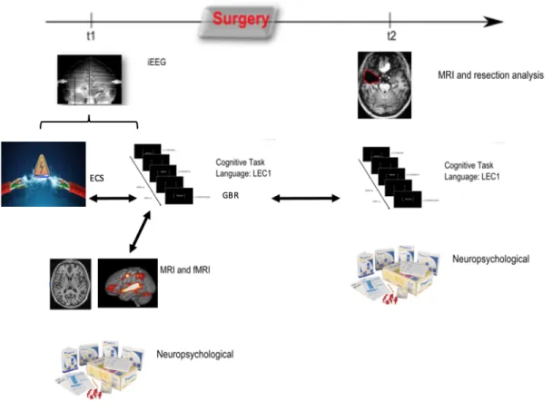
Figure 2. Schematic illustration of the experimental design. Left: pre-operative assessment (time 1 -t1-)
including iEEG evaluation including electro-cortical stimulation (ECS) and gamma band activity screening (GBR) through behavioural language task (LEC1), neuropsychological, anatomical (MRI) and functional MRI (fMRI). Right: post-operative assessment (time 2 -t2-) including LEC1, neuropsychological assessment and anatomical MRI (resection characteristics). Arrows represent the different comparison computed in the study.
2.8.1 ECS analysis
The clinical effect of ECS delivered over sites were retrospectively reviewed from patient’s video-SEEG files. Only speech disturbances, labelled as absent, incorrect or delayed responses or paraphasia, were taken into account. Other elicited clinical signs were not considered in this work. Thus, stimulated sites were binary classified into sites which showed language symptoms (ECS+) and those which did not (ECS-). As bipolar ECS were performed between two contiguous sites, each electrode was counted separately to allow comparison with GBR mapping. Any site that was common to two tested pairs, was regarded as ECS+ only
when it exhibited positive response in both paired stimulations that included the site. If the site was tested only in a single pair, it was labelled the same as the including pair.
2.8.2 Language-related GBR
To compute high-frequency activity amplitudes between 50 Hz and 150 Hz during Lec1 task, we applied a classical procedure performed in our team (see for instances Ossandon et al., 2011; Perrone-Bertolotti et al., 2014; and Vidal et al., 2011 for a review) and including the following processing steps: first, we bandpass filtered iEEG signals in multiple successive 10 Hz-wide frequency bands (e.g. 8–10 bands as [50–60 Hz], [60–70 Hz], etc.) using a zero phase shift non causal finite impulse filter with 0.5 Hz roll-off. Next, for each bandpass filtered signal we computed the envelope using a standard Hilbert Transform. The obtained envelope is down sampled to a sampling rate of 64Hz (i.e., one-time sample every 15,625ms). For each frequency interval, the time-varying amplitude was divided by its mean across the entire recording period of the experiment and multiplied by 100. This yields instantaneous amplitude envelope values as percentage-of-the-mean. Finally, the envelope signals computed for each consecutive band were averaged to provide one single time series (the GBR) across the entire recording session. The obtained envelopes had a sampling rate of 64 Hz. We also performed single trials GBR maps for each electrode contact. These single-trial maps were screened visually one-by-one. Hence, we determined electrodes sites which showed statistically significant GBR, labelled as GBR+, and those which did not show any statistically significant GBR, labelled as GBR-. Gamma band deactivations were not taken into account in this study.
The visual method used in this work was previously evaluated by two independent observers of our team: inter-rater agreement on detecting gamma activation was estimated on 17 666 single-trial maps using a Cohen’s kappa coefficient, which showed a substantial agreement (Cuisenier et al., 2016).
of Imaging Neuroscience, London, UK) implemented in MATLAB (MathworksInc., Sherborn, MA, USA). Each condition, language and rest, was modelled using a canonical hemodynamic function model. Data analysis started with the spatial pre-processing steps. The T1-weighted anatomical volume was co-registered to mean images created by the realignment procedure and was normalized to the MNI space using a trilinear interpolation. The anatomical normalization parameters were subsequently used for the normalization of functional volumes. Finally, to be able to compare fMRI results with the SEEG data, each functional volume was smoothed by a 3-mm FWHM Gaussian kernel. Time-series for each voxel were high-pass filtered (1/128 Hz cut-off) to remove low frequency noise and signal drift. Pre-processed data were then statistically processed and included into one design matrix including both sessions. Movement parameters derived from the realignment corrections (3 translations and 3 rotations) were also included in the design matrix as additional factors using ART toolbox for Matlab available on the Web at https://www.nitrc.org/projects/artifact_detect/. The block presentation allowed us to analyse data performing average response for all items, task vs control. Statistical parametric maps were generated from linear contrasts between the HRF parameter estimates for the different experimental conditions. Whole brain statistical analyses were first performed at an individual level in order to evaluate the language effect (K = 5; p value< .001 uncorrected, T = 3.55). Statistical analyses were subsequently performed at a group level (one sample t-test, k=5, p < .001 uncorrected; T=3.55). Finally, in order to compare fMRI results with GBR maps, we performed Region of Interest (ROI) analysis on fMRI data. We defined a sphere-type 3mm-radius ROI on each electrode contact from their MNI coordinates using SPM. We extracted the BOLD signal change for each of the ROI per patients. According to the statistical significance of the signal change on each site (p < .001, T = 3.55), we classified sites into two labels : fMRI + or fMRI -.
2.8.4 Functional data fusion
allows co-registration (using ANTs or SPM12) of the different imaging modalities (pre-implantation anatomical 3DT1 MRI, per-implantation 3DT1 MRI, post-operative 3DT1 MRI, fMRI) and visualization of respective surgery, electrode implantation and functional maps. 3-D modelized intracerebral electrodes were inserted on post-implantation MRI using IntrAnat Electrodes®. Cortex parcellation was achieved with Mars Atlas® (Brainvisa toolbox, UMR 7289, Marseille (Auzias et al., 2016)). Through this procedure, each electrode site was labelled according to i) its parcel in MarsAtlas, ii) its MNI (Montreal Neurological Institute) coordinates iii) its relation with resection mask (computed by subtracting post-operative 3D-T1 brain segmentation from the pre-implantation one) iv) fMRI ROI results v) clinical responses to ECS vi) language-related GBR. This allowed comparison between ECS, GBR and fMRI maps, as well as comparison of ECS and GBR maps with the extent of the resection.
2.8.5 Comparison between GBR, ECS and fMRI maps
As ECS is classically considered as the gold standard for localization of language, only anatomical sites where ECS were applied were taken into account.
GBR were first compared to ECS results, according to the methodology proposed by Sinai et al (2005). We thus determined the i) sensitivity, ii) specificity, and iii) positive and negative predictive values (PPV and NPV) of GBR maps in relation to ECS maps. GBR sensitivity was defined by the percentage of electrode sites that were positive for both methods (GBR+ and ECS+) among all ECS+ sites. GBR specificity was calculated as the percentage of electrode sites that were negative for both methods (GBR- and ECS-) among all ECS- sites. The PPV was defined by the probability that a GBR+ site showed language interference after ECS (ECS+) and was calculated as the percentage of sites that were both GBR+ and ECS+ among all GBR+ sites. Finally, the NPV was defined by the probability that a GBR- site did not exhibit
Then, GBR were compared with fMRI in the 6 patients in whom fMRI data was available. The sensitivity, specificity, PPV and NPV were determined according to the same methodology as described above.
Finally, to test the relation or agreement between GBR and the other two functional mapping methods (i.e., ECS and fMRI), a tetrachoric correlation (rt) was performed (see for instance Divgi, 1979). The rt allowed the evaluation of the correlation on binary data, thus the rt coefficient (ρˆ∗) showed the strength of the relationship or association between ratings for two raters (i.e., a “0” indicates no agreement and a “1” represents a perfect agreement between the measures). Thus, tetrachoric correlation coefficients (ρˆ∗) were computed.
2.8.6 Effect of surgery
Pre and post-operative behavioural performances during LEC1, in both phonological and semantic conditions, were compared using an analysis of variance (ANOVA) in terms of accuracy (percentage of correct responses) and reaction times (in ms).
Whether language post–operative outcome was related to the extent of the resection was evaluated using a Bravais-Pearson correlation (r) between the Delta performance (difference between pre- minus post-operative behavioural performances) and the volume of resection (in mm3).
Finally, to evaluate the prognostic value of removing brain regions displaying significant language-related GBR, we compared the Delta performance, for each experimental condition, between patients in whom at least one GBR + site was resected with the group of patients in whom none of the GBR+ site was resected (called control group). This comparison was performed using a Crawford and Howell’s modified t-test (Crawford et al., 1998), with a significance level set at p <0.05. A similar analysis was performed to compare post-operative language outcome of patients in whom at least one ECS+ site was resected with the group of patients in which no ECS+ site was resected. As the language task performed during ECS was a picture naming, close to the semantic condition of LEC1, only the performances obtained during the semantic
3. Results
3.1. Functional Maps
Among all 14 patients, 979 electrodes sites were explored through ECS and GBR (662 left, 317 right; mean: 70 sites/patient) and were thus considered in this study. This represented 58% of the total number of intracranial sites. The other 42% of intracranial sites (n=715; 360 left, 355 right) provided information only for GBR but were not stimulated, so that they were excluded from the analysis.
All patients exhibited GBR during the LEC1 task regardless of the experimental condition, at 81 of the 979 relevant sites (8%; mean: 6 GBR+ sites/patient). Twenty-five (30%) of the 81 GBR+ sites showed a selective activation for the semantic condition (n=12) or the phonological condition (n=13). ECS showed language interference in 9/14 patients at 51 of the 979 investigated sites (5 %, mean: 4 ECS+ sites/patient). BOLD signal changes were observed in all the 6 patients in whom language fMRI data was available: 59 of the 393 explored sites (as only sites where ECS were applied were considered) showed significant BOLD signal changes (15%, mean: 10 sites/patient). Figure 3 illustrates the cortical distribution of all investigated sites and sites that were positive for each method (ECS+, GBRs and fMRI+ sites).
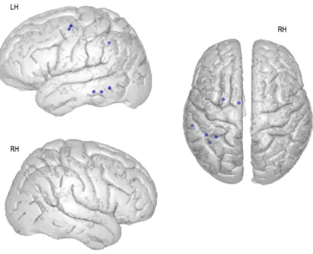
Figure 3 : Spatial distributions of the investigated sites, and positive responses during each method from
all 14 patients (P1-14) in common coordinates. Distributions are presented on axial and lateral views of MNI brain template. A. All electrode sites studied, where both ECS and GBR were performed (n=979) B. sites where ECS interfered with language. C. Electrode sites with significant GBR during Lec1. Different colours of the sites indicate the two different language processes: semantic (blue) and phonology (green). Red areas indicate sites which exhibited GBR in both conditions. D. Results from fMRI group analysis: Activated regions during the speech production task displayed on a 3D anatomical template and a 2D axial slice. k=5, p < .001 uncorrected; T=3.55, n=6. See Figure 6 in appendix for more details. LH: Left hemisphere, RH: Right hemisphere.
3.2. Comparison between GBR and ECS
superior parietal, dorsolateral somatosensory, medial visual, dorsomedial premotor and dorsomedial motor cortices. These sites showed GBR during LEC1 in both semantic and phonological conditions, except for one site located in the medial superior parietal cortex that showed GBR for the semantic condition only (see Table 3 and Figure 4 in Appendix for details).
ECS+ ECS - Total
GBR + 6 75 81
GBR - 40 858 898
Total 46 933 979
Table 2: Electrode sites distribution depending on ECS and GBR results.
Patient Site x y z Label GBR results
4 C'11 -59 -35 -13.3 LSPC Sem + Phono - 8 C'8 -31 -46 43 LSdl Sem + Phono + 8 F'6 -37 -56 -7 LSPC Sem + Phono + 9 D'5 -41 -47 -13 LVCm Sem + Phono + 12 L'6 -23 1 50 LPMdm Sem + Phono + 13 M'1 4 3 53 LMdm Sem + Phono +
Table 3: GBR+ and ECS+ sites. Including the name of the site (site), MNI coordinates (x, y, z) and the
anatomical label (Label). Results of GBR analyses for the two experimental conditions are presented. Abbreviations: LSPC: Left Superior Parietal Cortex; LSdl: Left Dorsolateral Somatosensory Cortex; LVCm: Left Medial Visual Cortex; LPMdm: Left Dorsomedial Premotor Cortex; LMdm: Dorsomedial Motor Cortex; Sem: semantic task; Phono: phonological task.
Sensitivity and specificity of GBR for language mapping relative to ECS were respectively 11.7% and 92.4%, with a PPV and NPV of 7.4% and 95.5%, respectively.
3.3. Comparison of GBR maps with fMRI activations
Only 6 patients had available data from a fMRI for language mapping, in whom a total of 393 sites were stimulated and were thus considered in this study. Five of these 393 sites were GBR+fMRI+ and 298 were
Table 5 and Figure 5 in Appendix for details).
fMRI+ fMRI - Total
GBR + 5 36 41
GBR - 54 298 352
Total 59 334 393
Table 4: Electrode sites distribution depending on fMRI and GBR results.
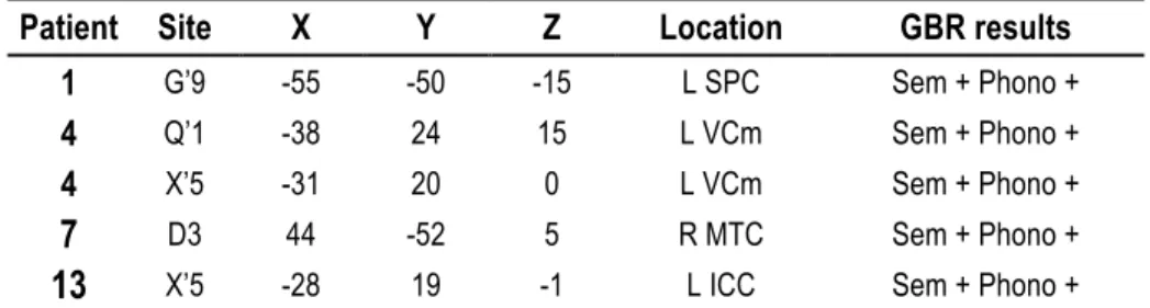
Patient Site X Y Z Location GBR results
1 G’9 -55 -50 -15 L SPC Sem + Phono +
4 Q’1 -38 24 15 L VCm Sem + Phono +
4 X’5 -31 20 0 L VCm Sem + Phono +
7 D3 44 -52 5 R MTC Sem + Phono +
13 X’5 -28 19 -1 L ICC Sem + Phono +
Table 5: MNI coordinates for the GBR+ IRMf+ sites. x, y, and z refer to the MNI coordinates of the sites, in
millimetres. L SPC: Left Superior Parietal Cortex; L VCm: Left Medial Visual Cortex; R MTC: Right Middle Temporal Cortex; L ICC: Left Isthmus Cingulate Cortex.
Sensitivity and specificity of GBR for language mapping relative to fMRI were respectively 8.4% and 89.2%, with a PPV and NPV of 12.2% and 84.7%, respectively.
3.4. Correlation between functional mapping methods
The tetrachoric correlation showed low correlation between GBR and fMRI, and between GBR and ECS as well, both for semantic and phonological conditions (see Table 6 for correlations values). The best correlation - although low - was observed between GBR and ECS for the semantic condition (ρˆ∗ = -.343).
GBR - ECS GBR - fMRI
Language
condition Phonological Semantic Phonological and Semantic Phonological Semantic Phonological and Semantic
ρˆ∗ -.285 -.343 -.217 -.053 .025 .104
3.5. Effects of surgery
Behavioural performances were considered as impaired post-surgery when they showed, as compared with pre-operative data, a statistically significant decrease of accuracy and/or statistically increase of reaction time. By opposition, Behavioural performances were defined as improved when they showed, as compared with pre-operative data, a statistically significant better accuracy and/or statistically shorter reaction time.
3.5.1. Effects of surgery on behavioural performances
Seven patients showed language changes in the semantic domain after surgery (see Table 7 and Table 8 in appendix for statistical details): 2 patients exhibited a significant impairment, one for reaction times (P11) and one for both accuracy and reaction times (P5), while 5 patients demonstrated a significant improvement, 3 in terms of accuracy (P7, P8 and P14) and 2 for reaction times. Behavioural comparison revealed no change in the other patients.
Seven patients showed language changes in the phonological condition after surgery (see Table 7 and Table 11 in appendix for statistical details): 2 patients (P5, P14) had a significant impairment of behavioural in terms of accuracy, while 5 patients were improved, 4 (P6, P7, P8, P11) in terms of accuracy and one (P13) in terms of reaction times.
No correlation was found between the Delta Performance (difference between pre- and post-operative behavioural performances) and the extent of the resections for both semantic and phonological conditions (see details in Table 10 in appendix).
Lec1 Speech
evaluation Resection S/A
(Months)
Semantic Phonological
P RT Acc RT Acc Pre/postDO 80 GBR+R Vlm
1 ns ns ns ns 77/49 Y 591 30 2 ns ns ns ns 80/ 78 N 1256 30 3 ns ns ns ns 74/77 N 8988 25 4 ns ns ns ns 79/NA N 24379 19 5 -- -- ++ -- NA/NA N 24141 15 6 ++ ns ++ ++ NA/NA N 23118 15 7 ns ++ ++ ++ NA/NA N 33221 18 8 ns ++ ++ ++ 77/NA N 5413 5 9 ns ns ns ns 78/NA N NR 3 10 ns ns ++ ns 80/NA Y 40623 3 11 -- ns ++ ++ NA/NA N 5025 4 12 ns ns ns ns 75/78 N 6576 4 13 ++ ns ++ ns 80/80 Y 6603 28 14 ns ++ ns -- 80/80 N 47210 24
Table 7 : Functional outcome of surgery and resection features: statistical significance of the behavioural
outcome of patients as assessed by LEC1 (including semantic and phonological condition), pre- and post-operative denomination score (DO80) assessed by speech therapist, resection details (relationship with GBR and volume in mm3) and time between surgery and post-operative assessment (S/A). Abbreviation:
RT: Reaction Time; Acc: Accuracy; ++: indicates a significant improvement in behavioural performances after surgery; --: indicates a significant decrement in performances after surgery; ns: no statistical difference between before and after surgery; GRB+R: indicates the resection of an GBR+ site or no; Y: yes, N: no; Vlm: volume.
3.5.2. Effect of GBR resection on behavioural performances
Two patients underwent a resection including at least one GBR+ site during the semantic condition (P1 and P13). P1 did not demonstrate any significant change after surgery, and its Delta performance did not statistically differ from the Delta performance of the control group (i.e., without GBR+ sites resected). P13 significantly improved after surgery for reaction times, but its Delta performance did not show any statistical difference with the Delta performance of the control group (see Table 8 in appendix)
Two patients underwent a resection including at least one GBR+ site during the phonological condition (P10 and P13). Both exhibited a significant post-operative improvement for reaction times but not for accuracy.
The Delta performance of these 2 patients was not statistically different from the Delta performance of the control group (See Table 9 in Appendix).
By comparison, 4 patients had a resection upon ECS+ sites (P4, P6, P12, P13), of whom 3 (P4, P12, P13) showed a significant decrease of accuracy after surgery as compared to the control group, and one (P6) showed a significant improvement of both accuracy and reaction times as compared to the control group (see Table 8 iin appendix for statistical details).
4. Discussion
The aim of this study was to assess whether task-related GBR might be used to anticipate cognitive deficits after surgery, and whether they might complement the classical methods for language mapping, namely ECS and fMRI
.
Indeed, GBR have been described as a putative biomarker of cognitive functions for almost 20 years (Lachaux et al., 2012) but their clinical relevance in predicting post-operative language outcome has never been formally assessed. To do so, we choose a language task that have the advantage to be compared with other modalities (ECS, fMRI) and provide quantitative features of behavioural performances to evaluate possible changes after surgery. The main results of the study, as well as its methodological limitations, are described in the followings sections.GBR mapping may help to choose the sites that have to be stimulated
Our results, though preliminary and extracted from a small cohort of patients, suggest first that GBR assessment can be a realistic complementary tool to ECS as part of iEEG recordings. Language-related GBR predicted ECS language interference with a high specificity (92.4%) and NPV (95.5%), meaning that if language tasks did not induce GBR on a cortical site, the likelihood of language impairment by stimulating the same site was very low. This highlights the possibility to first perform a functional map with GBR before starting the ECS procedure, and therefore save time and avoid stimulating sites that are a priori not relevant. These results compare favourably with the only two other studies having evaluated GBR and ECS during naming (n=13, Sinai et al. 2005) and word repetition (n=12, Towle et al. 2008) tasks, and which showed a specificity of 78% and 63%, respectively. These two studies, however, reported a much higher sensitivity (38% and 57%, respectively) than the one we found in our series (11.7%, with a PPV of 7.4%), a finding which can be related to the differences between the iEEG methods used. Sinai et al. (2005) and Towle et al. (2008) performed ECS and recorded GBR through subdural grids, that offered a broad coverage of neocortical areas. Compared with grids, SEEG electrodes provide narrower neocortical
Also, ECS using SEEG electrodes is very focal, since stimulation is applied bipolarily between contacts that are only distant from 2.5mm (center to center). Still, the current intensity used for functional mapping with grids is usually much higher (up to 15mA) than with SEEG electrodes (up to 3-4 mA), a difference which is related to the fact that subdural electrodes record the cortical surface while SEEG electrodes are intra-cortical. Previous studies suggested that ECS effects depend on several factors, such as the intensity of stimulation (Kahane & Dubeau, 2014; Trébuchon & Chauvel, 2016). Hence, it remains uncertain whether the ECS intensity applied in our study (ranging from 0,2 to 3mA) was sufficient enough to show obvious language disturbances. In any cases, there do not exist any studies having evaluated the ‘optimal’ ECS parameters to perform cognitive mapping.
GBR, ECS and fMRI do not reveal similar functional maps
Our results indicate that GBR and ECS were weakly correlated (R = - .22), suggesting a lack of concordance between GBR and ECS maps. A potential explanation for this result is related to the method itself. Indeed, ECS is a technique that disrupts functions (and therefore reveals key structures for a given cognitive process), while GBR is an activation method (which thus reveals the whole cortical network subserving a cognitive process). Still, subtle language dysfunctions triggered by ECS can be easily missed, leading to an underestimation of functional tissue. Consequently, language regions revealed by GBR are broader than - and partly overlap with - those defined with ECS (Brown et al., 2008). This is in line with the results of the present study, which showed that 81 GBR+ sites were identified, as compared with only 51 ECS+ sites. Furthermore, ECS may produce effects that are distant from the stimulated area, as the current may spread through axons or fibres to distant sites (Lesser et al., 1984; Blume, Jones, & Pathak, 2004). Indeed, current density models have showed that focal current application similar to ECS can interfere not only with cell body activity but also with axons transmission, thus influencing distant areas (Ranck, 1975).
methodological limitations that might explain these findings, including first the small number of patients assessed throughout both methods (n=6). Also, we deliberately choose small-sized (3mm-radius) ROI to compute comparisons between BOLD and GBR, this choice being motivated by the spatial resolution achieved by depth electrodes (3mm). In previous studies comparing GBR and BOLD activity (Conner et al., 2011; Esposito et al., 2013; Conner et al., 2011), greater ROI sizes were used (10mm) that guaranteed stronger correlation but reduced accuracy. Still, the restricted number of relevant cortical sites (i.e. only those sites where ECS were applied) represented another limitation, and a a better relationship between GBR and BOLD signal might have been obtained if consideration had been given to all implanted sites. A final limitation, detailed below, is related to the difference between the language tasks used during fMRI (speech production) and SEEG (visual presentation of single words for language comprehension).
Are GBR maps more accurate than ECS or fMRI maps?
The language system involves several linguistic processes and in clinical settings it may be important to discriminate and precisely localise these processes (see, for instance, Perrone-Bertolotti et al., 2012). Our results pinpointed one site among GBR+ECS+ sites, which illustrates nicely the capacity of GBR to specify language processing: C’11 (P 4) located in the medial superior parietal cortex. Indeed, this electrode site exhibited specific GBR induced only during the semantic condition, while ECS triggered a non-specific aphasia. This is in line with intracranial studies which established that GBR provided a powerful means to detect task-specific activations (Crone et al., 2001; Towle et al., 2008). Language mapping through ECS is based only on disruption of language and provides only rough “yes'' or ''no” responses. Stimulation therefore may appear as a less specific and as a less subtle mapping tool than GBR. Moreover, the slow time course of haemodynamic imaging technique (fMRI) makes it difficult to distinguish different linguistic processes (Towle et al., 2008). As neural activity during language occurs on a millisecond time scale, it appears to be better explored by intracranial EEG signal analysis than using fMRI.