Technical Notes & Surgical Techniques
Reasonability of implementation of the endoscopic technique for sellar
lesions in a low volume center
Jean-Yves Fournier, MD
a,⁎, Heidrun Lange, MD
a, Karen Huscher, MD
a, Sarah R. Haile, PhD
b,
Gerhard Hildebrandt, MD, PhD
a, Abel-Jan Tasman, MD, PhD
c, Doortje Engel, MD, PhD, MBA
aa
Clinic for Neurosurgery, Cantonal Hospital of St. Gall, CH-9007 St. Gall, Switzerland
b
Clinical Trial Unit, Cantonal Hospital of St. Gall, CH-9007 St. Gall, Switzerland
c
Clinic for Otorhinolaryngology, Cantonal Hospital of St. Gall, CH-9007 St. Gall, Switzerland
a b s t r a c t
a r t i c l e i n f o
Article history: Received 10 September 2014 Revised 8 October 2014 Accepted 12 October 2014 Keywords: Pituitary adenoma Endoscopic Microscopic TranssphenoidalWe evaluated the implementation of the endoscopic (E) technique by an interdisciplinary ENT/neurosurgeon team as compared to the established classical microscopic technique (M) performed by one experienced neurosurgeon for pituitary adenomas in a single center. A retrospective analysis of patients operated for newly diagnosed pituitary adenomas was performed between November 2004 and August 2012. Outcome and complications are presented. A total of 116 patients were operated, 64 microscopically (M) and 52 endoscopically (E). Mean follow up was 35 months (range 1.4–95), 1 patient was lost to follow-up. Most frequent pathology was hormone inactive adenoma (60% E, 51% M). Operating time was stable in the M-group (±94 min). The E-group showed a learning curve in mean operating time (2004–2007: 154 min, 2008–2012: 93 min). Postoperative CSF leaks were seen in 9.6% (E) vs. 3.1% (M) of cases. More E-cases were re-operated (5 vs. 1) and more M-cases received a lumbar drainage (8 vs. 19). Transient postoperative diabetes insipidus occurred more often after E-operations (17 vs. 5%, p = 0.03) without significant long term difference. Improved visual outcome showed a more favorable trend in E-cases. The implementation of the endoscopic technique was associated with more surgical complications in the learning phase, however with more improved visual outcome. Our observations should be of value for the more average neurosurgical department dealing with pituitary adenomas and aiming to switch from the microscopic to the endoscopic technique.
© 2014 The Authors. Published by Elsevier B.V. This is an open access article under the CC BY-NC-ND license (http://creativecommons.org/licenses/by-nc-nd/3.0/).
Introduction
Endoscopic neurosurgery is gaining popularity as first-choice technique for transsphenoidal resection of sellar lesions[1]. However in 2013 66% of pituitary tumours were still operated microscopically in the USA (personal communication at the North American Skull Base Society, San Diego, February 2014). From a technical point of view endoscopic (E) resections are expected to be better than microscopic (M) resections due to improved visibility. Changing a department's main technique from microscopic to endoscopic can be done mainly in two ways. One possibility is to use the endoscope at the end of or during a classical microscopic procedure; although this will not add supplementary risks for the patient, this can be quite cumbersome, because intraoperative switching techniques costs time. Another way is to work in a team with an experienced endoscopic rhinologist and conduct the operation purely endoscopically. This solution has the
advantage of accelerating some of the operative steps as the rhinologist performs the transnasal approach rapidly, and gives the team security in the manipulation of the endoscope itself during resection of the tumor in a three-hand fashion. In our institution two neurosurgeons (GH and JYF) perform transsphenoidal surgery. The less experienced was encouraged by the experienced to implement the endoscopic technique in cooperation with an endoscopic rhinologist. We retrospectively compared our endoscopic and microscopic cases of the past 8 years especially in respect to intra-and postoperative surgical complications in order to document this transition of one institution's practice. The observations made should be of value for the more average neurosurgical department dealing with pituitary adenomas and aiming to switch from microsurgical to endoscopic technique.
Methods
Data were collected retrospectively of all patients operated primarily for a sellar lesion microscopically or endoscopically in our institution between November 2004 and August 2012. Patients operated on transcranially were not included. Data collected included demographics, diagnosis, characteristics of the lesion, perioperative
Interdisciplinary Neurosurgery: Advanced Techniques and Case Management 1 (2014) 115–118
⁎ Corresponding author. Tel.: +41 71 494 3050; fax: +41 71 494 28 83. E-mail addresses:[email protected](J.-Y. Fournier),[email protected] (H. Lange),[email protected](K. Huscher),[email protected](S.R. Haile), [email protected](G. Hildebrandt),[email protected](A.-J. Tasman), [email protected](D. Engel).
http://dx.doi.org/10.1016/j.inat.2014.10.003
2214-7519/© 2014 The Authors. Published by Elsevier B.V. This is an open access article under the CC BY-NC-ND license (http://creativecommons.org/licenses/by-nc-nd/3.0/). Contents lists available atScienceDirect
Interdisciplinary Neurosurgery:
Advanced Techniques and Case Management
j o u r n a l h o m e p a g e :w w w . i n a t - j o u r n a l . c o mand postoperative course including complications, evaluation of the pituitary function and resection rate. Clinical data collected included age, sex, time of onset of the first symptoms, time of diagnosis, neurological signs, time offirst imaging and the presence of relevant associated diseases. All available images were reviewed by the authors to characterize the lesions for dimensions, presence of bleeding, compression of the chiasm, deviation of the pituitary stalk, cavernous sinus invasion, encasement of the internal carotid arteries and the Knosp–Steiner classification. Tumor volume was calculated by = (4/3)*pi*(length/20)*(width/20)*(height/20). Surgical details such as duration of the procedure, blood loss, intraoperative and postoperative complications including postoperative CSF leaks and the way they were managed, bleeding, diabetes insipidus, pituitary insufficiency, infection and presence of residual tumour were collected. Evaluation of the resection and tumour rest was done in 3 months postoperative MR-images. Residual tumor was measured and classified in small (b1 ml) and large (N1 ml).
Surgical techniques
The patients operated microscopically were operated by one of the authors (GH) with a classical microscopic technique beginning with a submucosal transseptal approach and placement of a nasal speculum. An otorhinolaryngologist, who leaves the theatre during the so-called
neurosurgical resection phase, conducts this approach. Fluoroscopic imaging is used to improve accuracy, giving sagittal information about the localization of the speculum in relation to the sellarfloor. The resection is conducted bimanually. The otorhinolaryngologist returns at the end of the procedure for the septal reconstruction.
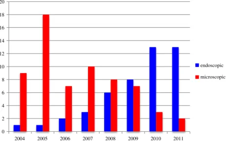
The patients operated endoscopically were operated by a team during the whole operation, which is composed of one experienced endoscopic rhinologist (JT) and a neurosurgeon (JYF) with some microscopic experience in pituitary surgery and more than 5 years of experience in cerebral endoscopy at the beginning of the series but no previous experience in transsphenoidal endoscopy. During the preparation phase the head is fixed in a Mayfield clamp and the nasal mucosa is decongested with cotton pledgets soaked in adrenaline 1:5000 (1 ml of adrenaline 1 mg/ml diluted in 4 ml saline) placed in the middle meatus. In addition, the mucoperiosteum of the lateral nasal wall and the mucoperiosteum and mucoperichon-drium of the septum are infiltrated with a lidocaine/adrenaline solution (1% lidocaine and 1/200.000 adrenaline). The procedure always started with a 0° endoscope (Karl Storz, Tuttlingen, Germany) and depending on the situation a 45° endoscope was used. The approach is performed under induced hypotension (b110 mmHg systolic blood pressure) and begins with lateralization without resection of the medial turbinate on both sides. If a large arachnoid defect is expected the sphenopalatine artery is identified and a nasoseptalflap prepared for watertight closure[2](Hadad, 2006). After widening of the ostium sphenoidale on both sides an anterior sphenoidotomy with posterior septectomy is performed. After opening of the sella with 2 and 3 mm Cloward punches, the resection of the adenoma is performed with two hands using suction and dissectors, rarely curettes. No endoscope holder was used in order to keep the pseudo-depth perception given by the dynamic movements of the endoscope and thus apply a three-hand technique. In most of 0 2 4 6 8 10 12 14 16 18 20 2004 2005 2006 2007 2008 2009 2010 2011 endoscopic microscopic
Fig. 1. Increasing proportion of endoscopic procedures during the observation period.
Table 1
Distribution of pathologies between the 2 groups.
Pathology Microscopic Endoscopic
Hormone inactive 33 31 PRLa 1 9 ACTHb 5 4 GHc 14 3 Pituitary hyperplasia 1 0 Aspergillosis 1 0
Metastasis breast cancer 2 0
Epidermoid cyst 0 1 Rathke's cyst 5 3 No certain diagnosis 2 1 Total 64 52 a Prolactinoma. b Cushing. c
Growth hormone secreting adenoma.
Table 2
Size and invasiveness of tumors.
Microscopic Endoscopic
Giant (sizeN40 mm) 8% 19%
Knosp grade IV 8% 20%
Bilateral cavernous sinus Invasion 16.1% 31.4%
Chiasm compressed 34.8% 52.9%
the procedures neuronavigation was applied. During the last 2 years of the series a micro-Doppler probe was systematically used to confirm the information of the navigation regarding the position of the internal carotid artery (ICA).
Statistics were conducted by Fisher's exact tests. Binary variables are described as n, % and significance 5%. For operation time linear regression lines were used to describe changes.
A special permission of the BAG (Federal Office of Public Health) was obtained due to the retrospective nature of this study, as well as regular approval of the ethics committee of St. Gall.
Results
In total 116 patients have been operated on transsphenoidally between November 2004 and August 2012; 52 were operated endoscopically and 64 microscopically (E: n = 52, M: n = 64; ± 0– 19 cases/year). There was no randomization of the patients between the two techniques; they were operated on according to the availability of the surgeons and, at the beginning, according to the feasibility of the procedure by the endoscopic team. Age and gender distribution did not differ between the two groups [mean endoscopic = 54 years (range E 19–79), microscopic =55 years (18–86]. Sixty-seven per cent were male. Mean follow up time was 35 months (range 1.4–95).
During the observation period the proportion of endoscopic procedures increased (Fig. 1). Neuronavigation was used in 80% of E-cases, but never in M-cases where fluoroscopic imaging was used systematically.
The most frequent pathology was hormone inactive adenoma (E: n = 31 60%, M: n = 33, 51%; seeTable 1). The E group consisted of larger and more invasive lesions, although not significant (Table 2).
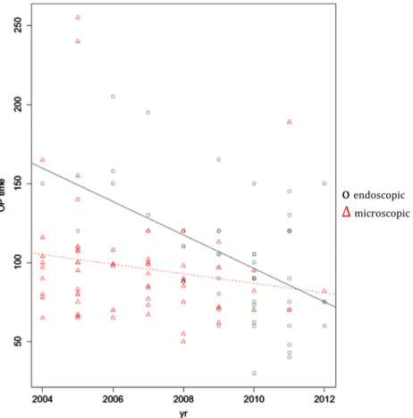
The duration of the procedures (Fig. 2) was stable in the experienced M-group (± 94 min). The E-group showed a clear learning curve in mean operation-time (2004–2007: 154 min, 2008–2012: 93 min).
Total resection was better in microscopic patients (39 vs. 44%) by trend (p = 0.65), whereas after endoscopic operations a small residuum (b1 ml) was seen as more often than after microscopic resection (38 vs. 29%). A large residuum (N1 ml) was seen in 31% (E) vs. 25% (M) of patients.
In both series only one postoperative infection occurred, resulting in infection rates of 1.9% (E) and 1.6% (M).
Intraoperative CSF leaks were seen in 31% (E) vs. 42% (M) of cases (Table 3). However, the data show 9.6% (E) and 3.1% (M) persistent postoperative leaks. More E-cases were reoperated (n = 5 vs. 1), while more M-cases received a lumbar drainage (19 vs. 8). In both groups 2 patients had to be reoperated because of large residual tumour. The ICA
o
endoscopic∆
microscopicFig. 2. Learning curve of the endoscopic versus the microscopic team. Endoscopic: R−10.6, CI 15.5–6.7, p = b0.01. Microscopic: R −3.0, CI 7.6–1.5, p = 0.19.
Table 3 Complications.
Complication Microscopic Endoscopic
CSF intraop. 27 (42.2%) 16 (30.8%) CSF leak postop. 2 (3.1%) 5 (9.6%) Infection 1 (1.6%) 1 (1.9%) DIa early postoperative 2 (3.1%)⁎ 9 (17.3%)⁎ DIachronic/persistent 3 (4.7%) 2 (3.8%) SIADHb 7 (10.9%) 2 (3.8%) ICAc injury 0 (0%) 1 (1.9%) a
DI: diabetes insipidus.
b SIADH: syndrome of inappropriate diuretic hormone. c ICA: internal carotid artery.
⁎ Statistically significant: p b 0.05.
117 J.-Y. Fournier et al. / Interdisciplinary Neurosurgery: Advanced Techniques and Case Management 1 (2014) 115–118
was damaged in 1 E-case, where after rapid tamponade the vessel lesion was treated successfully with endovascular embolization and the patient showed mild residual symptoms of a hemiparesis at 1-year follow-up. Transient postoperative diabetes insipidus (DI) occurred significantly more often after an endoscopic operation (17 vs. 3%, p = 0.03). However, there was no significant long-term difference in persistent DI (2 E vs. 3 M cases). Improved visual outcome was better in endoscopic patients (E: 47%; M: 34%, not significant), even though patients in the E-group had more chiasmatic compression [mild or severe chiasm compression in 73% (E) and 52% (M)].
Discussion
This retrospective study shows that the implementation of the endoscopic technique was associated with slightly higher incidence of complications. It presents several limitations, as it is a retrospective study without randomization of the assignment of the cases and it compares two surgical teams at different levels of experience in a low volume center. Next to that, the less experienced endoscopic team used technically more advanced support (neuronavigation, Doppler). However we found this comparison legitimate as it actually might reflect reality in many surgical units. Although from a statistical point of view there are no straightforward uni- or multivariate analyses permitted, thorough review of the cases is what was conducted.
Comparing our microscopic and endoscopic groups with each other and the literature several differences were identified. Resection rates showed larger residua in the E-group. This difference can be at least partially explained by the differences in the two groups, with a tendency for larger and more invasive tumours in the E-group. It can also be explained by the small sample size.
With an incidence of postoperative CSF leaks of 9.6% our endoscopic series is comparable with data of the literature, as shown by the systematic review by Rotenberg [3], who found a mean incidence of 14.3% or by the meta-analysis of Ammirati[4], where it was evaluated to be 7.0% (4.84 to 9.52%). Although the E-group had less intraoperative CSF leaks, there were more postop-erative leaks needing surgical treatment by either lumbar drainage or surgical revision. The different incidence of revision and lumbar drainage reflect different approaches of the two surgeons regarding their management. The experienced M surgeon preferred avoidance of postoperative CSF leaks with a more systematic application of a lumbar drain in the postoperative period if a CSF leak would persist or appear. The E team tried to avoid the patient's discomfort associated with a lumbar drainage for several days and thus decided more often to proceed to operative revision. The sellar floor reconstruction showed a learning curve as well, as 4 out of 5 postoperative CSF leaks occurred in thefirst 26 patients, in the second half of the series only one case had a postoperative CSF leak. The pediculated vascularized
septalflap after Hadad was performed in patients with very large lesions where the probability of a postoperative CSF leak was assessed as high. It seemed to have a protective effect, as only 1 out of 6 cases with large lesions with use of a Hadadflap had to be reoperated for a CSF leak.
The incidence of persistent DI (3.8%) was consistent with reported observations that vary between 2 and 31%.
In both series only one patient presented a postoperative infection so that infection rates remain low for both series (E = 1.9%, M = 1.6%) and within normal range for this type of surgery.
In spite of larger volumes, visual outcome was better in endoscopic, although 1 patient worsened, and 1 worsened due to other causes. This advantage could be explained by the fact that manipulation in the vicinity of the chiasm can be better visualized with the endoscope.
In the light of these observations we can recommend that neurosurgeons who treat sellar pathologies in a low volume center and are interested in switching to the endoscopic technique should start with adequate experience in microscopic transsphenoidal surgery. Beginning with simple pathologies such as cystic lesions or smaller, non-invading macroadenomas, will save time and stress, as well as adjoining themselves an experienced endoscopic rhinologist. Conclusions
In the past 8 years the documentation of the transition of our institution's practice from the microscopic to the endoscopic technique for pituitary adenomas was not associated with relevant complications. Our observations show that implementation from microscopic to endoscopic transsphenoidal technique in an average neurosurgical department is reasonable.
Disclosure
All authors report no conflicts of interest relevant to this article. References
[1]Tabaee A, Anand VK, Barrón Y, Hiltzik DH, Brown SM, Kacker A, et al. Endoscopic pituitary surgery: a systematic review and meta-analysis. J Neurosurg 2009;111 (3):545–54.
[2]Hadad G, Bassagasteguy L, Carrau RL, Mataza JC, Kassam A, Snyderman CH, et al. A novel reconstructive technique after endoscopic expanded endonasal approaches: vascular pedicle nasoseptalflap. Laryngoscope 2006;116(10):1882–6.
[3]Rotenberg B, Tam S, Hyung A, Ryu W, Duggal N, Ryu WH. Microscopic versus endoscopic pituitary surgery: a systematic review.pdf. Laryngoscope 2010;120: 1292–7.
[4] Ammirati M, Wei L, Ciric I. Short-term outcome of endoscopic versus microscopic pituitary adenoma surgery: a systematic review and meta-analysis. J Neurol Neurosurg Psychiatry 2012.http://dx.doi.org/10.1136/jnnp-2012-303194. 118 J.-Y. Fournier et al. / Interdisciplinary Neurosurgery: Advanced Techniques and Case Management 1 (2014) 115–118