Université de Montréal
Corticospinal Excitability, Mental Rotation Task, Motor Performance and Disability in Subjects with Musculoskeletal Disorders of the Wrist and Hand
par René Pelletier
École de réadaptation Faculté de médecine
Thèse présentée à la Faculté de médecine
en vue de l’obtention du grade de Philosophiae Doctor (Ph.D.) en Sciences de la réadaptation
Mai 2018
II
Université de Montréal Faculté des études supérieures
Cette thèse intitulée:
Corticospinal Excitability, Mental Rotation Task, Motor Performance and Disability in Subjects with Musculoskeletal Disorders of the Wrist and Hand
présentée par: René Pelletier
a été évaluée par un jury compose des personnes suivantes: Dre Brigitte Vachon
________________________________________
Président rapporteur
Dre Johanne Higgins
________________________________________
Directeur de recherche
Dr Daniel Bourbonnais
________________________________________
Codirecteur de recherche
Dre Lisa Carlesso
________________________________________
Membre interne du jury
Dr Guillaume Leonard
________________________________________
Examinateur externe
Dr Jonathan Tremblay
________________________________________
III
Résumé
L'objectif de cette thèse était de démontrer la présence de modifications des processus sensorimoteurs du système nerveux central (excitabilité corticospinale et schéma corporel tels que mesurés avec la Tâche de Reconnaissance de la Latéralité des Images droite gauche (TRLI)) chez des participants ayant des désordres musculosquelettiques au poignet et à la main. Un deuxième objectif était de déterminer la relation entre les changements de ces processus sensorimoteurs corticaux et des mesures sensorielles, de la fonction motrice, d'incapacité autodéclarée, de la douleur et des facteurs psychosociaux liés à la douleur.
Une étude observationnelle transversale a d'abord été menée pour mesurer l'excitabilité corticospinale des muscles de la main en utilisant la stimulation magnétique transcrânienne et la TRLI chez des participants en santé et des participants présentant des douleurs chroniques au poignet et à la main. L’excitabilité corticospinale du muscle court abducteur du pouce de la main affectée était augmentée chez les participants présentant une douleur chronique et ces changements étaient significativement corrélés avec l'intensité de la douleur, l'incapacité autodéclarée, et négativement corrélés avec l'excitabilité motoneuronale. Des différences de performances sur le TRLI, à la fois pour la précision et le temps de réaction, ont également été trouvées entre les participants du groupe contrôle et les participants avec douleur.
Dans une deuxième étude transversale, le TRLI, des mesures de motricité, sensibilité et des fonctions cognitives ont été administrées à soixante et un participants présentant des désordres musculosquelettiques du poignet ou de la main droite. Les modèles de régression linéaire multiple ont révélé que la prise de médicaments pour contrer la douleur, la participation à des activités (sociales, professionnelles, domestiques et récréatives), la discrimination tactile de deux points et le niveau de performance motrice expliquent les performances au TRLI. Les participants ayant pris des médicaments pour la douleur la journée de l’évaluation avaient une performance diminuée sur la précision et le temps de réaction sur le TRLI pour la main droite (affectée). Ces participants présentaient aussi une sévérité de douleur et d'incapacité plus élevée et une diminution de fonctions cognitives et motrices plus élevée que le reste des participants avec douleur qui ont été évalués.
IV
Dans l’ensemble, ces résultats suggèrent que les participants présentant des désordres musculosquelettiques hétérogènes du poignet ou de la main montrent des changements des processus sensorimoteurs corticaux. Alors que l'excitabilité corticospinale semble être liée à l'intensité de la douleur et à l’incapacité autodéclarée, le TRLI peut être associé à une confluence de facteurs (sensoriels, moteurs, cognitifs-affectifs et comportementaux). Ces résultats suggèrent aussi que les changements sensorimoteurs corticaux ne sont pas simplement le résultat du désordre musculosquelettique, mais impliquent plutôt une interaction complexe entre la douleur, les processus sensorimoteurs et cognitivo-affectifs, et peut-être aussi des réponses comportementales à l’atteinte musculosquelettique. Les résultats fournissent également des informations précieuses à propos des personnes qui pourraient bénéficier d'interventions orientées vers le rétablissement des processus centraux en plus des traitements de réadaptation axés sur les structures périphériques.
Mots-clés: désordres musculosquelettiques, douleur, neuroplasticité, excitabilité corticospinale, stimulation magnétique transcrânienne, imagerie motrice, performance motrice, incapacité, schéma corporel, tâche de rotation mentale
V
Abstract
The objective of the thesis was to investigate for the presence of changes in cortical sensorimotor processes (corticospinal excitability and the body schema measured with the Left Right Judgment Task (LRJT) performance), in participants with Musculoskeletal Disorders (MSD) of the wrist/hand. A second objective was to determine the relationship between these cortical sensorimotor processes and measures of sensory and hand motor function, disability, pain and pain related psychosocial factors.
First, an observational cross-sectional study was conducted to explore corticospinal excitability of muscles in the hand and cortical sensorimotor processes, utilizing transcranial magnetic stimulation and the LRJT in healthy, pain-free participants and participants with chronic wrist/hand pain. Increased corticospinal excitability for the abductor pollicis brevis of the affected hand in participants with chronic MSD of the wrist/hand was found and these changes were significantly correlated with pain intensity, disability, and negatively correlated with spinal motoneuronal excitability. Differences in LRJT performance were also found between healthy control participants and participants with pain for both LRJT accuracy and reaction time. In a second cross-sectional study, LRJT performance, motor, sensory and cognitive assessments were performed on sixty-one participants with MSD of the right dominant wrist/hand. The multiple linear regression model revealed that taking pain medication, participating in (social, work, household and leisure) activities, two-point discrimination, and motor performance explained performance on the LRJT of the right (affected) hand. Those participants that took pain medication on the day of the evaluation performed more poorly on both LRJT accuracy and reaction time of the right (affected) hand. These participants had higher pain severity and disability scores and decreased cognitive and motor function.
Collectively, these results suggest that participants with heterogeneous MSD of the wrist/hand display altered cortical sensorimotor processes. Whereas corticospinal excitability appears to be related to pain intensity and disability, the LRJT may be associated with a confluence of factors (sensory, motor, cognitive-affective, and behaviours). These findings suggest that cortical sensorimotor changes do not simply appear to be the result of the condition but involve a complex interaction between pain, sensorimotor and cognitive-affective processes, and possibly
VI
behavioural responses to the condition. The findings also provide valuable insight as to those persons who may benefit from cognitively directed interventions in addition to peripherally driven rehabilitative treatments.
Keywords: musculoskeletal disorders, pain, neuroplasticity, corticospinal excitability, transcranial magnetic stimulation, motor imagery, motor performance, disability, body schema, sensorimotor integration, mental rotation task, implicit motor imagery
VII
List of Tables
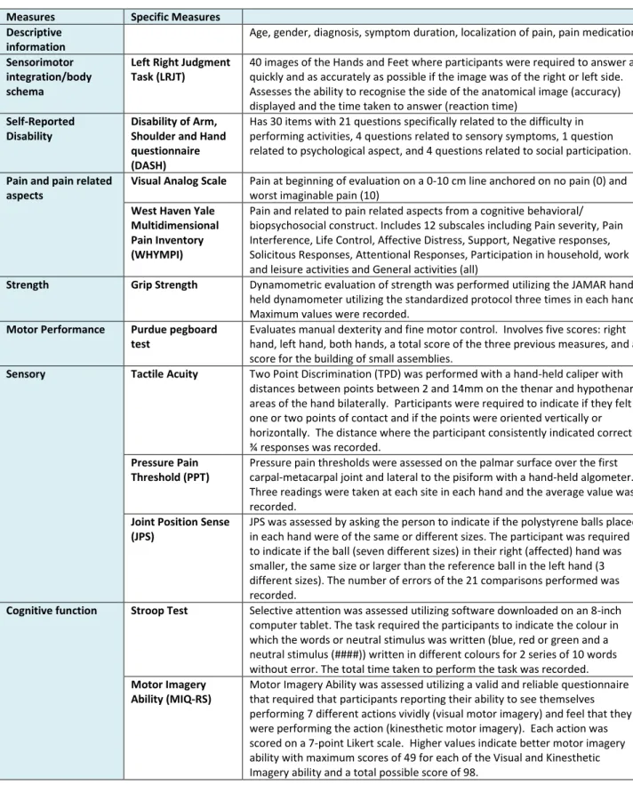
Chapter 4: Methodology and Methodological Considerations
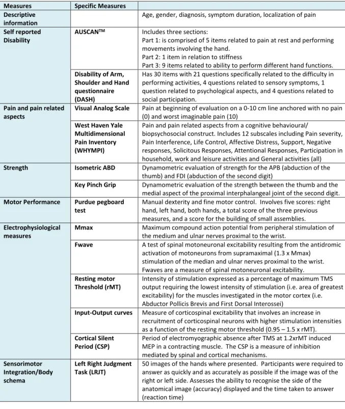
Table 4.1: Measures performed in study of corticospinal excitability in persons with
musculoskeletal disorders of the wrist/hand ... 78 Table 4.2: Measures performed in study of the Left Right Judgment Task, sensory, motor and cognitive assessment in participants with wrist/hand pain ... 89
Chapter 5: Results Section 5.1: Article 2
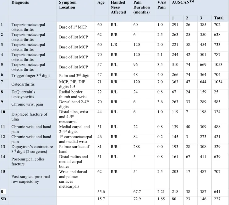
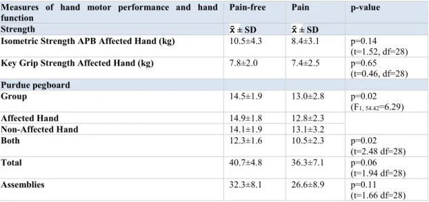
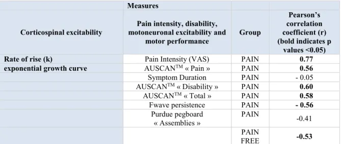
Table 5.1.1: Subject demographics and measures of pain and disability ... 103 Table 5.1.2: Strength and Purdue pegboard results ... 104 Table 5.1.3: Pearson correlation coefficients between measures of corticospinal excitability and electrophysiological and functional measures. ... 105 Table 5.1.4: Correlations between motor evoked potential amplitudes and isometric strength ... 118
Section 5.2: Article 3
Table 5.2.1: Differences in motor performance measures between participants in the Control and Pain groups ... 128 Table 5.2.2: Pearson product correlations (r) between Left Right Judgment Task performance and age ... 130 Table 5.2.3: Pearson product correlations (r) between Left Right Judgment Task reaction time and accuracy with measures of motor performance, disability and pain ... 131 Table 5.2.4: Pearson product correlations (r) between Left Right Judgment Task and motor performance (Purdue pegboard total scores) ... 132 Table 5.2.5: Pearson correlation coefficients (r) between motor performance and pain-related measures in participants with wrist/hand pain ... 132
VIII
Section 5.3: Article 4
Table 5.3.1: Descriptive and demographic information ... 158 Table 5.3.2: Linear regression model for Left Right Judgment Task accuracy for the hands 159 Table 5.3.3: Coefficients of best fitting linear regression models for Left Right Judgment Task hand accuracy... 160 Table 5.3.4: Left Right Judgment Task right hand reaction time ... 160 Table 5.3.5: Coefficients of best fitting linear regression models for Left Right Judgment Task right hand reaction time ... 161
Chapter 6: Discussion Section 6.3: Article 5
Table 6.3.1: Areas of neuroplastic changes associated with chronic musculoskeletal disorders and possible signs and symptoms manifested by the patient. ... 201
IX
List of Figures
Chapter 2: Review of literature
Figure 2.1: Nociception, pain and musculoskeletal disorders……….…..……… 15 Figure 2.2: Changes in the central nervous system associated with pain and musculoskeletal disorders ………...…... 20 Figure 2.3: Musculoskeletal disorders and sensorimotor integration ………. 38
Chapter 4: Methodology and Methodological considerations
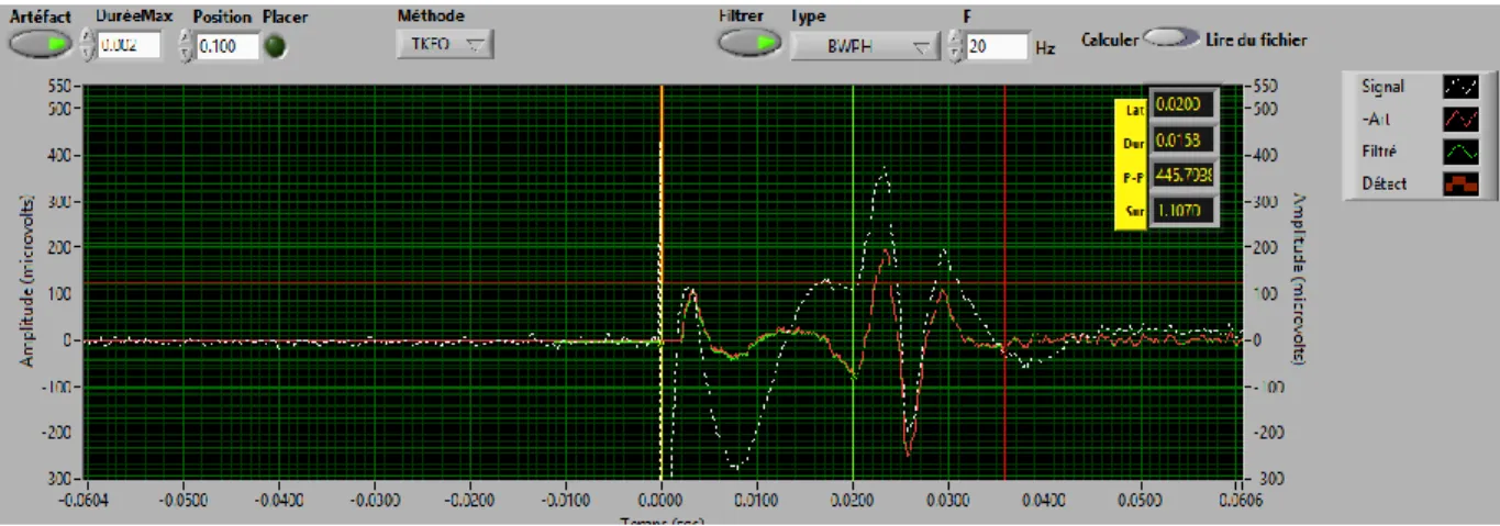
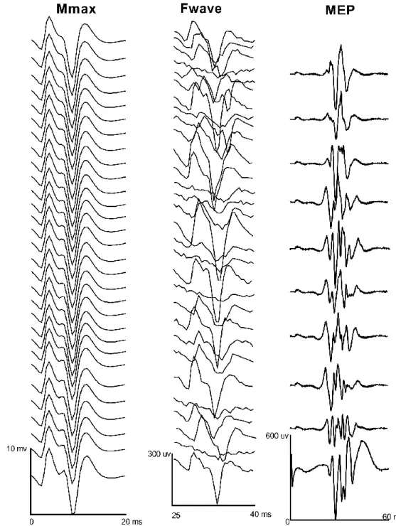
Figure 4.1: Isometric strength testing of the abductor pollicis brevis ………. 79 Figure 4.2: Cortical silent period for the abductor pollicis brevis ………... 82 Figure 4.3: Example of a motor evoked potential in the abductor pollicis brevis …………... 85 Figure 4.4: Representational traces of Mmax, Fwave and motor evoked potentials in one participant in the abductor pollicis brevis ……… 86
Chapter 5: Results Section 5.1: Article 2
Figure 5.1.1: Input-Output curves for the abductor pollicis brevis ………... 104 Figure 5.1.2: Correlation plots ………... 106 Figure 5.1.3: Resting motor threshold for the first dorsal interossei for the Control and Pain groups ……… 116 Figure 5.1.4: Input-Output curves for the first dorsal interossei ………...… 116 Figure 5.1.5: Cortical silent period duration (seconds) for the abductor pollicis brevis and first dorsal interossei ………. 117
Section 5.2: Article 3
X
Section 5.3: Article 4
Figure 5.3.1: Left Right Judgment Task performance in participants with musculoskeletal disorders of the wrist/hand ………. 158 Figure 5.3.2: Left Right Judgment Task accuracy and reaction time in participants who did and did not take pain medications on the day of the evaluation……….... 162
Chapter 6: Discussion
Figure 6.1: MSD, sensorimotor integration, corticospinal excitability and the left right
XI
Table of Contents
Résumé ... III Abstract ...V List of Tables ... VII List of Figures ... IX Abbreviations ... XIX Dedication ... XX Remerciements ... XXI Avant propos ... XXII
Chapter 1 Introduction ... 1
1.1 Epidemiology of Musculoskeletal Disorders (MSD) ... 1
1.2 Altered Sensorimotor Processes with MSD ... 2
1.2.1 Altered sensory processes ... 2
1.2.2 Motor control changes with MSD ... 2
1.2.2.1 Mechanisms responsible for motor control changes ... 3
1.3 Changes in Sensorimotor Cortical Properties and Organization with MSD ... 3
1.3.1 MSD and changes in the primary and secondary somatosensory cortices ... 4
1.3.2 Changes in the primary motor cortex associated with MSD ... 5
1.3.2.1 Experimentally induced acute pain and the primary motor cortex ... 5
1.3.2.2 Chronic MSD and the primary motor cortex ... 5
1.4 MSD, Rehabilitation, Function and Disability ... 6
1.5 Summary ... 6
1.6 Principal Objective/ Structure of the Thesis ... 7
Chapter 2 Review of Literature ... 9
2.1 Musculoskeletal Disorders, Nociception and Pain ... 9
2.1.1 Musculoskeletal Disorders (MSD) ... 9
2.1.2 Nociception and pain ... 10
2.1.3 Pain and MSD ... 12
2.1.3.1 Medial nociceptive system – cognitive, affective, motivational areas of the brain ... 13
2.1.3.2 Psychological factors associated with pain and disability ... 14
XII
2.1.3.3 Descending pain modulation system and sensitization ... 17
2.1.3.4 Constant vs intermittent pain in persons with MSD ... 21
2.1.3.5 Chronic pain ... 21
2.1.3.5 Lateral nociceptive system – sensory discrimination areas of the brain ... 22
2.2 Sensory Changes and MSD ... 23
2.2.1 Cortical sensorimotor changes and MSD ... 23
2.2.2 Body schema and MSD ... 24
2.3 MSD, Motor Control and Pain ... 27
2.3.1 Changes in the primary motor cortex associated with MSD ... 30
2.3.1.1 Transcranial Magnetic Stimulation (TMS) ... 30
2.3.3.2 MSD and the primary motor cortex ... 31
2.3.3.2.1 MSD and the motor threshold ... 32
2.3.3.2.2 MSD and intracortical inhibition and facilitation ... 32
2.3.3.2.3 MSD and the cortical silent period ... 34
2.3.3.2.4 MSD and corticospinal excitability measured with input-output curves .... 34
2.3.3.2.5 MSD and TMS mapping ... 35
2.3.3.2.6 Summary of TMS changes and MSD ... 36
2.3.3.2.7 MSD, corticospinal changes and motor function ... 36
2.4 Article 1: Is neuroplasticity in the central nervous system the missing link to our understanding of chronic musculoskeletal disorders? ... 39
2.4.1 Abstract ... 40 2.4.1.1 Background ... 40 2.4.1.2 Discussion ... 40 2.4.1.3 Summary ... 40 2.4.2 Keywords ... 41 2.4.3 Background ... 42 2.4.4 Discussion ... 44
2.4.4.1 Principles of experience dependent plasticity ... 45
2.4.4.2 Plasticity in the spinal cord and brain stem with chronic MSD ... 45
2.4.4.3 Neuroplastic changes in the primary somatosensory cortex and perceptual changes with MSD ... 48
XIII
2.4.4.5 Role of Pain in Central Nervous System (CNS) plasticity ... 52
2.4.4.6 Neuroplastic changes in meso-limbic and prefrontal structures in chronic pain states ... 53
2.4.4.7 Integrating CNS changes into a more comprehensive model of chronic MSD . 56 2.4.4.8 Impact of CNS plasticity in the rehabilitation of chronic MSD ... 57
2.4.4.9 Research ... 58 2.4.5 Summary ... 59 2.4.6 Abbreviations... 60 2.4.7 Competing Interests ... 60 2.4.8 Author’s Contributions ... 60 2.4.9 References ... 61
Chapter 3: Objectives and Hypothesis ... 75
3.1 General Objectives and Hypothesis ... 75
3.2 Specific Objectives of the Thesis ... 75
Chapter 4: Methodology and Methodological Considerations ... 77
4.1 Corticospinal Excitability, Left Right Judgment Task, Pain, Disability and Motor Performance ... 77
4.1.1 Measures ... 77
4.1.1.1 Demographic and baseline information ... 77
4.1.1.2 Pain related disability ... 79
4.1.1.3 Pain related psychosocial factors ... 79
4.1.1.4 Motor performance ... 79
4.1.1.4.1 Isometric strength ... 79
4.1.1.4.2 Purdue pegboard test ... 80
4.1.1.5 Disability ... 80
4.1.1.6 Assessment of corticospinal excitability ... 81
4.1.1.6.1 The hotspot and Resting Motor Threshold (rMT) ... 81
4.1.1.6.2 Input-Output curves ... 81
4.1.1.6.3 Cortical Silent Period (CSP) ... 82
4.1.1.6.4 Spinal motoneuronal excitability ... 83
4.1.1.6.5 Data acquisition and processing ... 84
XIV
4.2 Assessment of Left Right Judgment Task, Cognitive, Sensory, Motor, Disability and
Psychosocial Factors in Participants with Wrist/Hand Pain ... 87
4.2.1 Measures ... 88
4.2.1.1 Sensation ... 88
4.2.1.1.1 Tactile acuity ... 88
4.2.1.1.2 Pressure pain threshold... 88
4.2.1.2 Proprioception ... 90
4.2.1.3 Motor performance ... 90
4.2.1.3.1 Grip strength ... 90
4.2.1.4 Cognition/attention ... 90
4.2.1.4.1 Stroop test ... 90
4.2.1.4.2 Motor imagery ability ... 91
Chapter 5 Results ... 92
5.1 The relationship of corticospinal excitability with pain, motor performance and disability in subjects with chronic wrist/hand pain ... 93
5.1.1 Preface ... 94 5.1.2 Keywords ... 95 5.1.3 Abstract ... 95 5.1.4 Abbreviations... 96 5.1.5 Introduction ... 97 5.1.6 Methods ... 98 5.1.6.1 Subjects ... 98
5.1.6.2 Measures of pain intensity, hand motor performance and disability ... 98
5.1.6.2.1 Pain intensity and pain related disability... 98
5.1.6.2.2 Hand motor performance ... 99
5.1.6.2.3 Pain related disability of the hand ... 99
5.1.6.3 Measures of cortical excitability and Fwaves ... 99
5.1.6.3.1 Subject preparation ... 99
5.1.6.3.2 Data acquisition ... 99
5.1.6.3.3 Transcranial magnetic stimulation ... 99
5.1.6.3.4 Hotspot and Resting Motor Threshold (rMT) of the Abductor Pollicis Brevis (APB) ... 100
XV
5.1.6.3.5 Input-output (I-O) curves and motor evoked potential amplitudes during
active contractions ... 100
5.1.6.3.6 Maximum Compound Muscle Action Potential (Mmax) and Fwave evaluation ... 100
5.1.6.4 Analyses ... 100
5.1.6.4.1 Data analysis ... 100
5.1.6.4.2 Statistical analyses... 101
5.1.7 Results ... 102
5.1.7.1 Differences between groups in demographic, hand motor performance and disability ... 102
5.1.7.2 Differences between groups in corticospinal and Fwave measurements... 102
5.1.7.2.1 Resting motor threshold of the APB ... 102
5.1.7.2.2 Input-Output curves and motor evoked potential amplitudes during active contraction ... 102
5.1.7.3 Mmax and Fwaves ... 102
5.1.7.4 Association between measures of corticospinal excitability and measures of spinal motoneuronal excitability, pain intensity, hand motor performance and hand disability ... 105
5.1.8 Discussion ... 107
5.1.8.1 Corticospinal excitability and the association with measures of pain, hand motor performance and disability ... 107
5.1.8.2 Corticospinal and spinal motoneuronal excitability ... 108
5.1.9 Study Limitations ... 108
5.1.10 Conclusion ... 109
5.1.11 Acknowledgements ... 109
5.1.12 References ... 110
5.1.13 Supplemental Results ... 115
5.1.13.1 Methodology and analysis ... 115
5.1.13.2 Resting motor threshold of the First Dorsal Interossei (FDI) ... 115
5.1.13.3 Input-Output curves for the FDI ... 116
5.1.13.4 Spinal motoneuronal excitability and the FDI ... 117
5.1.13.5 Cortical silent period for the APB and FDI ... 117
XVI
5.2 Laterality recognition of images, motor performance, and aspects related to pain in participants with and without wrist/hand disorders: an observational cross-sectional study.
... 119 5.2.1 Preface ... 120 5.2.2 Abbreviations... 120 5.2.3 Abstract ... 121 5.2.4 Keywords ... 121 5.2.5 Highlights ... 122 5.2.6 Introduction ... 123 5.2.7 Methods ... 125 5.2.7.1 Participants ... 125 5.2.7.2 Measures ... 125
5.2.7.2.1 Left right judgment task ... 125
5.2.7.2.2 Pain ... 126
5.2.7.2.3 Hand motor performance ... 126
5.2.8 Statistical Analysis ... 127
5.2.9 Results ... 128
5.2.9.1 Demographics, baseline characteristics and motor performance ... 128
5.2.9.2 Left right judgement task - accuracy ... 129
5.2.9.3 Left right judgement task - reaction time ... 129
5.2.9.4 Accuracy and reaction time trade-off ... 129
5.2.9.5 Relationship between left right judgement task and measures of motor performance, disability and pain ... 131
5.2.10 Discussion ... 133
5.2.11 Limitations ... 136
5.2.12 Conclusion ... 136
5.2.13 References ... 138
5.3 Left Right Judgement Task and sensory, motor, cognitive and psychosocial assessment in participants with musculoskeletal disorders of the wrist/hand ... 144
5.3.1 Preface ... 146
5.3.2 Abstract ... 147
XVII 5.3.4 Keywords ... 147 5.3.5 Abbreviations... 148 5.3.6 Introduction ... 149 5.3.7 Methods ... 151 5.3.7.1 Dependent variable ... 152 5.3.7.2 Independent variables ... 152 5.3.7.2.1 Sensory measures ... 152
5.3.7.2.2 Motor performance measures ... 154
5.3.7.2.3 Disability measure ... 155
5.3.7.2.4 Cognitive measures ... 155
5.3.8 Sample size and statistical analysis ... 156
5.3.9 Results ... 157
5.3.9.1 Left right judgment task - hand accuracy and reaction time ... 157
5.3.9.2 Multiple Linear Regression (MLR) models ... 159
5.3.9.2.1 Left right judgment task right (affected) hand accuracy ... 159
5.3.9.2.2 Left right judgment task left hand accuracy ... 159
5.3.9.2.3 Left right judgment task right hand reaction time ... 160
5.3.9.2.4 Left right judgment task left hand reaction time ... 161
5.3.9.2.5 Left right judgment task feet accuracy and reaction time ... 161
5.3.9.3 PainMed vs NoPainMed ... 161
5.3.10 Discussion ... 163
5.3.10.1 Left right judgment task, motor imagery ability and the Stroop test ... 163
5.3.10.2 Left right judgment task, sensory and motor function ... 164
5.3.10.3 Left right judgment task, pain medication and general activities ... 165
5.3.12 Conclusion ... 168
5.3.13 References ... 169
Chapter 6 General Discussion ... 175
6.1 Changes in Corticospinal Excitability ... 176
6.1.1 Factors that may account for the increase in corticospinal excitability of the APB 180 6.1.2 Summary ... 184
XVIII
6.2.1 Left right judgment task performance and sensory, motor and cognitive factors .. 185
6.2.2 Left right judgment task and cortical processes ... 186
6.2.3 Left right judgment task, cognitive and sensorimotor changes ... 190
6.2.4 Left right judgment task and motor performance ... 191
6.2.5 Summary ... 194
6.3 Relevance in Rehabilitation Medicine ... 194
6.3.1 Addressing Neuroplastic Changes in Distributed Areas of the Nervous System Associated with Chronic Musculoskeletal Disorders ... 196
6.3.1.1 Abstract ... 197
6.3.1.2 Keywords ... 197
6.3.1.3 Introduction ... 198
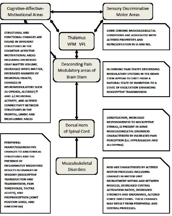
6.3.1.3.1 Neurophysiological changes occurring within peripheral receptors and the dorsal horn of the spinal cord ... 198
6.3.1.3.2 Neuroplastic changes within the brainstem ... 199
6.3.1.3.3 Neuroplastic changes in the cortical sensory discriminative areas ... 199
6.3.1.3.4 Neuroplastic changes in the cognitive-affective-motivational areas ... 200
6.3.1.4 Implications of distributed neuroplastic changes associated with chronic musculoskeletal disorders for rehabilitation ... 202
6.3.1.4.1 Interventions ... 203
6.3.1.5 Conclusion ... 211
6.3.1.6 References ... 213
6.4 Limitations ... 218
Chapter 7 Conclusion ... 218
Chapter 8 Future Directions ... 219
XIX
Abbreviations
APB: Abductor Pollicis Brevis
CNS: Central Nervous System
CSP: Cortical Silent Period
EMG: Electromyography
FDI First Dorsal Interossei
fMRI: Functional Magnetic Resonance Imaging
I-O: Input-Output
ISI: Inter-Stimulus Interval
LBP: Low Back Pain
LRJT: Left Right Judgment Task
M1: Primary Motor Cortex
MEP: Motor Evoked Potential
MSD: Musculoskeletal Disorders
MSO: Maximum Stimulator Output
MT: Motor Threshold
PPT: Pressure Pain Thresholds
PPG: Purdue Pegboard Test
rMT: Resting Motor Threshold
RT: Reaction Time
SI: Primary Somatosensory Cortex
SII: Secondary Somatosensory Cortex
SICF: Short Interval Cortical Facilitation SICI: Short Interval Cortical Inhibition TMS: Transcranial Magnetic Stimulation
XX
Dedication
To the persons who consult with me for their confidence and involving me in one of the most important aspects of their lives - their health
To those who believed in me and gave me a chance
Most of all to my family
To my wife for her love, support, friendship, patience and understanding. I look forward to making up for lost time together.
To my children for their love, support, understanding, and helping me grow as a person and a father
XXI
Remerciements
This process was a labour of love mixed with a tinge of frustration, exhilaration, despair, stress, fatigue. I loved (almost) every moment.
Thank you to my research directors Johanne Higgins, PhD and Daniel Bourbonnais, PhD for sharing generously of their time, knowledge and expertise.
Although I had limited time to interact with other students, each encounter was a pleasure and rewarding.
Thank you to the support staff at l’Institut de réadpatation Gingras-Lindasy de Montréal. Your goodwill, time, patience, and work where not only helpful but essential.
Thank you to all the people who graciously shared their precious time to participate in the studies.
Thank you to my family who sacrificed evenings, weekends, and vacations to allow me to undertake such a challenge at this point of my life.
XXII
Avant propos
Twenty-five years ago, I graduated with a M.Sc. degree in Biomedical Sciences (option réadaptation) at the University of Montréal under the supervision of Robert Forget, Ph.D. and Daniel Bourbonnais, Ph.D. The study in which I participated involved reflex modulation between synergistic muscles of the lower extremity with conditioned H-reflexes. After the completion of the master’s program, I had the full intention to pursue my doctoral studies. However, I was side-tracked for more than 20 years.
During the 20-year hiatus from research I taught as a part-time faculty member at Concordia University in the Exercise Science program and in osteopathic programs across Canada and Europe. However, most of my time was spent as a clinician working with individuals suffering from musculoskeletal conditions. Initially my practice was largely comprised of athletes and physically active individuals. The structural-pathology paradigm or biomedical model was the predominant model at the time of my undergraduate education and was an adequate conceptual and treatment model when working with acute musculoskeletal injuries in an athletic population. However, with time I began to see an ever-increasing number of persons suffering with chronic musculoskeletal injuries/conditions. Although treatment occasionally helps people to overcome their chronic musculoskeletal injuries/conditions, the biomedical model appears to be less efficacious and fails as a conceptual as well as a treatment model for these conditions. As a teacher and practitioner of a manual profession, given the heterogeneity in the application of interventions and poor intra- and inter-rater reliability of manual therapy diagnostic tests, I hypothesized that the much of what we do was related to neurophysiological, educational and behavioural changes associated with treatment more than biomechanical changes stemming from physical manipulation. Recently there has been an emergence of scientific literature of pain and of central neuroplastic changes associated with pain and musculoskeletal injuries across distributed areas of the nervous system that may provide additional insight into the neurophysiological underpinnings of both the clinical applications of treatment and manifestations of persons with chronic musculoskeletal injuries/conditions.
When I met with Daniel Bourbonnais and Johanne Higgins in 2012, I shared a review article, Boudreau et al (2010), The role of motor learning and neuroplasticity in designing
XXIII
research investigating central neurophysiological changes, usually associated with neurological conditions such as stroke and traumatic brain injuries, also demonstrated changes associated with musculoskeletal conditions. We went about elaborating a study investigating neurophysiological changes associated with musculoskeletal disorders of the hand, tapping into the area of expertise of Dr Bourbonnais (dynamometric evaluation of the hand) and Dr Higgins (transcranial magnetic stimulation). We spent the next year and half reconstituting Dr Bourbonnais’ lab at l’Institut de réadaptation Gingras Lindsay de Montréal, working with the biomedical engineer Michel Goyette at the research centre on the software, data acquisition and processing for the experiments. I enrolled in the Ph.D. in September 2013.
When I speak to friends, colleagues, patients, acquaintances I meet and tell them that I am pursuing my Ph.D. at the age of fifty, I am usually met with bemusement. I am inevitably asked why and what will I do with a Ph.D. My answer is usually somewhat long winded but involves three aspects: personal, professional and sociological. Personal motivations stem from an enjoyment of learning and belief that it is important for our health and well-being to undertake new challenges and live varied experiences. Professionally, I wished to gain increased knowledge of neurophysiological changes associated with pain and musculoskeletal disorders, and competencies to evaluate sensorimotor function. I believed these skills, knowledge and expertise would benefit my clinical practice and the comprehension of mechanisms that would hopefully result in better treatments for those persons suffering with chronic musculoskeletal disorders. Finally, the sociological motivation stems from my professional affiliation as an osteopath that is undergoing the process of regulation in the province of Quebec. I am motivated to augment my knowledge, expertise, and experience in the scientific process as well as sensorimotor processes associated with musculoskeletal disorders to contribute to the development of this profession. In areas of the world where the profession of osteopathy has been regulated there has been an evolution from private educational institutions to public/university programs. There will be a need for individuals with the knowledge and competencies for the scientific inquiry of physiological processes associated with osteopathic manual medicine, improved models resting upon current scientific knowledge and research principles, and proof of efficacy and the evolution of osteopathic concepts and interventions.
1
Chapter 1 Introduction
1.1 Epidemiology of Musculoskeletal Disorders (MSD)
Musculoskeletal Disorders (MSD) result from insult to muscles, tendons, tendon sheaths, ligaments, joints, cartilage and/or nerves (Barr et al. 2004). These injuries are associated with local and systemic inflammatory responses, cellular proliferative changes, altered sensorimotor processes and pain (Barr et al. 2004). MSD have a direct impact on a person’s ability to work, their quality of life, and are associated with important socioeconomic costs (Statistics 1998) accounting for roughly 3.4% of the gross domestic product in Canada (Coyte et al. 1998). Direct medical costs and indirect productivity losses account for 29% and 71% respectively of the total costs associated with MSD (Coyte et al. 1998). In Québec, MSD are responsible for 38% of occupational injuries compensated by the Commission des normes, de l'équité, de la santé et de la sécurité du travail and generate more than 40% of the compensation costs (Lamarche et al. 2011).
In a review of articles related to the incidence and prevalence of upper extremity MSD, Huisstede et. al. (2006) found upper extremity (shoulder, elbow, wrist and hand) MSD have point prevalence rates in the general population between 1.6 and 53% (Huisstede et al. 2006). Injuries to the wrist and hand had population prevalence rates in men and women of working age of 17.7% for men and 26.9% with women in the United Kingdom (Walker-Bone et al. 2004). Hand and wrist injuries account for approximately 20% of hospital visits to the emergency room in the Netherlands (de Putter et al. 2012). Of the musculoskeletal injuries for which persons present themselves to the emergency room in the Netherlands, lower extremity fractures and hand injuries rank first in total cost (direct and indirect) associated with injury (de Putter et al. 2012).
MSD may become chronic conditions associated with pain and disability. A systematic review found prevalence estimates of chronic pain range between 11.5% to 55.2% (Ospina et al. 2002). In Canada prevalence of chronic pain was found to be 18.9% in persons over 18 years of age, with roughly 6% of those suffering with chronic pain where experiencing MSD of the wrist/hand (Schopflocher et al. 2011). Direct and indirect costs related to chronic pain are estimated to be between $560 to $635 billion a year in the United States, more than the annual cost for cancer, heart disease and diabetes combined (Gaskin et al. 2012).
2
Given the personal and societal burden of chronic MSD, it is important to understand the pathophysiology of MSD to aid in the development of more efficacious interventions. Recent evidence suggest that altered neurological mechanisms may contribute to chronic MSD and their clinical manifestation of altered sensorimotor functions.
1.2 Altered Sensorimotor Processes with MSD
MSD result in rapid neurochemical/molecular changes at the site of the injury and within the spinal cord, and resultant functional and structural changes in subcortical and cortical structures (Wall et al. 2002). As a result of these changes, MSD result in reorganizational changes throughout the somatosensory system from the peripheral sensory neurons to cortical areas (Wall et al. 2002). Although mechanisms remain poorly understood, motor control changes are also characteristic of MSD (Barr et al. 2004; Hodges et al. 2011).
1.2.1 Altered sensory processes
Subjects with MSD characteristically display changes in peripheral sensorimotor processes and function. With acute MSD, changes in sensory afferent output are present resulting from the inflammatory response and neurochemical changes locally at the site of the MSD (Wall et al. 2002). Damage to musculoskeletal structures affects receptor transduction of both nociceptive and non-nociceptive neural receptors (Voscopoulos et al. 2010; Ward et al. 2015). Subsequent changes in structures and function are found within the spinal cord, brain stem (cuneate and gracilis nuclei), and thalamus (Wall et al. 2002). Altered sensory function includes increased transduction and transmission of nociceptive and non-nociceptive stimuli (Fernandez-Carnero et al. 2009; Skou et al. 2013; Chiarotto et al. 2013; Lluch et al. 2014). Peripheral sensory function including two-point discrimination thresholds (Luomajoki et al. 2011; Stanton et al. 2013; Catley et al. 2014), and decreased joint position sense are often manifested in persons with MSD (Sharma et al. 1997; Valeriani, Restuccia, Di Lazzaro, Franceschi, et al. 1999).
1.2.2 Motor control changes with MSD
Altered motor control patterns are well documented with MSD (Hodges and Tucker 2011). Consistent experimental evidence demonstrates that experimentally induced acute pain, a model associated with acute MSD, is associated with decreased maximum voluntary contraction, decreased muscle endurance, delayed muscle activation, and altered EMG activity in agonist/synergist muscles during agonist (decreased) and antagonist (increased) phases of
3
muscle activity in painful muscles (Arendt-Nielsen et al. 2008; Graven-Nielsen et al. 2008; Schabrun and Hodges 2012; Bank et al. 2013). With chronic MSD, changes in motor control include changes in strength (Dominick et al. 2005; de Oliveira et al. 2011), impaired Electromyographic (EMG) muscle activation between and within muscles (Tucker and Hodges 2009; Tucker, Butler, et al. 2009), decreased central activation ratios where participants with MSD display a decreased ability to maximally recruit spinal motoneurons when performing maximal voluntary contractions compared to healthy control participants (Verbunt et al. 2005; Hart et al. 2010), and increased co-contraction of agonist and antagonist muscles (Falla et al. 2008). Changes in motor activation have been related to both pain intensity and measures of psychological distress (Verbunt et al. 2005). These changes along the entire somatosensory system, behavioural priorities, cognitive/psychological factors (such as fear avoidance and catastrophization) and associated forebrain mechanisms influence motor control (Field 2009). Specific mechanisms underlying these motor control changes are, however, not well understood. 1.2.2.1 Mechanisms responsible for motor control changes
The changes in motor control in subjects with MSD may be the result of local and central factors. Motor control changes found in persons with MSD may be the result of altered sensory input arising from altered mechanoreceptor, chemoreceptor and muscle spindle activity to cortical structures (Brumagne et al. 1999; Thunberg et al. 2001; Panjabi 2006). Alternatively, motor control changes may result from behavioural changes to protect the area of pain (Field 2009), or central neurophysiological processes to minimize functional loss and protect the injured area (Hodges and Tucker 2011). Changes in muscle activation may also be a direct consequence of nociception both from spinal (Bank et al. 2013) and cortical processes (Frot et al. 2013).
1.3 Changes in Sensorimotor Cortical Properties and Organization with MSD
The changes in peripheral sensory and motor processes appear to be associated with changes in cortical sensory and motor structure and function. Although it is generally believed that neuroplastic changes in the Primary Somatosensory (S1) and Primary Motor (M1) cortices associated with MSD are driven by pain (Moseley and Flor 2012) the specific relationship between pain, cortical sensorimotor processes and motor control is unclear. Animal models have demonstrated that peripheral tissue compromise results in neurochemical and molecular changes locally at the site of injury and concurrent neuroplastic changes in S1 and M1 in the development
4
of upper extremity overuse injuries of the wrist and hand (Barr 2006; Barbe et al. 2006; Coq et al. 2009). However, these models have difficulty in parcelling cortical changes driven by the learning of new motor tasks, the repetitive movements performed, and those associated with pain and inflammation associated with the MSD.
1.3.1 MSD and changes in the primary and secondary somatosensory cortices
Changes in sensory afferent output resulting with MSD are associated with changes in cortical properties and organization in S1 and the Secondary Somatosensory (SII) cortices. Evoked potential and Functional Magnetic Resonance Imaging (fMRI) studies have found changes in structure and function within S1 and SII in persons with chronic pain conditions such as complex regional pain syndrome (Maihofner et al. 2003), carpal tunnel syndrome (Tecchio et al. 2002), focal hand dystonia (Elbert et al. 2004), and Low Back Pain (LBP) (Flor et al. 1997; Giesecke et al. 2004; Lloyd et al. 2008; Hotz-Boendermaker et al. 2016). These changes in properties and organisation in S1 and SII may be associated with behavioural findings of altered sensory perception, tactile acuity, and proprioceptive acuity found in subjects with MSD including LBP (Goossens et al. 2018). Tactile acuity specifically has been correlated with S1 reorganisation in persons with complex regional pain syndrome (Pleger et al. 2006).
There is also indirect evidence of sensorimotor changes in persons with MSD. Persons with MSD display changes in performance of the Left Right Judgment Task (LRJT) that requires the participant to determine as accurately and as quickly as possible if images of body parts presented are of the left or right side (Coslett et al. 2010a; Coslett et al. 2010b; Stanton et al. 2012; Schmid et al. 2012). The LRJT is believed to implicate cortical sensory processes, an internal representation of the body in peri personal space in real time that is derived from sensory input (i.e sensory, vestibular, visual). This internal representation of the body is referred to as the body schema. The precise anatomical position of the body parts in peri personal space is necessary to efficiently engage motor control processes required for the planning and execution of movement (Bray et al. 2011). The LRJT implicates cortical areas in the parietal cortex associated with sensorimotor integration and in cortical areas involved in attention, movement planning and execution (Parsons et al. 1995; Kosslyn et al. 1998). Decreased performance on the LRJT has been associated with pain intensity in some studies (Moseley 2004b; Hudson et
5
al. 2006; Linder et al. 2016) but not others (Coslett et al. 2010b; Bray and Moseley 2011; Schmid and Coppieters 2012; Stanton et al. 2012).
1.3.2 Changes in the primary motor cortex associated with MSD
Peripheral and central somatosensory changes, psychological factors, as well as behavioural responses to pain and injury appear to affect motor control (Field 2009). Cortical changes in M1 have been demonstrated in models of acute pain and with chronic MSD.
1.3.2.1 Experimentally induced acute pain and the primary motor cortex
Evidence of changes within M1 associated with acute MSD is lacking. However, experimentally induced pain is utilized as an experimental model for acute pain associated with MSD. Research demonstrates that within M1, experimentally induced pain is associated with decreased corticospinal excitability (Cheong et al. 2003; Fierro et al. 2010; Dube et al. 2011; Bank et al. 2013). Decreased corticospinal excitability, other factors being equal, would result in decreased motor activation and weakness characteristic of acute MSD (Arendt-Nielsen and Graven-Nielsen 2008; Graven-Graven-Nielsen and Arendt-Graven-Nielsen 2008).
1.3.2.2 Chronic MSD and the primary motor cortex
Although chronic MSD are also characterized by changes in motor control, there appears to be more variability, specifically of spinal motoneuronal and corticospinal changes within and between subjects and conditions (Hodges et al. 2003; Hodges and Tucker 2011) compared with experimentally induced acute pain. However, a number of studies demonstrate changes in corticospinal properties and organization in M1 in subjects experiencing chronic MSD including measures of corticospinal excitability such as Motor Thresholds (MT), Motor Evoked Potential (MEP) amplitudes, and representational changes in M1 assessed with Transcranial Magnetic Stimulation (TMS) in participants with patellofemoral pain (On et al. 2004; Te et al. 2017), anterior cruciate ligament injury (Héroux et al. 2006; Lepley et al. 2015), LBP (Strutton et al. 2005; Tsao et al. 2008; Tsao, Danneels, et al. 2011; Masse-Alarie et al. 2012; Elgueta-Cancino et al. 2015; Massé‐Alarie et al. 2017), sciatica (Strutton et al. 2003), rotator cuff tears (Ngomo et al. 2015) chronic shoulder pain (Bradnam et al. 2015), and lateral epicondylitis (Schabrun, Hodges, et al. 2014). These changes in corticospinal properties and organisation have been correlated with pain intensity (Elgueta-Cancino et al. 2015), symptom duration (Flor et al. 1997;
6
Ngomo et al. 2015), and the level of dysfunction (Ochi et al. 1999; Tsao et al. 2008; Kapreli et al. 2009; Tsao, Galea, et al. 2010). However, other studies have found no correlation between pain measures, function and changes in sensorimotor properties and organization (Bray and Moseley 2011; Ngomo et al. 2015; Bradnam et al. 2015; Te et al. 2017). Furthermore, altered cortical properties and representation in M1 may be present in the absence of pain and in the absence of peripheral nerve injury (Byl et al. 1996; Byl et al. 2000a; Byl et al. 2002).
1.4 MSD, Rehabilitation, Function and Disability
Conservative treatment for MSD is oriented to decreasing pain and restoration of motor and sensory function. Traditionally, rehabilitative care of MSD has been guided by a structural-pathology paradigm or biomedical model where local structural structural-pathology is believed to be the source of pain and disability and the target of intervention (Foster et al. 2003). Inherent within the biomedical model is the belief that pain and disability will resolve with restoration of normalized structure and function of compromised musculoskeletal structures and the patient will return to pre-injury levels of function and activities (Burton et al. 2008). However, conservative treatment (pharmacological, medical, physical therapies, behavioural therapies and complementary and alternative medical practices) inspired from the biomedical model involving peripherally driven treatment at the site of the MSD has several failings (Burton et al. 2008) and has not consistently produced positive outcomes specifically with regard to chronic MSD such as LBP (Foster et al. 2003). These findings have led some researchers to hypothesize that other mechanisms, including neurophysiological changes in the Central Nervous System (CNS), may be implicated in the pathophysiology of chronic MSD (Barr et al. 2004; Langevin et al. 2007; Wand et al. 2008; Wand, Parkitny, et al. 2011; Moseley and Flor 2012). Increased comprehension of underlying processes involved in sensory and motor deficits should in theory yield more efficacious interventions.
1.5 Summary
There is a growing body of evidence in animals and humans that neuroplastic changes in S1 and M1 occur simultaneously with MSD. MSD are also associated with abnormal sensory and motor processes. Altered central sensorimotor processes have been hypothesized to contribute to the development and ongoing chronicity of MSD (Hodges and Moseley 2003; Barr et al. 2004; Langevin and Sherman 2007; Forget et al. 2008). Although studies often investigate the
7
relationship between neurophysiological sensorimotor changes with pain intensity and duration, few have directly related these changes with measures of motor function, psychosocial aspects related to pain, and disability. More clarity in the relationship between sensorimotor cortical changes in structure, organization, and function with sensory and motor functions is necessary (Elgueta-Cancino et al. 2017). For example, changes in motor cortical properties and organization can be driven by behavioural changes related to cognitive and psychological processes mediated in the forebrain (Field 2009; Simons et al. 2014), use-dependent plasticity and motor learning (Nudo et al. 1996; Ziemann et al. 2001), as the result of coupling between sensorimotor areas stemming from altered sensory output from the area of injury (Schabrun, Ridding, et al. 2012), or from direct nociceptive transduction (Frot et al. 2013). Results from studies on the impact of interventions specifically addressing neurophysiological changes, although preliminary, have been accompanied by the return to normal neural structure and function in S1 (Flor et al. 2001; Napadow et al. 2007; MacIver et al. 2008) and in M1 (Tsao, Galea, et al. 2010) and improved clinical outcomes (Dilek et al. 2018). This knowledge is clinically important as rehabilitation interventions target pain reduction and restoration of sensorimotor function.
1.6 Principal Objective/ Structure of the Thesis
The primary objective of this thesis was to determine if sensorimotor neurophysiological processes are affected in persons with heterogeneous MSD of the wrist/hand. Secondly, to determine if there is a relationship between changes in cortical sensorimotor processes and changes in pain and pain related measures (such as pain interference, life control and affective distress), motor performance, and self-reported disability in a heterogeneous sample of participants with chronic wrist/hand pain. The thesis begins with a review of literature of nociception, pain and cortical sensorimotor changes associated with MSD followed by an article arguing that the neurophysiological changes associated with chronic MSD may be part of the pathophysiological processes associated with these injuries/conditions and may help explain why current interventions, which do not specifically address these neuroplastic changes, yield consistently small effects (Chapter 2). Chapter 3 presents the objectives and hypothesis. Chapter 4 presents the methodology and methodological considerations. Chapter 5 presents the results and includes three articles. An article of the corticospinal properties of the Abductor Pollicis
8
Brevis (APB) muscle in participants with and without chronic wrist/hand pain and their relationship with measures of pain, motor performance and disability. A second article is presented of the Left Right Judgment Task and the relationship with measures of pain, motor function and disability. A third article presents results from a study investigating the role of sensory, motor, cognitive, and pain-related factors with LRJT performance in participants with heterogeneous MSD of the wrist/hand. A general discussion is presented in Chapter 6 and includes in the implications for rehabilitation section a review article of interventions that can be utilized to attempt to renormalize neuroplastic changes in the CNS in persons with MSD. Chapter 7 is the conclusion and future directions are found in Chapter 8.
9
Chapter 2 Review of Literature
The review of literature will present information regarding sensorimotor processes and function associated with MSD. As pain is invariably a consequence of MSD, it is often difficult to parcel neurophysiological changes in sensory and motor function associated with the MSD (and associated damage to anatomical structures and muscular, tendinous and articular afferents) from those attributed specifically to the transduction and transmission of nociceptors and the pain experience. The review of literature therefore includes sections related to nociception, pain, and sensory and motor processes associated with MSD.
The review of literature begins with a review of MSD, nociception and pain. These include sections related to nociception transduction and transmission to subcortical and cortical areas. The subcortical and cortical processing of nociceptive information is divided into the medial and lateral nociceptive systems. The medial nociceptive system includes areas of the brain implicated in cognition, affect and motivation. Psychological influences on the pain experience, brain structures associated with these psychological factors also mediated in similar brain regions as those of the medial nociceptive system, are reviewed. Finally, the lateral nociceptive system implicated in cortical areas involved in the sensory discriminative aspects related to pain, specifically S1 and SII is described.
The second portion of the review of literature presents evidence on sensory changes associated with MSD and of changes in cortical sensory processes and function in subjects experiencing MSD. Motor changes associated with experimentally induced pain and with chronic MSD are described. Studies involving the LRJT and MSD are presented. The evidence of changes in M1 associated with chronic MSD is also presented with a particular emphasis on studies involving TMS. Finally, an article arguing that the cortical changes associated with MSD are part of the pathophysiological process and that the integration of treatment oriented towards the restoration of sensorimotor cortical function may improve outcomes is presented.
2.1 Musculoskeletal Disorders, Nociception and Pain 2.1.1 Musculoskeletal Disorders (MSD)
MSD involve loss of structural integrity to muscles, tendons, tendon sheaths, ligaments, joints, cartilage and nerves (Barr et al. 2004). Injury to musculoskeletal structures results in a cascade
10
of interrelated events designed to combat infection, limit further damage, and initiate repair (Voscopoulos and Lema 2010). Musculoskeletal structures are richly innervated with mechanoreceptors, chemoreceptors and nociceptors, and MSD alters sensory output from these receptors (Barr et al. 2004; Langevin and Sherman 2007). These changes in sensory output concomitant with MSD arise from both injury to anatomical structures and the presence of neurochemical/molecular changes including inflammatory mediators (i.e. cytokines, chemokines and neurotrophins such as adenosine triphosphate, tumour necrosis factor α, bradykinin, prostogandins, substance P), nerve growth factors and hormones (i.e. adrenaline) impacting both the site of injury and neurophysiological processing of sensory information in the dorsal horn of the spinal cord (Wall et al. 2002; Langevin and Sherman 2007). Functional and structural changes in the nervous system, including the spinal cord, brain stem, thalamus and cortical sensory areas occur rapidly in association with peripheral injuries (Wall et al. 2002). These physiological changes associated with MSD result in changes in sensory output, including nociception transduction and transmission, and appear to be associated with other alterations in sensory function such as tactile acuity and proprioception as well as pain (Goossens et al. 2018). Restoration of sensory and motor function and alleviation of pain is at the core of rehabilitation efforts in persons with MSD.
2.1.2 Nociception and pain
One specific consequence of MSD is the transduction of nociceptors. Nociceptors are free nerve endings located in the skin, mucosa, connective tissues, ligaments and articular capsules, periosteum, muscles, tendons, and arterial vessels that are transduced by mechanical, thermal (hot and cold), and chemical stimuli as well as polymodal nociceptors that respond to all noxious stimuli (Almeida et al. 2004). Nociception is defined as “the activity in the peripheral and
central nervous system elicited by mechanical, thermal, or chemical stimuli having the potential to inflict tissue damage” (Sherrington, 1906; Legrain et al. 2011). Nociceptive stimuli are
propagated along high threshold, fast conducting myelinated Aδ fibers and unmyelinated slower conducting C fibers. Aδ and C nociceptive neurons synapse predominantly in lamina I, II and V of the dorsal horn of the spinal cord. There is a direct relationship between noxious stimuli, nociceptor transduction, nociceptor transmission and actual or potential tissue injury in first order neurons conveying nociceptive information from the periphery to the spinal cord (Woolf
11
2011). Nociceptive stimuli are subject to inhibitory and excitatory influences in the dorsal horn of the spinal cord that results from both local factors (i.e. inflammatory mediators, nerve growth factors) and descending pain modulatory influences that can alter (i.e. enhance or attenuate) nociceptive transmission along second order neurons to higher subcortical and cortical centers (Heinricher et al. 2009). The origin and type of nociceptor and the neurons conveying nociceptive information are subdivided within lamina I and II of the dorsal horn of the spinal cord with neurons from each subdivision synapsing on second order neurons that project to different areas of the CNS (Almeida et al. 2004; Zylka 2005).
Nociceptive transmission from the spinal cord to subcortical structures is carried along six different pathways (Almeida et al. 2004). They are the spinothalamic, spinomesencephalic, spinoreticular, spinoparabrachial, and spinohypothalamic and spinocervical tracts (Almeida et al. 2004). Direct nociceptive information to higher centres of the CNS includes projections to the reticular formation, mesencephalic area including the periaqueductal gray region, parabrachial area, hypothalamus, amygdala, limbic structures and the thalamus (Price 2002; Almeida et al. 2004). Nociceptive information is therefore conveyed to subcortical areas involved in arousal and regulation of bodily processes within the brain stem and limbic areas (Price 2002). Nociceptive information is also conveyed to sensory areas involved in sensory-discriminative functions including S1 and SII, which in turn convey processed nociceptive information to limbic and prefrontal structures as well as motor areas (Almeida et al. 2004). The cognitive areas in the prefrontal cortex receive nociceptive input indirectly from projections from the thalamus, via sensory discriminative networks, and brainstem and limbic structures as to establish the response to nociceptive stimuli influenced by behavioural priorities (Brooks et al. 2005).
The processing of nociceptive information can be described as involving two systems (Almeida et al. 2004; Brooks and Tracey 2005). The medial nociceptive system is comprised of limbic, meso-limbic and cortical regions involved in the cognitive-affective-motivational areas processing nociceptive stimuli. The lateral nociceptive system is comprised of S1 and SII, parts of the sensory-discriminative network of pain (Almeida et al. 2004). The direct pathways to the forebrain and indirect pathways via the thalamus converge in the cingulate cortex and
12
subcortical structures that yield emotional valence to the stimuli and help establish response priorities in association with prefrontal cortical areas (Price 2002).
Cortical and subcortical areas involved in the transmission and processing of nociceptive stimuli and the perception of pain therefore include the thalamus, S1 and SII cortices, insula, cingulate cortex, amygdala, prefrontal areas and the cerebellum (Tracey et al. 2007; Perini et al. 2013). It is important to note that these structures do not respond uniquely to nociceptive stimuli but are activated in response to behaviourally relevant salient sensory input (Mouraux et al. 2011; Legrain et al. 2011). Other areas consistently activated during nociceptive processing include subcortical structures: the hippocampus, basal ganglia and amygdala. Evidence suggests that it is the interaction between the different structures that dictates the pain experience and behavioural responses to pain and MSD (see Figure 2.1) (Iannetti et al. 2010; Legrain et al. 2011).
2.1.3 Pain and MSD
One possible consequence to the transduction, transmission, and processing of nociceptive information is pain. Pain is defined as a ``Sensory and emotional experience associated with
real or potential injuries, or described in terms of such injuries`` (Merskey et al. 1994). It is
possible to experience nociception in the absence of pain as it is possible to feel pain in the absence of nociception (Legrain et al. 2011).
Conservative treatment for pain associated with MSD has largely been guided with a biomedical focus and it is anticipated that resolution of pain “will be achieved through reduction of
important biological mechanisms such as spasms, inflammation, or restrictions in motion”
(Burton et al. 2008). However, pain is a conscious precept subject to modulation depending upon both the evoking stimulus and the context (Lee, Nassikas, et al. 2011; Bushnell et al. 2013; Carlino et al. 2016), including anxiety, attention, memories/past experiences and/or the emotional state (Ossipov et al. 2010). Pain is a strong motivator of behaviours and is mediated/influenced by forebrain processes associated with context, cognitive and psychological factors all of which drive behaviours that may influence motor processes (Wiech et al. 2013; Navratilova et al. 2014).
13
2.1.3.1 Medial nociceptive system – cognitive, affective, motivational areas of the brain
Nociceptive information is conveyed to subcortical and cortical structures in series and parallel (Price 2002). Several of the nociceptive pathways project to subcortical and cortical regions in the forebrain involved in arousal and homeostatic regulation, but also in areas related to cognition, affect and motivation. The medial nociceptive system involves cognitive-affective-motivational centres of the forebrain including the prefrontal cortex, limbic and mesolimbic (reward centre) areas. Pain is a perceptual experience that is affected by a confluence of factors several of which are mediated by structures and function in the forebrain areas (Simons et al. 2014; Carlino and Benedetti 2016).
Each of these forebrain structures contributes to the pain experience and ensuing behavioural responses. The prefrontal structures are involved in cognitive aspects related to pain, including executive functions, working memory, attentional resources, cognitive appraisal, risk assessment, decision-making, and self-referential thought (Tracey 2010; Wiech and Tracey 2013). Other subcortical structures involved in nociceptive processing include the amygdala (imprinting of emotional salience to incoming sensory input) (Veinante et al. 2013), insula (involved in homeostatic monitoring, valuation of intensity, salience) (Baliki et al. 2009; Nelson et al. 2010; Segerdahl et al. 2015), and cingulate cortex (attention, error prediction, emotional valence, motor functions) (Paus 2001; Milham et al. 2003; Shackman et al. 2011). The insula receives thalamic projections from the posterior nuclei and the posterior division of the ventromedial nucleus as well as SII (Almeida et al. 2004). The posterior portion of the ventromedial nuclear thalamic projections are largely comprised of nociceptive inputs and project to the mid and anterior insula (Craig 1995). The anterior portion of the insula appears to play a role in viscerosensory and autonomic control tasks as well as in general attention (Nelson et al. 2010). The posterior insula is activated in response to nociceptive input and appears to play a fundamental role in pain processing (Segerdahl et al. 2015). The insula is also believed to play a role in valuation of intensity of nociceptive stimuli (Baliki et al. 2009). The insula therefore appears to play a role in orienting attentional resources to nociceptive input, in the valuation of pain, contributing to the autonomic responses to the stimuli, and via connections with the amygdala and hippocampus any contribute to learning and memory associated with nociceptive stimuli (Schnitzler et al. 2000).
14
Recent evidence has also demonstrated changes in the motivational-dopaminergic areas of the brain (ventral tegmental area and the nucleus accumbens) (Baliki et al. 2010; Baliki et al. 2012) that are integral to the reward circuitry of the brain. In patients with chronic low back pain, functional magnetic imaging blood oxygen level dependent responses to acute thermal noxious stimuli was altered compared to healthy control subjects (Baliki et al. 2010). Healthy subjects demonstrated positive phasic activity in the nucleus accumbens at the onset of the noxious stimulus and a negative peak when the stimulus was withdrawn. In subjects with chronic pain, the second peak, at the time of stimulus withdrawal was reversed (positive rather than negative) and there was an increase in nucleus accumbens tonic activity. The positive signal in healthy subjects was consistent with reward associated with pain relief. In the subjects with chronic pain, the negative deflection is consistent with punishment associated with attention directed towards the chronic pain. Increased functional connectivity assessed with functional magnetic
resonance imaging between the medial prefrontal cortex and the nucleus accumbens in subjects with back pain was also found to be predictive of those persons who would be experiencing back pain one year later (Baliki et al. 2012). A longitudinal study in rats in response to spared nerve injury demonstrated decrease connectivity between the nucleus accumbens and dorsal striatum, and decreased gene expression in the nucleus accumbens dopamine opioid receptors (Chang et al. 2014).The reward centres interact with other forebrain areas to affect motivational drive, influence cognitive appraisal, behavioural choices, and engage motor actions (Wiech and Tracey 2013; Navratilova and Porreca 2014). Activity within all these forebrain areas help to shape behavioural responses to the injury/condition (Wiech and Tracey 2013) and are implicated in self-regulatory and homeostatic processes involved in pain modulation (i.e. arousal, placebo response) (Benedetti et al. 2005).
2.1.3.2 Psychological factors associated with pain and disability
The response to nociceptive information in forebrain areas is dictated by psychological factors and associated neural processes that affect the interpretation, behavioural salience, and influence behavioural responses (Simons et al. 2014).
15
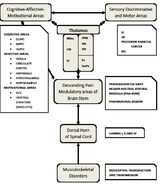
Figure 2.1: Nociception, central nervous system and musculoskeletal disorders
Nociception results from the chemical, mechanical and thermal stimulation and transduction of the nociceptors. Nociceptive information is processed in series and parallel within two brain areas. The lateral nociceptive system involves S1, SII involved in sensory discriminative aspects of the stimulus. The medial nociceptive system involves areas in the pre-frontal cortex, limbic and mesolimbic areas involved in the cognitive-affective and motivational aspects related to pain. Areas in the medial nociceptive system influence both descending modulatory systems and sensorimotor processes. DLPFC: Dorsolateral prefrontal cortex; VLPFC:
Ventrolateral prefrontal cortex; MPFC: Medial Prefrontal Cortex; OFC: Orbital Prefrontal Cortex; NACC: Nucleus Accumbens; VTA: Ventral Tegmental Area: VPL:
ventroposterolateral nucleus (VPL); VPM: ventroposteromedial nucleus; VPI: ventroposteroinferior nucleus; PO: posterior nuclei, VmPO: posterior division of the
ventromedial nucleus; MDvc: ventral region of the dorsal medial nucleus; CM: centromedial nucleus: lateral central nucleus.
16
An extensive body of evidence supports the presence of psychosocial risk factors associated with pain and disability. Perceived pain intensity (Truchon 2001; Denison et al. 2004; Casey et al. 2008), depression (Creamer et al. 1999; Pincus et al. 2002; Casey et al. 2008; Vranceanu et al. 2009; Wideman et al. 2012; Ross et al. 2015), low self-efficacy (Creamer et al. 1999; Arnstein et al. 1999; Asghari et al. 2001; Denison et al. 2004; Meredith et al. 2006; Wright et al. 2008; Vranceanu et al. 2009; Lee et al. 2015), resilience (Wright et al. 2008; Vranceanu et al. 2009), somatosization (Pincus et al. 2002), catastrophization (Denison et al. 2004; Vranceanu et al. 2009; Wideman and Sullivan 2012), fear (Denison et al. 2004; Vranceanu et al. 2009; Wideman and Sullivan 2012; Lee et al. 2015), distress (Pincus et al. 2002; Vranceanu et al. 2009; Lee et al. 2015), anxiety (Creamer et al. 1999; Vranceanu et al. 2009), passive coping (Truchon 2001; Vranceanu et al. 2009), work dissatisfaction (Truchon 2001), monotonous work (Truchon 2001; Östergren et al. 2005) and pain related cognitions (Casey et al. 2008; Vranceanu et al. 2009) are all documented risk factors for pain and disability. Factors such as catastrophization (Wertli et al. 2014), self-efficacy (Arnstein et al. 1999; Denison et al. 2004; Meredith et al. 2006; Wright et al. 2008; Ross et al. 2015; Lee et al. 2015), fear (Denison et al. 2004; Lee et al. 2015), anxiety (Meredith et al. 2006) and psychological distress (Lee et al. 2015) appear to play important mediating roles between pain and disability and the literature suggests that these factors are part of the causal process. Collectively, these studies demonstrate that psychological factors influence the pain experience and are implicated in the disability that persons with MSD experience.
2.1.3.2.1 Psychological factors and forebrain cortical activity
Psychological factors are mediated by forebrain processes, which often involve many of the same forebrain regions involved in the pain experience (Price et al. 2012; Simons et al. 2014). Brain regions involved with depression include a network of structures equally implicated in chronic pain including the medial prefrontal cortex, limbic, striatal, thalamic, and basal forebrain structures (Price and Drevets 2012; Simons et al. 2014). Increased activity in the cingulate cortex and hippocampus as well as the prefrontal cortex is found when a negative mood is induced in healthy subjects (Berna et al. 2010). Neural mechanisms involving the association of external stimuli with fear (i.e. fear learning), for example the association of