Université de Montréal
Regulation of the inositol 1,4,5-trisphosphate receptor 1 (IP3R1) by microRNA-26a in atrial fibrillation
Par
Faezeh Vahdatihassani
Faculté de Médecine
Mémoire présenté en vue de l’obtention du grade de Maître és Sciences (M.Sc.) en Sciences Biomédicales, option Médecine Expérimentale
Août 2020
Université de Montréal
Unité académique : Institut de Cardiologie de Montréal, Faculté de Médecine
Ce mémoire intitulé
Regulation of the inositol 1,4,5-trisphosphate receptor 1 (IP3R1) by microRNA-26a in atrial fibrillation
Présenté par
Faezeh Vahdatihassani
A été évalué(e) par un jury composé des personnes suivantes
Dr. Yahye Merhi Président-rapporteur Dr. Stanley Nattel Directeur de recherche Dr. Alvin Shrier Membre du jury
Résumé
Contexte: La physiopathologie de la fibrillation auriculaire (FA) a été caractérisée par des changements de concentration cellulaire de Ca2+ et des processus connexes menant à l'apparition
et au maintien de la maladie. Les récepteurs de trisphosphate d'inositol (IP3R) sont des canaux calciques ligand-dépendants pour lesquels la surexpression dans la FA a été liée à un remodelage cardiaque. Les microARN (miR, miARN), petits ARN non codants, sont d'une longueur d'environ 22 nucléotides et régulent l'expression des gènes par déstabilisation de l'ARN ou inhibition de sa traduction. De plus en plus de preuves ont été apportées sur le rôle des miARN dans la physiopathologie des troubles cardiaques, y compris le remodelage défavorable induit par la FA. Objectif: Notre laboratoire a montré que le niveau nucléaire IP3R1 est régulé à la hausse dans le modèle canin de FA, ce qui produit une augmentation de la charge nucléaire en calcium. Cette étude vise donc à étudier le rôle des miARN dans la régulation d'IP3R1 qui initie et/ou perpétue la FA dans les cardiomyocytes auriculaires du modèle de FA chez le chien.
Méthodes: Nous avons utilisé un modèle canin de AF établi par méta-cardiographie auriculaire pendant 600 bpm × une semaine; des cœurs perfusés par Langendorff pour isoler les cardiomyocytes auriculaires pour des expériences moléculaires; le criblage des miRs qui ciblent le gène ITPR1, codant IP3R1, en utilisant des bases de données en ligne; RT-qPCR pour mesurer l'expression de l'ARNm de ITPR1 et confirmer le niveau d'expression des miARN criblés; l'analyse Western Blot pour évaluer le niveau de protéine d’IP3R1; le test de la double luciférase reporter, la surexpression et l'abattement des miARN en culture primaire de cardiomyocytes isolées ou de lignées cellulaires appropriées; et l'imagerie par fluorescence calcique Fluo-4 AM pour évaluer le rôle potentiel des miARN sur la manipulation du Ca2+. Pour les expériences de manipulation des
miARN, les cellules ont été transfectées avec 1) un miARN non codant (miR-NC, groupe témoin), 2) un miARN mimétique et 3) un inhibiteur du miARN (AMO). La signification statistique est calculée avec le test t de Student ou l'analyse unidirectionnelle de variance (ANOVA) suivie par le test de Tukey à comparaisons multiples en utilisant le logiciel GraphPad Prism version 6.00.
Résultats: Nos données indiquent une augmentation du niveau de la protéine IP3R1 sans changement apparent de l'expression du gène ITPR1 dans les cardiomyocytes de l'oreillette gauche par rapport à notre modèle canin de FA. Sur la base de l'analyse informatique, il a été prédit que miR-26a ciblerait l'ARNm de l'ITPR1. La FA a considérablement réduit la régulation du miR-26a dans les cardiomyocytes de l'oreillette gauche. Le dosage de la double luciférase reporté dans les cellules H9C2 a montré que le miR-26a agissait directement sur la région non traduite 3′ (3′UTR) de l'ARNm ITPR1. De plus, la surexpression de miR-26a a réduit le niveau de la protéine IP3R1 et a diminué le taux diastolique [Ca2+] dans le noyau et le cytosol des cardiomyocytes de
chien, des transistors de Ca2+ stimulés électriquement; tandis que le knockdown de miR-26a a
inversé ces effets. L'expression de l'ARNm de l'ITPR1 est restée inchangée dans les cardiomyocytes de chien isolées après la transfection avec l'imitateur et l'inhibiteur de l'ARNm. Conclusion: La régulation à la hausse d'IP3R1 dans la FA est due à l'inhibition de la traduction par le miR-26a, qui est régulé à la baisse dans les cardiomyocytes auriculaires du modèle canin de FA. Ce changement est associé à une altération de la manipulation du Ca2+, qui se traduit par une
augmentation des taux de Ca2+ diastolique nucléaire. Nos résultats suggèrent que la régulation à
la baisse de miR-26a augmente l'expression de l’IP3R1, contribuant au remodelage pro-arythmique dans la FA.
Mots-clés: microARN, homéostasie calcique, physiopathologie cardiaque, fibrillation auriculaire, remodelage cardiaque, IP3R1.
Abstract
Background: The pathophysiology of atrial fibrillation (AF) has been characterized by changes in the cellular concentration of Ca2+ and related processes leading to the initiation and maintenance
of the condition. Inositol trisphosphate-receptors (IP3Rs) are ligand-gated calcium channels for which overexpression in AF has been linked to cardiac remodeling. microRNA (miR, miRNA)s, small non-coding RNAs, are around 22 nucleotides in length and regulate gene expression by mRNA destabilization or inhibition of its translation. A growing body of evidence has emerged about miRNA's role in the pathophysiology of cardiac disorders, including AF-induced adverse remodeling.
Objective: Our laboratory has shown that nuclear IP3R1 level is upregulated in the dog AF model, producing increased nuclear calcium loading. Hence, this study aims to investigate the role of miRNAs in the regulation of IP3R1 initiating and/or perpetuating AF in atrial cardiomyocytes of the dog AF model.
Methods: We used AF dog model established by atrial-tachypacing for 600 bpm × one week; Langendorff-perfused hearts to isolate atrial cardiomyocytes for molecular experiments; screening miRs that target ITPR1 gene, encoding IP3R1, using online databases; RT-qPCR to measure ITPR1 mRNA expression and confirm the expression level of the screened miRNAs; western blot analysis to evaluate the protein level of IP3R1; dual-luciferase reporter assay, overexpression and knockdown of miRNAs in primary culture of isolated cardiomyocytes or appropriate cell lines; and Fluo-4 AM calcium fluorescence imaging to assess the potential role of the miRNA on Ca2+ handling. For miRNA manipulation experiments, cells were transfected with 1)
non-coding miRNA (miR-NC, control group), 2) miRNA mimic, and 3) inhibitor of the miRNA (AMO). Statistical significance is calculated with Student's t-test or one-way analysis of variance (ANOVA) followed by Tukey's multiple comparisons test using GraphPad Prism software version 6.00.
Results: Our data indicated a rise in IP3R1 protein level with no apparent change in ITPR1 gene expression in left atrial cardiomyocytes from our dog AF model. Based on the computational
analysis, 26a was predicted to target the ITPR1 mRNA. AF significantly downregulated miR-26a in left atrial cardiomyocytes. The dual-luciferase reporter assay in H9C2 cells showed that miR-26a directly acted on the 3′ untranslated region (3′UTR) of ITPR1 mRNA. In addition, miR-26a overexpression reduced the IP3R1 protein level and decreased the diastolic [Ca2+] in both nucleus
and cytosol of the electrically-stimulated Ca2+ -transients, dog cardiomyocytes, while miR-26a
knockdown reversed these effects. ITPR1 mRNA expression remained unaltered in isolated dog cardiomyocytes after transfection with the miRNA mimic and inhibitor.
Conclusion: IP3R1 upregulation in AF is due to translation inhibition by miR-26a, which is downregulated in the atrial cardiomyocytes of the dog AF model. This change is associated with altered Ca2+ handling, reflected as enhanced nuclear diastolic Ca2+ levels. Our results suggest that
miR-26a downregulation enhances the IP3R1 expression, contributing to pro-arrhythmic remodeling in AF.
Keywords: microRNA, calcium homeostasis, cardiac pathophysiology, atrial fibrillation, cardiac remodeling, IP3R1.
Table of contents
Résumé ... 5 Abstract ... 7 Table of contents ... 9 List of tables ... 13 List of figures ... 15List of acronyms and abbreviations ... 17
Acknowledgments ... 21
Chapter 1 –Introduction ... 23
Atrial Fibrillation: definition and potential consequences ... 23
AF risk factors ... 23
AF and concomitant cardiac diseases ... 24
AF Pathophysiology ... 25
Autonomic neural dysregulation ... 27
Structural remodeling ... 27
Electrical remodeling or ion channel dysfunction ... 28
Ca2+-handling abnormalities ... 28
Inositol 1,4,5 trisphosphate receptor (IP3R) ... 29
Structure... 29
Distribution ... 30
IP3R role in cardiac function and pathological cardiac remodeling ... 31
IP3 signaling cascade ... 32
IP3R function in cardiomyocytes ... 33
Role of IP3R signaling in cardiac diseases ... 33
miRNAs and post-translational regulation in cardiac disease ... 34
miRNA nomenclature... 35
Canonical biogenesis pathway and regulation of gene expression ... 35
Roles of miRNAs in cardiac development and function ... 36
miRNAs in AF pathophysiology: electrical and structural remodeling ... 37
miRNAs as biomarkers in AF ... 40
Rationale, hypothesis, and objectives ... 42
Research Framework ... 43
Contribution of the Author ... 43
Chapter 2 – Methods ... 45
Canine atrial fibrillation model... 45
Isolation of dog atrial cardiomyocytes ... 46
Cell culture ... 47
Synthesis and manipulation of miR-26a mimic and inhibitor ... 47
Confirmation of miR-26a targeting site by luciferase assay ... 48
Protein expression analysis ... 48
Gene and miRNA expression analysis ... 49
Confocal imaging of nucleoplasmic and cytoplasmic calcium transients ... 50
Statistical analysis ... 50
Chapter 3 – Results ... 53
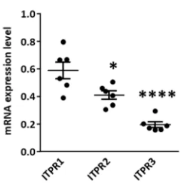
ITPR1 shows higher expression in atrial cardiomyocytes ... 53
Bioinformatic analysis of candidate miRNAs and selection of miR-26 ... 56
Validation of miR-26a as a potential regulator of ITPR1 ... 58
miR-26a and Calcium transients... 62
Chapter 4 – Discussion... 65
Possible causative mechanism of IP3R upregulation in AF ... 65
miRNA dysregulation in AF and contribution to IP3R upregulation ... 66
miRNA dysregulation of calcium in cardiomyocytes: possible roles in AF pathophysiology .... 71
Novel elements and significance ... 73
Limitations ... 74
Chapter 5 – Future research and Conclusions ... 77
Future research ... 77
In vivo consequences of miR-26a dysregulation and role in AF: ... 77
Probing the consequences of nuclear Ca2+ loading and HDAC export for the molecular control of AF ... 78
Conclusions ... 80
References ... 81
List of tables
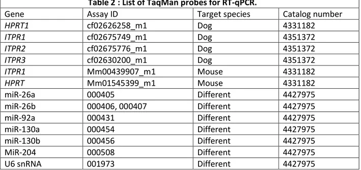
Table 1: List of RNA/DNA sequences (5′-sequence-3′) used in luciferase activity assay and transfection studies. ... 48 Table 2: List of TaqMan probes for RT-qPCR. ... 50 Table 3: List of miRNAs that their roles have been described in AF ... 69
List of figures
Figure 1: Interactions between atrial fibrillation (AF) and underlying illnesses (10, 11). ... 24
Figure 2: Major pathophysiological processes contributing to AF. ... 26
Figure 3: Upregulation of IP3R1/2 protein levels and alterations in resting [Ca2+] in atrial cardiomyocytes of the canine AF model. ... 31
Figure 4: Schematic representation of the research framework. ... 43
Figure 5: In vivo model of atrial fibrillation. ... 45
Figure 6: Left atrial cardiomyocyte isolation experimental setup and protocol ... 46
Figure 7: Differential expression of the 3 isoforms of ITPR in the left atrial cardiomyocytes of control dogs. ... 53
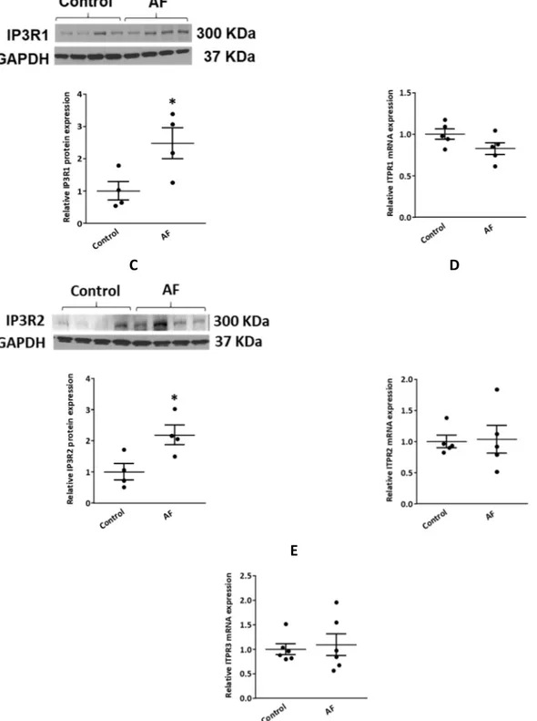
Figure 8: Upregulation of IP3R1/2 protein expression in the AF dog model. ... 55
Figure 9: Schematic of selection criteria for candidate miRNAs predicted to target ITPR1. ... 57
Figure 10: Expression of various miRNAs predicted to target ITPR1 in AF and control atrial cardiomyocytes. ... 58
Figure 11: miR-26a is a potential candidate predicted to target ITPR1. ... 59
Figure 12: Confirmation of the regulation of IP3R1 expression by miR-26 in the HL-1 cell line. .. 60
Figure 13: Confirmation of regulation of IP3R1 expression after transfection with miR-26a and AMO-26a in atrial cardiomyocytes. ... 62
Figure 14: Effect of miR-26a overexpression and knockdown on Ca2+ transients (CaTs) in atrial cardiomyocytes. ... 64
Figure 15: Molecular consequences of downregulated miR-26a in AF. ... 74
List of acronyms and abbreviations
2-APB: 2-Aminoethoxydiphenyl borate 3′UTR: 3′ untranslated region
AF: atrial fibrillation AGO: Argonaute
AMO: anti-miRNA oligonucleotide AP: action potential
APD: action potential duration AT-II: angiotensin II
BNP: brain natriuretic peptide
CABG: coronary artery bypass grafting
CACNA1C: voltage-gated channel subunit alpha1 C CaMKII: Ca2+/calmodulin-dependent protein kinase II
CaT: Ca2+ transient
CHD: congenital heart disease CTL: control
Cx: connexin
DAD: delayed afterdepolarizations DAG: diacylglycerol
DGCR8: DiGeorge syndrome critical region gene 8 DNA: deoxyribonucleic acid
ER: endoplasmic reticulum ET-1: endothelin-1
GAPDH: glyceraldehyde-3-phosphate dehydrogenase HDAC: histone deacetylase
HEPES: (4-(2-hydroxyethyl)-1-piperazineethanesulfonic acid HF: heart failure
HPRT1: hypoxanthine-guanine phosphoribosyltransferase ICaL: L-type Ca2+ current
IK1: inward rectifier background K+ current
IKACh: acetylcholine-regulated K+ current
IP3: inositol 1,4,5-trisphosphate IP3R: IP3 receptor
IV: intravenous
KCNJ2: potassium inwardly-rectifying channel subfamily J member 2 LA: left atrium
LTCCs: L-type Ca2+ channels
MEF2: myosin enhancer factor 2 MHC: myosin-heavy chain miR, miRNA: microRNA miR-NC: non-coding miRNA mRNA: mature ribonucleic acid NCX: sodium/calcium exchanger
NFAT: nuclear factor of activated T cells PLC: phospholipase C
POAF: postoperative atrial fibrillation RISC: RNA-induced silencing complex RyRs: ryanodine receptors
SERCA2a: SR Ca2+-ATPase 2a
SR: sarcoplasmic reticulum
Acknowledgments
I would like to express my deepest gratitude to professor Stanley Nattel who is “a man of heart, in every sense of the word”. He has been the best mentor for me. He has encouraged me in all aspects of my research and allowed me to grow as a student. I would like to thank him for the opportunity he has given me in his laboratory and the invaluable guidance and feedback.
I would like to pay my special regards to professor Yahye Merhi, director of the program of Biomedical Sciences, for the invaluable guidance and continuous encouragement.
I am also grateful to my committee member, professor Catherine Martel at Montreal Heart Institute. I am thankful to her for her guidance, support, and encouragement extended to me. I would like to thank my thesis jury member, professor Alvin Shrier for the comments and feedback that have been absolutely invaluable.
I would like to thank Dr. Xiaoyan Qi, Dr. Jiening Xiao, Dr. Feng Xiong, Dr. Roddy Hiram, and Louis Villeneuve for teaching invaluable knowledge and experience in the laboratory, all their support, being a friend, and for all the discussions in science. I wish to thank Nathalie L’Heureux, Chantal St.-Cyr, and Jennifer Bacchi for helping me with many things during my work in the laboratory and my study and whose technical and secretarial assistance were a milestone in the completion of this project. I would also like to acknowledge my colleagues Dr. Mozhdeh Mehdizadeh, Dr. Raidan Alyazidi, Fatima Hubaishi, and Dr. Anna Garcia-Elias, Dr. Patrice Naud, Xixiao Li, Dr. Abhijit Takawale, Yasemin Altuntas, and Dr. Donghai Liu for their help, support, and discussions in science.
I would like to thank my family, especially my mother and father, my sister, Fatemeh, and my husband, Mohammad, for their support, attention, and encouragement during my study.
Chapter 1 –Introduction
Atrial Fibrillation: definition and potential consequences
Atrial fibrillation (AF) is the most common type of sustained arrhythmia, with an estimated lifetime risk of 22% to 26%. AF is associated with pronounced population morbidity and mortality and reduction in quality of life and functional status (1). According to the 2014 AHA/ACC/HRS Guideline, AF is defined as “a supraventricular tachyarrhythmia with uncoordinated atrial activation and consequently ineffective atrial contraction” (2). In the electrocardiogram, AF is recognized by the absence of repeating P waves replaced by irregular atrial activity and irregular R-R intervals unless the patient has atrioventricular (AV) block (2). Atrial fibrillation can result in hemodynamic changes that are often clinically significant. Potential consequences vary for individual patients and are fatigue, which is the most common symptom, palpitations, fall in blood pressure, syncope, and dyspnea (2). AF is associated with heart failure (HF), and individuals with either condition could develop the other (3). Patients with AF can develop either too rapid or too slow ventricular rate, uncoordinated atrial contraction, and beat-to-beat variability in the ventricular filling, leading to hemodynamic consequences (2).
AF risk factors
Numerous risk factors are associated with an increased risk of developing AF. Nevertheless, various studies have shown that many of these risk factors are reversible, making it possible to prevent and manage some cases of AF through risk modification. Non-cardiac and cardiothoracic surgery, obesity, pneumonia, hyperthyroidism, hypertension, diabetes, pericarditis, myocarditis, HF, electrocution, diabetes mellitus, binge drinking, tobacco use, obesity, and many more have been reported as reversible causes of AF (2, 4).
Age is regarded as the most critical risk factor for AF in the general population. Evidence suggests that over 30% of patients diagnosed with AF are 80 years of age and older, and up to 16% are 75 to 84 years old, while approximately 1% of affected individuals are aged <60 years (2).
The results of the ATRIA study in the US showed that the prevalence of AF in the study population (1.89 million) was 0.95%, of which 45% were 75 years of age. This study projects that more than 5.6 million people will have AF by the year 2050, and 50% will be ≥80 years old (5). There is also a gender-dependent difference in the incidence of AF and, accordingly, the Framingham Heart Study has reported that the lifetime risk for development of AF after 40 years of age is 26% for men and 23% for women (6). Similar findings were also reported by the ATRIA study (5).
AF and concomitant cardiac diseases
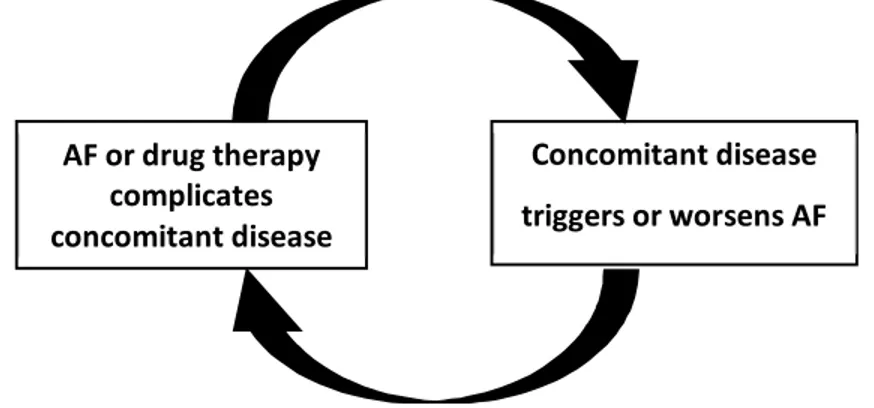
Although AF is known to be a final common endpoint of atrial remodeling resulting from heart diseases, it can also be, in turn, a cause of electrophysiological changes and cardiac remodeling that explain the progressive nature of arrhythmia (7-9).
AF often complicates or exacerbates the underlying heart diseases or non-cardiac illnesses (2, 10). It seems possible that other critical illnesses cause changes in the underlying rhythm, which complicate the patient's problem and triggers recurrent AF (Figure 1) (10, 11). In asymptomatic AF patients as initially, the ventricular rate is not adequately controlled; they may develop tachycardia-induced ventricular dysfunction and HF (12). Worsened HF increases atrial stretch and heightens sympathetic tone making the AF more resistant to rate-control or rhythm-control treatments (11).
Figure 1: Interactions between atrial fibrillation (AF) and underlying illnesses (10, 11).
AF is a potent and independent risk factor for stroke, increasing its risk by approximately 5–fold throughout all ages. Detection of AF, even in its asymptomatic stage, is the basis of the decision
AF or drug therapy complicates concomitant disease
Concomitant disease triggers or worsens AF
to implement anticoagulant therapy to prevent stroke (13). It has been shown that the left atrial appendage is the area where the thrombi are usually formed in AF (14). Impaired left ventricular (LV) ejection fraction (≤35%) and/or HF are important risk factors for stroke in AF patients as predicted by ACC/AHA/ESC guidelines (15).
AF can complicate valvular heart diseases, particularly left-side lesions. Aortic or mitral regurgitation/stenosis results in left atrial volume and pressure overload and, subsequently, progressive atrial dilation. The chronic structural changes in the left atrium promote AF by increasing atrial fibrosis and electrophysiological remodeling. Importantly AF emerges as a marker of adverse cardiovascular events, and AF patients with mitral valve disease face an elevated risk of mortality related to an increased risk of stroke (10).
Hypertrophic cardiomyopathy (HCM) is a genetic disorder that results from several mutations in genes involved in muscle contraction and characterized by unexplained LV hypertrophy (16). AF is also common in HCM patients and is related to left atrial dilatation and remodeling (17). Patients with congenital heart disease (CHD), which affects 4-10 per 1000 live births (18), seem to be at higher risk of developing AF. Atrial arrhythmias occur in CHD patients due to comorbidities, persistent volume/pressure overload, and scars of surgical procedures and increase the risk of morbidity and mortality to great extent. Recent studies have reported a significantly higher risk of ischemic stroke, developing HF, and death in CHD patients who developed AF compared with patients who did not (19).
AF Pathophysiology
AF is maintained by multiple-circuit re-entry and/or repetitive and rapid firing of ectopic atrial foci, which mostly arise from delayed depolarizations (DADs) and, in some cases, early after-depolarizations (1). DADs are thought to be caused during diastole by the abnormal release of calcium from the sarcoplasmic reticulum (SR). Ryanodine receptors (RYRs) are the SR Ca2+ release
channels that release Ca2+ during excitation-contraction coupling and are normally closed during
diastole. Channel dysfunction and Ca2+ overload in SR contribute to the Ca2+ leakage from RyR2
DAD. This diastolic calcium release by leaky RyR2 activates a net depolarizing inward positive-ion current and DADs through the sodium/calcium exchanger (NCX), which exchanges intracellular 1 Ca2+ ion for 3 extracellular Na+ ions (20).
Four major pathophysiological processes have been suggested to contribute to AF and account for the induction of focal ectopic activity and re-entrant circuit, and include electrical remodeling or ion channel dysfunction, structural remodeling, autonomic neural dysregulation, and Ca2+
-handling abnormalities (Figure 2) (1).
Findings of different studies imply that in addition to other cardiac diseases and conditions, AF itself can be the cause of these underlying contributors, which promotes the development of AF, and this vicious cycle is called “AF begets AF” (8). There has been a pile of evidence that confirms the central role of abnormal Ca2+-handling in AF-pathophysiology. Regarding this, we review the
studies in the area of AF-related Ca2+-handling abnormalities, with particular focus on the
contributions of IP3Rs. Also, the other contributing mechanisms are briefly summarized.
Figure 2: Major pathophysiological processes contributing to AF.
Ca2+ handling abnormalities have emerged as the central mechanism to the pathophysiology of
AF, leading to delayed afterdepolarizations (DADs) that cause focal ectopic firing. Inositol trisphosphate-receptors (IP3Rs) are ligand-gated calcium channels overexpressed in AF,
contributing to cardiac remodeling. RyRs: ryanodine receptors; SR: sarcoplasmic reticulum; CaMKII: Ca2+/calmodulin-dependent protein kinase II (8).
Autonomic neural dysregulation
The relationship between intrinsic cardiac nerve activity and spontaneous arrhythmias has attracted considerable attention. Some investigations have suggested that atrial arrhythmia can be generated due to the synergistic work and close anatomical association between muscle fibers and nerves. Accordingly, rapid atrial pacing in an animal model could produce autonomic remodeling (21). Shortening of the atrial effective refractory period by vagal and sympathetic activation can also be regarded as an important mechanism promoting AF induction and maintenance. Furthermore, left atrial hyperinnervation has been suggested to be important in AF induced by atrial tachycardia remodeling (22).
Structural remodeling
Extensive evidence indicates that AF-related structural remodeling is characterized by atrial enlargement and tissue fibrosis. Transforming growth factor (TGF)-β1, angiotensin II (AT-II), connective tissue growth factor, and platelet-derived growth factor are important profibrotic molecules involved in the atrial tissue fibrosis (23, 24). Although it has not been fully understood, several explanations have been put forward to justify the deleterious effects of atrial fibrosis in AF. Atrial fibrosis has been shown to cause local conduction disturbances and unidirectional block. It increases the number of fibroblasts, which produce large quantities of collagen, disturbing electrical continuity (25). Co-cultured preparations of fibroblasts and cardiomyocytes revealed that fibroblast coupling to cardiomyocytes could alter the electrical activity of cardiomyocytes leading to spontaneous impulse formation, abnormalities in cardiac conduction, and arrhythmias (25). Ca2+ signals are also essential for diverse functions in fibroblasts. For example, the receptor
binding of AT-II activates the phospholipase C (PLC) pathway leading to diacylglycerol (DAG) and IP3 production. IP3 binds to the IP3R inducing Ca2+ release from the endoplasmic reticulum (ER is
analogous to the SR in cardiomyocytes), and DAG can directly activate specific Ca2+-permeable
transient receptor potential (TRP) channels leading to Ca2+ entry into fibroblasts (26). Findings of
membrane or increased Ca2+ release from the ER cause enhancement of proliferation and
fibroblast differentiation into myofibroblasts promoting fibrosis in AF (27).
Electrical remodeling or ion channel dysfunction
Pumps, ion channels, and exchangers are considered essential components of cardiac electrophysiology that can be altered by atrial remodeling.
Atrial tachycardia during AF has been shown to downregulate L-type Ca2+ current (I
CaL) carried by
L-type Ca2+ channels (LTCCs). The Ca2+ overload, which is caused by a high atrial rate, activates
the Ca2+-dependent calmodulin–calcineurin–nuclear factor of activated T cells (NFAT) system and
enhances NFAT translocation into the nucleus. NFAT is a transcription factor that decreases the transcription of the calcium voltage-gated channel subunit alpha1 C (CACNA1C) gene encoding
Cav1.2 LTCCs, thus reducing ICaL. Downregulation of ICaL mitigates inward Ca2+ current, which
causes shortening of the action potential (AP) duration (APD) and thereby promotes re-entry (28). Another principal component of electrical remodeling is the upregulation of inward-rectifier K+
current (IK1) and acetylcholine-regulated K+ current (IKACh). IK1 is mainly composed of Kir2.1
subunits contributing to the resting potential and phase 3 repolarization. Evidence shows that IK1
is increased in chronic AF, which is an important determinant of AF-maintaining re-entry (29). As seen in AF, atrial tachycardia has been suggested to increase the IKACh leading to reductions in APD
(30). Alternation in expression and distribution of main atrial gap junction protein such as connexin (Cx) 40 and Cx43 connecting cardiomyocytes electrically can also occur in AF and contribute to electrical remodeling. Connexin gene transfer has been shown to improve atrial conduction and reduce AF (31).
Ca
2+-handling abnormalities
Despite the considerable physiologic potential, Ca2+ signaling has a central role in atrial
remodeling and AF pathophysiology. During each AP in a healthy heart, Ca2+ enters
cardiomyocytes through LTCCs and then triggers Ca2+ release from SR mainly through RYR2.
Atrial tachycardia and increased reactive oxygen species in AF result in the persistent Ca2+
overload, which activates Ca2+/calmodulin-dependent protein kinase II (CaMKII). This kinase
regulates the activities of the proteins involved in Ca2+ handling LTCC, RyR2, and phospholamban
(33, 34). CaMKII-dependent RyR2 hyperphosphorylation results in pronounced SR Ca2+-leak,
which, together with greater depolarizing inward NCX current and increased intracellular Ca2+
membrane voltage coupling gain, enhance the risk of potentially-arrhythmogenic DADs in AF (35). Two forms of intracellular Ca2+release channels regulate the intracellular Ca2+concentration: RyR
and IP3Rs. RYRs have been suggested to have a key role in Ca2+ release from SR during
excitation-contraction coupling. IP3 induces intracellular Ca2+ release from IP3Rs that mediate the hormonal
regulation of cardiac contraction. Cardiomyocytes express IP3Rs less abundantly compared to RYRs. However, the upregulation of IP3R expression in atrial tissue of patients with chronic AF is thought to contribute to intracellular Ca2+abnormalities and Initiation and perpetuation of AF (36,
37).
As previously stated, the intracellular calcium level plays a central role in the activation of cell signaling, which mediates pro-fibrillatory atrial remodeling. In this regard, we aim to determine if a specific miRNA can contribute to the dysregulation of IP3Rs in AF. Below is a description that briefly summarizes the roles of IP3R in cardiac physiology and pathology with the emphasis on AF.
Inositol 1,4,5 trisphosphate receptor (IP3R)
Structure
The IP3R is a ligand-gated calcium channel activated by inositol trisphosphate (IP3). Molecular cloning studies have demonstrated that IP3R is comprised of three types that are encoded by distinct genes: ITPR1, ITPR2, and ITPR3. Each isoform is about 300 KDa and forms homo- and heterotetramers, divided into three functionally active domains: N-terminal ligand-binding domain, C-terminal helical domain, and modulatory domain. The N-terminus has a ligand-binding domain for IP3 and a suppressor domain. C-terminal domain has six transmembrane α-helices that are packed together to form the ion-conducting pore and C-terminal tail. The C-terminal tail
modulates gating, and It is also a site for post-translational modifications and binding of multiple ligands that modulate the channel function. The modulatory domain, located between N-terminus and the C-N-terminus channel domain, is a binding site for a variety of molecules regulating the channel function and for post-translational modifications. Three IP3R isoforms share similarities, and the channel domain is well conserved (65-70%) among them (38, 39).
Distribution
IP3Rs are ubiquitously expressed in all tissues and primarily localized to the ER/SR. It became apparent that most cell types express more than one type of IP3R, and the subcellular distribution of IP3Rs depends on the cell type. The Golgi apparatus, secretory vesicles, plasma membrane, or other specialized membranes such as perinuclear or nuclear membrane may contain IP3Rs (40). Western blot, IP3-binding, and PCR analysis in Lipp et al. study indicated that the three IP3R isoforms are expressed in both atrial and ventricular myocytes, and types I and II IP3Rs are the most abundant species (41). It has been suggested that IP3R1 is dominant in human atrial myocytes and rat Purkinje myocytes, whereas IP3R2 is predominantly expressed in ventricular and atrial myocytes of the other animal species (42). Although all types of IP3Rs are expressed in the heart, only a few investigations assessed the role of type 1 and 3 (39).
Results of a co-immunostaining of atrial myocytes suggest that IP3R2 is mainly expressed in the subsarcolemmal region and is colocalized with the junctional RyR2. Also, in electrically paced atrial myocytes, the IP3 ester increased the action potential-evoked Ca2+ transient (CaT),
indicating that IP3Rs could modulate excitation-contraction coupling (41). Electron microscopy results suggested that IP3R1 is highly found in the intercalated discs of the ventricular and atrial cardiomyocytes (43). Previous work has also shown that IP3Rs are highly concentrated in the nuclear envelope and perinuclear membranes. Local Ca2+ release from IP3Rs activates the gene
expression or drives pathological cardiomyocyte growth (44, 45).
Results of a recent study suggest that IP3Rs are expressed in both nuclear and non-nuclear fractions of isolated canine atrial cardiomyocytes. Upon IP3 stimulation in this system, increased resting [Ca2+]
Figure 3: Upregulation of IP3R1/2 protein levels and alterations in resting [Ca2+] in atrial
cardiomyocytes of the canine AF model.
nuclear pore complex
ine-scan imaging of cytosolic and nucleoplasmic of resting Ca2+in one permeabilized
CTL atrial CM before and after 20 µM IP3. (C): Effect of IP3 and IP3+2-aminoethoxydiphenyl borate (2-APB) (50 µM) on CTL atrial CM resting [Ca2+]
nuc and [Ca2+]Cyto. (D): Line - scan imaging
of cytosolic and nucleoplasmic CaTs in permeabilized AF atrial myocyte before and after IP3.
(F): Effect of IP3 and IP3 + 2APB (50 µM) on AF atrial CM resting [Ca2+]nuc and [Ca2+]Cyto. Data are
shown as mean±SEM.
IP3R role in cardiac function and pathological cardiac remodeling
IP3Rs are involved in the regulation of cardiomyocytes function and the pathogenesis of heart diseases. Although IP3Rs are the main channels regulating intracellular Ca2+ in many cells, RYRs
are the dominant channels of calcium release in the heart (39). Therefore, many investigations A
have focused on the RYRs signaling in cardiomyocytes and made the studies on IP3Rs roles challenging.
IP3 signaling cascade
In cardiomyocytes, hormonal factors such as endothelin-1 (ET-1), many transmitters, and stimuli such as stretch activate the IP3-induced Ca2+ release. IP3 is an intracellular messenger and is
generated through the activation of plasma membrane receptors coupled to PLC. Receptors such as receptor tyrosine kinases and G protein-coupled receptors can activate PLCs and increase the Ca2+ release downstream of PLC activation. The PLC enzyme liberates the signaling molecules IP3
and DAG from phosphatidylinositol 4,5-bisphosphate (PIP2). The hydrophilic IP3 then translocates from the membrane to the cytoplasm and binds to its main target IP3R mostly on SR and causes a conformational change leading to Ca2+ release (42). The nuclear envelope is also able to store
Ca2+ and has been shown to have the entire components of the IP3-mediated Ca2+ signaling
network (47). Previous studies have revealed that G protein-coupled receptors are also found in the nuclear envelope and involved in Ca2+ release from the nuclear membrane into the
nucleoplasm associated with IP3 signaling (48).
Role of IP3R signaling in the development of the heart
A plethora of investigations in embryonic stem cell or prenatal cardiac cells have suggested substantial roles for IP3Rs in cardiac development. IP3Rs have been demonstrated to be responsible for the early cycling of Ca2+ before the excitation-contraction coupling maturation in
developing myocytes. There is evidence indicating that the periodic Ca2+ oscillation inducing
spontaneous contractions in murine embryonic cardiomyocytes are dependent on the IP3 and sensitive to IP3R blocker 2-aminoethoxydiphenyl borate (2-APB) (49). In another survey on mice embryos using the IP3R1 probe, it was reported that IP3R mRNA is highly expressed in all tissues and the developing heart tube in the early stages of development (E8.5), while RYRs are not abundant. Data from the same study also suggests that the IP3-mediated Ca2+ signaling pathway
during the early stage of embryogenesis may be involved in cellular differentiation and apoptosis as part of the developmental programs (50). In addition, IP3R-mediated Ca2+ release in embryonic
differentiating cardiomyocytes (51). The role of iP3Rs in nodal cell automaticity in the developing atria has also been observed in the isolated atrial cells from E14.5 mouse embryos (52). Also, IP3R1-/- and IP3R2-/- double-mutant mice could not survive due to heart defects showing that they
play a crucial role in cardiogenesis (53). IP3R function in cardiomyocytes
IP3Rs have been shown to modulate cytosolic calcium levels in the heart, despite the lower mRNA levels (~50-fold) than RYRs (54). Application of IP3 to permeabilized chick atrial muscle was shown to induce transient increase in tension, which is suggested to be involved in the positive inotropic effect and contraction in the atrium (55). Studies also have highlighted the role of IP3R-dependent Ca2+ release in positive inotropic effects in ventricles by enhancing the systolic CaTs. In
permeabilized rabbit ventricular myocytes, IP3R activation by direct application of IP3 could increase the Ca2+ spark frequency (56). IP3Rs are located in the perinuclear and nuclear
membranes of cardiomyocytes. This localization of IP3Rs restricts the CaT to the nuclear matrix and activates a variety of transcription factors, subsequently initiating cardiac remodeling in response to hormonal stimuli (e.g., ET-1) (44).
Role of IP3R signaling in cardiac diseases
The IP3Rs expression levels are altered during pathological remodeling. Owing to their functions in Ca2+ release from the SR and the nuclear resources, IP3Rs can trigger arrhythmias and
hypertrophic gene transcription via several mechanisms.
Several lines of evidence have shown increased IP3R2 expression in hypertrophy observed in patients with ischemic dilated cardiomyopathy or animal models of hypertensive rats and aortically-banded mice. It is important to note that increased Ca2+ release through IP3Rs in
hypertrophic cardiomyocytes sensitizes neighboring RyRs opening leading to enhancement of extrasystolic Ca2+ transients giving rise to cardiac arrhythmia (57). As previously discussed, IP3Rs
are also responsible for the hypertrophic response of hormonal factors such as ET-1 and AT-II. The potent constrictor ET-1 activates the transcription of specific cardiac genes such as atrial natriuretic factor (ANF), a key marker of hypertrophy, and increases the assembly of myosin light chain-2 into sarcomeric units, which is a hallmark of the hypertrophic phenotype. Besides, these
effects of ET-1 are accompanied by the stimulation of inositol phospholipid hydrolysis, which results in the accumulation of inositol phosphates, including inositol monophosphate, inositol bisphosphate, and IP3 and DAG in cardiomyocytes (58). In a study by Higazi et al., induction of hypertrophic remodeling by ET-1 was shown to be dependent on IP3-induced Ca2+ release. They
reported that ET-1 induced elevation of the nuclear Ca2+ content by stimulating the Ca2+ release
from prenuclear IP3R2 and increased ANF expression by activating the calcineurin /NFAT pathway (59). In an investigation in IP3R2-deficient mice, the administration of ET-1 failed to increase diastolic Ca2+ concentration. It should be noted that no compensatory changes in protein
expression of type 1 and 3 were also observed in the global knockout of IP3R-2. Therefore, lack of IP3R2 was protective against the positive inotropic and arrhythmogenic effects of ET-1 (60). However, IP3R-2 knockout in a mouse model of dilated cardiomyopathy could not alter the hypertrophic response in pressure overload (61). Cao et al. also indicated higher levels of IP3R1 mRNA in the atrial tissue of AF patients, which correlated with AF duration (62). Similar results were also reported by Yamada et al. on IP3R1 protein and mRNA levels in right atrial samples from AF patients with mitral valvular disease (37). 2-APB, an inhibitor of both IP3Rs and TRP channels, has been shown to suppress the incidence and probability of sustained cardiac arrhythmia in the rabbit AF model (63). In a recent investigation in our laboratory, assessment of IP3R expression in left atrial cardiomyocytes from AF dogs indicated that IP3R1/2 levels were higher in cytosolic and nucleoplasmic fractions compared with control samples. IP3R1 level was markedly increased in both nuclear and non-nuclear regions, while IP3R2 was significantly upregulated in the non-nuclear fraction. The physiological agonist IP3 increased the resting [Ca2+]
in cytoplasm and nucleus of control and AF saponin-permeabilized atrial cardiomyocytes. However, the effect was greater in AF cells, especially in the nuclear region. In the presence of 2-APB, IP3 failed to increase the resting [Ca2+] in control and AF cardiomyocytes (Figure 3). These
results suggest that atrial cardiomyocyte Ca2+ handling is deranged in AF and that the principle
underlying mechanism is upregulation of IP3Rs.
miRNAs and post-translational regulation in cardiac disease
miRNAs are a class of genes that were discovered in Caenorhabditis elegans in 1993 by Lee et al. (64) and Wightman et al. (65). They suggested that lin-4 does not encode any protein and is
complementary to the LIN-14 3′UTR and negatively regulates its protein level and controls the postembryonic developmental events in C. elegans (64, 65).
Based on the latest data released by the miRBase database (v22), 38,589 hairpin precursor miRNAs have been found in 271 organisms. Precursor miRNAs (pre-miRNAs) are capable of producing 48,860 different mature miRNAs. The human genome is estimated to contain 1917 annotated pre-miRNAs and 2654 mature miRNAs sequences (66). miRNAs can be categorized based on their locations in the genome relative to intron and exon positions. Approximately half of the miRNAs are transcribed from polycistronic transcription units in which miRNAs loci are located in close proximity to each other. miRNAs loci are located in the intronic and exonic regions of protein-coding or non-protein-coding transcription units (67). The residing of the miRNAs within the introns of protein-coding genes depicts the dual performance of the genes and could explain the coordinated expression of mRNA and miRNAs modulating the same biological processes (68, 69).
miRNA nomenclature
According to a standard nomenclature system, the mature miRNAs are named with the prefix “miR-” followed by a number indicating the order of the miRNA discovery (e.g., miR-26). The first three letters, such as "hsa: human", before the prefix “miR”, indicate the species (hsa-miR-26). A letter after the number reflects the identical sequences differing in only one or two nucleotides (e.g., miR-26a or miR-26b). An additional dash-number suffix shows different pre-miRNAs that are processed into an identical miRNA (e.g., miR-26-1). The 3p and 5p demonstrate the 3’ and 5’ miRNAs (e.g. miR-26a-5p). A uniform system to annotate miRNAs has been described in Ambros et al. short article (70).
Canonical biogenesis pathway and regulation of gene expression
miRNAs are a class of small (~22 nucleotides [nt] in length) non-coding RNAs that negatively regulate gene expression and play key roles in biological processes and pathological conditions in plants and metazoans. Most miRNAs have multiple isoforms or isomiRs that can be generated through different mechanisms modifying mature miRNAs and precursor miRNAs (pre-miRNAs) (67, 71). The first step in miRNAs biogenesis is the generation of a hairpin-shaped transcript called
primary miRNA (pri-miRNA, 100 to 1,000 base pairs). Transcription is mostly mediated by RNA polymerase II (Pol II) and for a minor group of miRNAs by Pol III. The hairpin structures are cleaved into a ∼65 nucleotide pre-miRNAby nuclear RNase III enzyme Drosha and with the help of the cofactor DiGeorge syndrome critical region gene 8 (DGCR8) and then exported from the nucleus into the cytoplasm by the exportin 5. The cytoplasmic RNase III Dicer catalyzes the pre-mRNA splicing producing a mature ∼22-nt miRNA duplex. The RNA duplex incorporates into the Argonaute (AGO) to generate RNA-induced silencing complex (RISC). The RNA duplex dissociates into a guide or seed strand, which remains in the AGO, and a passenger strand, which is degraded (8, 67). miRNA-induced silencing complex binds to 3′ UTR of mRNA, leading to translational repression and/or RNA destabilization. When the base pairing between miRNA and mRNA is perfect, mRNA cleavage and degradation are induced. However, imperfect miRNA complementary with mRNA targets is the dominant case in metazoans and results in mRNA deadenylation, inhibition of translation initiation, elongation block, or proteolysis of the nascent polypeptides (72). miRNAs recognize the mRNA targets with the nucleotides 2–8 in their 5' region called “seed” region, which nucleates the miRNA–mRNA interaction. However, the seed region pairing is not always a necessity for miRNAs function (73).
Roles of miRNAs in cardiac development and function
One miRNA has numerous targets with different affinity, and each miRNA family that shares the same 7–8 nt in the seed region has 300 conserved targets (74). From the very beginning, miRNA roles in biological processes such as lin-4 and let-7 regulatory functions in C. elegans larval developmental timing have been investigated (75). Studies on cardiac-specific gene knockout and knockdown models have also suggested essential roles for miRNAs in modulating cardiac development and functions (69, 76). Results of a deep sequencing analysis in the murine adult heart revealed that a small number of miRNAs, including let-7 family members, 29a, miR-26a, and miR-133a were highly abundant, and miR-1 accounted for nearly 40% of all miRNAs in the heart (77).
Mice genetically deleted for DGCR8 displayed penetrant and severe phenotypes and developed ventricular dysfunction responsible for dilated cardiomyopathy and premature death. The
analysis of the gene expression profile of the hearts in DGCR8 mutant mice showed upregulation of 14% of the genes predicted to be targeted by 10 cardiomyocyte-enriched miRNAs. These observations emphasize the importance of miRNAs in maintaining cardiac function (77). Cardiac-specific deletion of the RNase enzyme Dicer gene essential for miRNA biosynthesis; was used to demonstrate the role of miRNAs in development. However, these results signify the overall functions of many miRNAs rather than a specific one in the cardiac system. In a survey by Zhao et al., the cardiac deletion of Dicer led to pericardial edema, deficiently developed ventricular myocardium, and death. Cardiac and muscle-specific miR-1-2 was shown to be affected abundantly in the Dicer mutant heart. Targeted deletion of miR-1-2 had profound consequences, including cardiac morphogenetic and electrophysiologic defects and cardiomyocyte hyperplasia induced by cell-cycle abnormality (78). Kwon et al. found that miR-1 was involved in the differentiation of cardiac progenitors and cardiac development in the Drosophila embryos. They
reported thatmiR-1 overexpression in cardiac mesoderm reduced the number of cardiac cells. In addition, the deletion of miR-1 was lethal during embryogenesis and hatching (79). Chen et al. reported that lack of Dicer in the heart caused alterations in myocardial structure and cardiac conduction and reduced cardiac contractile proteins and heart rate. All these events led to dilated cardiomyopathy and death shortly after birth (80). miR-133 is another cardiac and muscle-specific miRNA. miR-1 and miR-133 are bicistronic miRNAs, and their expressions are controlled by the same regulators of muscle lineages such as myosin enhancer factor 2 (MEF2), serum response factor, and MyoD. Both miRNAs are present and enriched in early cardiac progenitors derived from embryonic stem cells. They have shown opposing effects during further differentiation of embryonic stem cells into cardiac muscle progenitors. miR-133 inhibited, whereas miR-1 promoted cardiomyocyte differentiation (81). Upon knockdown of miR-143 or disruption of the miR-143- adducin3 pathway, encoding an F-actin capping protein, dramatic changes in the cardiac chamber including atrial dilation and ventricular collapse were also displayed in zebrafish (82).
miRNAs in AF pathophysiology: electrical and structural remodeling
Several cardiovascular diseases are associated with differential gene expression driven by miRNAs function. Specific miRNAs are expressed and enriched in tissues and/or cells, reflecting their involvement in pathophysiologic mechanisms of diseases. In pathological conditions, up- or
downregulated miRNAs target genes of signaling pathways underlying arrhythmia, contractility defects, hypertrophy, cardiomyopathies, fibrosis, and other pathogenic phenotypes. Further analysis through molecular research, including miRNAs overexpression or inhibition of their function, has highlighted their essential roles as disease biomarkers and new therapeutic targets in cardiovascular disease treatments.
Several studies have reported regulatory roles for miRNAs in cardiac excitability, cardiac conduction, automaticity, and repolarization (83, 84).Deregulated miRNAs significantly altered the expression of cardiac ion channels such as calcium channels, sodium channels, potassium channels, and Cx43 in certain cardiac conditions associated with AF (83).
In the Luo et al. study, interesting results have been obtained on the potential roles of miR-26 in the pathophysiology of AF. miR-26 was shown to target potassium inwardly-rectifying channel subfamily J member 2 (KCNJ2) gene encoding Kir2.1 (IK1), which is upregulated in AF (85). They found that miR-26 expression was significantly decreased, and IK1 was upregulated in atrial samples of AF patients and the dog model of persistent AF. miR-26a overexpression and knockdown with miR-26 antisense were successful in suppressing and enhancing the KCNJ2/Kir2.1 expression. Adenovirus-mediated expression of miR-26 reduced AF vulnerability in mice, and the opposite effect was achieved upon knockdown of endogenous miR-26 by antagomiR. The transcription factor NFAT was found to be the repressor of the miR-26 gene. The results suggest that increased NFAT activity mitigates the transcription of miR-26, which results in the downregulation of KCNJ2/Kir2.1, creating a substrate for atrial remodeling and promoting AF (85). In another study, they showed that miR-26 could regulate TRPC3 in fibroblasts, contributing to atrial structural remodeling. TRPC3 protein expression was upregulated in freshly isolated left atrial fibroblasts from dogs and atria from AF patients, and miR-26 was decreased in canine AF atria. Experimental miR-26a overexpression in canine left atrial fibroblasts was shown to decrease TRPC3 protein expression and fibroblast-number (27).
The secretome of transfected primary murine cardiac fibroblast with pre-miR-29b was analyzed by proteomics. miR-29b was shown to target several proteins involved in fibrosis, such as collagen and matrix metalloproteinase-2 secretion, and blocked the response of fibroblasts to TGF-β as a
potent mediator in fibrosis (86). In another study, miR-29b expression was found to be decreased in the left atrial tissue and atrial fibroblasts of dogs with congestive heart failure (CHF) developing AF. Expression of the extracellular matrix genes, including collagen-3A1, collagen-1A1, and fibrillin, increased significantly in canine CHF fibroblasts and cultured left atrial fibroblasts subjected to miR29b knockdown (87). As mentioned in the text, ICaL generated by LTCCs is reduced
in AF, which is a hallmark of electrical remodeling. In a survey by Zhao et al., miR-29a was increased in atrial tissues of AF patients, whereas the expression of CACNA1C, a subunit of LTCC, was downregulated. miR-29a transfection into HL-1 cells negatively regulated CACNA1C expression and was successful in reducing ICaL (88).
There is also evidence that miR-135a negatively regulates NCX1 expression and contributes to arrhythmogenic cardiac remodeling. As investigated in the Duong et al. study, the complete atrioventricular block was associated with miR-135 downregulation and increased NCX1 activity in left ventricular samples. miR-135a overexpression in neonatal rat ventricular myocytes reduced NCX1 expression and spontaneous beating frequency and altered caffeine-induced Ca2+
transients (89).
miR-1 is a muscle-specific miRNA and is expressed in high levels in cardiomyocytes. Girmatsion et al. reported that miR-1 could regulate Kir2 subunit expression, and IK1 density increased in the left
atria of patients with persistent AF. Rapid pacing of the human atrial slices from SR patients led to increased Kir2.1 expression while decreased miR-1 expression (90). miR-1 was also increased in myocardial samples from patients with coronary artery disease and a rat model of myocardial infarction (MI). Transfection of miR-1 into healthy myocardium slowed cardiac conduction and resulted in potential abnormality leading to arrhythmias. Injection of the anti-miRNA oligonucleotide (AMO)-1 into infarcted hearts suppressed arrhythmias and alleviated the downregulated levels of Kir2.1 and Cx43 in MI rats (91).
miR-133 has been reported to play a crucial role in cardiac hypertrophy. Decreased expression of miR-133 was shown in samples from interventricular septum from patients with hypertrophic cardiomyopathy and different murine models of cardiac hypertrophy (92). In the study by Drawnel et al., IP3R2 expression was increased while the miR-133a level was diminished in
ventricular samples from rats with pressure overload-induced hypertrophy (93). In concordance with these results, miR-133a overexpression in neonatal rat ventricular myocytes downregulated IP3R2 expression. AMO-133a transfection prevented the repressive effect of miR-133a on IP3R2 expression and increased the cell surface area and expression of the hypertrophic marker ANF. The pro-hypertrophic effect of miR-133a was suggested to result from IP3-induced calcium release, which regulates the transcription of 133a. These results indicate that decreased miR-133a level in hypertrophic myocardium results in the upregulation of IP3R2, after which the excessive Ca2+ release from this receptor may promote arrhythmic events (93). Downregulation
of miR-133a and miR-590 was observed in the canine model of nicotine-induced atrial fibrotic remodeling. Long term administration of nicotine to dogs increased AF vulnerability and duration. Nicotine also stimulated collagen content and fibrosis, upregulated TGF-β1 and TGF-β receptor type II, and decreased miR-133 and miR-590 expression. TGF-β1 and TGF-β receptor, which regulate collagen production and deposition, have been identified as the target genes of miR-133 and miR-590 (94).
miRNAs as biomarkers in AF
Very stable forms of miRNAs can be detected in plasma or serum in contrast to mRNAs (95). Therefore, this stability allows the retrospective analysis and detection of pathologic conditions using miRNAs as molecular biomarkers in conserved tissue and blood samples (96). It is not fully determined how miRNAs enter the circulation. Hypotheses have proposed that they are secreted in forms of membrane-bounded-vesicles such as exosomes, apoptotic bodies, or microvesicles. Another theory is that they are secreted as protected protein–miRNA complexes such as AGO or other RNA-binding proteins. miRNAs are also released as by-products of dead cells (97). A growing body of evidence indicates that circulating miRNAs might also be used as biomarkers in AF (95). CHF-induced atrial fibrotic remodeling is a substrate for AF maintenance. An investigation by Dawson et al. has shown that miR-29b levels were decreased in atrial tissues of chronic AF patients and plasma of patients with CHF or AF and further reduced in plasma of patients with both conditions. The results suggested that miR-29b could serve as both biomarker and therapeutic target in AF and CHF (87).
In a pilot study by da Silva et al., expression levels of six miRNAs were measured in plasma from patients with acute new-onset AF and well-controlled AF. Results showed increased levels of miR-133b, miR-328, and miR-499 and decreased expression of miR-21 in patients with acute new-onset AF compared with well-controlled AF. Bioinformatics data demonstrated that these miRNAs target mRNA of genes such as Smad7 involved in fibrosis and directly related to AF (98).
Postoperative atrial fibrillation (POAF) is the most postoperative arrhythmia affecting approximately 1:3 patients undergoing coronary artery bypass grafting (CABG). Harling et al. indicated that among the 16 miRNAs differentially expressed in the atrial tissue of POAF patients, miR-483-5p and miR-208a were the most upregulated and downregulated miRNAs, respectively. miR-483-5p was also significantly overexpressed in the preoperative serum of POAF patients (99). In another study, serum levels of miR-23a and miR-26a were lower in POAF patients compared to non‐POAF patients after CABG (100).
The expression levels of serum miRNAs in patients with hyperthyroidism (Graves’ disease [GD]) with or without AF were also determined in Wang et al. study. Among the eight candidate miRNAs, miR-1a expression level was significantly decreased in GD + AF patients and negatively correlated with the increased left atrial diameter compared to the GD group (101). The association between plasma miRNAs levels and AF was also tested in the miRhythm study. Plasma and atrial tissue levels of miR-21 and miR-150 miRNAs, which have pro-fibrillatory potential and affect mRNAs of atrial fibrosis, were decreased in AF patients compared to those without AF. Plasma levels of both miRNAs increased after AF ablation. The Results also demonstrated a further decrease in plasma levels of miR-21 and miR-150 in persistent AF compared to paroxysmal AF, reflecting that miRNA levels were affected by the duration and intensity of AF (102). Similar findings on the effect of radiofrequency catheter ablation on miRNA expression profile have also been reported in AF patients. The authors in this study demonstrated that 409-3p and miR-432 levels were downregulated in plasma samples of AF patients and upregulated in the postoperative plasma samples. No significant difference was observed in miRNAs levels between healthy individuals and postoperative patients (103). In a recent investigation by Zhang et al., the plasma level of miR-155 was shown to be related to AF recurrence. miR-155 was remarkably
upregulated in the recurrent AF group compared to the non-recurrent group and positively correlated with the left atrial diameter (104).
Rationale, hypothesis, and objectives
AF is an important arrhythmia, and its mechanisms are still poorly understood. Levels of IP3Rs are significantly higher in the atrial tissue of patients with AF, as reviewed above (37, 62). In recent work from our laboratory in a canine AF model, IP3R1/2 was upregulated in nuclear and non-nuclear fractions prepared from cardiomyocytes isolated from left atrial tissue, producing increased nuclear calcium loading (Figure 3). Assessment of IP3R expression by immunostaining showed that in left atrial cardiomyocytes from AF dogs, IP3R1/2 expression is increased, particularly in nucleoplasmic fractions. IP3R1 was markedly upregulated in both nuclear and non-nuclear regions, while IP3R2 was significantly upregulated only in the non-non-nuclear fraction. Detailed analysis related increased nuclear Ca2+ loading to upregulated IP3R1. Furthermore, IP3R1
knockdown in atrial cardiomyocytes prevented the downregulation of L-type Ca2+ current, a
central mechanism involved in AF-related remodeling. These findings lend direct support to the idea that IP3R upregulation plays a causal role in atrial arrhythmogenesis in AF.
Analysis of the changes of IP3R1 expression suggested greater increases in protein than in mRNA expression in the dog AF model. This observation suggests the possibility of underlying miRNA dysregulation because miRNA prominently interferes with mRNA translation. I designed this study to investigate the hypothesis that miRNAs play a role in the regulation of IP3R1 expression in atrial cardiomyocytes of dogs with atrial remodeling caused by AF.
The principal objectives were to:
Confirm the protein expression of IP3R1 and expression level of the ITPR1 gene, which encodes IP3R protein, in cardiomyocytes isolated from left atrial tissues of the AF dog model.
Identify miRNA candidates that target the ITPR1 gene through bioinformatic studies. Select a small number of candidate miRNAs for further study based on bioinformatics to
Assess the ability of selected candidate miRNAs to regulate ITPR1 expression and Ca2+
handling in cardiomyocytes isolated from left atrial tissues of control dogs.
Research Framework
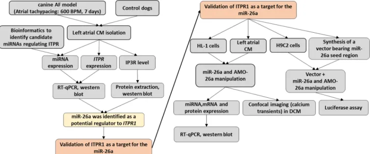
Figure 4 is a schematic representation of the experimental procedures in the current study.
Figure 4: Schematic representation of the research framework. AF: atrial fibrillation, DCM: dog cardiomyocytes.
Contribution of the Author
The AF-dog model for the present study is generated in the animal facility of the Montreal Heart Institute. Nathalie L’Heureux performed the surgeries on dogs, as described in Liu et al. publication (105). Xiaoyan Qi did all the work related to the left atrial cell isolation. I, together with Jiening Xiao, performed the construction of RNA/DNA sequences used in transfection studies. I determined the miRNAs targeting the ITPR1 gene through the in silico studies. I cultured freshly isolated cardiomyocytes and H9C2 cells and performed the transfection and biochemistry and molecular biology experiments, including cloning, (micro)RNA and protein extraction, qRT-PCR, and western blot. I, together with Louis Villeneuve, performed the nucleoplasmic and cytoplasmic calcium transient experiment. I, together with Xiaoyan Qi and Feng Xiong, performed calcium transient analysis.
Chapter 2 – Methods
Canine atrial fibrillation model
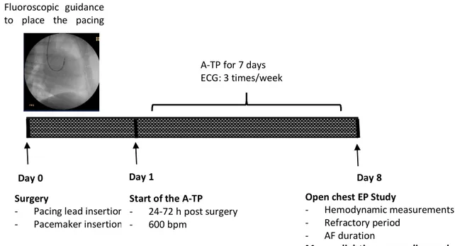
Adult Mongrel dogs of either sex, weighing between 18 and 32 kg, were obtained from LAKA Inc (2016-47-01, 2019-47-03 for control dogs, 2015,47-01 for AF dogs) and assigned to atrial fibrillation (AF) or control (CTL) groups. Dogs used in this study were handled in accordance with the “Guide for the Care and Use of Laboratory Animals” established by the National Institutes of Health as approved by the Montreal Heart institute Ethics Committee. Animals were anesthetized with 0.07mg/kg acepromazine (IM), 5.3 mg/kg ketamine (IV), 0.25 mg/kg diazepam (IV), and then followed by 1.5% isoflurane and were ventilated. A bipolar pacing lead with fluoroscopic guidance was placed in the right atrial appendage via a jugular vein and connected to a subcutaneous pacemaker implanted in the neck (right side). Twenty-four to seventy-two hours after surgery, dogs in the AF group were subjected to atrial tachypacing at 600 bpm for seven days (Figure 5). In animals of the CTL group, no pacemaker was inserted (105).
Figure 5: In vivo model of atrial fibrillation.
AF: atrial fibrillation, A-TP: atrial tachypacing; EP: electrophysiological.
Day 0 Surgery
- Pacing lead insertion - Pacemaker insertion
Day 1
Start of the A-TP - 24-72 h post surgery
- 600 bpm
- EP Study
Day 8 Fluoroscopic guidance
to place the pacing lead
A-TP for 7 days ECG: 3 times/week
Open chest EP Study
- Hemodynamic measurements
- Refractory period - AF duration
Myocardial tissue sampling and cell isolation
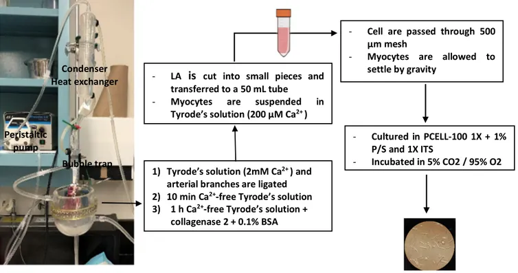
Isolation of dog atrial cardiomyocytes
Cardiomyocytes were isolated from the left atrium (LA) with enzymatic digestion through the Langendorff system. Briefly, dogs were anesthetized with 2 mg/kg morphine (IV) and 120 mg/kg alpha-chloralose. Hearts were aseptically and quickly removed after intra-atrial injection of 10,000 U heparin and placed in Tyrode’s solution containing 136 mM NaCl, 5.4 mM KCl, 2 mM CaCl2, 1 mM MgCl2, 10 mM dextrose, 5 mM HEPES, 0.33 NaH2PO4 (pH was adjusted to 7.3 with NaOH). The left coronary artery of the isolated heart was cannulated, and the LA was dissected free and perfused with 100% oxygenated Tyrode’s solution (37°C, 1.8 mM Ca2+). The arterial
branches were ligated to have a leak-free system, and LA tissues were perfused with Ca2+-free
Tyrode’s solution for 10 minutes, followed by 1-hour perfusion with 0.45 mg/mL collagenase (CLS II, Worthington) and 0.1% bovine serum albumin (MilliporeSigma) in Ca2+-free Tyrode’s solution
for enzyme digestion. Digested tissue was removed from the cannula and cut into small pieces, and atrial cardiomyocytes were harvested (Figure 6) (105).
500
Figure 6: Left atrial cardiomyocyte isolation experimental setup and protocol BSA: bovine serum albumin; ITS: insulin-transferrin-selenium-X.
Cardiomyocyte isolation system
1) Tyrode’s solution (2mM Ca2+ ) and
arterial branches are ligated 2) 10 min Ca2+-free Tyrode’s solution
3) 1 h Ca2+-free Tyrode’s solution +
collagenase 2 + 0.1% BSA Peristaltic
pump
Bubble trap Condenser
Heat exchanger - LA is cut into small pieces and transferred to a 50 mL tube
- Myocytes are suspended in Tyrode’s solution (200 µM Ca2+ )
- Cell are passed through 500 µm mesh
- Myocytes are allowed to settle by gravity
- Cultured in PCELL-100 1X + 1% P/S and 1X ITS

![Figure 3: Upregulation of IP3R1/2 protein levels and alterations in resting [Ca 2+ ] in atrial](https://thumb-eu.123doks.com/thumbv2/123doknet/2036085.4447/31.918.136.773.105.649/figure-upregulation-ip-protein-levels-alterations-resting-atrial.webp)