Full Terms & Conditions of access and use can be found at
https://www.tandfonline.com/action/journalInformation?journalCode=yacb20
Acta Clinica Belgica
International Journal of Clinical and Laboratory Medicine
ISSN: 1784-3286 (Print) 2295-3337 (Online) Journal homepage: https://www.tandfonline.com/loi/yacb20
Treatment of a patient with severe
cytomegalovirus (CMV) infection after
haploidentical stem cell transplantation with
donor-derived CMV-specific T cells
Joline Ingels, Saskia De Smet, Kelly Heyns, Nele Lootens, Jonas Segaert,
Tom Taghon, Georges Leclercq, Karim Vermaelen, Evelyne Willems, Etienne
Baudoux, Tessa Kerre, Frédéric Baron & Bart Vandekerckhove
To cite this article: Joline Ingels, Saskia De Smet, Kelly Heyns, Nele Lootens, Jonas Segaert, Tom Taghon, Georges Leclercq, Karim Vermaelen, Evelyne Willems, Etienne Baudoux, Tessa Kerre, Frédéric Baron & Bart Vandekerckhove (2020): Treatment of a patient with severe
cytomegalovirus (CMV) infection after haploidentical stem cell transplantation with donor-derived CMV-specific T cells, Acta Clinica Belgica, DOI: 10.1080/17843286.2020.1752446
To link to this article: https://doi.org/10.1080/17843286.2020.1752446
Published online: 14 Apr 2020.
Submit your article to this journal
Article views: 105
View related articles
CASE REPORT
Treatment of a patient with severe cytomegalovirus (CMV) infection after
haploidentical stem cell transplantation with donor-derived CMV-speci
fic T
cells
Joline Ingelsa,b, Saskia De Smetb, Kelly Heynsc, Nele Lootensb, Jonas Segaertd, Tom Taghona,
Georges Leclercqa, Karim Vermaelenc, Evelyne Willemse, Etienne Baudouxe, Tessa Kerrea,d, Frédéric Barone
and Bart Vandekerckhovea,b
aDepartment of Diagnostic Sciences, Ghent University, Ghent, Belgium;bCell Therapy Unit, Department of Regenerative Medicine, Ghent University Hospital, Ghent, Belgium;cDepartment of Respiratory Medicine, Ghent University Hospital, Ghent, Belgium;dDepartment of Internal Medicine, Ghent University Hospital, Ghent, Belgium;eDepartment of Medicine, Division of Hematology, University of Liège, Liège, Belgium
ABSTRACT
Objectives: Cytomegalovirus (CMV) infection is one of the most common complications in allogeneic hematopoietic stem cell transplant (allo-HSCT) recipients. The classic antiviral treat-ments have shown clinical efficacy but are often associated with drug resistance. Reconstitution of CMV-specific cellular immunity is essential in controlling CMV infection; therefore, adoptive transfer of CMV-specific T cells is a promising treatment option. We treated a patient with a multidrug resistant CMV infection after haploidentical HSCT with CMV-specific T cells. Methods: The T cells were derived from the HSCT donor who was CMV seropositive. We generated the T cells by a short-term Good Manufacturing Practice (GMP) grade protocol in which a leukapheresis product of the HSCT donor was stimulated with the immunodominant antigen pp65 and interferon-γ secreting cells were isolated. A total of 5 × 10⁵ T cells were
administered to the patient within 30 hours after leukapheresis.
Results: The patient was closely monitored for reconstitution of antiviral T cell immunity and viral replication after adoptive T cell transfer. We observed an in vivo expansion of both CD4+
and CD8+CMV-specific T cells associated with a significant decrease in viral burden and clinical
improvement.
Conclusion: This case report further supports the feasibility and effectiveness of adoptive donor T cell transfer for the treatment of drug resistant CMV infections after allo-HSCT.
KEYWORDS
CMV infection; adoptive T cell transfer; stem cell transplantation
Case report
Cytomegalovirus (CMV) infection has remained an important cause of morbidity and mortality after allo-geneic hematopoietic stem cell transplantation (allo-HSCT). This is especially the case in recipients of HLA-mismatched or HLA-haploidentical HSCT, as well as in those given T cell depleted grafts [1–3]. Standard anti-viral agents such as ganciclovir have significantly reduced the incidence of early-onset CMV disease. However, prolonged administration is associated with significant hematopoietic toxicity, drug resistance, delayed CMV-specific T cell reconstitution, and devel-opment of late-onset CMV infection [4]. Previous reports have demonstrated that T-cell immunity is essential in controlling CMV infections [5]. Therefore, a promising approach to treat refractory CMV infection after allo-HSCT in patients who lack anti-CMV immu-nity has been the adoptive transfer of CMV-specific T cells from the original stem cell donor [6,7].
We here report the adoptive transfer of CMV-specific T cells in a 9-year-old girl for the treatment of CMV
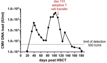
infection after allo-HSCT. The girl received CD34-selected peripheral blood stem cells from her HLA-haploidentical mother as treatment for a refractory neu-roblastoma. Conditioning regimen consisted of melpha-lan 140 mg/m2, Thiotepa 10 mg/m2,fludarabine 200 mg/ m2 and thymoglobuline 10 mg/kg. The infused graft contained 16.6 × 10⁶CD34+cells/kg recipient with resi-dual T cells of 0.02 × 10⁶ CD3+ cells/kg recipient. She experienced CMV infection 24 days after transplantation (Figure 1.). Ganciclovir, foscavir and cidofovir all failed to eradicate CMV infection and CMV sequencing demon-strated a L773V substitution in the CMV polymerase domain (a mutation known to cause resistance to ganci-clovir, foscavir and cidofovir) [8].
Because of the rapid increase in viral load (Figure 1) and the demonstration of the mutation in the CMV polymerase domain, it was decided the treat the patient with CMV virus-specific T cells of the mother HSC donor on day 104 after transplantation. At the same time, the patient was started on letermovir in addition to cymevene + foscavir combination. The CMV serostatus of the mother was repeated and
CONTACTBart Vandekerckhove bart.vandekerckhove@ugent.be Department of Diagnostic Sciences, Ghent University, Ghent, Belgium https://doi.org/10.1080/17843286.2020.1752446
confirmed to be seropositive: CMV IgM-, IgG+. Next, we assessed the specific cellular immunity by quantifying the peripheral blood T cells reactive to the immuno-dominant antigen of CMV phosphoprotein-65 (pp65). Isolated PBMC were incubated with Peptivator CMVpp65, a 15-mer peptide pool covering the com-plete primary structure of the pp65 antigen and sub-sequently IFNγ production was visualized by intracellular staining andflowcytometric analysis. We detected a robust CMVpp65-specific CD4+ T cell
population (Figure 2(a)), while virtually no specific CD8+T cells were observed. The frequency of specific CD4+T cells was clearly above background (criterium: at least twice background levels) and was considered adequate to obtain a sufficient number of CMV-specific T cells for adoptive transfer. An apheresis of the mother was scheduled 4 days later.
As our patient was <18 years, she failed the inclu-sion criteria of our clinical trial protocol (Eudra CT 2013-004953-26). We therefore decided to perform the
Figure 1.Time course of viral load. CMV-DNA load was assessed in peripheral blood by PCR at different time points after HSCT. Time point of infusion of CMV-specific T cells is indicated in the graph.
Figure 2.Detection of CMVpp65-specific T cells in PBMC and isolation from a leukapheresis of the family HSCT donor. (a) PBMC of the HSCT donor were stimulated for 6 hours with CMVpp65 Peptivator and as a control without, intracellularly stained for IFNγ and analyzed byflowcytometry. Dot plots represent IFNγ expression in the CD3+CD4+or CD3+CD8+population. (b) IFNγ expression in CD3+T cells and CD4+and CD8+T cell distribution before isolation and in the isolated fraction (final product). (c) Schematic overview of the production process for the isolation of CMVpp65-specific T cells by the IFNγ Capture technology.
procedure conforming to the hospital exemption pro-cedure legislation and notified the federal agency for medicines and health products (FAMHP) thereof. This procedure is meant to enable the production of a particular medicinal product specifically designed for a particular patient for which no similar medicine is available on the market. This allowed us to produce the product in a timely fashion.
The leukapheresis product was collected from 7.7 liters of blood volume of the mother donor and con-tained 5.7 × 10⁹nucleated cells. The leukapherate was processed the following day using the CliniMACS cyto-kine capture technology in a licensed Good Manufacturing Practice (GMP) facility (Figure 2(c)). Following a standardized production protocol, a total of 1 × 10⁹nuclear cells were stimulated with Peptivator CMVpp65 to induce IFNγ production. After 4 hours, cells were labelled with a bispecific antibody (Catchmatrix Reagent) that binds to the cells using its CD45 specificity and is able to catch secreted IFNγ on the cell membrane using its IFNγ specificity. After a wash step and appropriate dilution, the coated cells were incubated for exactly 45 minutes at 37°C to allow for secretion of IFNγ and capture on the surface of the cells by the Catchmatrix Reagent. Next, cells were labelled with a second IFNγ-specific antibody coupled to a magnetic bead resulting in bead covered CMVpp65-specific T cells. The cell mixture was then passed through a magneticfield to separate the bead-coated CMVpp65-specific cells from the uncoated cells. We recovered 5.4 × 10⁵viable CD3+T cells, of which 2.7 × 10⁵ were CD4+IFNγ+ and 0.1 × 10⁵CD8+IFNγ+ T cells (Figure 2(b)). The cells were washed and resus-pended in Plasmalyte with 4% human serum albumin. Total T cell dose was 24.4 × 10³T cells/kg patient. We performed extensive quality testing on process
intermediates as well as on the final product. Mycoplasma testing was performed using a PCR method according to the European Pharmacopoeia immediately after culture and proved negative. Endotoxin levels were <4.4 IU/kg patient. Sterility test-ing was performed on the apheresis product, after Peptivator stimulation and on the final product. However, as the definitive results were not available at the time of release, a gram-stain was performed on thefinal product and was negative. The whole produc-tion process from stimulaproduc-tion to release was com-pleted within 12 hours.
The CMV-specific T cells were infused intravenously as a single dose on the same day of isolation, 114 days after HSCT. There were no infusion-related acute toxi-cities. Although donor and recipient are HLA-haploidentical, no GvHD related to the T cell product was induced up to 14 weeks after infusion.
Survival and in vivo expansion of CMVpp65-specific T cells was assessed at 2, 4 and 8 weeks following adoptive T cell transfer (Figure 3). CMV-specific CD4+ T cells were readily detectable in the peripheral blood of the patient 2 weeks after infusion, and expanded thereafter reaching a similar frequency as found in CMV seropositive healthy individuals. This response remained stable during the follow-up period of 8 weeks. Interestingly, although a low number of CD8+ T cells were present in the T cell product (Figure 2(b)), CMVpp65-specific CD8+T cells strongly expanded over time and even exceeded the frequen-cies found in healthy CMV seropositive individuals at 8 weeks post-infusion. The expansion of CMVpp65-specific T cells was accompanied by a significant reduc-tion of CMV DNA copies in peripheral blood to <500 IU/ml blood which is the lower limit of detection of this assay (Figure 1).
Figure 3.Expansion of CMVpp65-specific T cells after adoptive T cell transfer. CMVpp65-specific T cell response was assessed in peripheral blood of the recipient at 2, 4 and 8 weeks after adoptive T cell transfer. PBMC were stimulated with CMVpp65 Peptivator for 6 hours and intracellularly stained for IFNγ. The percentage of IFNγ+T cells obtained after stimulation with CMVpp65 Peptivator was corrected for background staining by subtracting the percentage of IFNγ+cells measured in the absence of stimulation.
Discussion
In this case report, we describe the successful adoptive transfer of CMV-specific T cells derived from a haploidentical HSCT family donor in a patient with a refractory CMV infection after HSCT.
The successful production and transfer of CMV-specific T cells requires a suitable donor, a validated pro-duction protocol and coordinated logistics. As our patient presented with a life-threatening CMV infection, the cell therapy product had to be administered as rapidly as possible. Therefore, the throughput time from donor screening to collection of apheresis and infusion of the T cells had to be minimized. From the moment the intent to treat was decided, it took 2 days to screen the donor for CMV-specific T cells and another 2 days for the apher-esis and isolation of the cells. The final product was transported immediately after production to the trans-plant centre and administered within 4 hours after pro-duction. This very short throughput time is only possible when fully validated procedures and trained dedicated production and QC personnel is available at all time. In addition, for the isolation of the T cells, we applied the GMP-compatible IFNγ Capture technology in which the CMV-specific T cells are directly isolated from a leukapheresis without the need for prolonged cultures. This technology allowed us to isolate the T cells and release the product within one day after the apheresis.
We administered a low dose of 24.4 × 10³T cells/kg body weight and observed an expansion of CMV-specific T cell immunity, associated with a strong reduction in CMV copies. Clinical effects with low T cell doses have also been observed in a study by Feuchtinger et al., in which haploidentical T cells admi-nistered at a median dose of 7.1 × 10³T cells/kg were associated with strong reductions in CMV viremia [6]. As in our case, these low doses did not result in GvHD, which is a potential life-threatening toxicity when administering T cells in a haploidentical setting.
CMV-specific T cell immunity has been demon-strated to be crucial in the effective control of CMV infection in posttransplant patients. After T cell infu-sion, we observed an expansion of both CD4+ and CD8+ T cells. Cytotoxic CD8+ T cells destroy virus-infected cells, while helper CD4+ T cells regulate the induction of effector CD8+T cells and maintenance of CD8+ T memory cells [9,10]. Multiple studies suggest that functionality of both T cell subsets is required to achieve an effective control of CMV infection [11,12]. The strong reduction of CMV copies we observed is thus most likely attributable to the combined recon-stitution of CD4+and CD8+T cell immunity. Of note, the viral load in blood was already decreasing at the time of T cell infusion following the introduction of letermovir. Eighteen days before infusion the patient
had profound T-lymphopenia (4.9 CD3+/µl with no detected naive CD4+T cells) and T cell counts normal-ized concurrent with the clearance of the CMV infec-tion, to 452 CD3+/µl 3 days after infusion and 2077 CD3+/µl 1 month after T cell infusion. We believe that the increase in peripheral T cell levels is a consequence of the viral clearance and of peripheral expansion of infused T cells in this context of profound lymphope-nia. Further, it is unlikely that the posttransplant-generated naïve T cells would contain such a high percentage of CMV reactive T cells at the measured time points. It is therefore implausible that endogen-ous reconstitution of CMV reactive T cells contributed to the decrease in viral load.
Acknowledgments
We thank prof. dr. Jerina Boelens for sterility testing and Gram stain.
Disclosure statement
The authors report no declarations of interest.
Funding
This work was supported by the Kinderkankerfonds.
References
[1] Green ML, Leisenring W, Xie H, et al. Cytomegalovirus viral load and mortality after haemopoietic stem cell transplantation in the era of pre-emptive therapy: a retrospective cohort study. Lancet Haematol.
2016;3:e119–e127.
[2] Chang Y-J, Zhao X-Y, Huo M-R, et al. Immune recon-stitution following unmanipulated HLA-mismatched/ haploidentical transplantation compared with HLA-identical sibling transplantation. J Clin Immunol.
2012;32:268–280.
[3] Liu J, Kong J, Chang YJ, et al. Patients with refractory cytomegalovirus (CMV) infection following allogeneic haematopoietic stem cell transplantation are at high risk for CMV disease and non-relapse mortality. Clin Microbiol Infect.2015;21:1121.e9-1121.e15.
[4] Ahmed A. Antiviral treatment of cytomegalovirus infection. Infect Disord - Drug Targets. 2011;11: 475–503.
[5] Cwynarski K, Ainsworth J, Cobbold M, et al. Direct visualization of cytomegalovirus-specific T-cell recon-stitution after allogeneic stem cell transplantation. Blood.2001;97:1232–1240.
[6] Feuchtinger T, Opherk K, Bethge WA, et al. Adoptive transfer of pp65-specific T cells for the treatment of chemorefractory cytomegalovirus disease or reactiva-tion after haploidentical and matched unrelated stem cell transplantation. Blood.2010;116:4360–4367. [7] Einsele H, Roosnek E, Rufer N, et al. Infusion of
cyto-megalovirus (CMV)–specific T cells for the treatment of 4 J. INGELS ET AL.
CMV infection not responding to antiviral chemother-apy. Blood.2002;99:3916–3922.
[8] Chou S, Ercolani RJ, Sahoo MK, et al. Improved detection of emerging drug-resistant mutant cyto-megalovirus subpopulations by deep sequencing. Antimicrob Agents Chemother. 2014;58:4697– 4702.
[9] Laidlaw BJ, Craft JE, Kaech SM. The multifaceted role of CD4+ T cells in CD8+ T cell memory. Nat Rev Immunol.
2016;16:102–111.
[10] Janeway’s Immunobiology. Ninth Edition. By Kenneth Murphy and Casey Weaver; with contributions by Allan
Mowat, Leslie Berg, and David Chaplin; with acknowl-edgment to: charles A. Janeway, Jr., Paul Travers, and Mark Walport. New York: Garland Science. Tay Q Rev Biol.2018;93:59.
[11] Crough T, Khanna R. Immunobiology of human cyto-megalovirus: from bench to bedside. Clin Microbiol Rev.2009;22:76–98.
[12] Walter EA, Greenberg PD, Gilbert MJ, et al. Reconstitution of cellular immunity against cytomega-lovirus in recipients of allogeneic bone marrow by trans-fer of T-cell clones from the donor. N Engl J Med.