Brucella abortus is prevalent in both humans and animals in Bangladesh 1
2
A. K. M. Anisur Rahman1,2,3
, Claude Saegerman2, Dirk Berkvens3, Falk Melzer4, Heinrich Neubau-3
er4, David Fretin5, Emmanuel Abatih6, Navneet Dhand7, Michael P. Ward7* 4
5
1 Department of Medicine, Bangladesh Agricultural University, Mymensingh-2202, Bangladesh. 6
2 Research Unit of Epidemiology and Risk Analysis applied to the Veterinary Sciences (UREAR-7
Ulg), Fundamental and Applied Research for Animals & Health (FARAH), Department of Infec-8
tious and Parasitic Diseases, Faculty of Veterinary Medicine, University of Liège, Liège, Belgium. 9
3 Department of Biomedical Sciences, Institute of Tropical Medicine, Antwerpen, Belgium. 10
4 Federal Research Institute for Animal Health, Reference Laboratory for Brucellosis and CEM, 11
Naumburger Str. 96a, 07743 Jena, Germany. 12
5 Department of Bacteriology and Immunology, Veterinary and Agrochemical Research Center, 13
Brussels, Belgium. 14
6 Department of Applied Mathematics, Computer Science and Statistics, Faculty of Sciences, Ghent 15
University, 281 Krijgslaan, B-9000, Ghent, Belgium. 16
7 Faculty of Veterinary Science, The University of Sydney, 425 Werombi Road, Camden, 2570 17
NSW, Australia. 18
19
* Corresponding author at: michael.ward@sydney.edu.au 20 Tel: +61-2-93511607 21 Fax: +61-2-93511618 22 23
Summary 1
To determine the role of different Brucella (B.) spp. in Bangladesh, 62 animal samples and 500 2
human sera were tested. Animal samples from cattle, goats and sheep (including milk, bull semen, 3
vaginal swabs and placentas) were cultured for Brucella spp. Three test positive human sera and all 4
animal samples were screened by Brucella genus specific real time PCR (rt PCR) and positive sam-5
ples were then tested by IS711 rt PCR to detect B. abortus and B. melitensis DNA. Only B. abortus 6
DNA was amplified from 13 human and six animal samples. This is the first report describing B. 7
abortus as the aetiological agent of brucellosis in occupationally exposed humans in Bangladesh. 8
Of note is failure to detect B. melitensis DNA, the species most often associated with human brucel-9
losis worldwide. Further studies require to explore the availability of Brucella melitensis in Bangla-10
desh. 11
Keywords: Brucella abortus, humans, animals, clinical samples, Bangladesh, real-time PCR 12
13
Impacts 14
Only Brucella abortus was detected from humans and domestic ruminants 15
Brucellosis may not be the principle reason for abortion in domestic ruminants 16
Brucella melitensis was not detected from any of the human and animal sample. 17
Introduction 1
Brucellosis caused by a range of Brucella spp. is a widespread bacterial zoonosis which 2
impacts both human health and animal production in endemic countries. It is a severely debilitating 3
illness, characterized by fever, sweating, fatigue, weight loss, headache and arthralgia which can 4
persist for weeks to months in humans (Ariza et al., 2007). Chronic or life-threatening sequelae can 5
result. WHO considers brucellosis a neglected zoonosis. It is a serious occupational hazard for live-6
stock farmers, dairy workers, butchers, hired animal caretakers, veterinarians, laboratory workers 7
and for consumers of raw livestock products (Anon., 2005). The isolation of B. abortus in Bangla-8
desh was first reported in 1981 (Pharo et al., 1981) from two seropositive cows. However, the pro-9
cedures used to type these isolates as B. abortus were not fully described. Moreover, Brucella genus 10
and B. abortus DNA have recently been amplified from seropositive humans (Rahman et al., 2012) 11
and bovine sera (Rahman et al., 2014), respectively, using real time (rt) PCR. Brucella abortus 12
DNA has also been detected from patients with prolonged fever who were serologically positive 13
(Rahman et al., 2016). The Brucella spp. responsible for infections in occupationally exposed hu-14
mans have not been defined. The seroprevalence of brucellosis in domestic ruminants ranges from 15
3.6 to 7.3% (Islam et al., 2013). In humans, 18.2% of dairy workers, 2.6% of livestock farmers, 16
21.6% of goat farmers, 2.5% of butchers and 5.3% of veterinary practitioners were found to be se-17
ropositive in Bangladesh (Rahman et al., 2012; Rahman et al., 1983; Rahman et al., 1988). It indi-18
cates this disease in endemic in Bangladesh but still the species of Brucella prevalent in both hu-19
mans and different species of animals are not known completely. 20
Laboratory detection and identification of Brucella species is still based on culture and pheno-21
typic characterization, respectively, performed in laboratories with level 3 biosafety facilities. The 22
isolation of Brucella spp. helps in understanding the prevalent biovars of the dominants species and 23
also the source of infection in outbreaks (Al Dahouk et al., 2007). PCR techniques have become 24
popular for rapid detection of brucellae from clinical (blood or serum) samples (Zerv et al., 2001; 25
Queipo-Ortuño et al., 2005). The IS711 based rt PCR is reported to be specific and highly sensitive 26
(Bounaadja et al., 2009). Most RT PCR assays developed to date are designed to detect brucellae at 1
genus level, enabling early implementation of treatment. Brucella IS711 species specific multiplex 2
rt PCRs for B. abortus and B. melitensis also exist (Probert et al., 2004). 3
A recent study estimated the cost of brucellosis on livestock production in neighbouring India 4
to be USD 3.4 billion (Singh et al., 2015), and brucellosis is a disease of public health importance 5
in both developed and developing countries (Dean et al., 2012). The global health burden caused by 6
brucellosis has been recently estimated to be >250,000 Disability Adjusted Life Years (DALYs) 7
(Kirk et al., 2015). The aim of the current study was to determine the Brucella species responsible 8
for brucellosis in animals and humans in Bangladesh. 9
Materials and Methods 10
Ethical statement 11
The study protocol was peer reviewed and approved by the Ethical Review Committee of 12
Mymensingh Medical College. Informed written consent was taken from all individuals prior to the 13
collection of blood. Animal research was approved by the Faculty of Veterinary Science of Bangla-14
desh Agricultural University. 15
Human samples 16
Collection of human sera has been previously described (Rahman et al., 2012). Briefly, a sur-17
vey of livestock farmers, milkmen, butchers, and veterinary practitioners in the Mymensingh, Sher-18
pur, and Dhaka districts of Bangladesh was conducted between September 2007 and August 2008. 19
In addition, workers at two government-owned farms in Dhaka District were sampled. A total of 20
500 individuals were sampled. In this survey the prevalence of Brucella antibodies (based on paral-21
lel interpretation of three serological tests – Rose Bengal Test [RBT], Standard Tube Agglutination 22
Test [STAT] and an Indirect ELISA) was estimated to be 4.4% (Rahman et al., 2012). Twenty-two 23
human sera were positive in at least one serological test. Out of 22, 13 were positive in three sero-24
logical tests and in a Brucella genus specific RT PCR (Rahman et al., 2012) targeting the BCSP31 25
gene encoding a 31-kDa antigen conserved among Brucella spp (Navarro et al., 2004) and included 26
in this study. The BCSP31-PCR assay was carried out using standard procedure (Bounaadja et al., 1
2009; Baily et al., 1992), and IS711-PCR was done using the procedure described by Halling et al. 2
(1993). 3
Animal samples 4
Randomly collected milk ring test (MRT) positive bulk milk samples (cattle, goat and gayal 5
[Bos frontalis]), convenience samples of placentas, vaginal swabs from different animals (cattle, 6
goats and sheep) post-abortion (within 0-3 days) and semen samples from bulls (with orchitis) were 7
collected and used to isolate and detect Brucella genus and species specific DNA. 8
Staining 9
Impression smears of vaginal swabs and placentas were stained by the Stamp method (Alton et 10
al., 1988). In brief, the impression smears are dried by flame and stained with working carbol fuch-11
sin solution for 10 min. Then decolorized by 3% acetic acid solution for 1 min and counterstained 12
with 1% malachite green solution for 20 seconds. After washing in tap water, dried and observed 13
under microscope using 100X objective (oil immersion), Brucella organisms appear pale red in a 14
blue background. 15
Bacteriology 16
For the isolation of Brucella organisms, clinical samples were cultured in Farrell’s medium us-17
ing standard methods (Alton et al., 1988). Briefly, milk samples were centrifuged at 6000 g for 15 18
min; cream and sediment were spread on half of the plate and streaked on the other half. Swab and 19
semen (150 microliter) samples were also spread and streaked in the same way. 20
The cotyledons of the placentas were cut into small pieces (about 5 g). Approximately 4–5 ml 21
of normal saline was added and a homogenate was prepared in a stomacher or blender. About 150 22
microliter of the homogenate was spread and streaked as for milk, swab and semen samples. Plates 23
were incubated at 37°C in 7.5% CO2 and first inspected after 48 hours. A sample was considered 24
culture negative if no growth occurred within 7 days. DNA was extracted from samples using the 25
DNeasy spin column kit (QIAGEN) according to the manufacturer's protocol. 26
IS711 genus specific and Brucella abortus and Brucella melitensis specific real-time PCR 1
The IS711 rt PCRs originally described as a multiplex PCR assay (Probert et al., 2004) were 2
performed as single assays to detect Brucella spp. DNA and to distinguish between B. melitensis 3
and B. abortus DNA, respectively, without further modification. The species-specific assays were 4
applied when Brucella DNA had been detected in a sample in the genus-specific assay. The same 5
primers and probes were obtained from TIB MOLBIOL (Berlin, Germany). Amplification reaction 6
mixtures were prepared in volumes of 25 μl containing 12.5 μl TaqManTM Universal Master Mix 7
(Applied Biosystems, New Jersey, USA), 0.75 μl of each of the two specific primers (0.3 μM) and 8
0.5 μl TaqMan probe (0.2μM), 5 μl of template, and 6.25 μl nuclease-free water. The rt PCR reac-9
tion was performed in duplicate in optical 96-well microtiter plates (qPCR 96-well plates, Micro 10
AmpTM, Applied Biosystems) using a Mx3000P thermocycler system (Stratagene, La Jolla, Cali-11
fornia) with the following run conditions, 2 min at 50°C, 10 min at 95°C, followed by 50 cycles of 12
95°C for 15s and 57°C for 1 min. Cycle threshold values below 40 were considered positive. The 13
threshold was set automatically by the instrument. In addition, the samples scored positive by the 14
instrument were confirmed by visual inspection of plots of cycle numbers versus fluorescence val-15
ues. 16
Results 17
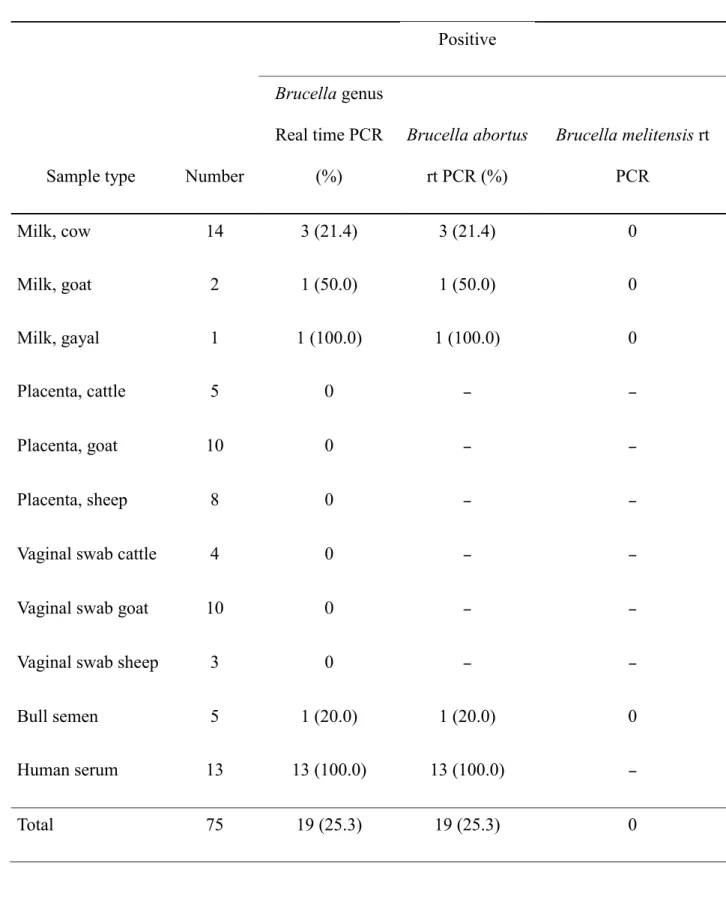
Brucella-like bacteria were not seen in any of the stained smears. No growth of Brucella spp. 18
was noted in any of the clinical samples, but six (9.8%) of 62 animal samples investigated were 19
positive in the genus specific BCSP31 rt PCR assay (Table 1). Among animals, only five milk 20
(three cattle, one goat and one gayal) and one semen sample were positive in the B. abortus specific 21
rt PCR. 22
Brucella abortus DNA was amplified from all of the 13 human serum samples tested. The clin-23
ical characteristics of these 13 patients and treatment received are shown in Table 2. Among brucel-24
losis infected patients 76.9%, 61.5%, 46.2% and 53.8% had fever, arthralgia, backache and sweat-25
ing respectively. 26
No B. melitensis DNA could be amplified from human or animal samples. 1
Discussion 2
We have described the successful amplification of B. abortus DNA from human serum, milk 3
(cattle, goat, gayal) and bull semen from Bangladesh. Animals and their products are the sole source 4
of human infection. Consequently, the presence of B. abortus DNA in human samples together with 5
their history of contact demonstrates that dairy animal populations in Bangladesh are a source of 6
brucellosis in humans, and that B. abortus is the most common (and potentially only) aetiological 7
agent of human brucellosis in Bangladesh. The presence of B. abortus DNA in milk and semen 8
supports this conclusion and moreover, the recent detection of B. abortus DNA from bovine sera in 9
Bangladesh (Rahman et al., 2014) provides further evidence of the causal role of B. abortus. Detec-10
tion of B. abortus DNA from five districts of Bangladesh (Bandarban, Chittagong, Dhaka, My-11
mensingh and Sirajganj) suggests that infection of dairy animals is widespread. However, due to 12
small size and non-randomness of the sample our result does not represent whole Bangladesh. 13
Brucella abortus DNA was detected from milk samples of gayal in the hilly district of Bandar-14
ban, Bangladesh. The gayal (Bos frontalis syn.: mithan or mithun) is a semi-domestic ruminant 15
found in the hill regions of northeast India, Myanmar, Bhutan, Bangladesh, China and Malaysia 16
(Simoons and Simoons 1968; Mason, 1988). The source of infection of these animals is not clear. 17
Bandarban district shares a common border with Mizoram state, India and Chin state, Myanmar. 18
Although no report of the isolation of Brucella spp. from gayal in India exists, high Brucella sero-19
prevalence in gayal in Mizoram has been reported (Rajkhowa et al., 2005). Moreover, it is reported 20
that gayal populations from Tripura and Mizoram in India and from Bangladesh mingle due to the 21
porous nature of this border (Choudhury, 2002). Another possible source of Brucella in this gayal 22
population is crossbreeding with cattle. Holstein Friesian bull semen originating from one of the 23
government farms included in the current study – and found to be Brucella-infected – has been used 24
for this crossbreeding purpose (Huque et al., 2011). B. abortus DNA was also detected from one 25
goat milk sample. Although B. melitensis is the most common agent of caprine brucellosis, B. abor-26
tus infection of goats has been reported especially in countries where B. melitensis is absent, for 1
example Brazil (Lilenbaum et al., 2007). Including the current study, B. melitensis has not been 2
reported from Bangladesh. The failure to detect B. melitensis DNA may also be due to very small 3
number of goat samples we tested. 4
Brucella DNA was detected in human serum samples even though these had been collected a 5
long time after clinical symptoms had resolved in patients (Navarro et al., 2006). All 13 patients, 6
which were seropositive in the RBT, SAT and iELISA, presented with clinical symptoms consistent 7
with brucellosis and indeed all had recovered after a ‘typical' brucellosis treatment had been admin-8
istered. Confirmatory diagnosis by species-specific rt PCR is sufficient for timely initiation of bru-9
cellosis treatment. Probert et al. (2004) developed a multiplex rt PCR to detect B. abortus and B. 10
melitensis from culture growth. Modifying this technique, we were able to show that IS711 species-11
specific rt PCR is capable of amplifying Brucella DNA from human sera and animal samples at 12
species level. Estimating the positive predictive value of combinations of serological tests – using rt 13
PCR as the gold standard – was not an objective of this study. Where feasible, use of rt PCR to con-14
firm brucellosis in patients with clinical symptoms consistent with this disease and suspected dairy 15
animal contacts is warranted. 16
Abortion is the most common clinical sign of brucellosis in female domestic ruminants and 17
usually aborted fetuses, fetal membranes and fluids contain high bacterial loads contaminating the 18
environment and causing a high risk of infection to other animals (Saegerman et al., 2009). In our 19
study, none of the 40 fetal membranes and vaginal swabs originating from cattle, sheep and goats 20
contained Brucella DNA. Although the sample size is small, it indicates that Brucella may not be a 21
major cause of abortion in domestic ruminants in Bangladesh. 22
Brucella DNA was detected from some of the MRT positive, culture negative milk samples. 23
The possible reason for unsuccessful recovery of isolates may be that the samples had been stored 24
for 2–3 years after collection prior to shipment to Belgium (for isolation and molecular detection). 25
Indeed, isolation is most likely during the acute phase of infections caused by B. melitensis or B. 26
suis and less successful in B. abortus infections. Brucellae are rarely isolated from samples with a 1
competing microflora (Al Dahouk et al., 2002). The presence of competing organisms (observed 2
during culture) may be another potential reason for isolation failure (Das et al., 2010). 3
This study further confirms that B. abortus is a cause of human brucellosis in Bangladesh and 4
that infection is likely endemic in livestock. Contaminated milk represents a potential source of 5
infection. Testing representative number of goat fetal membranes from different parts of the country 6
will help understanding the presence of B. melitensis in Bangladesh. 7
The strength of this study is that for the first time it described the presence of Brucella abortus 8
in occupationally exposed humans, goats and gayals in Bangladesh. This study has also some limi-9
tations including small size and non-randomness of the samples. Blood culture to isolate Brucella 10
from seropositive humans was not possible due to lack of facility. The results of this study also do 11
not represent whole of Bangladesh. 12
13
Acknowledgements 14
This study was supported by the Belgian Directorate General for Development Cooperation 15
(DGDC). 16
References 17
Anonymous, 2005: Livestock sector brief of Bangladesh. Food and Agriculture Organization of 18
the United Nations; Available at 19
http://www.fao.org/ag/againfo/resources/en/publications/sector_briefs/lsb_BGD.pdf (accessed 20
on 28 November 2015). 21
Al Dahouk, S., P. Le Flèche, K. Nöckler, I. Jacques, M. Grayon, H. C. Scholz, H. Tomaso, G.
22
Vergnaud, and H. Neubauer, 2007: Evaluation of Brucella MLVA typing for human brucellosis. J
Mi-23
crobiol Methods. 69, 137-145. 24
Al Dahouk, S., H. Tomaso, K. Nöckler, H. Neubauer, and D. Frangoulidis, 2002: Laboratory 25
based diagnosis of brucellosis–a review of the literature. part 1: Techniques for direct detection 26
Alton, G.G., L. M. Jones, R. D. Angus, and J. M. Verger, 1988: Techniques for the brucellosis 1
laboratory. Institut National de la recherche Agronomique (INRA). 2
Ariza, J., M. Bosilkovski, A. Cascio, J. D. Colmenero, M. J. Corbel, M. E. Falagas, Z. A. Mem-3
ish, M. R. H. Roushan, E. Rubinstein, N. V. Sipsas, and J. Solera, 2007: Perspectives for the 4
treatment of brucellosis in the 21st century: the Ioannina recommendations. PLoS Med. 4, e317. 5
Baily, G.G., J. B. Krahn, B. S. Drasar, and N. G. Stoker, 1992: Detection of Brucella melitensis 6
and Brucella abortus by DNA amplification. J Trop Med Hyg. 95, 271-275. 7
Bounaadja, L., D. Albert, B. Chénais, S. Hénault, M. S. Zygmunt, S. Poliak, and B. Garin-8
Bastuji, 2009: Real-time PCR for identification of Brucella spp.: A comparative study of IS711, 9
bcsp31 and per target genes. Vet Microbiol. 137, 156-164. 10
Choudhury, A., 2002: Distribution and conservation of the gaur Bos gaurus in the Indian sub-11
continent. Mammal Rev. 32, 199–226. 12
Das, T., M. Ershaduzzaman, K. K. Islam, M. M. Haque, M. M. Rahman, and K. S. Islam, 2010: 13
Surveillance of Brucella melitensis and Brucella abortus from aborted Bengal Goats in Bangla-14
desh. Res J Vet Sci. 3(1), 40-48. 15
Dean, A.S., L. Crump, H. Greter, E. Schelling, and J. Zinsstag, 2012: Global burden of human 16
brucellosis: a systematic review of disease frequency. PLoS Negl Trop Dis. 6(10), e1865. 17
Halling, S.M., F. M. Tatum, and B. J. Bricker, 1993: Sequence and characterization of an inser-18
tion sequence, IS711, from Brucella ovis. Gene 133(1), 123-127. 19
Huque, K. S., M. A. Jalil, and M. M. Rahman, 2011: Development of beef cattle breed using 20
gayal (Bos frontalis). Final report. 2011; Animal Production Research Division, Bangladesh 21
Livestock Research Institute, Savar, Dhaka. 22
Islam, M.A., M. M. Khatun, S. R. Werre, N. Sriranganathan, and S. M. Boyle, 2013: A review 23
of Brucella seroprevalence among humans and animals in Bangladesh with special emphasis on 24
epidemiology, risk factors and control opportunities. Vet Microbiol.166(3), 317-326. 25
Kirk, M.D., S. M. Pires, R. E. Black, M. Caipo, J. A. Crump, B. Devleesschauwer, D. Döpfer, 1
A. Fazil, C. L. Fischer-Walker, T. Hald, and A. J. Hall, 2015: World Health Organization esti-2
mates of the global and regional disease burden of 22 foodborne bacterial, protozoal, and viral 3
diseases, 2010: a data synthesis. PLoS Med. 12(12), e1001921. 4
Lilenbaum, W., G. N. de Souza, P. Ristow, M. C. Moreira, S. Fráguas, V. da Silva Cardoso, and 5
W. M. R. Oelemann, 2007: A serological study on Brucella abortus, caprine arthritis--6
encephalitis virus and Leptospira in dairy goats in Rio de Janeiro, Brazil. Vet J. 173, 408-412. 7
Mason, I. L., 1988: World Dictionary of Livestock Breeds, 3rd Edn, C. A. B. International, 8
Wallingford. 9
Navarro, E., M. A. Casao, and J. Solera, J., 2004: Diagnosis of human brucellosis using PCR. 10
Expert Rev Mol Diagn. 4(1), 115-123. 11
Navarro, E., J. C. Segura, M. J. Castaño, and J. Solera, J., 2006: Use of real-time quantitative 12
polymerase chain reaction to monitor the evolution of Brucella melitensis DNA load during 13
therapy and post-therapy follow-up in patients with brucellosis. Clin Infect Dis. 42, 1266-1273. 14
Pharo, H.J., A. Motalib, S. Alam, G. C. Fraser, and S. F. Routledge, 1981: Preliminary infor-15
mation on the prevalence of bovine brucellosis in the Pabna milk-shed area of Bangladesh. 16
Bangladesh Vet J. 15, 43-51. 17
Probert, W.S., K. N. Schrader, N. Y. Khuong, S. L. Bystrom, and M. H. Graves, 2004: Real 18
Time Multiplex PCR Assay for Detection of Brucella spp., B. abortus, and B. melitensis. J Clin 19
Microbiol. 42, 1290-1293. 20
Queipo-Ortuño, M.I., J. D. Colmenero, G. Baeza, and P. Morata, 2005: Comparison between 21
Light Cycler real-time polymerase chain reaction (PCR) assay with serum and PCR–enzyme-22
linked immunosorbent assay with whole blood samples for the diagnosis of human brucellosis. 23
Clin Infect Dis. 40, 260-264. 24
Rahman, A. K. M. A., D. Berkvens, D. Fretin, C. Saegerman, M. U. Ahmed, N. Muhammad, A. 1
Hossain, and E. Abatih, 2012: Seroprevalence and risk factors for brucellosis in a high-risk 2
group of individuals in Bangladesh. Foodborne Pathog Dis. 9, 190-197. 3
Rahman, A. K. M. A., D. Berkvens, C. Saegerman, D. Fretin, N. Muhammad, A. Hossain, and 4
E. Abatih, 2016 : Seroprevalence of brucellosis in patients with prolonged fever in Bangladesh. 5
J Infec Dev Ctries. 10(09), 939-946. 6
7
Rahman, M.M., T. I. M. F. R. Chowdhury, A. Rahman, and F. Haque, 1983: Seroprevalence of 8
human and animal brucellosis in Bangladesh. Indian Vet J. 60, 165–168. 9
Rahman, M.M., M. Haque, and M. A. Rahman, 1988: Seroprevalence of caprine and human 10
brucellosis in some selected areas of Bangladesh. Bangladesh Vet J. 22, 85–92. 11
Rahman, M.S., M. A. S. Sarker, A. K. M. A. Rahman, R. R. Sarker, F. Melzer, L. D. Sprague, 12
and H. Neubauer, 2014: The prevalence of Brucella abortus DNA in seropositive bovine sera in 13
Bangladesh. Afr J Microbiol Res.8(48), 3856-3860. 14
Rajkhowa, S., H. Rahman, C. Rajkhowa, and K. M. Bujarbaruah, 2005: Seroprevalence of bru-15
cellosis in mithuns (Bos frontalis) in India. Prev Vet Med. 69, 145-151. 16
Saegerman, C., D. Berkvens, J. Godfroid, and K. Walravens, 2009: Bovine brucellosis. In: In-17
fectious and Parasitic Disease of Livestock. Editions Tec et Doc. pp. 971-1001. 18
Simoons, F.J. and E. S. Simoons, 1968: A ceremonial Ox of India. The Mithan in nature, cul-19
ture and history. The University of Wisconsin Press, Madison. Wisconsin, USA. 20
Singh, B.B., N. K. Dhand, and J. P. S. Gill, 2015: Economic losses occurring due to brucellosis 21
in Indian livestock populations. Prev Vet Med. 119(3), 211-215. 22
Zerva, L., K. Bourantas, S. Mitka, A. Kansouzidou, and N. J. Legakis, 2001: Serum is the pre-23
ferred clinical specimen for diagnosis of human brucellosis by PCR. J Clin Microbiol. 39(4), 24
1661-1664. 25
Table 1. Summary of results of testing of human and animal samples for Brucella in Bangla-1
desh using real time PCR 2
Positive
Sample type Number
Brucella genus Real time PCR (%) Brucella abortus rt PCR (%) Brucella melitensis rt PCR Milk, cow 14 3 (21.4) 3 (21.4) 0 Milk, goat 2 1 (50.0) 1 (50.0) 0 Milk, gayal 1 1 (100.0) 1 (100.0) 0 Placenta, cattle 5 0 – – Placenta, goat 10 0 – – Placenta, sheep 8 0 – –
Vaginal swab cattle 4 0 – –
Vaginal swab goat 10 0 – –
Vaginal swab sheep 3 0 – –
Bull semen 5 1 (20.0) 1 (20.0) 0 Human serum 13 13 (100.0) 13 (100.0) – Total 75 19 (25.3) 19 (25.3) 0 3 rt PCR, Real Time PCR 4 –, not tested 5 6
Table 2. Characteristics of 13 patients diagnosed positive with a Brucella abortus real-time PCR in 1
Bangladesh. 2
Patient Fever Arthralgia Backache Sweating
1 Yes Yes No Yes
2 No Yes Yes No
3 Yes No Yes Yes
4 Yes Yes No Yes
5 No Yes No No
6 Yes Yes Yes No
7 Yes No No No
8 Yes No No Yes
9 Yes Yes No No
10 No Yes Yes No
11 Yes Yes No Yes
12 Yes No Yes Yes
13 Yes No Yes Yes
% 76.9 61.5 46.2 53.8
All patients were treated by Doxycycline 100 mg twice daily plus rifampicin 600 mg daily for 6 3
weeks 4