University of Montreal
Polymorphism of cutaneous human papillomavim ses
By
Laila Ibrahim Motaibi Bio-Medical Science program
Faculty of Medicine
A dissertation submifted to the Faculty ofthe Superior studies for the Degree of Master (M. Sc.)
in Bio-Medical Science
Montréal, QC lune 2005
f’
© Laila Ibrahim Alotaibi V
)
University of Montreal V
w
O5
Q
QC6
.
col
(J1b
de Montréal
Direction des bibliothèques
AVIS
L’auteur a autorisé l’Université de Montréal à reproduire et diffuser, en totalité ou en partie, par quelque moyen que ce soit et sur quelque support que ce soit, et exclusivement à des fins non lucratives d’enseignement et de recherche, des copies de ce mémoire ou de cette thèse.
L’auteur et les coauteurs le cas échéant conservent la propriété du droit d’auteur et des droits moraux qui protègent ce document. Ni la thèse ou le mémoire, ni des extraits substantiels de ce document, ne doivent être imprimés ou autrement reproduits sans l’autorisation de l’auteur.
Afin de se conformer à la Loi canadienne sur la protection des renseignements personnels, quelques formulaires secondaires, coordonnées ou signatures intégrées au texte ont pu être enlevés de ce document. Bien que cela ait pu affecter la pagination, il n’y a aucun contenu manquant.
NOTICE
The author of this thesis or dissertation has granted a nonexclusive license allowing Université de Montréal to reproduce and publish the document, in part or in whole, and in any format, solely for noncommercial educational and research purposes.
The author and co-authors if applicable tetain copyright ownership and moral rights in this document. Neither the whole thesis or dissertation, nor substantial extracts from it, may be printed ôr otherwise reproduced without the author’s permission.
In compliance with the Canadian Privacy Act some supporting forms, contact information or signatures may have been removed from the document. While this may affect the document page count, it does flot represent any loss of content from the document.
University of Montreal faculty of Superior Studies
This entitled memory:
Polymorphism of cutaneous human papillomavim ses
Presented by: Laila Ibrahim Motaibi
Was evaluated by a jury composed ftom the following persons:
Roger, Michel Coultice, Francois
fortin, Claude
ABSTRACT
Human papillomaviruses (HPV) are etiologic agents of many epithelial tumors in humans. The broad spectrum of HPV-induced pathologies ranges from common warts to neoplastic lesions of the cervix uteri. Numerous recent studies reported the presence of HPV sequences in precancerous and malignant skin tumors. A particularly high prevalence of HPV DNA could be demonstrated in skin tumors of immunosuppressed transplant recipients. Surprisingly, a substantial proportion ofl{PV types involved tumed out to belong to the larger group of I{PV types originally believed to be exclusively associated with tumors in patients with epidermodyspiasia verruciformis (EV). 0f total 81 of cutaneous samples were collected from renal transplant recipients (RTR), skin squamous ceil carcinoma (SCC), actinic keratosis (AK) and participants without skin lesion. Samples were analyzed using two published primer pairs (FAP59/64 and I{VP2fB5) to detect HPV DNA in cutaneous samples. We were able to detect HPV DNA in 91% (68/75) from RTR, SCC, AK and participants without skin lesion. Direct sequencing and sequencing of cloned amplicons were performed and compared. By comparing FAP59/64 versus HVP2/B5 primer pairs we were able to detect HPV DNA in 100% (8/8) of RTR with FAP59/64 versus 13% (1/8) with HVP21B5, in 38% (7/8) versus 25% (2/8) of participants with 5CC, in 100% (8/8) versus 17% (2/12) of participants with AK and in 87% (41/47) versus 9% (4/47) of participants without skin lesion. Three novel types were described (LIOl, L102, L103). Multiples types were found in 75% (6/8) of RTR samples, 83% (10/12) of AK samples, 71% (5/7) of 5CC samples, 50% (6/11) of samples from normal participants older than 50 years, 50% (15/30) of samples from normal participants younger than 50 years old.
RÉSUMÉ
Les papillomaviruse humains (HPV) sont les agents étiologiques du cancer du col de l’utérus. Le spectre des pathologies induites par HPV s’ étend des verrues communes aux lésions néoplasiques du col de l’utérus. Des études récentes ont rapporté la présence de séquences de NPV dans les lésion précancéreuses et malignes de la peau. Un taux élevé de détection d’ ADN de HPV est démontré dans les tumeurs de la peau de patients ayant recu une greffe de rein. Etonnamment, une proportion substantielle des types de HPV impliqués est révéléé appartenir au plus grand groupe de types de HPV associés aux tumeurs des patients avec epidermodysplasia verruciformis (EV). 81 échantillons cutanés recueillis chez des patients receveurs d’une greffe rénale (RTR), avec carcinome squameu de la peau (8CC), de kératose actinique (AK) et sans lésion de la peau, ont été
analysé par PCR avec les amorces dégénérés FAP59/64 et HVP21B5. Ces paires
d’amorces ont été utilisée pour détecter I’ ADN de HPV dans de échantillons cutanés. Nous pouvions détecter de l’ADN de HPV dans 91% (68/75) des spécimens. Le typage par séquençage direct, après clonage des amplicons a été exécuté et les resultats comparés. En comparant fAP59/64 et HVP2/B5, nous pouvions détecter de l’ADN de HPV pour 100% (8/8) des patients avec RTR avec FAP59/64 contre 13% (1/8) avec HVP2/B5, pouf 88% (7/8) contre 25% (2/8) des participants avec $CC, pour 100% ($18) contre 17% (2/12) des participants avec AK et pour 87% (41/47) contre 9% (4/47) des participants sans lésion de la peau. Trois types originaux ont été décrit (LIOl, L102, L103). Plusieurs types de HPV ont été trouvé dans 75% (6/8) des échantillons de RTR, 83% (10/12) des échantillons de AK, 71% (5/7) des échantillons de 8CC, 50% (6/11) des échantillons des participants normaux âgés de 50 ans et plus, 50% (15/30) des échantillons des participants normaux plus jeunes que 50 ans.
TABLE 0F CONTENTS
ABSflACT iii
RÉSUN[É iv
TABLE 0F CONTENTS V
LIST 0F FIGURES viii
Literature Review viii
Article ix
LIST 0F TABLES x
Literature Review x
Article xii
LIST 0F ABBREVIATIONS xiii
Dedicates xv Acknowledgements xvi LITERATURE REVIEW 1 Introduction 2 1 RUMAN PAPILLOMAVifiUS: 3 1.1 Histoiy 3
1.2 HPV structure andbio1ogy 4
1.2.1 Generat biotogy ofHP V 4
1.2.2 Motecular BioÏogy ofpapiltomavirus oncogenes: 5
1.2.2.1 The E6 oncoprotein: 6
1.2.2.2 The E7 Oncoprotein: 7
1.2.2.3 TheE5oncoprotein 9
1.2.2.3.1 Interaction ofES with cettularfactors: 14
1.3 Classification of HPV 15
1.3.1 Classfication according to sequences nucteotides: 15
1.3.1.1 Papillomavirustypes 16
1.3.1.3 Variants• . 17
1.3.2 CÏassfication according the site of infections: 17
1.3.3 CÏassfication according to the high risk or Ïow risk 19
1.4 Mechanism ofhuman papillomavirus infectiow 20
1.4.1 Entiy: 20
1.4.2 Shedding: 21
1.4.3 Coordination of the viral replication cycle: 22
1.4.4 Replication ofthe viral genome: 24
2 Human papillomavirus and skin cancer: 25
2.1 Normal skinS 25
2.2 Types ofnonmelanoma skin cancer 27
2.3 Precancerous and preinvasive skin conditions 2$
2.3.1 Actinic keratosis: 2$
2.3.2 Squamous ce!! carcinoma in situ: 29
3 Ultraviolet Rays (UVR) and skin: 30
3.1 UVA 30
3.2 UVB 30
3.3 UVC 30
3.4 Measurement ofUVR 31
3.5 UVR in human skin 32
3.6 UVR and skin cancer 34
3.7 Other risk factors ofnon-melanoma skin cancer 34
3.8 Psoriasis: 35
3.8.1 Psoriasis and UVR and HPV: 35
3.9 Epidermodysplasia verruciformis: 36
4 Cel]ular defense mechanisms against oncogenesis: 38
4.1 Apoptosis: 3$
4.1.1 Bel-2 family 39
4.1.2 Bakprotein 39
5 Prevalence of IIPV on the skin: 42
5.2 Prevalence ofHPV DNA in immunocompetent individuals with skin lesions:44Error! Bookmark flot defined.
5.3 Prevalence ofHPV DNA in healthy people 44
6 Detection assays for cutaneous BPV primers pairs: 46
6.1 Overview ofPCRassays for cutaneousHPVs 4$
6.1.1 FAP59/64 primer pair 48
6.1.2 HVP2/C andfl4/B15 primer pairs: 49
6.1.3 f and G primer sets: 50
6.1.4 HD andAMprimer sets inhuman: 50
6.1.5 GP5+/GP6 and CF65 / CP7O +CP66 /CP69: 53
6.1.6 MYO9/1J and CPprimers: 55
6.1.7 HVP2/B5 andCFprimers: 57 6.1.8 fAP6085/6319: 58 6.1.9 Others primers: 58 STUDY OBJECTiVE 60 ARTICLE 61 DISCUSSION 97 CONCLUSION 112 REFERENCES 113
LI$T 0F FIGURES
Literature Review
Figure 1: Human papillomavirus. Reference: http :llwww. prn.orgIimages/prn_nbcntnt_images/models/hpv_model.png Figure 2:Organisation ofthe linearised genome of HPV-16.
Reference: http:I/www-ermm.cbcu.cam. ac.uk/smc/images/figOO2smc. gif
Figure 3:
E7 Effects on Rb. E7 binding to Rb lead to release of sequestered E2F, enabling the celi cycle to progress.
Reference: http ://www.baclesse. ft/cours/fondamentale/7-carcino-virale/
figure 4:
Schematic representation of a skin wart (papilloma).
Reference: iitpj//gsbs.utmb. edu/microbooklimages/fig66_4.JPG
Figure 5:
HPV infection in epithelial layers.
Reference: http :llwww.nimr. mrc. ac.uklvirology/doorbar/images/pap_full.jpg.
Figure 6: Section of skin.
Figure 7:
$quamous ccli carcinoma ($CC).
Reference http://a248.e.akamai.net/7/248/4350/www.merck.com/mrksh aredlmmanual/plates/p 126
Figure 8:
Effect ofthe sun on the skin.
figure 9:
Epidermodyspiasia verruciformis. Reference: http://www.emedicine.com/
Figure 10:
Apoptotic pathways in the skin.
Reference: Trends in Molecular Medicine 8, Storey A. Papillomaviruses: death-defying acts in skin cancer, 417-42 1.
Figure 11:
LIST 0F TABLES
Literature Review
Table 1:
IIPV gene products andtheirfunction.
Table 2:
Cutaneous types and the disease. Reference: emedicine.com
Table 3:
Mucosal types and the diseases. Reference: emedicine. com
Table 4:
Mucosal types and the diseases. Reference: emedicine. com
Table 5:
List of primers.
Reference: Harwood et al. 1999.
Table 6:
HPV DNA positive using the both sets ofprimers Reference: de Villier et ai, 1997.
Table 7:
JWV DNA in tumors and perilesional skin ftom immunocompetent patients using both primer sets.
Reference: Astori et al 1998
Table 8:
Detection ofHPV DNA in samples of normal and psoriatic skin Reference: Weissenbom et al. 1999
Table 9:
HPV primer sequences used to detect HPV types 1, 2, 5 and 8 Reference: Dano et al 1982; fuchs et al, 1986; Zachow et al., 1987
Table 10:
Detection ofHPV in human skin lesions by PCR Reference: Biliris et ai, 2000.
Table 11:
Sequences of oligonucleotides used as primers of specific PCR products. Reference: Meyers et al 2000
Table 12:
Frequency ofHPV DNA detection in cutaneous SCC using different PCR. Reference: Meyers et al 2000
Table 13:
Detection ofNPV DNA with each degenerate primer sets. Reference: Surentheran et al 199$.
Article
Table 1:NPV detection rates in swab samples from renal transplant recipients, individuals with various cutaneous lesions and healthy controls.
Table 2:
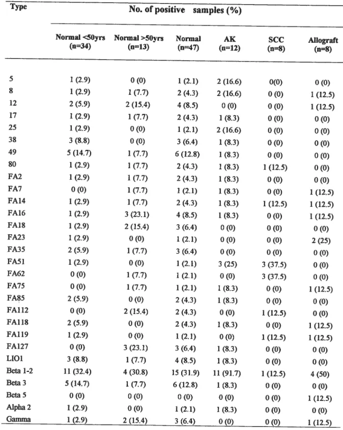
Detection rates ofHPV types in 75 3-globin-positive swab samples.
Table 3:
New putative types and the closest related kown HPV types
Table 4:
Detection rates ofHPV types in 75 f3-globin-positive skin samples selecting HPV types detected in more than two samples.
Table 5:
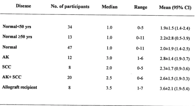
Burden ofHPV infection in 75 3-globin-positive skin samples measured as the number of types detected per sample and underlying disease.
LI$T 0F ABBREVIATION$
AK: Actinic keratosis
BPV: Bovine papillomavims
BCC: Basal ccli carcinoma
Bp: Base pair
C1N: cervical intraepithelial neoplasia
CSf-1R Colony-stimulating factor-1 receptor
DNA: Deoxy-riboneuclic acid
EGF: Epidermal growth factor
EGFR: Epidermal growth factor receptor
EV: Epidermodysplasia verruciformis
FISH: Fluorescence in situ hypridization
HPV: Human papillomavirus
Kd: Kilodalton
LCR: Long control region
MED: Minimal erythema dose
NMSC: Non-melanoma skin cancer
NCR: Non-coding region
Nt: Nucleotide
NIKS cells: previousIy named Bcl-Ep/$L ceils
ORF: Open reading frame
PCR: Polymerase chain reaction
PV: Papillomavims
PDGFR: Platelets-derived growth receptor B
PUVA: Psoralen and UVA light treatment
RTR: Renal transplant recipients
$V40: Simian vacuolating Virus 40
SCC: Squameous ceil carcinoma
UVR: Ultraviolet rays
UV: Ultraviolet UVA: Ultraviolet A UVB: Ultraviolet B UVC: Ultraviolet C VW: Viral warts WT: Wild type
DEDICATE
I dedicate this memory to myparentsfor their encouragement andsupport, tomy husband ami my kids.
ACKNOWLEDGMENTS
My thanks go to my research director Dr. Francois Coutlee, forhis availability ail during the course ofmy master. My thanks go also to the attending research Simon Gagnon and
Introduction
Human papillomavimses (HPV) are small double-stranded DNA vimses found in a wide variety of proliferative lesions of epithelial origin. In recent years there has been a considerable increase in the number of identffied human papillomavims types. There are currently more than 100 distinct types based on DNA sequence homology, but several groups and partially characterized novel sequences have now been described, predicting the existence of many more. Accumulating epidemiological and experimental data strongly support that there is a relation between HPV infection and benign or malignant neoplasia. Mthough the nature ofthe association is flot definitive, certain cutaneous HPV types are found in a high percentage of non-melanoma skin cancers (NMSC) but no HPV types have been associated specifically with NMSC in the general population. Some cutaneous HPV types (I{PV-1, 2, 4, 7, 57) are associated with benign planter/palmer and common skin warts [1]. On the other hand, evidence suggesting an etiologic role for HPV in NMSC comes from studies on the rare disorder “epidermodyspiasia verruciformis” (EV). About 30% of patients with this disease deveÏop squamous celi carcinomas (SCC) ofthe skin, especially on sun-exposed sites [2].
In recent years, several consensus primer-mediated PCR techniques that allow for the detection of a wide range of HPV genotypes have been designed. Many of these methods have been used for detection ofIIPV types in healthy skin as well as in NMSC. These methods have included single round PCR using one pair ofdegenerate primers [3], combinations ofdegenerate primers
[4,5,61
or nested PCRs using two pairs of degenerate primers [7]. In our study we compared two primer pairs (FAP59/64 and HVP2/B5) optimized to detect cutaneous HPV infection [3,4,8,9,10,111.1 ITUMAN PAPILLOMAVIRUS:
1.1 History
Human papillomaviruses (HPV) are small, non-enveloped double-stranded DNA epitheliotropic viruses [1]. They belong to the papillomaviridae family, members of which infect squamous epithelia and cause proliferative diseases (papilloma) in a number of vertebrates, including man, non-hum an primates, caille, rabbits and dogs, in a highly species-specific manner.
MOPEI. 0F HLYMAN IAPI.OrVTRUs
ijorCxidf’mtein (u)
Viral Nud’c P.cid (oN)
Figure (1): Human papillomavirus
HPV was the first tumor virus to be transmitted experimentally from one host to another. This was accomplished in 1894 by Licht [2] who transmitted warts ftom his brother to himself by inoculation of crude wart material. Ciuffo [3] in 1907 and Serra
[41
one year later demonstrated that warts could be induced by celi- free filtrates of wart material. In 1919, Wile and Kingrey [5] successfully transmitted warts through a succession of human volunteers using sterile extracts of wart material, Electronmicroscopic studies by Strauss et al [6J, Melnick et al [7], Bunting [8] and Mmeirta et al [9] confirmed a viral etiology for cutaneous warts.
Melinck in 1962 classified the papillomaviruses together with the polyomavimses and 5V40 (Simian Vacuolating Virus 40) in the papovaviridae family because they were small DNA vii-uses sharing ultra-structural features [10]. Reports of transmission of fihrates ofwarts, laryngeal papillomas, and genital tract condylomas to human volunteers who developed typical cutaneous warts at the sites of inoculation were interpreted as indicating that there was one type of human wart virus and that the site of infection and perhaps, the genetic makeup of the patient, determined the clinical appearance of cutaneous warts and mucosal papillomas [11]. However, recognition that there were different papillomavirus types ami subtypes stimulated a new interest in the i-ole of these vii-uses in hyperpiasia and neoplasia arising in squamous epithelia of the anogenital and digestive tracts [12,13,14,15,161.
12 HPVStructure andBiotogy:
1.2.1 General biology offlPV
HPV virons are non-enveloped icosahedral capsids (50 to 55 nm in diameter) of 72 capsomeres [17]. They do flot contain lipids and are inactivated by treatment with 0.4% formalin for 72 hours at 4°C [18]. Both complete and empty particles may be found in tissue samples [19,20].
Molecular analysis of PV DNA was first determined by Crawford [21] and Crawford [19,20]. Using stringent hybridization techniques without the aid of various restriction endonucleases, it was concluded that although PV from various species had similar structures and molecular weights, there was no polynucleotide sequence shared among the different genomes analyzed. The PV genome is found in virions and infects cells in three forms: a covalently closed, supercoiled molecule with a sedimentation coefficient of 23 S, uncoiled circulai- molecule with a sedimentation coefficient of 17 S,
and a linear molecule with a sedimentation coefficient of 16 S. [22,23]. The molecular size ofthe genome based on agarose gel electrophoresis and contour length measurement of DNA molecules by electron microscopy reveals a molecular weight of approximately 5x10⁶ Daltons, corresponding to 8000 base pairs, which is sufficient to code for proteins of 300,000 Daltons [12,131.
1.2.2 Mokecular biology of papillomavirus oncogenes:
Papillomavirus genomes for each type contain 9-10 open reading frames. With variable spiicing pattems, they have the potential to synthesize 12-15 gene products. The open reading frames labeled “E,” for early, represent those genes in bovine papillomavirus (BPV) which were thought to be involved in episomal replication in cultured ceils. The late, or “L,” genes encode the viral capsid proteins. The number afier E or L refers to the size of the peptide coded by the open reading frame, one being given to the largest peptide [24].
. I
—
Eb E L
E1 E E Et’ L2
4 14
t1on-b:ruCur.itprOEr Virion ro:in, G.nor o
O h bIbp
I I I I
Simplified organic1tion (Iinectrised) of human papiIIomaviru type 16 (HPV-16) génome
n MOIeŒJIOT cioine
figure (2): Simplified organization ofthe linearised genome of human papillomavirus type 16. The scale bar is in kilobase pairs. The rectangles represent the positions ofvarious open reading frames (ORFs). The E genes encode proteins that are produced early in the infectious cycle (the non-structural proteins), whereas the L genes encode proteins that are produced late in infection (virion proteins that are necessary for virus assembly). The actual protein products of the genes are complex owing to the production of multiple messenger RNA(mRNA) transcrÏpts.
Table (I): HPV gene products and their function.
Gene Function
El Initiation ofDNA replication
E2 Transcriptional regulationmNA replication
E3 ?
E4 Disrupts cytoskeleton?
E5 Transforming protein, interacts with growth factor receptors
E6 Transforming protein, binds to p53, leading to degradation of
p53
E7 Transforming protein, binds to pRB
E8 ?
LI Major capsid protein L2 Minor capsid protein
1.2.2.1 The E6 oncoprotein:
Papillomavirus E6 protein consists of about 150 amino acids believed to bind a zinc atom through two sets of cysteine repeats (cysteine X-X cysteine zinc fingers, where X is any amino acid) [25,26]. The human papillomavims E6 proteins have moderate homology at the amino acid level, indicating that while they share functions, they may also differ. HPV-16 E6 has a haif-life of 30-60 min and is present in transformed and cancer-derived celi unes at extremely low levels [27]. Several reports have demonstrated that high and low-risk E6 genes can stimulate transcription equally, suggesting that some E6 fiinction may be relevant to viral replication rather than correlate with oncogenic potential [28,29,30].
The high-risk HPV-16 and 12 F6 proteins interact with p53, as do $V 40 large T antigen and adenovirus Elb [31]. HPV and other vimses presumably interfere with the ability of p53 to block celi division and DNA synthesis so that viral DNA can replicate to high levels. Indeed these mutations could permit genetic drift that can circumvent immune defenses. SV40 and adenovirus prevent p53 mediated ceil cycle arrest by synthesizing large amounts of large T and Eib, which effectively hold p53 in inactive complexes. F6 protein eliminates p53 functions tbrough a novel mechanism. It lias been shown that formation of the F6-p53 complex in vitro induced p53 degradation through an ubiquitin-dependent mechanism [32]. High-risk F6 binds a 100-kDa protein, called F6-AP (for F6 Associated pfotein), which appears to be required for the binding of E6 to p53, and is necessary for degradation of p53 [33]. The gene for E6-AP is a member ofthe ubiquitin pathway for protein degradation [34]. It lias been reported that although low risk (HPV-6 and 11) E6 also binds p53 with reduced efficiency, it is flot capable of inducing p53 degradation [35].
Despite these important findings, there are several unes of evidence that suggest that F6 possesses other ffinctions. BPV F6 does flot bind p53, yet fiuly transforms murine C127 cells [36]. BPV F6 and HPV6 F6, which do not induce p53 degradation in vitro or in vivo, can immortalize human mammary epithelial celis, although they are much less efficient than HPV-16 [37]. HPV-$, which is found in cutaneous $CC in patients suffering from epidermosysplasia verruciformis, does not bind or degrade p53 in vitro but, similarly to BPV F6, transforms mouse cells [32,39].
1.2.2.2 The f7 Oncoprotein:
The F7 oncoprotein is an acidic 98 amino acid phosphoprotein that has been localized in the nuclear matrix [40]. Two cysteine-X-X-cysteine motifs in the carboxy terminus of the protein mediate zinc binding and dimerization [41]. The recognition of the amino acid similarities between the ITPV F7 protein and the DNA tumor virus transforming proteins SV4O Large T and adenovims Fia facilitated elucidation of the biochemical properties of F7 [42,43]. These regions of similarity were shown to bind the
tumor suppressor gene product Rb and the reiated p107 proteins [44,45,46,47]. Both Rb and p107 regulate ceil cycle division but act at different steps. The p107 binding region
ofE7 overlaps with but canbe distinguished from the Rb binding domain [48,49].
It is though that when E7 binds Rb or p107, a transcription factor nonnally bound to Rb/p107 is released, because E7 and the factor bind the same pocket [50,51]. This factor, called E2F, is a sequence-specific DNA binding protein, and the DNA motif it recognizes is found in many genes essential for celi division [52]. There is evidence that the events induced by the Rb/ p107 association with F7 can lead to a complex yet coordïnated cascade of positive and negative signais that allow a celi to replicate its DNA and divide. Other regions of F7 bind additional growth-related cellular factors. HPV-16 F7 has been identifled in complexes with histone kinase, p33 and cydliui A and casein kinase II, which phosophorylation may in part relate to the oncogenic potential of high riskE7 [53,54,55,56].
Both HPV-16 and 18 F7 induced focus formation in murine ceil une transformation assays [57,5$]. Consistent with its interaction with RbIp 107, addition of F7 alone into primary human keratinocytes resulted in an increased rate of proliferation for an extended period oftime, although celis eventually senesced [59]. Using very high efficiency retroviral-mediated infection of primai-y human keratinocyes, it has been shown that low-risk HPV-6 F7 cancooperate with high-risk E6 to induce immortalization
ofprimary human keratinocyes, and in the alternative mixing experiment, HPV-6 F6 cooperated with HPV-16 F7 to induce immortalization, although in both instances, the efficiency was less than with high-risk E6 and E7 [60]. As with E6, there remain unidentified properties ofE7 that arerequired for its transforming activities. For example, mutations in F7 that do not affect Rb/p107 association interfered with its ability to transform and immortalize ceils [61].
figure (3): E7 Effects on Rb. E7 binding to Rb Ïeads to the release of sequestered E2f, enabling the ceil cycle to progress
1.2.2.3 The E5 oncoprotein:
The F5 protein represents another fascinating and ingenious means developed by papillomaviruses to prime cells for viral replication. An F5 gene has been identified in bovine, deer, elk, and some human papillomaviruses. These animal viruses cause dermal fibroblast proliferation (fibropapillomas) along with an epithelial component in their hosts. E5 protein is small: the BPV E5 gene product contains only 44 amino acids. The I{PV E5 gene is ofien deleted or its gene flot expressed in human cervical carcinomas, whereas E6 and F7 are always transcribed. This suggests that F5 plays a mie at an early stage of viral carcinogenesis. BPV-1 and HPV-6c ES have been reported to transform established murine fibroblast ceil unes, whereas HPV-L6 ES did flot [36,62,631.
Introduction of HPV-16 E5 into established murine keratinocyte unes induced their tumorigencity in nude mice, suggesting a potential role in human malignancy [64].
In contrast to the HPVs, the major transforming protein of bovine papillomavirus type 1 (BPVI) is the F5 protein, a 44-amino-acid highly hydrophobic protein that localizes predominantly to the Golgi and exists as homodimers [65,66,67]. The BPV1 E5 protein is able to transform both murine fibroblasts and keratinocytes in transformation assays in vitro [64,68]. The BPV1 F5 protein is able to bind to and activate the platelet derived growth factor
f3
receptor in the absence of exogenous ligand [69,70], and this has been shown to correlate with cellular transformation [71]. BPV1 E5 also binds to the 16-kDa pore-forming membrane component of the vacuolar proton ATPase (v-ATPase), a protein essential for the acidification of intraceliular compartments such as lysosomes, endosomes, and the Golgi [72]. The binding ofBPV1 E5 to the 16-kDa protein is able to cause aikalization of the Golgi, and this has been shown to correlate with cellular transfonnation [73]. Because of the structural similarities between the BPV1 E5 and HPV-16 E5 proteins, and because BPV-1 F5 has strong transforming potential, work was begun to determine if HPV- 16 ES was also an oncogene.E5 seems to be important early in the course of infection. It stïmulates ccli growth by forming a complex with the epidermal growth-factor receptor, the platelet-derived growth-factor-B receptor and the colony-stimuiating factor-1 receptor [74]. Recently, F5 has also been shown to prevent apoptosis following DNA damage [75]. However, as HPV-infected lesions progress to cervical cancer, the episomal viral DNA ftequently becomes integrated into host-cell DNA. And a substantial part ofthe genome, commoniy including the ES coding sequence, is deleted [76]. So, ES is flot obligatory in late events of HPV-mediated carcinogenesis.
Unlike E6 and E7, the major viral oncoproteins, the E5 pfotein of HPV-16 is flot commonly found in cervical carcinoma celis [77,78]. However, it is considered an oncogene given its ability to transform mouse fibroblasts and keratinocytes, cause mitogenic stimulation of human keratinocytes, and cooperate with E7 to stimulate
proliferation of human keratinocytes [64,79,80,81,82]. The E5 gene of HPV-16 is a 83-amino-acid hydrophobic membrane protein [$3,841 that localizes to the Golgi apparatus, endoplasmic reticulum, and nuclear membrane [85].
Multiple studies have suggested that the HPV-16 E5 gene could interact with epidermal growth factor receptor (EGFR) signaling. Studies indicate that HPV-16 ES causes an increased activation of the EGFR in the presence of ligand [79,80,81], and coimmunoprecipitation experiments indicate that BPV-16 E5 can form a complex with growth factor receptors [74]. The HPV-16 E5 protein also binds to the 16-kDa membrane component of the v-ATPase [$5] and delays endosomal acidification in human keratinocytes [86]. It has been argued that in binding with the 16-kDa protein, E5 disrupts the 16-kDa protein-v-ATPase complex [87,8$], which resuits in the inhibition of endosomal acidification.
E5 acts during the productive stage ofthe HPV-16 life cycle. A study showed that
HPV- 16E5 mutants infecting basal celis resuits in a lower percentage of supra-parabasal ceils undergoing DNA synthesis compared with ceils harboring the (Wild Type) WT HPV-16 genomes [91]. Previously, it had been reported that E7 plays a critical role in the productive stage of the HPV- 16 life cycle [89]. In that study, it was demonstrated that HPV-16 E7 mutants do not reprogram supra-parabasal ceils to support DNA synthesis, and this defect correlates with the absence of viral DNA amplification. That study also established that ceils harboring HPV-16 F7 mutants failed to modulate the differentiation program ofrafi cultures ofNIKS celis and displayed reduced expression of Li, the major viral capsid protein expressed in the productive stage of the HPV- 16 life cycle. In contrast, the HPV-16 E5 mutant genomes did modulate the differentiation program in rafi cultures ofMKS ceils, as was seen in NIKS harboring the WT HPV-16 genome. HPV-16 E5 mutants also expressed late viral proteins in the productive stage of the HPV- 16 life cycle at the same levels as seen with HPV-16 WT. HPV-16 infected ceils retained viral DNA amplification as analyzed by fISH. Thus, E5 plays a more subtie role during the productive stage ofthe viral life cycle than does F7.
A parallel study of E5 in the context of the HPV-3 I life cycle by Fehrmann et al. [90] has also shown a subtle effect of F5 during the productive stage of the viral life cycle. In both studies, the disruption of ES had no observable effect on the non-productive stage of the viral life cycle. Specifically, HPV-16 and IIPV-3 1 with E5 mutations [90,911 could be maintained as nuclear plasmids. The E5 variation in HPV-16 was a deletion of an adenosine at position 30 of the F5 sequence. Celis harboring these E5 mutant genomes displayed similar growth kinetics in the absence or presence ofEGF in monolayer cultures compared to cells harboring the WT HPV genome. In contrast, both studies noted defects during the productive stage ofthe viral life cycle.
Cells harboring HPV-16 E5 mutants displayed a significant reduction in the percent of supra-basal cells undergoing DNA synthesis in raft culture. They also found a lower induction of cyclins A and B and lower retention of proliferative potential in ceils harboring HPV-3 1 E5 mutants, compared to those harboring WT genomes, upon suspension of those cdl populations in semi-solid medium. In both studies, celis harboring F5 mutants retained the ability to amplify viral DNA upon induction of cellular differentiation. The investigators discerned a two-fold decrease in the degree of amplification in celis harboring E5 mutants compared to those harboring WI’ HPV-3 1 by Southem analysis. They also observed a twofold decrease in the ftequency of celis supporting amplification of the E5 mutant genome, based on FISH analysis of organotypic rafi cultures. However, this difference was flot statistically significant. Southem analysis of DNA extracted from HPV-16 WT and HPV-16 ES mutant rafis cultures was performed as described by Ozbun and Meyers [92] but they could flot detect viral DNA amplification in the HPV-16 WT rafts or in rafts generated with a clone of cervical epithelial cells (W12E cells) that harbored episomal HPV-16 DNA. They believed that this absence ofdetectable amplification was due to the very low percentage of celis (0.09 to 3.48%) within a rafi supporting viral DNA amplification as shown by their fISH analysis [91].
Studies have indicated that HPV-16 E5 is able to cooperate with E7 to induce proliferation, enhance immortalization, and promote anchorage-independent growth of
baby rat kidney ceils [79,93]. In these studies, it was found that transfection of F5 alone into primary rodent ceils had littie effect on proliferation of these ceils and that F7 alone was able to increase proliferation in comparison to the vector alone. However, cotransfection of ES and F7 resulted in a significant increase in the amount of proliferating colonies over that of F7 alone. Considering that F5 contributes to the capacity of HPV-16 to reprogram differentiating celis to support DNA synthesis, a property also reliant on E7 [89,91], F5 could play a cooperative role with F7 in the productive stage ofthe viral life cycle [91].
The mechanism by which HPV-16 F5 is contributing to the productive stage of the viral life cycle is not yet clear. Many studies have suggested a link between HPV- 16 ES protein and the EGFR signaling pathway. These studies suggest that when treated with EGF, E5-expressing ceils display anchorage-independent growth [$1], increased mitogenic potential [62,82], and increased growth factor receptor signaling (with or without FGF) [67,94]. Whereas Laimins et al found littie, if any, EGFR present in keratinocytes following suspension in semi-solid medium, other investigators clearly found that FGFR was present in the superficial layers of rail cultures of early-passage human foreskin keratinocytes or NIK$, albeit at lower levels than those observed in the basal layers [91,95]. A similar expression pattem ofthe EGFR in basal as well as supra-basal compartments of rail cultures has been reported in the context of an HPV-3 1-positive CIN 1 lesion-derived population, the CIN 612 9F ceils [96]. In that study, the authors also monitored expression of HPV-3 1 F5 in the context of rail cultures and found that E5 protein levels were induced in a time-dependent manner, suggesting that its expression is tied to the differentiation and stratification of epithelial ceils. Consistent with this observation, they detected F5-positive celis within the more superficiai layers of the CN 612 9F rail cultures. These data indicate that both E5 and one of its known targets, EGFR, are expressed within the terminally differentiating ccli compartment in which we have observed an effect of F5 during the productive stage of the viral life cycle.
The binding of HPV-16 F5 to the 16-kDa component of the v-ATPase may also be important in F5’s contribution during the productive stage ofthe viral life cycle [85]. It
has been shown that endosomal acidification ofHPV-16 E5-expressing celis is inhibited and that this can lead to increased receptor recycling to the celi surface [86]. Another study has suggested that HPV-16 E5 affects trafficking from endocytic compartment rather than endosomal acidification [97]. Studies under way will allow us to determine which ofthese mechanisms are involved in E5!s contribution during the productive stage of the viral life cycle.
1.2.2.3.1 Interaction ofES with cettutarfactors:
BPV-1 F5 forms complexes with different transmembrane proteins. Thus, E5 directly binds to the transmembrane domain to the platelet-derived growth receptor B (PDGFR) and ffinctionally interacts with the epidermal growth factor receptor (EGFR) and the colony-stimulating factor-1 receptor (CSF-1R), [69,98,99]. Through these interactions, the receptors may be activated in a ligand-independent manner. Moreover, their signais, e.g. increased receptor phosphoiylation, mitogen activated protein kinase activity and phospholipase C-y-1 activity are enhanced even in the absence of a physiological ligand [67,100]. This may resuit from interference with receptor degradation and intemalisation [101,1021.
NPV-16 and HPV- 6 E5 also have been shown to co-operate with the EGFR and PDGFR [$0,8 1,82]. Moreover, HPV-16 E5 enhances endothelin receptor signaling [103]. Whereas HPV-6 F5 also associates with the EGFR, the related erbB2 receptor and the PDGFR, HPV-16 E5 does flot bind to cellular growth factor receptors [104]. Another common target of both BPV and HPV E5 proteins is a 16 kDa membrane pore protein representing a subunit of the ff-dependent vacuolar ATPase [72,85]. For ATPase binding, the glutamine residue within the hydrophobic domain seems to play an important role [105]. As a result of this interaction, acidification of endosomes is inhibited. This has been suggested to be responsible for the prolonged retention and reduced degradation of the EGFR in the presence of the F5 protein [$6]. However, the binding ofNPV-16 E5 to the 16 kDa ATPase subunit couid be dissociated from the F5-mediated EGFR overactivation [1061.
1.3 CtassWcation ofHPV:
Until the late 1 970s, papillomaviruses affracted littie interest as they were only known as the causal agents of “warts”, benign cutaneous lesions in some mammals and humans. Since warts are normally only a cosmetic problem but flot a major threat to public health, they were mostly of academic interest. In the 1 980s, newly developed powerful molecular biology techniques led to the detection of dozens of human papillomaviruses in benign and malignant mucosal lesions [14,107], such as cervical cancer and its precursor lesions, as well as in genital and laryngeal warts. Present data support the existence of more than 100 HPV types. The whole genomes of about 100 HPV types have been isolated and completely sequenced, while we have only indirect evidence for the other types, most ofien through the sequence of polymerase chain reaction (PCR) amplicons, which unambiguously identified the presence of HPV-related sequences. Description ofHPV types has changed several times in parallel with technical progresses and phylogenetic analysis. An HPV type is now defmed as an isolate whose Li gene sequence is at least 10% dissimilar to that ofany other known HPV types.
1.3.1 C]assification according to sequences nucleotides:
In the mid-1950s to 1960s, papillomaviruses and polyomaviruses were analyzed
by electron microscopy and basic nucleic acid analyses. These two groups of viruses were found to be the only one with double stranded circular DNA genomes, and non enveloped particles consisting of icosahedral capsids. As a consequence, they were considered closely related and were placed ïnto a common family, the papovaviruses Papoviridae. Sequence and functional studies in the 1 9$Os showed that these similarities were too superficial to establish relationship [10$]. Ml polyomaviruses have a genome size around 5 kb, while the genome of papillomaviruses is close to $ kb. Polyomaviruses transcribe mRNA from both strands, while papillomavirus transcription occurs only in one direction. And lastly, and most importantly, polyomavimses and papillomaviruses do flot share any substantial amount of nucleotide or amino acid sequence similarity, with the exception ofa small homologous segment in the T-antigen and Fi genes, respectively
[109]. Since taxonomic classification should reflect natural relationships, it was concluded that these vimses form two separate families. Only fairly recently, the famiiy “papillomaviruses” (Papillomaviridae) became officially recognized by the International Council on Taxonomy ofViruses (ICTV) [1101.
1.3.1.1 Papillomavirus types:
Papillomaviruses are identified by the abbreviation PV and one or two letters indicating the host species. This can be derived from an English word, for example
“HPV’ for human papillomavirnses ami “CRPV” for coffontail rabbit papillomaviruses, or the scientific name of the host, e.g. MnPV for Mastomys natalensis papiliomavims, which infects an African rat. HPV types are identified by numbers considering the historie sequence of their description, e.g. HPV- 1, HPV-2, etc. [111]. Presently, and for the last 20 years, E.M. de Villiers at the Reference Center for Papiilomaviruses at the German Cancer Research Center in Heidelberg has controlled this process [112]. New HPV types have to be registered by this center to confirm completeness of the genomic isolate. New types have to show 10% nucleotide sequence diversity in the Li gene from ail known HPV types. Thereafier, assignment of a new number and publication of this new HPV types is possible. HPV types, whose genome was generated by PCR rater than traditional cloning techniques, are identified by addition of the abbreviation “cand” (for candidate) before their number, e.g. candHPV-86 [110].
1.3.1.2 Subtypes:
The term “subtypes” was used in the 1980s to identifS? isolates of an HPV type with different restriction nuclease digestion patterns. Subsequently, this term became redefined as referring to an isolate whose Li sequence is 2-10% different from that of any known type. A consequence ofthis redefinition was that several subtypes (e.g. HPV 6a, HPV-6b and HPV-6c) had to be eliminated, as they showed less than 2% sequence diversity. $urprisingly, today only three HPV isolates are known to fulfihi this latter
subtype definition. HPV-46, HPV-55, and HPV-64 had been originally described as separate types, but their types status has now been cancelled, as they are subtypes of HPV-20, HPV-44, and HPV-34, respectively [110].
1.3.1.3
Variants:“Variants” ofHPV types differ by at most 2% in the ORF sequences or by at most 5% in the LCR of the original isolate characterized, also referred as “prototype” or “reference genome”. Variants are identffied by PCR-sequencing mainly. This strategy has been applied to numerous HPV types from isolates throughout the world [113,114,115,116,117,118J. The two principal observations from these studies are that there is apparently only a limited number (for example 20-100) of common variants for each ITPV type, and that variants showed maximal divergence when they were sampled from different groups living in different countries. HPV types have these coevoived with the human species [1191. HPVs did not infect humans from an animal reservoir such as for some other vimses, like Ebola, the SARS coronavirus, or influenza. There are indications that variants of the same HPV type differ biologically and etiologically [120,121]. Such differences may contribute to the disparities in the incidence of cervical cancer throughout the world, although this question stiil requires substantial research before conclusions can be drawn [122].
1.3.2 Classification according the site of infections (Genital, cutaneous or mucosal):
It was found that different HPV types associated with similar lesions are sometimes only very distantly related to one another. For example, HPV-1, HPV-2, HPV 4 and HPV-41 are ail found in wart-iike cutaneous lesions, but are on remote branches of evolutionary trees. HPV-16 and HPV-18, the two HPV types that have become paradigms in the research on cervical carcinogenesis, are less related to one another than to some HPV types that are neyer found in cervical malignancies.
Table (2): Cutaneous types ofHPV ffPV Type 1, 2, 4, 26, 27, 29, 41, 57, 65 1, 2, 4, 63 3, 10, 27, 28, 38, 41, 49 1, 2, 3, 4, 7, 10, 2$ 2, 27, 57 16 2, 3, 10, 12, 15, 19, 36, 46, 47, 50 5, 8, 9, 10, 14, 17, 20, 21, 22, 23, 24, 25, 37, 3$ 37, 38
Non-genital Cutaneous Disease Common warts (verrucae vulgaris) Plantar warts (myrmecias)
flat warts (verrucae plana)
Butcher’s warts (common warts of people who handie meat, poultry, and fish)
Mosaic warts
Ungual squamous cell carcinoma
Epidermodyspiasia verruciformis (benign) Epidermodysplasia verruciformis (malignant or benign)
Non-warty skin lesions
Table (3): Mucosal types ofHPV (non-genital). HPV Type 6, 11 6, 11, 16, 1$ 6, 11,30 16,18 57 16,1$ 6,11 16 13,32 16,18 16,12 16,18
Non-genital Mucosal Disease Respiratory papillomatosis
Squamous cdl carcinoma ofthe lung Laryngeal papilloma
Laryngeal carcinoma Maxillary sinus papilloma
Squamous ccli carcinoma ofthe sinuses Conjunctival papiliomas
Conjunctival carcinoma
Oral focal epithelial hyperplasia (Heck disease) Oral carcinoma
Oral leukoplakia
Table (4): Mucosal types ofHPV (Anogenital).
HPV Type Ano-genital Disease
6, 11, 30, 42, 43, 44, 45, 54 Condylomata acuminate
16, 18, 34, 39, 42, 45 Bowenoid Papulosis
16, 18, 31, 34 Bowen disease
6,11 Giant condylomata (Buschke-Lôwenstein tumors)
30, 34, 39, 40, 53, 57, 59, 61, 62, Unspecified intraepithelial neoplasia 64, 66, 67, 6$, 69
6, 11, 43 Low-grade intraepïthelial neoplasia
31, 33, 35, 42, 44, 45, 51, 5216, 18, High-grade intraepithelial neoplasia 56, 58, 59, 66
6, 11, 16, 18 Carcinoma ofvulva Malignant Vulvar Lesions
16 Carcinoma ofvagina
16, 18, 31, 33, 35, 45, 51, 52, 56, Carcinomaofcervix 58, 59, 66
16, 18, 31, 33, 35, 45, 51, 52, 56, Carcinomaofanus 58, 59, 66
16 Carcinoma in situ of penis (erythroplasia of Queyrat)
16,18 Carcinoma ofpenis
1.3.3 Classification according to the high risk or Iow risk
As a consequence of infection and viral persistence in the epithelium, a variety of proliferative lesions may arise in the skin or mucosa according to their preferential sites of infection. These include benign warts and condylomas but also dyspiastic lesions, which may ffirther de-differentiate and progress to cancer. Distinct clinical entities have been found to be associated with distinct HPV types. Based on the biological or clinical properties, HPV types have therefore been classified into “low-risk” types mainly
associated with benign disease only and “high-risk” types also associated with malignant disease (124).
In general, sometimes we are classifying types according to site of infection could led to confiising contradictions. Examples ofthese contradictions include:
• HPV-6 and HPV-l I are typically found in genital warts or condylomata acuminate in the genital tract, and they were therefore considered “genital” HPVs. However, they are also found in non-genital sites, for example in papillomas of the larynx [123].
• Genital warts can be of mucosal as well as cutaneous origin. HPV types involved include mucosal types as well as HPV-2, HPV-27, and HPV-57, which are causes ofcommon warts, and ITPV-7 [123].
1.4 Mechanism of human papiiomavirus infection:
1.4.1 Entry:
Papillomavimses gain access to the basal celis through physical breaks in the epithelial barrier. Basal ceils are infected by one or two copies ofthe episomal viral DNA per celi. The extrachromosomal viral DNA in basal ceils replicates in concert with normal cdl division thus maintaining a constant number of episomal HPV genome per cdl. Presumably, the viral DNA is maintained in the daughter celis in upper levels ofthe epithelium. It is in these strata of ceils undergoing differentiation that viral RNAs are expressed at substantial levels [125]. The differentiation-specific cellular events promote viral transcription and DNA replication. Thus papillomavirus is shed to high numbers only in expendable, terminally differentiated ceils sloughed off the epithelium. NPV are not lytic viruses [24].
1.4.2 Shedding:
Aller many copies of the circular viral DNA are synthesized in the upper strata, these genomes are incorporated into a particle, or capsid, which consists ofthe Li and L2 proteins. The viral capsid protects its DNA as the keratinocytes terminally differentiate as well as after being shed into the envfronment. linportantly, papillomaviruses do flot bud from the cell’s plasma membrane and thus do not incorporate a membrane-derived lipid envelop. HPV particles are thus flot sensitive to environmental stresses such as heat, soaps, or desiccation. Aller assembly, papillomaviruses are carried along with differentiated ceils to the stratum corneum, where they are released with cell death due to apoptosis [24].
EpidermalCeil Differenti tian Pathway
j Capsidpmteir.s VirusprticIes RepIictng virdflNA
(
ExpreIon of)
early PapIII orna VirusLite Cc1eStamo
-N
Slrawmsplnosum ciiecnts)Figure (4): Schematic representation of a skin wart (papilloma). The papillomavims life cycle is tied to epithelial cell differentiation. The terminal differentiation pathway of epidermal ceils is shown on the lefi. Events in the virus life cycle are noted on the right. Late events in viral replication (capsid protein synthesis and virion morphogenesis) occur only in terminally differentiated celis.
1.4.3 Coordination ofthe viral replication cycle:
After entering the appropriate host ccii, viral gene will be expressed in an ordered fashion. Within the non-coding region (NCR) of each virus are DNA sequences that are recognized by cellular transcription factors [126,127,128,129,130]. The papillomavirus genotypes vary in the type, array, and position ofthese sites, which could have important consequences for pathogenicity.
In addition to these binding sites for cellular factors, papillomaviruses encode a DNA binding protein designated E2. E2 binds the inverted palindrome 5’-ACCG NNNN CGGT-3’ with vely high affinity (—10’1M) [131], and multiple copies of this DNA
palindrome are found in every papillomavirus genome [132]. Their position ofien varies among different genotype groups. E2 can stimulate, and under certain instances repress, viral transcription [133,134,135]. E2 is also required for viral DNA replication. When F2 binds its recognition site, it usually stimulates transcription from the nearby promoters in
a classical “enhancer” mode.
The E2 protein averages about 400 amino acids with a monomeric molecular weight of approximately 50 kilo-Daltons (KD) and has distinct functional domains [136,137,138]. The carboxy terminal 100 amino acids of ail papillomavirus F2 proteins constitute the DNA binding domain. This small region is sufficient for sequence-specific DNA binding and dimerization, which E2 must do to bind DNA [139]. The atomic structure of the BPV F2 protein bound to its cognate DNA site was found to fold as an unusual B- barrel dimer with a DNA recognition helix crossing each monomer [140].
The amino terminal haif of the F2 protein is necessary for activation of gene expression, so these amino acids must interface with the ccli’ s transcription machinery. This region ofE2 represents its transcription activation domain. When F2 binds a specïfic segment on the viral DNA through its DNA binding domain, its transcription activation domain recruits the cellular factors that lead to synthesis of the viral mRNA. One such
factor may be Spi, although there is evidence that E2 activates transcription in concert with a variety of basal promoter factors [141,1421.
The E2 DNA binding and transcription activation domains share considerable homology among the papillomaviruses. Between these two regions is a variable stretch of residues that are flot conserved, and are generally thought to represent a “flexible hinge.” The E4 reading frame overlaps this E2 hinge region and the papillomaviruses differ substantially in their E4 protein. E4 is flot essential for viral transcription, replication, or transformation in vitro and is believed to serve a role in maturation of the viral capsid and escape ofthe viral particle from the dense intermediate filament network of the epithelial cell [143,1441.
Papillomaviruses do not express high levels of their RNAs and proteins until the late stages of epithelial differentiation. To restrict expression of the viral genes, BPV and HPV generates tmncated E2 proteins to act as transcriptional repressors [145,146,147,148]. These repressors lack the amino terminal transcription activation domain but retain a functional DNA binding domain [149]. Therefore they compete with full-length F2 proteins for binding the E2 recognition sites on the viral genome. In addition, the E2 protein normally is a dimer, and formation of a heterodimer between a full-length and truncated F2 protein represents another mechanism for the inactivation of E2-induced transcription [150]. Both full-length and tmncated E2 can repress viral transcription factors to their recognition sites in the LCR Dominance of the latter function may reduce viral expression in basal and parabasal kerationcytes.
1.4.4 Reptication ofthe viral genome:
The viral DNA must be selectively replicated in differentiated celis to produce a higli level of infectious progeny in each ce!!. This would normally be very detrimental except that PV replication occurs in terminally differentiated cells. This highlights the central paradox of papillomavirus replication: it begins in a non-replicating cdl layer in which the multitude of enzymes necessary for DNA synthesis is thought flot to be present. Because papi!!omavirus do flot encode a DNA polymerase or the associated factors necessary to duplicate DNA, they must induce the ccli to replicate while differentiating. Consequently, the virus must mobilize these ce!lular factors to repiicate the viral genome.
Figure (5): The $000 base pair papillomavirus genome is shown diagrammatically on the right. Vira! proteins are expressed in a highly organized paftern as an infected cel! migrates towards the epithelial surface. The timing of viral protein expression in infected epithe!ium is shown on the left.
Papillomavims utilizes two proteins, E2 and El, to identify their genomes among the mass of host DNA. Mutations in the viral El gene interfere with autonomous replication ofthe viral DNA [151]. The El protein binds to E2 protein [152,153,154]. It is believed that E2 and El each bring a set of cellular factors to the viral DNA, and these
factors replicate the viral DNA. E I wealdy binds a specific DNA sequence in the viral regulatoiy region, and this activity of El is greatly enhanced when it is complexed with E2 [155,156]. In BPV, the El binding site is adjacent to E2 sites, and this segment of viral DNA is sufficient for autonomous replication in murine ceils when Fi and F2 proteins are expressed [157]. The El protein has helicase activity, which is necessary for separating theHPVDNA strands prior to their replication [158]. Both BPV and HPV Fi and F2 are necessary for viral DNA replication [159,160,161].
2 Human papïllomavirus and skin cancer:
2.1 Normal skin:
The skin is considered the largest organ of the body and has many different functions. The skin functions in thermoregulation, protection, metabolic functions and sensation. The skin is divided into two main regions, the epidermis, and the dermis, each providing a distinct role in the overali function of the skin. The dermis is aftached to an underlying hypodermis, also called subcutaneous connective tissue, which stores adipose tissue and is recognized as the superficial fascia of gross anatomy.
The epidermis is the most superficial layer of the skin and provides the first barrier of protection from the invasion of foreign substances into the body. The principal celi of the epidermis is called a keratinocyte. The epidermis is subdivided into five layers or strata. The outermost part of the epidermis is called the stratum comeum, or homy layer. li is composed of dead keratinocytes (the main type of ceil of the epidermis) that are continually shed. Below the stratum comeum are layers of living keratinocytes, also called squamous ceils. These ceils form an important protein called keratin. Keratin contributes to the skin’s ability to protect the rest ofthe body.
The lowest part of the epidermis, the basal layer, is formed by basal ceils. These celis continually divide to form new keratinocytes, which replace older keratinocytes that wear off of the skin surface. The basement membrane separates the epidermis from the deeper layers of skin. Melanocytes are also present in the epidermis. These skin cells produce the protective brown pigment called melanin. Melanin makes the skin tan or brown. It protects the deeper layers ofthe skin from the hannful effects ofthe sun.
The middle layer ofthe skin is called the dermis. The dermis is much thicker than the epidermis. It contains hair follicles, sweat glands, blood vessels, and nerves that are held in place by collagen. Collagen, which is made by skin fibroblasts, gives the skin its resilience and strength.
Lymphvesse!
Figure (6): Section of skin showing epidermis and dermis layers.
The last and deepest layer of the skin is called the subcutis. The subcutis and the lowest part ofthe dermis form a network of collagen and fat ceils. The subcutis conserves heat and lias a shock-absorbing effect that helps protect the bodys organs from injury.
2.2 Types of nonmetanoma skin cancer (NMSC):
Skin cancers are cÏassified into 2 general categories: nonmelanoma and melanoma. Nonmelanoma skin cancers (NMSC) are the most common cancers of the skin. Melanocytes can also form malignant melanoma and benign growths (moles). There are many types of NMSCs, but 2 types are most common--basal cefl carcinoma and squamous cell carcinoma.
Squamous ccli carcinomas deveiop in higher levels of the epidermis and account for about 20% of ail skin cancers. They commonly appear on sun-exposed areas of the body such as the face, ear, neck, hp, and back ofthe hands. They can also develop within scars or skin ulcers elsewhere. Less ofien, they develop in the genital area. SCCs tend to be more aggressive than basal ceil cancers. They are more likely to invade tissues beneath the skin, and slightiy more likely to spread to lymph nodes andlor distantparts of the body.
Basal ceii carcinoma begins in the iowest layer of the epidermis, called the basai ccii layer. About 75% of ail skin cancersare basal ccli carcinomas. They usuaily develop on sun-exposed areas, especially the head and neck. Basal cdl carcinoma was once found almost exclusively in middle-aged or older people. Now it is also being seen in younger people, probably because theyare spending more time in the sun with their skin exposed. Basal celi carcinoma is slow growing. It is highly unusual for a basal ccli cancer to spread to lymph noUes or to distant parts of the body. However, if a basal ccli cancer is lefi untreated, itcan grow into nearby areas and invade the bone or other tissues beneath
the skin. Affer treatment, basal ccli carcinoma can recur in the same place on the skin. Mso, new basai ccli cancers can start elsewhere on the skin. Thirty-five to fifty percent of people diagnosed with one basai ccli cancer develop a new skin cancer within 5 years of the first diagnosis.
Less common types of NM$C include: Kaposi’s sarcoma, cutaneous lymphoma, skin adnexal tumors, various types of sarcomas, and Merkel ccli carcinoma. Together, these types account for less than 1% ofNMSCs.
2.3 Precancerous andpreinvasive
skin
conditions:
2.3.1 Actinic keratosis:
Actinic keratosis (AK) is thickened, scaly (keratotic) growth of the skin, also
flesh-coiored. Usuaily they deveiop on the face, ears, back of the hands, and arms of middle-aged or older people with fair skin, although they can arise on other sun-exposed areas ofthe skin. Individuais with one actinic keratosis usually develop many more.
Actinic keratosis represents atypical keratinocytic proliferations confined to the epidermis. Histological changes of altered ccli polarity, variation in ceil size and basophilicity, enlarged nuclei, prominent nucleoi, and mitosis are present in AK, ail of which are aiso seen in SCC [162,163,164]. The difference between AK and SCC is strictly architectural. AKs by definition are confïned to foci in the epidermis, whereas SCC involves the full thickness of the epidermis with extension into adnexai epithelium and the dermis.
80% ofAKs appear on sun-exposed skin ofthe head, neck, arms, and hands. They are more common in whites (17 per 1,000) than blacks (0.2 per 1,000). The clinicai course ofAK is unpredictable and ranges from spontaneous disappearance to progression to SCC with potential for metastasis. Estimates of progression ftom AK to SCC over 10 years have varied from 13% to 20% [165,166J. Mthough AKs have traditionaliy been described as premalignant, evidence now indicates that AKs and $CCs lie on a clinical, histological, cytological, and molecular continuum [162,167,168,1691. Thus, AKs are flot premalignant but are truiy malignant in that the cells comprising AK have alrcady undergone neoplastic transformation, and the evolution from AK to SCC involving the dermis and deeper structures represents progression rather than transformation [170].
2.3.2 Squamous celi carcinoma in situ:
Squamous ccli carcinoma in situ, also called Bowen’s disease, is the earliest form of squamous ccli skin cancer. The ceils of these cancers are contained entirelywithin the
epidermis and have not invaded the dermis. Bowen’s disease appears as reddish patches. Compared with actinic keratosis, Bowen’s disease patches tend to be larger, redder, more scaly, and crusted. Like invasive squamous ccli skin cancers, the major risk factor is
overexposure to the sun. Bowen’s disease of the anal and genital skin is ofien related to sexually transmitted infection with HPVtypes-16, 18, 31) the types that can also cause genital warts or preinvasive cervical disease.
3 Ultraviolet rays (UVR) and skin:
Most skin cancers are linked to sunburn or prolonged exposure to the sun. Skin celis are damaged by the electromagnetic radiation that makes up sunshine. The dangerous rays contain UV radiation and can penetrate deep into our ceils and cause gene damage, the trigger for cancer. There are three types ofUV: UVA, UVB and UVC.
3.1
([VA
UVA is the predominant type of UV radiation from the sun. It increases production of melanin in the skin, resulting in a temporary tan. UVA doesn’t burn the skin. UVA rays cause aging, wrinlding, and loss of elasticity. UVA also increases the damaging effects of UVB, including skin cancer and cataracts. Long term exposure can lead to skin cancer.
3.2
UVB
UVB radiation makes up a very small proportion of the sun’ s UV radiation. But it can cause redness and burning. Prolonged exposure can resuit in blistering and second degree burns. Exposure to UVB rays is a risk factor for both NMSCs and malignant melanoma. UVB rays cause a much greater risk of skin cancer than UVA.
3.3
UVC
UVC radiation gets filtered out by the ozone layer and does flot reach the earth. It can be artificially produced for example in arc welding lamps and is extremely damaging to the skin.
Pro1oiçed Expsu Damao dntz& co1ezflb?rs
.3,
Wrkzk1d/ii skiii to pzdmza14
Actink Kritosis4,
Skia CtnzcFigure (8): Effect ofthe sunlight on the skin
3.4 Measurement of UVR:
In cutaneous photobiology, radiant exposure is ftequently expressed as ‘exposure dose’ in units of J/cm2 (or 11m2) ‘Biologically effective dose’, derived from radiant exposure weighted by an action spectmm, is expressed in units of 1/cm2 (effective) or as multiples of ‘minimal erythema dos& (MED).
3.5 UVR in humai, skin
Responding to a variety of stimuli resulting in DNA damage, the sequence-specific transcriptional activator p53 is well known as a “guardian ofthe cell cycle” [124]. The E6 proteins of the high-risk HPV types, HPV-16 and HPV-1$, bind to wild-type p53 in vitro and target its degradation via the ubiquitin pathway. Mutant p53 proteins which do flot complex with F6 are flot degraded [310], whereas those mutations leading to conformational change of the p53 protein, are degraded [311]. The E6 proteins of the low-risk genitai NPV types do bind top53, although degradation is flot induced [32,312]. p53 mutations are frequently present in actinic keratosis and malignant tumors (squamous ccli carcinoma and basal celI carcinoma) ofthe skin. The majority ofthe mutations are C-T or CC-C-TC-T transitions, with mutationai hot-spots occur-ring predominantly in the DNA binding region of the p53 gene [188,313-3171. These mutations are mainly induced by UVB exposure, although similar mutations have been demonstrated in malignant tumors from PUVA-treated (psoralen and UVA) psoriasis patients [31$]. The mechanism by which cutaneous HPV types interact with cellular proteins in the pathogenesis ofNMSC la poorly understood. The transcriptional transactivation activity of p53 following DNA damage by UV radiation is inhibited by the HPV-1$ E6 [319] as well as the HPV-1 F6, but flot by the E6 ofthe cutaneous types, HPV-5, 8 and 47 [320] or HPV-77 [298].
On the other hand, little is known about the functional significance of the mutations present in the p53 proteins in NMSC. The clonai expansion of p53-mutated normai keratinocytes (as demonstrated by microdissection) indicates that the mutationai event may be an early event [321,322]. An arginine substitution ofa praline at the codon 72 of the p53 gene, resuiting in electrophoretically distinct forms of the protein [323], was recently shown to be significantiy more susceptible to high-risk HPV E6-mediated degradation [211]. This polymorphism was demonstrated in a large number of actinic keratosis samples [18$] and squamous ccli carcinomas ofthe skin [2111, but a functional correlation with skin tumors remains to be determined. Codon 24$ is regarded as one of the mutational hotspots in NMSC [314,316,317]. In vitro studies demonstrated that this p53 mutant was unable to transactivate a reporter gene, to inhibit ccli proliferation or to
interfere with c-jun activity even though no change in its conformation was noted [324]. The introduction of human p53 proteins with mutations at codons 175, 248 or 273 into mouse ceils with no endogenous p53 genes induces a tumorigenic potential in these celis and thus leading to a gain of fiinction. p53 mutant proteins also act as a trans-dominant mutation influencing the oligmeric protein complexes with wild-type p53 proteins [325]. Wild-type p53 proteins are less stable with a haif-life of approximately 20 min compared to several hours for the mutant p53 proteins. This resuits in a much higher level of mutant p53 in tumor tissue, the presence ofwhich is demonstrated by specific antibodies [325].
Base substitutions in the tumour suppressor gene p53 found in human squamous ceil skin carcinomas that had developed at sites exposed to the sun, were similar to those found in experimental systems exposed to UVR, and especially to UVB.
I. UVA radiation is mutagenic to prokaiyotes and induces DNA damage in ffingi. It is mutagenic to and induces DNA damage, chromosomal aberrations and sister chromatid exchange in mammalian cells. It also induces DNA damage and mutation in human celis in vitro [3281.
II. UVB radiation is mutagenic to prokaiyotes and induces chromosomal aberrations in plants. It is mutagenic and induces DNA damage, sister chromatid exchange and transformation in mammalian cells. It is also mutagenic and induces DNA damage and transformation in human celis in vitro and induces DNA damage in mammalian skin celis irradiated in vivo [328].
ifi. UVC radiation induces DNA damage and is mutagenic to prokaryotes, ffingi and plants, induces DNA damage in insects and aneuploidy in yeast. It induces sister chromatid exchange in amphibian and avian cells in vitro. It is mutagenic and induces DNA damage, chromosomal aberrations, sister chromatid exchange and transformation in mammalian and human cells in vitro. It also induces DNA damage in mammalian skin cells irradiated in vivo [328].
3.6
UVR
and skin cancer:
NMSC is the most ftequently occurring malignancy worldwide in the Caucasian population [171]. Resuits ftom epidemiological studies suggest that exposure to sunlight increases the risk of NM$C. NMSC in a normal population occurs mainly on sun exposed sites areas to UV radiation as a major environmental factor in the pathogenesis of these tumors [172]. Differences in incidences of 50-fold for BCCs and 100-fold for SCCs have been noticed among the white population in northem Europe and Australia. The rates observed in the Hawain white population are substantially lower than those for Australian whites, indicating that additional factors other than sun exposure may be important for the induction of NMSC [173]. This idea is substantiated by observations made in Finland where BCC could not be associated with outdoor occupations such as farming, fishing and forestry [174], as well as in cases of BCC developing on less exposed areas [175]. Solar UVB radiation represents one of the major environmental impacts for humans [176] resulting in about 40,000 new cases ofNMSC arising annually in the UK and 1,000,000 in the USA.
3.7 Other riskfactors of non-metanoma skin cancer (NMSC):
There are many etiological factors that has been proposed to cause NMSC, chemical exposure (arsenic, tar, coal, paraffin), long-term or severe skin inflammation or injury, immunodeficiency, human papillomavirus, psoriasis patients treated with psoralen and ultraviolet light (PUVA), smoking, male gender, histoiy of previous skin cancer, radiation therapy (such as radiation treatments for leukemia, goiters, ankylosing spondylitis), Xerodenna pigmentosum (very rare inherited condition that reduces the skin’s ability to repair damage to DNA caused by sun exposure) and Basal cell nevus syndrome (rare congenital condition present at birth) and age.