Université de Montréal
Study of a novel evolutionarily conserved pattern of
histone acetylation
par
Roshan Elizabeth Rajan
Programmes de biologie moléculaire Faculté de médecine
Thèse présentée à la Faculté de médecine
en vue de l’obtention du grade de Philosophiae Doctor (Ph.D.) en biologie moléculaire
option biologie des systèmes
Décembre, 2017
ii
Resumé
Le génome eucaryote est empaqueté dans une structure hautement ordonnée appelée chromatine. Même si la structure de la chromatine est importante pour le maintien de l'intégrité génomique, elle constitue une barrière à de nombreux processus basés sur l'ADN tels que la réplication de l'ADN, la transcription et la réparation de l'ADN. Les histones contiennent une diversité déconcertante de modifications covalentes qui sont concentrées principalement, mais non exclusivement dans leurs queues amino-terminales. Les modifications des histones jouent un rôle central dans la régulation de la structure et de la fonction de la chromatine. Cependant, la détermination de la stoechiométrie des modifications à des sites spécifiques, l'identification des motifs de modifications et l'établissement de leurs rôles physiologiques restent des défis redoutables.
Dans cette étude, nous avons utilisé la spectrométrie de masse pour déterminer la stoechiométrie de l'acétylation de résidus lysine spécifiques des histones. En général, les résidus lysine des histones dépourvus d'acétylation sont dérivatisés pour rendre les peptides résultants chimiquement équivalents à leurs homologues acétylés, mais pouvant être distingués par spectrométrie de masse. Cependant, cette méthode est insuffisante pour étudier les peptides contenant plus d'une lysine acétylable, tels que ceux dérivés de la queue N-terminale des histones H3 et H4. La digestion trypsique de tels peptides génère des «isomères de position», des isomères qui ont la même masse, mais qui portent des groupes acétyle à des positions différentes. La quantification précise de l'acétylation d'un site spécifique dans ces peptides est donc un défi analytique majeur.
iii
Dans le deuxième chapitre, nous décrivons une nouvelle méthode, pour quantifier l'acétylation à un site spécifique dans les peptides co-éluants isomériques et isobariques, qui combine des données LC-MS / MS à haute résolution avec un nouvel algorithme bioinformatique, Iso-PeptidAce. En utilisant des spectres de masse en tandem (MS/MS) de peptides synthétiques, les produits de fragmentation caractéristiques de chaque isomère de position ont été identifiés et utilisés pour déconvoluer des spectres provenant de mélanges d'isomères de position et quantifier l'abondance de chaque isomère. Nous avons ensuite testé l'applicabilité de l'Iso-PeptidAce pour quantifier les augmentations en fonction du temps de l'acétylation des histones des cellules d'érythroleucémie K562 traitées avec des inhibiteurs d’histone déacétylase (HDAC). En utilisant notre méthode, nous avons également trouvé que les histones H3 et H4 associées à CAF-1, un facteur d'assemblage de la chromatine, ont une stoechiométrie élevée d’acétylation sur plusieurs résidus de H3 et H4, par rapport aux histones totales.
Dans le chapitre 3, nous avons appliqué Iso-PeptidAce pour déterminer la stoechiométrie de l'acétylation chez la levure de fission présentant un mutant d’histone désacétylase. Conformément aux études antérieures impliquant Clr3 et Sir2 dans la régulation de l’hétérochromatine, nous avons observé que les cellules dépourvues de ces HDAC présentaient une augmentation de l'acétylation H3-K14 uniquement sur les peptides coexistant avec H3-K9 di / tri méthylé, une marque caractéristique de l'hétérochromatine.
Au chapitre 4, nous décrivons la découverte de très hauts niveaux d'acétylation sur deux résidus de lysine. Nous avons trouvé qu'une stoechiométrie élevée d’acétylation
iv
(20-25%) à H3-K14 et H3-K23 et une faible stoechiométrie d’acétylation à H3-K9 et H3-K18 (<10%) est un profil global de H3 conservé sur le plan évolutif d’acétylation. En utilisant des souches de levures de fission (S. pombe) où la seule source de gènes d'histone porte des mutations H3-K14R et / ou H3-K23R qui empêchent l'acétylation, nous avons démontré que H3-K14 et H3-K23 ont des fonctions distinctes. De façon surprenante, nous avons trouvé que les phénotypes observés dans les cellules mutantes H3-K14R sont largement dus à la mutation du résidu lysine, plutôt qu'à la perte d'acétylation. En utilisant des souches de S. pombe dépourvues d'histone acétyltransférases (HAT), nous avons identifié Mst2 et Gcn5 comme étant les acétyltransférases qui contribuent à H3-K14ac et à H3-K23ac in vivo.
Très peu d'études ont cherché à déterminer spécifiquement les stoechiométries d'acétylation des histones. Nos résultats suggèrent qu’en moyenne, sur l'ensemble du génome, chaque deuxième ou troisième nucléosome contient une molécule H3 avec une acétylation K14 et / ou K23. Cela nous amène à penser que l'acétylation de l'histone H3 à l'échelle du génome peut jouer un rôle important dans la fonction chromosomique. Il est impératif de comprendre la signification fonctionnelle de ce modèle d'acétylation étant donné que la thérapie épigénétique est activement étudiée comme stratégie pour traiter de nombreuses maladies.
Mots-clés
Histones, acétylation, spectrométrie de masse, histone acétyltransférases, histone déacétylases, Schizosaccharomyces pombe
v
Abstract
The eukaryotic genome is packaged into a highly ordered chromatin structure. Even though chromatin structure is important for maintaining genomic integrity, it is a barrier to numerous DNA-based processes such as DNA replication, transcription and DNA repair. Histones contain a bewildering diversity of covalent modifications
that are mostly but not exclusively concentrated within their amino-terminal tails. Histone modifications play a central role in regulating chromatin structure and function. However, determining the stoichiometry of site-specific modifications, identifying patterns of modifications and establishing their physiological roles remain formidable challenges.
In this study, we exploited mass spectrometry to determine the stoichiometry of acetylation at specific histone lysine residues. In general, histone lysine residues lacking acetylation are derivatized to render the resulting peptides chemically equivalent but distinguishable by mass from their acetylated counterparts. However, this method is insufficient to study peptides that contain more than one acetylatable lysine, such as those derived from the N-terminal tail of histones H3 and H4. Tryptic digestion of such peptides generates ‘positional isomers’, isomers that have the same mass but bearing acetyl groups located at different positions. Accurate quantification of site-specific acetylation in those peptides is, therefore, a major analytical challenge.
vi
In the second chapter, we describe a novel method for quantifying site-specific acetylation of co-eluting isomeric and isobaric peptides that combines high-resolution LC-MS/MS data with a novel bioinformatics algorithm, Iso-PeptidAce. Using tandem mass spectra (MS/MS) of synthetic peptides, fragmentation products diagnostic of each positional isomer were identified and were used to deconvolute spectra that arise from mixtures of positional isomers and quantify the abundance of each isomer. We then tested the applicability of Iso-PeptidAce to quantify time-dependent increases in histone acetylation of K562 erythroleukaemia cells treated with histone deacetylase (HDAC) inhibitors. Using our method, we also found that histones H3 and H4 associated with CAF-1, a chromatin assembly factor, have a high stoichiometry of acetylation on multiple residues of H3 and H4, compared with total histones.
In Chapter 3, we applied Iso-PeptidAce to determine the stoichiometry of acetylation in fission yeast histone deacetylase mutants. Consistent with previous reports implicating Clr3 and Sir2 in heterochromatin function, we observed that cells lacking these HDACs showed an increase in H3-K14 acetylation only on those peptides where it co-exists with di/trimethylated H3-K9, a mark of heterochromatin.
In chapter 4, we describe the discovery of very high levels of acetylation on two lysine residues. We found that a high stoichiometry of acetylation (20-25%) at H3-K14 and H3-K23, and low stoichiometry of acetylation at H3-K9 and H3-K18 (<10%), is an evolutionarily conserved global pattern of H3 acetylation. Using fission yeast (S.
vii
pombe) strains harboring histone mutations H3-K14R and/or H3-K23R that prevent
acetylation, we demonstrate that H3-K14 and H3-K23 have separable functions.
Surprisingly, we found that the phenotypes observed in H3-K14R mutant cells are largely due to mutation of the lysine residue, rather than loss of acetylation. Using S.
pombe strains that lack histone acetyltransferases (HATs) we identified Mst2 and
Gcn5 as the acetyltransferases that contribute to H3-K14ac and H3-K23ac in vivo.
Very few studies have aimed at specifically determining the acetylation stoichiometries of histones. Our results suggest that, on average, over the entire genome, every second or third nucleosome contains an H3 molecule with K14 and/or K23 acetylation. This leads us to surmise that genome-wide acetylation of histone H3 may have an important role in chromosome function. It is imperative to understand the functional significance of this acetylation pattern given that epigenetic therapy is actively pursued as a strategy to treat many diseases.
Key Words:
Histones, acetylation, mass spectrometry, histone acetyltransferases, histone deacetylases, Schizosaccharomyces pombe
viii
Table of Contents
Résumé ii
Abstract v
Table of contents viii
List of tables xiii
List of figures xiv
List of abbreviations xvi
Acknowledgements xxi
1. Introduction 1
1.1. Eukaryotic chromatin: an architectural marvel 2
1.1.1. The nucleosome core particle 2
1.1.2. Chromatin structure 4
1.1.3. Euchromatin and heterochromatin 4
1.2. Histones 6
1.2.1. Histone H3 variants 8
1.3. Post-translational modifications of histones 10
1.3.1. Are all histone modifications ‘epigenetic’? 11
1.3.2. Writers, Readers and Erasers 12
1.4. Histone acetylation 13
1.4.1. Histone acetyltransferases 15
1.4.1.1. Gcn5-related N-acetyltransferases 16 1.4.1.2. MYST family acetyltransferases 18
1.4.2. Histone deacetylases 19
1.4.3. Histone deacetylase inhibitors 21
1.4.4. Cellular functions of histone acetylation 23
1.4.4.1. Chromatin assembly 23
1.4.4.2. DNA damage response 28
ix
1.5. Studying histone modifications in model organisms 39 1.5.1. Schizosaccharomyces pombe as a model for
studying histone modifications 42
1.6. Replication-dependent nucleosome assembly 45
1.7. Quantifying histone modifications 48
1.8. Thesis Objectives 54
1.9. References 57
2. Discovery of protein acetylation patterns by deconvolution of
peptide isomer mass spectra 75
2.1. Abstract 76
2.2. Introduction 77
2.3. Materials and Methods 79
2.3.1. Synthetic peptide preparations 79
2.3.2. Synthetic peptide mixtures 80
2.3.3. Detection efficiency of H3 and H4 acetylated peptides 80
2.3.4. Cell treatment with HDAC inhibitors 80
2.3.5. Purification of yeast CAF1 81
2.3.6. Fractionation of core histones by RP-HPLC 82 2.3.7. Propionylation and trypsin digestion of histones 83 2.3.8. LC–MS/MS analyses of histone digests 83
2.3.9. Data analysis 84
2.4. Results 86
2.4.1. Deconvolution of mixed MS/MS spectra by Iso-PeptidAce 86 2.4.2. Extraction of elution profiles and fragment ion patterns 87 2.4.3. Histone acetylation following HDAC inhibition 92 2.4.4. Acetylation patterns in CAF1-bound histones 96
2.5. Discussion 100
x
2.7. Author Contributions 102
2.8. References 103
2.9. Supplementary Information 107
3. Unraveling site-specific and combinatorial acetylations of histones using high-resolution mass spectrometry in HAT and HDAC mutants of fission yeast 153
3.1. Abstract 154
3.2. Introduction 155
3.3. Materials and Methods 158
3.3.1. Strains and Media 158
3.3.2. Histone Extraction 158
3.3.3. Histone Fractionation by RP-HPLC 160
3.3.4. Propionylation and Tryptic Digestion of Histone Proteins 160
3.3.5. LC–MS/MS Analysis of Histone Peptides 161
3.3.6. Data Analysis 162
3.4. Results and Discussion 163
3.4.1. LC–MS/MS analysis of H3 and H4 peptides 163
3.4.2. Deconvolution of coeluting isomeric peptides and changes in acetylation at specific sites 164
3.4.3. Effects of HDAC mutations on global histone acetylation levels 167
3.4.4. Clr6 mutation affects the acetylation levels of multiple sites on the H3 tail 170
3.4.5. Loss of Clr3 activity specifically affected the level of H3K14ac co-occurring with a di- or trimethylated H3K9 172
3.4.6. Sir2 and Hos2 mutation caused moderate increase in acetylation of H3 174
3.4.7. Loss of Clr6 activity had a striking impact on the in vivo abundance of a tri-acetylated H4 tail 175
xi
3.4.8. Loss of Hos2 activity mainly affects H4 acetylation
motifs that contain H4K16ac 177
3.4.9. Sir2 and Clr3 Affect the Methylation Status of the H3 Tail 178
3.5. Conclusion 184
3.6. Acknowledgements 185
3.7. Author contributions 185
3.8. References 186
3.9. Supplementary Information 190
4. A novel evolutionarily conserved pattern of histone acetylation 204
4.1. Abstract 205
4.2. Introduction 206
4.3. Results 208
4.3.1. High stoichiometry of acetylation on histone H3-K14 and H3-K23 208
4.3.2. Phenotypes of fission yeast cells that cannot acetylate H3-K14 or H3-K23 212
4.3.3. Mst2 and Gcn5 contribute to H3-K14 and H3-K23 acetylation 216
4.3.4. The phenotypes of mst2 and H3-K14R mutant cells are not equivalent 219
4.4. Discussion 221
4.5. Materials and Methods 225
4.5.1. Cell culture and SILAC labeling 225
4.5.2. Isolation and in-vitro proliferation of peripheral blood mononuclear cells (PBMCs) 226
4.5.3. Cell cycle analysis by flow cytometry 226
4.5.4. Yeast strains and media 227
xii
4.5.6. Drug sensitivity assays 228
4.5.7. Histone isolation 228
4.5.8. Histone fractionation by RP-HPLC 230
4.5.9. Propionylation and tryptic digestion of histones 230
4.5.10. LC−MS/MS analysis of histone peptides 231
4.5.11. Data Analysis 232
4.6. Figure Legends 233
4.7. Figures 236
4.8. References 240
4.9. Supplementary Information 244
5. Discussion and perspectives 250
5.1. Iso-PeptidAce as a valuable tool to analyze isomeric peptides 251
5.2. A novel evolutionarily conserved pattern of histone acetylation 259
5.3. Future Perspectives 271
xiii
List of tables
Table 1.1. Readers of histone modifications 14
Table 1.2. MYST acetyltransferases in model organisms 19
Table 1.3. Histone deacetylase inhibitors 22 Table S2.1. Lists of synthetic H3 and H4 peptides and their concentration in mM 133
Table S2.2. Synthetic H4 peptide standard dilutions 134
Table S2.3. Deconvolution of mixture spectra by Iso-PeptidAce 135
Table S2.4. Calculated acetylation site occupancies of H3 and H4 peptides 136
Table S2.5. LC/MS intensity data for CAF-1-bound and total H3 and H4 peptides 137
Table S2.6. List of known acetylation sites in selected bromodomains substrates 138
Table S2.7. Raw intensity data for control and HDACi treated samples 140
Table S3.1. Fission yeast strains used in this study 190
Table S3.2. Parallel Reaction Monitoring (PRM) inclusion list 191
Table S3.3. Analysis of global H3 and H4 acetylation in HDAC mutants 193
Table S3.4. Analysis of changes in the level of acetylated isoforms of H4 in response to HDAC deletion 194
Table S3.5. Analysis of site-specific acetylation of H3 in HDAC mutants 195
Table S3.6. Analysis of acetylation stoichiometry of H4 lysine residues in HDAC mutants 197
Table S3.7. Analysis of the methylation levels of H3K9 and H3K36 in the HDAC mutants 199 Table S4.1. Fission yeast strains used in the study
xiv
List of figures
Figure 1.1. Nucleosome core particle 3
Figure 1.2. Chromatin fibers 5
Figure 1.3. Secondary structure of core histones 7
Figure 1.4. Alignment of amino acid sequences of human histone
H3 isoforms 9
Figure 1.5. Simplified checkpoint activation pathway in fission yeast 31 Figure 1.6. Fission yeast mitotic cell cycle 44 Figure 1.7. Replication-coupled nucleosome assembly 47 Figure 1.8. Summary of methods used for mass spectrometric
analysis of histone modifications 50
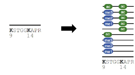
Figure 1.9. Proteoforms of the H3 tryptic peptide -9KSTGGKAPR17- 53
Figure 2.1. Deconvolution of co-eluting acetylated isomers by Iso-PeptidAce 91 Figure 2.2. Analysis of acetylation site occupancies in histones
H3 and H4 following HDAC treatment 95
Figure 2.3. Analysis of acetylation site occupancies in
CAF1-associated histones H3 and H4 97
Figure S2.1. Peptides within the same isomeric groups exhibit
very narrow retention 107
Figure S2.2. MS signal responses of synthetic H3 and H4 peptides 108 Figure S2.3. Acetylated isomers co-elute from reverse-phase column
and produce composite MS/MS spectra upon fragmentation 109 Figure S2.4. Isomeric peptides produce distinct fragment ion patterns 110 Figure S2.5. Deconvolution of di- and tri-acetylated isomers of histone H4 111 Figure S2.6. Fractionation of total histones by RP-HPLC 112 Figure S2.7. MS/MS spectra of H3 and H4 peptide isomers 113 Figure 3.1. LC-MS/MS analysis of histone peptides 165 Figure 3.2. Analysis of global H3 and H4 acetylation in response to HDAC
depletion 169
Figure 3.3. Analysis of changes in site-specific acetylation of H3
xv
Figure 3.4. Analysis of changes in site-specific acetylation of H4
in response to HDAC depletion 176
Figure 3.5. Analysis of the impact of HDAC depletion on the methylation states of H3K9 and H3K36 179
Figure 3.6. Summary of HDAC activities in fission yeast 183
Figure S3.1. Representative ion chromatogram of separated isobaric peptides 201 Figure S3.2. Analysis of global H4 acetylation in response to HDAC depletion 202 Figure S3.3. Analysis of the impact of Clr3 mutation on the PTM isoforms of H3 peptide 9-KSTGGKAPR-17 203
Figure 4.1. High stoichiometry of acetylation of H3K14 and H3K23 is evolutionarily conserved 236
Figure 4.2. H3-K14 has diverse functions compared to H3-K23 237
Figure 4.3. Gcn5 and Mst2 contribute to H3-K14 and H3-K23 acetylation in Schizosaccharomyces pombe 238
Figure 4.4. Loss of mst2+ does not phenocopy H3K14R 239
Figure S4.1. High stoichiometry of acetylation on H3-K14 and H3-K23 246
Figure S4.2. Representative growth curves of histone mutants 247
Figure S4.3. Heatmap representation of the log2 fold changes of normalized LC−MS signals 248
Figure S4.4. Representative growth curves of HAT mutants 249
Fig 5.1. Indirect approach to define the function(s) of H3-K14 and H3-K23 acetylation in S. pombe 274
xvi
List of abbreviations
ac acetyl
Acetyl-CoA acetyl coenzyme A
ACN acetonitrile
ADP adenosine diphosphate
ATP adenosine 5´-triphosphate
ATRX α-thalassemia/mental retardation syndrome X-
linked
BCA Bicinchoninic Acid
BET bromodomain and extra terminal domain
bp base pair
BRD bromodomain
CAF-1 Chromatin Assembly Factor 1
CENP-A centromere protein-A
ChIP Chromatin immunoprecipitation
CO2 carbon dioxide
CPT Camptothecin
C-terminus carboxyl terminus
DAXX Death domain–associated protein 6
DDR DNA damage response
DMSO dimethylsulphoxide
DNA deoxyribonucleic acid
dNTP deoxyribonucleoside triphosphate
DSB double-strand break
DTT dithiothreitol
EDTA Ethylenediaminetetraacetic acid
Esa1 Essential Sas family acetyltransferase 1
FACS fluorescence-activated cell sorting
FACT facilitates chromatin transcription
FASTA FAST-All
xvii
Gcn5 General control non-repressible 5
GNAT Gcn5-related N-acetyltransferase
H2SO4 Sulfuric acid
HAT histone acetyltransferase
HCD High-energy collision dissociation
HCl hydrogen chloride
HDAC Histone deacetylase
HDACi Histone deacetylase inhibitor
HEPES N-(2-hydroxyethyl)
piperazine-N´-(2-ethanosulfonic acid)
His histidine
HJURP Holliday junction recognition protein
HPLC High performance liquid chromatography
HR homologous recombination
HU hydroxyurea
i.d. Internal diameter
IPTG isopropyl-β-D-thiogalactopyranoside
KAT lysine acetyltransferase
KDAC lysine deacetylase
KDM lysine demethylase
LC-MS/MS Liquid chromatography tandem mass
spectrometry
Leu leucine
LOH loss of heterozygosity
LRR Leucine-rich repeat
m/z mass-to-charge
MgCl2 magnesium chloride
MMS methyl methanesulfonate
MOZ Monocytic leukemia Zinc finger
MS/MS tandem mass spectra
xviii
NaCl sodium chloride
NAD+ nicotinamide adenine dinucleotide
NCP nucleosome core particle
NHEJ non-homologous end joining
nLC Nano-liquid chromatography
N-terminus amino terminus
NuA3 nucleosomal acetylation of H3
NuA4 nucleosomal acetylation of H4
O.D. optical density
PCAF p300/CBP associating factor
PcG Polycomb group
PCNA Proliferating Cell Nuclear Antigen
PCR polymerase chain reaction
PEV position effect variegation
PH pleckstrin-homology
PHD plant homeodomain
PIKK phosphoinositide 3-kinase related kinase
ppm parts-per-million
PMSF phenylmethylsulfonyl fluoride
PRM parallel reaction monitoring
PRMT protein arginine methyltransferases
PTM post translational modification
PSM peptide spectrum matching
RBBP7 retinoblastoma binding protein 7
RFC replication factor C
RNA ribonucleic acid
RNR ribonucleotide reductase
RP Reverse phase
RPA replication protein A
xix
RP-HPLC reverse phase high performance liquid chromatography
RSC remodel the structure of chromatin
Rtt109 Ty1 transposition gene product 109
SAGA Spt-Ada-Gcn5-acetyltransferase
SAHA suberoylanilide hydroxamic acid
SAS something about silencing
SC synthetic complete
SDS sodium dodecyl sulfate
SDS-PAGE sodium dodecyl sulfate polyacrylamide gel
SILAC Stable isotope labeling by amino acid in cell
culture
SLIK SAGA-like
SRM Single reaction monitoring
ssDNA single-strand DNA
STAGA SPT3-TAFII31-GCN5L acetylase
SWI/SNF SWItch/Sucrose NonFermentable
TAP tandem affinity purification
TCA Trichloroacetic acid
TEV tobacco etch virus
TFA Trifluoroacetic acid
TFTC TBP-free TAFII complex
TIP60 Tat-interacting protein
Top1 topoisomerase I
TRRAP Transformation/transcription domain-associated
protein TrxG Trithorax group Ts thermosensitivity TSA trichostatin A URA uracil UV Ultraviolet
xx
WT wild-type
XIC Extracted ion chromatogram
YES yeast extract supplemented
YPD yeast peptone dextrose
5-FOA 5-Fluoroorotic acid
xxi
Acknowledgments
This work could not have been completed without the constant support of my supervisor, family, and friends. Words cannot express my gratitude to you all, but this is a humble attempt.
First, I would like to express my special thanks to my supervisor Dr. Alain Verreault for giving me the opportunity to pursue my Ph.D. under his supervision. Alain has been a wonderful teacher and an excellent mentor. He broadened my horizon of thought by encouraging me to think critically and testing every result with the ‘baloney detection kit.’ My deepest gratitude to you, Alain, for all the insightful discussions, your unconditional support through the years and especially for not losing faith in me when the going got tough.
I would like to extend my sincere thanks to my co-supervisor Dr. Pierre Thibault who patiently explained the basics of mass spectrometry to me, a novice in this analytical technique. Pierre’s ability to think outside his area of expertise and critically question the results have been of tremendous value to my research. He also gave me access to his lab space for work.
The results presented here could not have been made possible without Dr. Nebiyu Abshiru. Nebiyu and I had a fruitful collaboration on all three projects of this thesis, which lean on mass spectrometry-based analyses. Along with Pierre, Nebiyu has given me the ability to appreciate MS-based methods and their applications in biology. Thank you Nebiyu, for all your help and discussions.
Moreover, I would like to thank the members of my Ph.D. committee, Dr. Lea Harrington, and Dr. Jason Tanny, for their valuable advice on my research project. I am hugely grateful to Dr. Richard Bertrand and Dr. Martine Raymond, and for all their support during my Ph.D. Special gratitude to Dr. Julie Mantovani, Suzanne Renaud, Vivianne Jodoin, Pascale Le Therizien, Lucie Yan Liu, Mirela Pascariu and Sandra Weber who have helped me during this journey. I would also like to acknowledge IRIC
xxii
Members Ph.D. awards and the Molecular Biology Program of the Université de Montréal for their scholarships recognizing my efforts.
I extend my special thanks to the past and current members of Verreault lab and Thibault lab for all their support. Each one of you who have contributed to my learning experience. I especially acknowledge the students of IRIC, past, and present, who have been an important part of my life at IRIC. Thank you for your smiles, kind words, reagents and help with instruments.
My dear friends, Arun, Simi, Dezi, Becky and Chris who were always there for me, through thick and thin, this would not have been possible without you.
A few years ago, I carried a dream of doing my Ph.D. It came to fruition only because of the continual support of my family. I started my Ph.D. merely two weeks after getting married, something unthinkable for an Indian girl who had an arranged marriage. My family, which includes my husband Bobby, my parents, parents-in-law, my brothers-in-law, their families and my sweet sisters, have been my strength during the last six years. Special thanks to Papa, Mama, Dada and Amma for their unconditional love and encouragement. I owe it to my husband the most for making my dream come true. Thank you beyond words, my beloved Achacha, for all your sacrifices, words of comfort, encouragement, and prayers. You are indeed the greatest blessing in my life! It gives me great pleasure to dedicate this thesis to my family.
xxiii
"We are like dwarfs sitting on the shoulders of giants. We see
more, and things that are more distant, than they did, not because
our sight is superior or because we are taller than they, but
because they raise us up, and by their great stature add to ours."
John of Salisbury
2
1.1. Eukaryotic chromatin: an architectural marvel
The eukaryotic genome showcases an elaborate and dynamic architecture. In human cells, roughly two meters of deoxyribonucleic acid (DNA), is packaged into the confines of the cell nucleus which is only a few micrometers wide. This incredible feat is accomplished through hierarchical folding and compaction of DNA into chromatin. The chromatin structure poses challenges to all cellular processes that require access to DNA such as DNA replication, transcription and DNA repair. Cells have evolved to overcome DNA compaction while maintaining regulation of these processes through a myriad of effectors that belong into four broad categories: ATP-dependent chromatin remodelers, DNA and histone modifying enzymes and histone chaperones.
1.1.1. The nucleosome core particle
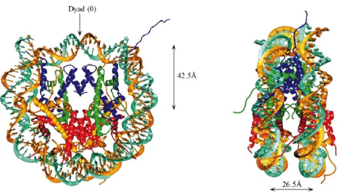
The ‘quantum’ of chromatin structure is the nucleosome. Major discoveries that led to the elucidation of chromatin structure came in 1973-74 (Olins and Olins, 2003). An insightful proposition by Roger Kornberg that the repeating unit of chromatin has 200 base pairs (bp) of DNA and eight histone molecules lent a new perspective to the puzzling structure of chromatin (Kornberg, 1974; Thomas and Kornberg, 1975). Oudet and colleagues provided the first biochemical and microscopic evidence for a repeating structure in chromatin in 1975, confirming Kornberg’s ideas (Oudet et al., 1975). The 2.8 Å resolution structure of the nucleosome reconstituted with alpha satellite repeat DNA provided intricate details of histone arrangement and histone-DNA interactions (Luger et al., 1997). The nucleosome core particle consists of 147 bp of DNA wrapped around an octameric protein core in 1.65 turns of a left-handed superhelix. The protein
3
core is composed of two molecules each of four core histone proteins, known as H2A, H2B, H3 and H4, which form a central H3-H4 tetramer (H3-H4)2 flanked by two
H2A-H2B dimers (Fig 1.1). Histone H3 makes additional contacts with histone H2A and with the DNA and plays an important role in the stabilization of NCP.
Figure 1.1. Nucleosome core particle: ribbon traces for the 147 bp DNA phosphodiester backbones (brown and turquoise) and eight histone protein main chains (blue: H3; green: H4; yellow: H2A; red: H2B. The views are down the DNA superhelix axis for the left particle and perpendicular to it for the right particle. For both particles, the pseudo-twofold axis of symmetry (shown at the top) is aligned vertically with the center of the 147 bp of nucleosomal DNA (Dutnall and Ramakrishnan, 1997).
4 1.1.2. Chromatin structure
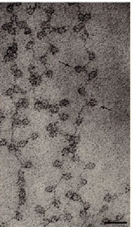
Chromatin consists of two structural entities: the nucleosome core particle and the ‘linker’ region: short DNA segments of varying length that connect nucleosome core particles. One molecule of linker histone H1 binds to the linker DNA. Nucleosomes are connected to each other through linker DNA to form linear nucleosomal arrays that have the appearance of ‘beads-on-a-string’, the primary structure of chromatin. The packaging of DNA with histones yields a chromatin fiber that is approximately 10 nm in diameter and shortens its length approximately six-fold (Fig 1.2). Short-range interactions between neighboring nucleosomes result in a higher-order structure of chromatin, commonly known as the 30 nm fiber (Luger et al., 2012; Woodcock and Dimitrov, 2001). Linker histone H1 plays an important role in stabilizing the 30 nm fiber. However, using ChromEMT, a method combining electron microscopy tomography (EMT) with ChromEM labeling which enhances the contrast of DNA, Ou et al. has recently shown that chromatin exists as a flexible 5- to -24 nm diameter chain that can be packed at different densities in interphase cells and in mitotic chromosomes (Ou et al., 2017). Our knowledge of the dynamics and formation of higher-order structures remains limited despite technological advances.
1.1.3. Euchromatin and heterochromatin
One of the earliest observations of distinct chromatin structures was made by German botanist Emil Heitz in 1928 (Straub, 2003). He observed cytologically distinguishable chromosome regions that he called euchromatin and heterochromatin based on differential staining and proposed that the heterochromatin is functionally inactive.
5
Heterochromatin is highly condensed chromatin characterized by repetitive DNA elements, is gene-poor and less transcribed. Conversely, euchromatin is rich in genes, more accessible to the transcription machinery and hence easily transcribed.
Figure 1.2. Chromatin fibers: A) At low ionic strength chromatin appears as 'beads on a string'. Black bar: 30 nm. B) Moderate ionic strength forms the higher-order 30-nm fiber. Black bar: 50 30-nm (Olins and Olins, 2003)
Extensive methylation of histone 3 at lysine 9 (H3K9me) is a characteristic of heterochromatin domains shared by most eukaryotes (Huisinga et al., 2006). This posttranslational modification is catalyzed by the evolutionarily conserved H3K9-specific histone methyltransferase SU(VAR)3-9 (Suv39h1&2 in mammals, Clr4 in
6
fission yeast). Another conserved feature of heterochromatin is the presence of non-histone chromosomal protein heterochromatin protein 1 (HP1, Swi6 in fission yeast). Moreover, heterochromatin regions contain histones that are hypoacetylated on H3 and H4 (Trojer and Reinberg, 2007). Heterochromatin is broadly classified as either Constitutive heterochromatin or Facultative heterochromatin. Constitutive heterochromatin is silenced throughout the cell cycle and occurs at centromeres, telomeres, and transposable elements. Facultative heterochromatin is found at developmentally regulated loci such as the mating type loci and certain meiotic genes in yeast. Facultative heterochromatin can be converted into euchromatin in response to cellular signals. Heterochromatin can propagate in a sequence-independent manner and spread across domains. Heterochromatin structure interferes with recombination (Grewal and Jia, 2007).
1.2. Histones
Histones are highly conserved proteins enriched in the basic amino acids arginine and lysine. They are extremely abundant in eukaryotic cells; their total mass (4 core histones and H1) is roughly equal to that of a cell’s DNA. In addition to their role as architectural proteins, histones contribute to chromosome function through three principal mechanisms: ATP-dependent remodeling complexes can alter the properties of nucleosomes or their position relative to specific DNA sequences, variant or ‘replacement’ histones can alter the composition of nucleosomes by replacing the most abundant forms of the core histones and a rich variety of covalent histone modifications can regulate their function. There are four canonical core histones: H2A, H2B, H3, and H4. Variant histones exist for core histones H3, H2A and H2B and for the linker histone
7
H1. Intriguingly, no variant of histone H4 seems to exist. All core histones have two structurally and functionally distinct domains, a globular histone-fold domain and a conformationally flexible and highly charged amino-terminal domain, commonly known as the histone tail. The histone-fold domain consists of three α-helices (α1-α3) connected by short loops L1 and L2 (Fig 1.3). The histone-fold domain aids in the assembly of histone dimers through a so-called ‘handshake’ motif. The histone-fold domains mediate histone-histone and histone-DNA interactions. The N-terminal tails of histones are unstructured and protrude out of the nucleosomal DNA (Luger 2000). A striking feature of the N-terminal tails is that they are the targets of many post-translational modifications, elaborated in section 1.3.
Figure 1.3. The secondary structure of core histones showing the globular histone-fold domain and the unstructured ‘tail’ domains. α-helices are depicted as solid boxes and are numbered according to Luger et al., 1997 (Dutnall and Ramakrishnan, 1997).
8 1.2.1. Histone H3 variants
The regulatory repertoire of chromatin structure and function is expanded through the replacement of canonical histones by variants. Variant histones can be incorporated into chromatin throughout the cell cycle, unlike canonical histones that are deposited onto nascent DNA during replication. All core histones except H4 have variants, the H3 variants being the least diverse. In human cells, histone H3 is present as eight different variants: H3.1, H3.2, H3.3, H3.1t (H3.4), H3.3C (H3.5), H3.Y.1 (H3.Y), H3.Y.2 (H3.X) and CenH3 (see Fig. 1.4). H3.4 and H3.5 are testis-specific H3 variants. The H3 variants can be grouped into two classes. Canonical forms of histone H3 provide the supply of H3 molecules needed during replication. In humans, there are two flavors of replicative histone H3, known as H3.1 and H3.2 that differ by a single amino acid (Fig 1.4). Replacement variants are incorporated in a replication-independent manner. H3.3 is the replication-independent histone H3 in humans. CenH3 also known as CENP-A is a centromeric H3 variant exclusively found in the nucleosomes present at centromeres.
The highly basic nature of histones can lead to non-specific interactions between soluble (non-nucleosomal) histones and DNA, generating aggregates. Cells delegate dedicated escort proteins, also known as chaperones, to prevent such non-specific interaction of histones. Chaperones selectively associate with either canonical or variant histone forms. For example, chromatin assembly factor-1 (CAF-1) specifically associates with core histones H3.1 and H3.2, but not with the variant H3.3 (Akiyama
9
et al., 2011; Latreille et al., 2014; Tagami et al., 2004). There are two H3.3-specific chaperones, the HIRA complex deposits H3.3-H4 complexes at transiently nucleosome-free regions. A protein complex formed by the Death domain-associated protein 6 (DAXX) and the -thalassemia/mental retardation syndrome X-linked (ATRX), incorporates H3.3-H4 into pericentric heterochromatin and sub-telomeric chromatin regions (Banumathy et al., 2009; Goldberg et al., 2010; Lewis et al., 2010; Rai et al., 2011; Ray-Gallet et al., 2002; Tagami et al., 2004). The Holliday junction recognition protein (HJURP) is a chaperone specific for the centromeric variant CenH3, which it incorporates at centromeres in early G1 (Dunleavy et al., 2009; Foltz et al., 2009; Jansen et al., 2007).
Figure 1.4. Alignment of amino acid sequences of human histone H3 isoforms. The sequences are compared to H3.1 and the differences are highlighted (Filipescu et al., 2014)
10
1.3. Post-translational modifications of histones
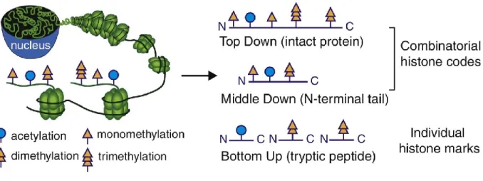
In the mid-1960s, seminal work by Allfrey and Murray demonstrated that histones are post-translationally modified (Allfrey, Faulkner, & Mirsky, 1964; Allfrey & Mirsky, 1964; Murray, 1964). The last few decades saw the discovery of a staggering number of histone post-translational modifications (PTMs). Researchers in the field have demonstrated tremendous enthusiasm to identify, catalog, classify and interpret cross-talk between various histone modifications. These include extensively studied PTMs such as acetylation, methylation, phosphorylation, ubiquitination, sumoylation, poly (ADP) ribosylation, deimination and proline isomerization (Kouzarides, 2007). Many newly identified modifications such as butyrylation, crotonylation, malonylation, succinylation, formylation, hydroxylation and O-GlcNAcylation have increased the complexity of studying covalent histone modifications and their functions (Huang, Sabari, Garcia, Allis, & Zhao, 2014). The majority of PTMs are located on the histone tails but the globular domains also carry key modifications. The study of covalent modifications in the N-terminal tail domains dominated research until the early 2000s. This was mainly because the primary method for discovering histone modifications, Edman degradation, permitted the analysis of only 20–30 amino acids from the N-terminus of a protein. Covalent modifications that occur within histone-fold domains were identified with the advent of mass spectrometry.
Histone PTMs have been implicated in the regulation of numerous aspects of chromatin structure and function. The dynamic regulation of chromatin function by histone modifications occurs through two main mechanisms. Many histone
11
modifications weaken the non-covalent interactions between histones and DNA. For example, acetylation of specific residues reduces the positive charge of histones and their interaction with DNA (Bannister and Kouzarides 2011). Second, PTMs create surfaces that bind to proteins with PTM-specific binding domains. These proteins, collectively known as ‘readers’ of histone PTMS, in turn, mediate the biological effects that are associated with specific PTMs (Yun et al. 2011). Many lines of evidence point towards an active interplay between different histone marks in the regulation of chromatin function. Our current understanding of histone modifications was revolutionized by the ‘Histone Code hypothesis’ which suggests that distinct histone modifications may form combinational readouts which, in turn, expand the repertoire of chromatin effectors that can act at a given locus (Jenuwein et al., 2001; Strahl and Allis, 2000). Although there are many proponents of ‘histone code’ hypothesis, there lacks a consensus opinion of exactly what this entails (Rando, 2012).
1.3.1. Are all histone modifications ‘epigenetic’?
A brief history of the use of the term ‘epigenetics’ is given here to illustrate that histone modifications and epigenetic modifications have been used interchangeably in the literature. The term ‘epigenetics’ was coined by Waddington in 1942 to define ‘complex developmental processes that connect phenotypes to genotypes (Waddington, 2012). This term has however generated much controversy in recent years. One of the popular definitions of epigenetics was given by Riggs, Martienssen and Russo as “The study of mitotically and/or meiotically heritable changes in gene function that cannot be explained by changes in DNA sequence (Riggs et al., 1996). Bird defined the term
12
‘epigenetics’ as: “the structural adaptation of chromosomal regions so as to register, signal or perpetuate altered activity states” (Bird, 2007). According to Mark Ptashne, “the use of the word epigenetic implies a fundamental property of the system - self-propagation - that is, so far as we now know, not true of nucleosome modifications” (Ptashne, 2007). A more narrow definition that has been widely accepted defines ‘epigenetics’ as “a stably heritable phenotype resulting from changes in a chromosome without alterations in the DNA sequence” (Berger et al., 2009). Thus, the researchers in the field are divided in their definition of which histone modifications should be considered epigenetic and which should not. In this thesis, histone modifications that are stably inherited across generations are referred to as epigenetic modifications while those that are dynamically regulated during different cellular processes are referred to as histone modifications.
1.3.2. Writers, Readers and Erasers
Regulation of chromatin structure and function is a dynamic and complex process. Extracellular and intracellular cues trigger a cascade of downstream events that result in localized or genome-level alterations in chromatin structure mediated by chromatin modifiers. Chromatin regulators can be broadly classified into: ‘writers’, ‘readers’ and ‘erasers’. Writers introduce modifications on DNA or histones while erasers, as the name suggests, remove modifications. Histone acetyltransferases (HATs), Histone methyltransferases (HMTs), kinases and protein arginine methyltransferases (PRMTs) are examples of writers. Examples of erasers include histone deacetylases (HDACs),
13
phosphatases and lysine demethylases (KDMs). See section 1.4.1. and 1.4.2. for a detailed description of ‘writers’ and ‘erases’ pertinent to this thesis.
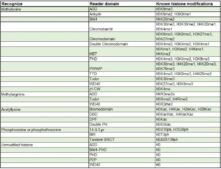
Readers recognize modifications and use them as docking sites through specialized domains. Most often, readers are part of multi-subunit protein complexes. Bromodomains, chromodomains, WD40 repeats, Tudor domains and PHD fingers are all chromatin-binding domains that recognize modified histone tails. Some of the most common histone readers are listed in Table 1.1. Bromodomains recognize and bind acetylated lysine residues. Gcn5 contains a bromodomain that is important for the binding, recognition and retention of the histone acetyltransferase protein complex SAGA on acetylated promoter nucleosomes (See section 1.4.1.). Chromodomains specifically bind methylated lysine tails. In Drosophila melanogaster and vertebrates, the chromodomain of heterochromatin protein 1 (HP1) binds methylated H3K9, whereas the chromodomain of Polycomb binds methylated H3K27 (Filippakopoulos and Knapp, 2012; Marmorstein and Zhou, 2014; Patel, 2016; Patel and Wang, 2013).
1.4. Histone acetylation
Histone lysine acetylation discovered in 1964 is one of the best studied post-translational modifications (Allfrey et al., 1964). Acetylation refers to the covalent addition of an acetyl group from acetyl-coenzyme A (acetyl-CoA) to the epsilon (ε) amino group of lysine. This reversible modification is catalyzed by two families of enzymes, histone acetyltransferases (HATs) and histone deacetylases (HDACs). Acetylation removes the positive charge of the ε-amino group of the lysine side chain. Lysine acetylation has been implicated in many cellular processes such as
14
nucleosome assembly, DNA replication, transcription, DNA damage response (i.e. DNA damage signaling) and DNA repair (Bannister and Kouzarides, 2011; Shahbazian and Grunstein, 2007). It is interesting to note that the relationship between histone acetylation and transcription was first observed prior to the discovery of nucleosomes as the building blocks of chromatin.
15 1.4.1. Histone acetyltransferases
Histone acetyltransferases catalyze the addition of acetyl groups to lysine side chains. In the early 1990s, two acetyltransferases, Nat1-Ard1, were purified. They acetylated the N-terminal amino group of H2A and H2B and not internal lysines like other HATs do (Park and Szostak, 1992). The first histone acetyltransferase to be purified was Saccharomyces cerevisiae Hat1 which acetylates histone H4 molecules at lysine residues 5 and 12 prior to H4 deposition onto DNA (Kleff et al., 1995; Parthun et al., 1996). Most HATs are part of multi-subunit complexes. The combinations of these subunits contribute to the unique features of each HAT complex. HATs are divided into two major classes, type-A and type-B, based on their substrate specificity. Type-A HATs are nuclear HATs that catalyze transcription-related acetylation in the context of chromatin (nucleosomal histones). The first A-type HAT to be purified was Gcn5, a homolog of the yeast transcriptional co-activator Gcn5, from the macronuclei of the ciliate protozoan Tetrahymena thermophila (Brownell et al., 1996). The A-type HATs can be classified into at least three subfamilies: GCN5/PCAF, MYST and CBP/p300. The families of nuclear HATs show very high sequence divergence. In addition to the well-characterized nuclear HAT families, there are others that are less studied such as TAF250 (Hodawadekar and Marmorstein, 2007). The type-B HATs are distinguished by the fact that they acetylate histones prior to their deposition into chromatin. Histone acetyltransferase 1 (Hat1) and yeast Rtt109 are examples of B-type HATs. They acetylate new H3 and H4 molecules, a mark important for their deposition onto chromatin. Even though Hat1 was initially thought to be cytoplasmic, evidence shows
16
that it is also found in the nucleus (Parthun, 2007). Many studies have shown that several HATs also have non-histone substrates (Sterner and Berger, 2000).
1.4.1.1. Gcn5-related N-acetyltransferases
The General control non-repressible 5 (Gcn5)-related N-acetyltransferases (GNATs) belong to a superfamily of enzymes. The Gcn5/PCAF family consists of yeast Gcn5, Hat1, Elp3, metazoan Gcn5, p300/CBP associating factor (PCAF) and related proteins. The metazoan Gcn5 homologs have a metazoan-specific N-terminal PCAF homology domain absent in yeast. HATs of the GNAT family are characterized by four conserved sequence motifs, A-D, of which motif A, which houses the acetyl-CoA binding domain, is the most highly conserved. The A, B and D domains of GNAT HATs form a central core. The N- and C-terminal domains flanking the core confer substrate specificity (Hodawadekar and Marmorstein, 2007; Kimura et al., 2005; Marmorstein and Zhou, 2014; Ud-Din et al., 2016). Denu and coworkers have elucidated the catalytic mechanism of GNAT HATs. They showed that Gcn5 functions through a ternary complex mechanism in which both substrates (i.e., lysine and acetyl-CoA) must be bound to the enzyme before catalysis can occur. This involves deprotonation of the lysine substrate by Glu173 (of yGcn5), facilitating the direct transfer of the acetyl group from acetyl-CoA to the lysine side chain (Tanner et al., 1999).
Gcn5 is the catalytic subunit of several multi-subunit lysine acetyltransferase complexes with well-established roles in gene transcription. In S. cerevisiae, these complexes include Spt-Ada-Gcn5-acetyltransferase (SAGA), ADA, and SAGA-like (SLIK) (Grant et al., 1997; Pray-Grant et al., 2002). Orthologous complexes with similar
17
subunit composition include the SAGA and ATAC complexes in Drosophila and the STAGA (SPT3-TAFII31-GCN5L acetylase), PCAF and TBP-free TAFII complex
(TFTC) complexes in humans (Lee and Workman, 2007; Nagy and Tora, 2007). In S. cerevisiae, Gcn5 has been shown to play an important role in replication-coupled nucleosome assembly, in parallel with Rtt109, the HAT that acetylates H3-K56. This study showed that deletion of some subunits common to the various Gcn5-containing HAT complexes (Ada1, Ada2, Ada3, Spt7 or Spt20) phenocopy the deletion of GCN5 in cells lacking Rtt109 but the authors were not able to ascribe the role of Gcn5 in replication-coupled nucleosome assembly to a well-established protein complex that contains Gcn5 as catalytic subunit (Burgess et al., 2010).
Gcn5 has both histone and non-histone substrates. The substrate specificity is regulated by non-catalytic proteins present in each complex. However, interactions between Gcn5 and side chains in the +2 and +4 positions relative to the substrate lysine is conserved across divergent protein substrates (Clements et al., 2003; Poux and Marmorstein, 2003). Recombinant Gcn5 has little or no activity toward nucleosomal histones but predominantly acetylates lysine 14 of H3 and lysines 8 and 16 of H4 (Kuo et al., 1996). When part of multi-subunit complexes, Gcn5 can acetylate both free and nucleosomal histones. For example, S. cerevisiae ADA preferentially acetylates K14 and K18 of nucleosomal H3 whereas SAGA acetylates K14 and K18 and, to a lesser degree, K23 and K9 (Grant et al., 1999).
18 1.4.1.2. MYST family acetyltransferases
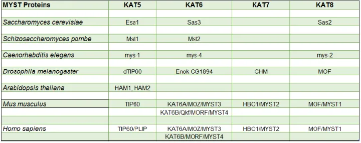
The MYST family acetyltransferases are highly conserved, nuclear HATs that play important roles in DNA replication, transcriptional regulation, the DNA damage response and DNA repair. They always function in multi-subunit complexes that are also evolutionarily conserved. The term MYST family was coined after founding members of the family: MOZ (MOnocytic leukemia Zing finger), Ybf2-Sas3, Sas2 (Something about silencing protein 2) and TIP60 (Tat-interacting protein 60). All MYST family HATs contain a highly conserved MYST domain, which consists of an acetyl-CoA binding domain and a zinc finger. In addition, some members also have additional domains such as chromodomain and plant homeodomain (PHD)-linked zinc fingers (Sapountzi and Côté, 2011; Yang, 2015). The MYST acetyltransferases from major model organisms are listed in Table 1.2
Several structural studies of MYST HATs from yeast to humans have been reported. Even though the amino- and carboxy-terminal domains are divergent, MYST family members possess a core region involved in acetyl-CoA binding that is conserved across the members of various acetyltransferase families. The structures of MYST HATs superimpose well with Hat1 and Gcn5/PCAF (Marmorstein and Zhou, 2014). The MYST family HATs have a conserved glutamate residue which overlays with the catalytic glutamate residue of Gcn5/PCAF. In addition, MYST family HATs have a cysteine residue in the active site. MYST HATs have a ping-pong catalytic mechanism, in which the enzyme first forms an acetylated intermediate involving a conserved cysteine in the catalytic site before the formation of an acetylated histone product (Yan
19
et al., 2000, 2002). Studies of the MYST family enzymes Esa1 and MOZ indicate that efficient acetylation is influenced by interactions with regions of the substrate located at a distance from the lysine to be acetylated. For example, piccolo (Nucleosomal Acetylation of H4) NuA4 is predominantly an H4 N-terminal tail specific enzyme. Full-length histone H4 is acetylated 2000-fold faster than histone tail peptides, indicating that interactions with the histone-fold domain of H4 are critical for the high catalytic efficiency (Bernsden and Denu, 2008).
Table 1.2. MYST acetyltransferases in model organisms
1.4.2. Histone deacetylases
Histone deacetylases (HDACs) catalyze the removal of acetyl groups from lysine side chains and have been implicated in gene repression. Based on phylogeny histone deacetylases have been classified into two families, the classical HDAC family and the yeast silent information regulator 2 (Sir2)-related deacetylases, commonly known as sirtuins. The classical family can be further sub-divided into three classes of HDACs:
20
Class I, Class II and Class IV. Class I HDACs are homologous to S. cerevisiae Rpd3 and include human HDAC1, HDAC2, HDAC3 and HDAC8. Enzymes that have sequence homology to S. cerevisiae Hda1 constitute the Class II HDACs. They can be further divided into two subclasses based on their phylogeny: Class IIa and Class IIb (Frye, 2000; Gregoretti et al., 2004). Class IIa includes human HDAC 4,5, 7 and 9 while Class IIb includes HDAC 6 and 10. Class III HDACs are also called sirtuins after the founding member of the family, the S. cerevisiae Sir2 enzyme (Seto and Yoshida, 2014). In S. cerevisiae, the class III HDACs include Sirt2, Hst1, Hst2, Hst3 and Hst4 and SIRT1 through SIRT7 in humans. Human HDAC 11 is thus far the sole member of class IV HDACs.
Sirtuins do not share sequence or structural homology to other HDAC families. They use a distinct catalytic mechanism that is dependent on the oxidized form of nicotinamide adenine dinucleotide (NAD+) as a co-substrate. Class I, II and IV HDACs require a zinc ion (Zn), for deacetylase activity. They share structural similarity in their catalytic domains (Hodawadekar and Marmorstein, 2007).
HDACs are also part of multi-subunit complexes. HDACs have been shown to have protein substrates other than histones. Taking into consideration the increasing number of non-histone substrates for HATs and HDACs, a new naming system has been put forward (Allis et al., 2007). These enzymes are now called lysine acetyltransferases (KATs) and lysine deacetylases (KDACs) under the new nomenclature.
21 1.4.3. Histone deacetylase inhibitors
Chromatin regulators are frequently mutated in cancer, neurological disorders, metabolic disorders and inflammatory diseases. Changes in DNA and histone modifications are reversible and the ability to modulate enzymatic properties and/or structural properties of chromatin modifying enzymes have led to the development of a repertoire of pharmacological inhibitors. The predominant class of these inhibitors target HDACs and are known as histone deacetylase inhibitors (HDACi). Even though most of these inhibitors were originally developed to combat cancers, several of them are being used to treat immune and neurological disorders (Falkenberg and Johnstone, 2014; Marks and Xu, 2009).
The most commonly used HDAC inhibitors are so-called "pan" inhibitors that target multiple HDACs. This broad range of specificity makes it difficult to determine whether the biological consequences of HDAC inhibition are due to inhibition of a specific HDAC, or the combined effect of inhibiting multiple HDACs. HDACi can also inhibit histone deacetylases that are part of multi-protein complexes that incorporate them as key enzymatic components.
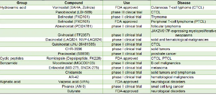
Based on their chemical structure, HDACis are classified into the following groups; hydroxamic acids (trichostatin A, vorinostat), carboxylic acids (valproate, butyrate), benzamides (entinostat, mocetinostat), cyclic peptides (apicidin, romidepsin), epoxyketones (trapoxin), and hybrid molecules. Hydroxamate derivatives generally inhibit HDACs from multiple classes whereas benzamide derivatives are generally
22
restricted to inhibiting class I HDACs. (Falkenberg and Johnstone, 2014; Marks, 2010; West and Johnstone, 2014).
In addition to HDACi, inhibitors of chromatin modulators (e.g. small molecules that prevent bromodomains from recognizing acetylated lysine residues), are also in development and clinical trials (Filippakopoulos and Knapp, 2014; Muller et al., 2011). US Food and Drug Administration (FDA)-approved DNA-demethylating agents azacitidine (also known as 5-azacytidine) and decitabine (also known as 5-aza-2′-deoxycytidine) which are used to treat myelodysplastic syndromes (MDS) and a range of other malignancies are by far the most successful and long-standing inhibitors of epigenetic processes. Inhibitors of histone methyltransferases such as DOT1L and EZH2 as well as inhibitors of protein arginine methyltransferase (PRMT) show promise for the treatment of cancer and immune disorders (Falkenberg and Johnstone, 2014 and references within).
23
1.4.4. Cellular functions of histone acetylation
Histone acetylation has been implicated in several important cellular functions including replication-coupled nucleosome assembly, DNA damage response, DNA repair and regulation of gene expression. In this section, evidence that supports the role of histone acetylation in these processes is discussed.
1.4.4.1. Chromatin assembly
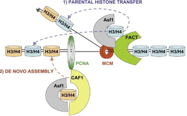
During DNA replication, duplication of DNA is accompanied by a transient disruption of chromatin structure and concomitant histone synthesis. The chromatin structure is rapidly re-established in a step-wise manner through two key steps. First, pre-existing or parental histones located ahead of replication forks are distributed among the two nascent sister chromatids. Parental histone segregation is followed by deposition of newly synthesized histones, a process called replication-coupled (DNA synthesis-coupled or de novo) nucleosome assembly (Verreault, 2000). In addition to chromatin assembly that occurs throughout the genome during S-phase, chromatin structure is dynamically transformed during transcription. This constitutes the replication-independent or DNA synthesis-replication-independent chromatin assembly. In addition, at sites of DNA repair, a PCNA-dependent chromatin assembly occurs.
One question that has been of interest to early researchers was the timing of nucleosome assembly with respect to DNA synthesis. Using a very short pulse-labeling time of 1 min with tritiated thymidine, Seale showed that newly replicated chromatin is sensitive to digestion by DNase I, but recovers its resistance characteristic of mature
24
chromatin after about 15 min (Seale, 1975). However, electron microscopic observations showed that both arms of the replication fork contained nucleosomes and lacked stretches of nucleosome-free DNA, implying that chromatin maturation continues even after de novo nucleosome assembly (reviewed in Annunziato, 2012). A likely explanation for the gradual maturation came with the identification of acetylation on newly synthesized histones (Jackson et al., 1976; Ruiz-Carrillo et al., 1975). Acetylation of new H4 molecules was shown to have a half-life of 20-30 min suggesting that acetylation was partly responsible for the increased DNase I sensitivity of new chromatin (Cousens and Alberts, 1982; Jackson et al., 1976; Ruiz-Carrillo et al., 1975). It was later demonstrated that chromatin replicated in the presence of sodium butyrate had higher magnesium-solubility, characteristic of acetylated chromatin, compared to bulk chromatin. The increased solubility in magnesium coincided with a five to six-fold decrease in linker histone H1, thus establishing that deacetylation of new histones is necessary for chromatin maturation (Annunziato and Seale, 1983; Perry and Annunziato, 1989).
Pioneering studies in the mid-70s showed that new histone H4 is acetylated in the cytoplasm based on biochemical fractionation of cytosolic and nuclear proteins. One caveat of this approach is that numerous proteins that are soluble in the nucleus end up in the "cytosolic" extract. In a nutshell, these authors could not conclude that the acetylation of new histone molecules took place in the cytoplasm based on the fractionation experiments that they conducted at the time (Jackson et al., 1976; Louie et al., 1974; Ruiz-Carrillo et al., 1975). There are four lysine residues in the amino-terminal tail of H4 that can be acetylated: lysine 5, 8, 12 and 16. Using Edman
25
sequencing of newly synthesized H4 pulse-labeled with tritiated lysine, the sites of acetylation were found to be H4K5 and H4K12 in yeast, flies and mammalian (HeLa) cells (Benson et al., 2006; Sobel et al., 1995). In Tetrahymena, H4K4 and H4K11 are the deposition-related acetylation sites, due to a deletion of arginine at position 3 (Chicoine et al., 1986). Even though specific functions of H4K5 and H4K12 acetylation remain enigmatic, this evolutionarily conserved pattern of H4 diacetylation is considered a hallmark of replication-coupled chromatin assembly.
Histone H3 is also acetylated rapidly after synthesis (Jackson et al., 1976). The pattern and sites of acetylation in new H3 molecules have been less conserved than those of H4. For example, in the fruit fly, newly synthesized H3 is acetylated on lysine 14 and 23 while in the ciliate Tetrahymena, lysine 9 and 14 are acetylated (Sobel et al., 1995). In budding yeast, Saccharomyces cerevisiae, four of the five N-terminal tail lysine residues (K9, K14, K23 and K27) are acetylated, with H3K9 showing the highest level of acetylation, followed by H3K27 (Kuo et al., 1996). In mammalian cells, about ten percent of new histone H3 molecules are acetylated at lysine residues 14, 18 and 23 (Benson et al., 2006). Unlike H4 which is predominantly acetylated on lysines K5 and K12, a large proportion of new H3 molecules are nonmodified, particularly in mammalian cells (Benson et al., 2006; Sobel et al., 1995).
The N-terminal tail acetylations of H3 and H4 are important for nucleosome assembly. In S. cerevisiae, the N-terminal tails of H3 and H4 have redundant functions in nucleosome assembly both in vitro and in vivo. In addition, cells lacking N-terminal tail acetylation of both H3 and H4, through mutation of multiple lysine residues to arginine, are not viable. The extracts from these cells are defective in chromatin assembly in
26
vitro. (Ling et al., 1996; Ma et al., 1998). Surprisingly, in S.cerevisiae cells lacking the N-terminal of H3, the conserved deposition-related H4-K5, K12 acetylation is not required for histone deposition in vitro and in vivo, as long as H4 lysine 8 is intact (Ma et al., 1998). It has been demonstrated that the N-terminal tails of H3 and H4 are not necessary for CAF-1 dependent chromatin assembly in vitro (Shibahara et al., 2000). The presence of multiple copies of histone genes makes it difficult to perform comprehensive studies in S. pombe and higher eukaryotes. Using fluorescent protein-tagged histones under the control of heat shock inducible promoters, Ahmed and Henikoff showed that the N-terminal tail of H3.3, but not that of H3.1, is dispensable for its deposition onto DNA (Ahmad and Henikoff, 2002). However, it is unclear whether post-translational modifications of H3.1 are necessary for its assembly into chromatin in higher eukaryotes.
In addition to the acetylation that occurs on the N-terminal tails, new histones are also acetylated in the globular domain prior to their deposition into chromatin. The best well-characterized globular domain acetylation occurs on lysine 56 of histone H3. H3K56 acetylation is found in yeast (budding yeast, fission yeast and Candida albicans) and Drosophila (Masumoto et al., 2005; Recht et al., 2006; Wurtele et al., 2010; Xu et al., 2005). Even though H3 lysine 56 acetylation has been detected in humans, the abundance is very low (Drogaris et al., 2012; Jasencakova et al., 2010). Lysine 56 is the last residue of the α-N helix of histone H3 and the non-acetylated form interacts with DNA at the entry/exit point of the nucleosome (Masumoto et al., 2005; Ozdemir et al., 2005; Xu et al., 2005).The acetylation of K56 is thought to weaken the histone-DNA interactions by neutralizing the positive charge of lysine (Bernier et al., 2015; Neumann
27
et al., 2009; Su et al., 2012). H3 lysine 56 acetylation occurs predominantly on newly synthesized histones, peaks during S-phase and, in the absence of DNA lesions, is removed from histones in G2/M (Celic et al., 2006; Maas et al., 2006; Masumoto et al., 2005). Evidence suggests that H3K56 acetylation plays an important role in both replication-coupled and replication-independent chromatin assembly. A critical function of H3 lysine 56 acetylation in these processes may be to regulate interactions with histone chaperones as this modification has been shown to promote the association of new H3 molecules with histone chaperones CAF-1 and Rtt106 (Li et al., 2008). H3 molecules bound to CAF-1 and Rtt106 during S-phase are acetylated at H3K56 (Li et al., 2008; Masumoto et al., 2005). In cells lacking H3K56ac, the amounts of new H3 molecules bound to CAF-1 and Rtt106 are considerably reduced which, in turn, delays the deposition of new histones behind replication forks (Li et al., 2008). The reduced amounts of histones bound to Rtt106 when H3-K56 cannot be acetylated can be, at least partially explained by the fact that Rtt106 binds with higher affinity to H3 molecules that are K56-acetylated. Together, these results indicate that the acetylation of new histone H3 at K56 promotes H3-H4 deposition by CAF-1 and Rtt106 onto nascent chromatin during S phase. Mutations that abolish H3K56ac show synthetic lethality with mutation of lysine residues in the N-terminal tails of H3 or H4 in the presence of genotoxic agents that cause DNA damage during S phase (Li et al., 2008). It could be possible that new histone deposition behind replication forks which is important for DNA damage survival is partially compromised when mutations that abolish K56 acetylation or H3 N-terminal tail acetylations are present but more severely compromised when both forms of acetylations are compromised.