HAL Id: hal-01916755
https://hal.archives-ouvertes.fr/hal-01916755
Submitted on 8 Nov 2018
HAL is a multi-disciplinary open access
archive for the deposit and dissemination of
sci-entific research documents, whether they are
pub-lished or not. The documents may come from
teaching and research institutions in France or
abroad, or from public or private research centers.
L’archive ouverte pluridisciplinaire HAL, est
destinée au dépôt et à la diffusion de documents
scientifiques de niveau recherche, publiés ou non,
émanant des établissements d’enseignement et de
recherche français ou étrangers, des laboratoires
publics ou privés.
Structural evidence for a programmed general base in
the active site of a catalytic antibody
Beatrice Golinelli-Pimpaneau, Olivier Gonçalves, Thierry Dintinger,
Dominique Blanchard, Marcel Knossow, Charles Tellier, Richard Lerner
To cite this version:
Beatrice Golinelli-Pimpaneau, Olivier Gonçalves, Thierry Dintinger, Dominique Blanchard, Marcel
Knossow, et al.. Structural evidence for a programmed general base in the active site of a catalytic
antibody. Proceedings of the National Academy of Sciences of the United States of America , National
Academy of Sciences, 2000. �hal-01916755�
Structural evidence for a programmed general base
in the active site of a catalytic antibody
Be´atrice Golinelli-Pimpaneau*†, Olivier Gonc¸alves‡, Thierry Dintinger‡, Dominique Blanchard§, Marcel Knossow*, and Charles Tellier*
*Laboratoire d’Enzymologie et Biochimie Structurales, Centre National de la Recherche Scientifique, Bâtiment 34, 1 Avenue de la Terrasse, 91198 Gif-sur-Yvette Cedex, France;‡Centre National de la Recherche Scientifique-Formation de Recherche en Evolution 2230 Biocatalyse,
Faculte´ des Sciences et des Techniques, 2 Rue de la Houssinie`re, B.P. 92208, 44322 Nantes Cedex 03, France; and§Laboratoire de
Biotechnologie, Etablissement de Transfusion Sanguine, 34 Boulevard Jean Monnet, 44011 Nantes Cedex 01, France
Edited by Richard A. Lerner, The Scripps Research Institute, La Jolla, CA, and approved June 27, 2000 (received for review April 24, 2000)
The crystal structure of the complex of a catalytic antibody with its cationic hapten at 1.9-Å resolution demonstrates that the hapten amidinium group is stabilized through an ionic pair interaction with the carboxylate of a combining-site residue. The location of this carboxylate allows it to act as a general base in an allylic rearrangement. When compared with structures of other antibody complexes in which the positive moiety of the hapten is stabilized mostly by cation– interactions, this structure shows that the
amidinium moiety is a useful candidate to elicit a carboxylate in an antibody combining site at a predetermined location with respect to the hapten. More generally, this structure highlights the advan-tage of a bidentate hapten for the programmed positioning of a chemically reactive residue in an antibody through charge comple-mentarity to the hapten.
A
llylic rearrangements play a fundamental role in the biosyn-thesis of terpenes and steroid hormones and in the biodegra-dation of fatty acids (1). The enzymatic rearrangement of -␥ unsaturated ketones requires a general base, usually a carboxylate, to abstract the␣-proton of the ketone (Fig. 1; refs. 1 and 2). This reaction leads to a dienol or dienolate high-energy intermediate 3 that does not possess a charge complementing that of the carbox-ylate. Therefore, antibodies elicited against a hapten that mimics the transition state or the dienol intermediate of allylic rearrange-ment would acquire a properly positioned general base carboxylate to catalyze this reaction only by serendipity. However, the extensive experience available on catalytic antibodies (3, 4) and the structures of catalytic antibodies elicited by transition state analogue haptens show that properly positioned chemically reactive residues are rarely present in the active site (5). An alternative approach that aims to generate functional residues, also termed ‘‘bait and switch’’ (6, 7), uses a charged hapten to induce the required complementary charged residue (8, 9). Haptens containing a positive charge have provided antibody catalysts for important reactions such as acyl transfer (7), elimination (9, 10), and phosphodiester hydrolysis (11). However, in the absence of structural data, it is not possible to establish unambiguously the nature and identity of the catalytic residue that has been induced and the relationship between the location of the haptenic charge and the position of the catalytic residue in the antibody combining site.Indeed, in the few cases in which the use of a hapten containing a positively charged moiety successfully induced catalytic antibodies and in which the structure of the hapten–antibody complex was determined, there was no negatively charged residue in the active site directly facing the positive charge, but stabilization of the haptenic charge was mediated mostly by cation– interactions (12–14). Herein, we report the structure, at 1.87-Å resolution, of the complex of an antibody catalyzing an allylic rearrangement with its cationic hapten. We provide direct evidence for an ionic pair interaction between the amidinium function of the hapten and a combining site carboxylate, which allows the precise positioning of this group, and show that this carboxylate is the general base responsible for catalysis.
Materials and Methods
Fab Preparation, Purification, and Crystallization.The 4B2 antibody was purified from the ascitic fluid as described (15). The Fab was generated by papain digestion of the antibody under standard conditions (30 mM Tris, pH 7.4兾138 mM NaCl兾1.25 mM EDTA兾1.5 mM 2-mercaptoethanol) by using a 3% (wt兾wt) papain-to-antibody ratio and a 9-h digestion time. Undigested IgG and Fc fragment were removed by DEAE anion exchange chromatography and gel filtration on a Sephacryl S100 HR column, and the Fab was purified further by ion exchange chromatography on a mono Q FPLC column by a NaCl gradient in 20 mM ethanolamine buffer at pH 9.3.
Crystals were grown at 4°C by using the hanging-drop procedure in wells containing 1 ml of 16% (vol兾vol) polyeth-ylene glycol 4000, 3% (vol兾vol) dioxan, 20% (vol兾vol) glycerol, 0.2 M ammonium sulfate, 5 mM strontium chloride, and 20 mM sodium acetate (final pH 5). Drops consisting of a 2-l aliquot of a protein solution with hapten (0.25 mM hapten and 11.6 mg of Fab per ml in 0.15 M NaCl兾0.05% NaN3) were
mixed with 2l of the well solution. Despite the simultaneous growth of thin needles and polyhedral-shaped crystals, this procedure yielded, in some drops, monocrystals of dimensions up to 0.7⫻ 0.45 ⫻ 0.35 mm3.
X-Ray Data Collection and Structure Determination. Diffraction data were recorded by using one crystal kept at 4°C on the W32 station of the Laboratoire pour l’Utilisation du Rayonnement Electromagne´tique (Orsay, France) synchrotron with a MAR Image Plate system. Data were processed with DENZO and SCALEPACK (16), and statistics are shown in Table 1. The structure of the 4B2 Fab (IgG1, ) was solved by molecular replacement with the programAMORE(17); the models used
were the Fv domain of Fab D23 (PDB code 1yec) and the CL-CH1 dimer of Fab 36-71 (PDB code 6fab). The atomic model was refined by alternating cycles of model reconstruc-tion with the programO(18) and of refinement withCNS(19). The final refinement statistics are given in Table 1. Fig. 2 A–C was drawn with the programO(18).
Results
Catalytic Activity and Mechanism. Antibody 4B2 was generated against a cyclic amidinium hapten 1a (Fig. 1). This antibody catalyzes with significant rate enhancement (kcat兾knon⫽ 1,500)
the allylic rearrangement of-␥ unsaturated ketone 2, which is structurally related to the hapten (15), and the ring opening of
This paper was submitted directly (Track II) to the PNAS office.
Data deposition: The atomic coordinates have been deposited in the Protein Data Bank, www.rcsb.org (PDB ID code 1F3D).
†To whom reprint requests should be addressed. E-mail: beatrice.golinelli@lebs.cnrs-gif.fr. The publication costs of this article were defrayed in part by page charge payment. This article must therefore be hereby marked “advertisement” in accordance with 18 U.S.C. §1734 solely to indicate this fact.
5-nitro-benzisoxazole in the Kemp elimination (kcat兾knon ⫽
18,000; ref. 20). Both reactions would be catalyzed by a residue acting as a general base to abstract the␣-proton of -␥ unsat-urated ketone in allylic isomerization or the proton of the isoxazole ring in the Kemp elimination. Three lines of evidence strongly suggest that a carboxylate residue acts as a base in the catalytic mechanism of allylic isomerization: the stereoselectivity
of the␣-proton exchange showed by deuterium NMR, the pH dependence of the reaction rate, and the effect of specific chemical modification of carboxylates (15).
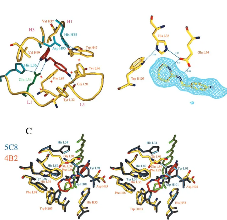
Interaction of the Hapten with the Active Site.The structure of 4B2 complexed to 1b, an analogue of hapten 1a lacking the linker arm to the carrier protein (Fig. 1), shows that the hapten is buried in a funnel-shaped deep cleft. The amidine is accommodated at the bottom of the pocket, which constitutes a highly hydrophobic environment where no water molecule was detected (Fig. 2A). Such a cavity at the interface of the heavy- and light-chain variable domains has been described in antibodies catalyzing various reac-tions such as ester hydrolysis (21), pericyclic reacreac-tions (22, 23), epoxyalcohol cyclization (12), or polyene cyclization (14).
The hapten establishes three hydrogen bonds with residues of the antibody. The terminal amine function, which is linked to the carrier protein for immunization is accessible to solvent and establishes a hydrogen bond with a main chain carbonyl oxygen and with a water molecule. The two NH groups of the amidinium are hydrogen bonded to the two oxygens of the Glu L34 carboxylate (Fig. 2B). At the crystallization pH (pH⫽ 5), which is close to the pH for optimal catalysis of allylic isomerization (pH 4.6), the L34 carboxylate deprotonated form is stabilized by a hydrogen bond network with His L36 and Trp H103, which also likely positions the carboxylate side chain at the proper location to abstract a proton from the substrate stereoselectively. There are three other polar residues in the combining site (Fig. 2 A). They cannot play the role of a general base either, because they point toward the external medium (Asp H95) or because their protonated nitrogen points toward the hapten (His H35 and His L36) (Fig. 2 A and B). Taken together with the biochemical data, the structure identifies Glu L34 as the general base that abstracts the␣-proton in the first step of allylic isomerization of 2 to yield the dienol intermediate 3 (Fig. 1).
Glutamate occurs at position L34 of only 5% of antibody sequences (24). It is therefore tempting to suggest that the significant stabilization of the antibody–hapten complex pro-vided by the glutamate–amidinium interaction because of the bidentate nature of both groups has favored the selection of a glutamate at this position during antibody maturation. As a consequence of its bidentate character, the interactions that the amidinium group establishes are anisotropic. This property confers on the amidinium the potential to generate a precisely positioned complementary charged residue.
Fig. 1. Scheme of the reaction catalyzed by antibody 4B2. 4B2 catalyzes allylic rearrangement of␣-cyclopent-1-en-1-yl-p-acetamidophenone 2 to ␣-cyclopentylidien-p-acetamidophenone 4 via the enediol intermediate 3, 2-[4-(1-carboxy)propylamidobenzylamino]-3,4,5,6-tetrahydropyridinium. 1a is the hapten used to generate 4B2. The structure that was determined is that of the complex of 4B2 with 2-(4-aminobenzylamino)-3,4,5,6-tetrahydropyridinium 1b. Antibody 5C8 was generated against hapten 5a, and its x-ray structure was determined in the presence of inhibitor 5b (12).
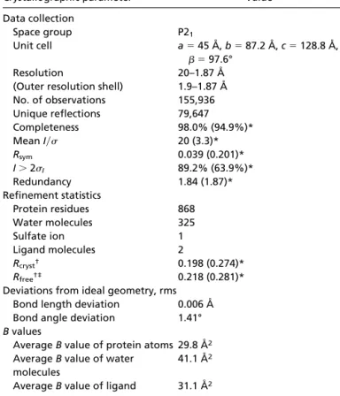
Table 1. Data collection and refinement statistics for Fab 4B2 complexed with 1b
Crystallographic parameter Value
Data collection
Space group P21
Unit cell a⫽ 45 Å, b ⫽ 87.2 Å, c ⫽ 128.8 Å,
 ⫽ 97.6°
Resolution 20–1.87 Å
(Outer resolution shell) 1.9–1.87 Å
No. of observations 155,936 Unique reflections 79,647 Completeness 98.0% (94.9%)* Mean I兾 20 (3.3)* Rsym 0.039 (0.201)* I⬎ 2I 89.2% (63.9%)* Redundancy 1.84 (1.87)* Refinement statistics Protein residues 868 Water molecules 325 Sulfate ion 1 Ligand molecules 2 Rcryst† 0.198 (0.274)* Rfree†‡ 0.218 (0.281)*
Deviations from ideal geometry, rms
Bond length deviation 0.006 Å
Bond angle deviation 1.41°
B values
Average B value of protein atoms 29.8 Å2
Average B value of water molecules
41.1 Å2
Average B value of ligand 31.1 Å2
*Values for highest-resolution shell are given in parentheses. †All data (F⬎ 2
F).
‡Of the data, 10% were set aside for the Rfreecalculation during the entire refinement.
Golinelli-Pimpaneau et al. PNAS 兩 August 29, 2000 兩 vol. 97 兩 no. 18 兩 9893
Fig. 2. (A) Schematic view of the active site of the 4B2-1b complex. The ligand is in red; water molecules are indicated as red crosses; Glu L34 is indicated in green; the other polar residues are represented in blue. The C␣s of residues L32–L36, L89–L97, H35–H37, and H94–H99 in hypervariable loops L1, L3, H1, and H3 are shown in yellow. The aromatic residues (Trp H47, Phe L89, Tyr L96, Tyr L32, Trp H103, and Phe L98) that have hydrophobic interactions with the hapten have been represented, except Phe L98 and Trp H103 for reasons of clarity. Nitrogen N␦1 of His H35 is involved in a conserved H bond with N1 of Trp H47. His H35 is therefore neutral but cannot play the role of a general base, because its protonated nitrogen N2 points toward the inside of the cavity. (B) Hydrogen bonding network established with catalytic residue Glu L34. Hydrogen bonds are shown as dashed lines. A Fobs-Fcalc electron density map calculated without the amidinium ligand is superimposed on the structure. The map is contoured at the level of two standard deviations. One of the oxygens of Glu L34 is hydrogen bonded to the protonated nitrogen N2 of His L36. This tautomeric form of His L36 is stabilized by an additional hydrogen bond between its N␦1 and the NH1 of Trp H103. Residue His L36 is therefore neutral but cannot play the role of a general base, because its unprotonated nitrogen N1 points away from the substrate. (C) Comparison of the environment of the charge of the hapten in antibodies 5C8 (in blue) and 4B2 (in yellow). 5C8 catalyzes the disfavored cyclization of an epoxyalcohol (12). The␣-carbons of residues of the combining site (L32–L38, L43–L50, L86–L93, L96, L98, H32–H39, H91–H95, and H102–H104) of 5C8 have been superimposed on those of 4B2 (the rms deviation is 0.645 Å). The bottom of the active sites is formed by identical residues in both antibodies (Phe L98, Trp H47, His H35, Trp H103, and Val H37). The distance between the nitrogen of 5b of 5C8 (light green) and the carbon between the two nitrogens of 1b of 4B2 (red) is 1.05 Å. In addition to the ionic interaction with glutamate L34, the hapten amidinium charge of 4B2 is stabilized through a cation– interaction with Phe L89, which is also involved in a stacking interaction with the amidine cycle. Residues of 5C8 involved in cation– interaction with the quaternary amine of 5b are (distance to the nitrogen) Tyr L91 (4.97 Å), Trp H103 (5.59 Å), His H35 (4.55 Å), His L89 (4.96 Å), and Tyr L36 (5.39 Å). In addition, in 5C8, Asp H101 (for which there is no equivalent in 4B2 because of the short H3 loop of this antibody) and Asp H95, which are, respectively, 4 Å and 3.7 Å away from the positive charge, provide a second sphere polar environment to the quaternary amine (12). Trp H47, Val H37, and Tyr L96 are not represented for reasons of clarity.
The Environment of the Charge of the Hapten. The directional property of the amidinium group is highlighted by a comparison of the 4B2 combining site to that of another catalytic antibody, 5C8, elicited against hapten 5a (Fig. 1) where the amidine group of 1a is replaced by a quaternary amine (12). The piperidinium ring of inhibitor 5b and the amidinium ring of 1b occupy very similar positions at the bottom of the combining sites (Fig. 2C). Both haptens carry positively charged nitrogens that are posi-tioned close to aromatic rings, as often observed in proteins (25). In 4B2, the charge of the amidinium group of the hapten is stabilized mainly by the ion pair with glutamate L34. By contrast, in antibody 5C8, no carboxylate is available to stabilize the quaternary ammonium of 5b through a direct ionic interaction. In the 5C8–5b complex, the isotropic distribution of the charge of the quaternary ammonium group induces an environment consisting of distant polar and -electron-containing residues that stabilize the charge in a diffuse fashion (Fig. 2C). Such charged nitrogen–aromatic interactions have also been observed in the structures of small molecules (26), of antibody McPC603 bound to phosphorylcholine (27), of antibody catalysts com-plexed with their aminophosphonic acid (13) or N-oxide hapten (14), and of acetylcholinesterase complexed with various qua-ternary amine ligands (28).
Discussion
The induction of a precisely positioned carboxylate residue in an antibody combining site is of general use to generate catalysts for reactions requiring either a nucleophile (29, 30) or a general base or acid as in enzyme-catalyzed proton transfer reactions (2, 31, 32). The hapten–4B2 complex defines amidinium as a useful candidate for that purpose and provides a starting point for the design of haptens capable of eliciting more sophisticated
con-stellations of catalytic residues in antibody combining sites. The induction of a second, planned, catalytic residue in the combin-ing site of an antibody, which would enhance its catalytic efficiency, would be favored by incorporation in the same hapten structure of two bidentate charged groups. Such molecules that have been developed as specific protein binders (33) would be logical hapten candidates to achieve this goal.
Methods capable of eliciting chemically reactive groups in anti-body combining sites such as those using haptenic charge (6–9), for which an unambiguous demonstration is presented here, or reactive immunization (34) are particularly useful to produce efficient catalytic antibodies, especially when transition state analogues are difficult to synthesize or when reactions proceed via multiple transition states (35). Indeed, these methods allow the use of a hapten that does not need to match precisely the transition state of the reaction but rather elicits a strategically positioned functional residue. Therefore, antibodies resulting from these strategies have been found to catalyze different reactions that use the same mechanism, provided that the various substrates possess the com-mon chemical function with which the induced residue can react (10, 36, 37). This finding is also illustrated by antibody 4B2, which catalyzes both the Kemp elimination (20) and the allylic rearrange-ment of a-␥ unsaturated ketone.
We thank L. Tchertanova (Institut des Substances Naturelles, Centre National de la Recherche Scientifique, Gif-sur-Yvette) for surveying the Cambridge Database, J. Perez for helping us to use facilities at Labo-ratoire pour l’Utilisation du Rayonnement Electromagne´tique, Orsay, France, and J. Janin for reading the manuscript. Partial financial support was provided by the European Union Training and Mobility Research Grant ERBFMXCT 98-0193 (to M.K.), and the cost of this article was covered by the Centre National de la Recherche Scientifique, Groupe-ment de Recherche 897.
1. Schwab, J. M. & Henderson, B. S. (1990) Chem. Rev. 90, 1203–1245. 2. Kim, S. W., Cha, S.-S., Cho, H.-S., Kim, J.-S., Ha, N.-C., Cho, M.-J., Joo, S.,
Kim, K. K., Choi, K. Y. & Oh, B.-H. (1997) Biochemistry 36, 14030–14036. 3. Schultz, P. G. & Lerner, R. A. (1995) Science 269, 1835–1842.
4. Blackburn, G. M., Datta, A., Denham, H. & Wentworth, P. (1998) Adv. Phys. Org. Chem. 31, 249–392.
5. Thayer, M. M., Olender, E. H., Arvai, A. S., Koike, C. K., Canestrelli, I. L., Stewart, J. D., Benkovic, S. J., Getzoff, E. D. & Roberts, V. A. (1999) J. Mol. Biol. 291, 329–345.
6. Janda, K. D., Weinhouse, M. I., Schloeder, D. M., Lerner, R. A. & Benkovic, S. J. (1990) J. Am. Chem. Soc. 112, 1274–1275.
7. Janda, K. D., Weinhouse, M. I., Danon, T., Pacelli, K. A. & Schloeder, D. M. (1991) J. Am. Chem. Soc. 113, 5427–5434.
8. Shokat, K. M., Leumann, C. J., Sugasawara, R. & Schultz, P. G. (1989) Nature (London) 338, 269–271.
9. Thorn, S. N., Daniels, R. G., Auditor, M.-T. M. & Hilvert, D. (1995) Nature (London) 373, 228–230.
10. Romesberg, F. E., Flanagan, M. E., Uno, T. & Schultz, P. G. (1998) J. Am. Chem. Soc. 120, 5160–5167.
11. Wentworth, P., Jr., Liu, Y., Wentworth, A. D., Fan, P., Foley, M. J. & Janda, K. D. (1998) Proc. Natl. Acad. Sci. USA 95, 5971–5975.
12. Gruber, K., Zhou, B., Houk, K. N., Lerner, R. A., Shevlin, C. G. & Wilson, I. A. (1999) Biochemistry 38, 7062–7074.
13. Hsieh-Wilson, L. C., Schultz, P. G. & Stevens, R. C. (1996) Proc. Natl. Acad. Sci. USA 93, 5363–5367.
14. Paschall, C. M., Hasserodt, J., Jones, T., Lerner, R. A., Janda, K. D. & Christianson, D. W. (1999) Angew. Chem. Int. Ed. Engl. 38, 1743–1747. 15. Gonc¸alves, O., Dintinger, T., Lebreton, J., Blanchard, D. & Tellier, C. (2000)
Biochem. J. 346, 691–698.
16. Otwinovsky, Z. & Minor, W. (1997) Methods Enzymol. 276, 307–325. 17. Navaza, J. (1994) Acta Crystallogr. A 50, 157–163.
18. Jones, T. A., Zhou, J.-Y., Cowan, S. W. & Kjeldgaard, M. (1991) Acta Crystallogr. A 47, 110–119.
19. Adams, P. D., Pann, N. S., Read, R. J. & Bru¨nger, A. T. (1997) Proc. Natl. Acad. Sci. USA 94, 5018–5023.
20. Genre-Grandpierre, A., Tellier, C., Loirat, M.-J., Blanchard, D., Hodgson,
D. R. W., Hollfelder, F. & Kirby, A. J. (1997) Bioorg. Med. Chem. Lett. 7, 2497–2502.
21. Charbonnier, J.-B., Carpenter, E., Gigant, B., Golinelli-Pimpaneau, B., Eshhar, Z., Green, B. S. & Knossow, M. (1995) Proc. Natl. Acad. Sci. USA 92, 11721–11725. 22. Ulrich, H. D., Mundorff, E., Santarsiero, B. D., Driggers, E. M., Stevens, R. C.
& Schultz, P. G. (1997) Nature (London) 389, 271–274.
23. Heine, A., Stura, E. A., Yli-Kauhaluoma, J. T., Gao, C., Deng, Q., Beno, B. R., Houk, K. N., Janda, K. D. & Wilson, I. A. (1998) Science 279, 1934–1940. 24. Kabat, E. A., Wu, T. T., Perry, H. M., Gottesman, K. S. & Foeller, C. (1991)
in Sequences of Proteins of Immunological Interest (U.S. Public Health Service, National Institutes of Health, Bethesda, MD), 5th Ed.
25. Gallivan, J. P. & Dougherty, D. A. (1999) Proc. Natl. Acad. Sci. USA 96, 9459–9464.
26. Verdonk, M. L., Boks, G. J., Kooijman, H., Kanters, J. A. & Kroon, J. (1993) J. Comput. Aided Mol. Des. 7, 173–182.
27. Dougherty, D. A. & Stauffer, D. A. (1990) Science 250, 1558–1560. 28. Harel, M., Schalk, I., Ehret-Sabatier, L., Bouet, F., Goeldner, M., Hirth, C.,
Axelsen, P. H., Silman, I. & Sussman, J. L. (1993) Proc. Natl. Acad. Sci. USA
90,9031–9035.
29. Verschueren, K. H., Seljee, F., Rozeboom, H. J., Kalk, K. H. & Dijkstra, B. W. (1993) Nature (London) 363, 693–698.
30. Zechel, D. L. & Withers, S. G. (2000) Acc. Chem. Res. 33, 11–18.
31. Davenport, R. C., Bash, P. A., Seaton, B. A., Karplus, M., Petsko, G. A. & Ringe, D. (1991) Biochemistry 30, 5821–5826.
32. Mitra, B., Kallarakal, A. T., Kozarich, J. W., Gerlt, J. A., Clifton, J. G., Petsko, G. A. & Kenyon, G. L. (1995) Biochemistry 34, 2777–2787.
33. Albert, J. S., Peczuh, M. W. & Hamilton, A. D. (1997) Bioorg. Med. Chem. 5, 1455–1467.
34. Wagner, J., Lerner, R. A. & Barbas, C. F., III (1995) Science 270, 1797–1800. 35. Koch, T., Reymond, J.-L. & Lerner, R. (1995) J. Am. Chem. Soc. 117,
9383–9387.
36. Barbas, C. F., III, Heine, A., Zhong, G., Hoffman, T., Gramatikova, S., Bjo¨rnestedt, R., List, B., Anderson, J., Stura, E. A., Wilson, I. A., et al. (1997) Science 278, 2085–2092.
37. Lin, C.-H., Hoffman, T. Z., Wirshing, P., Barbas, C. F., III, Janda, K. D. & Lerner, R. A. (1997) Proc. Natl. Acad. Sci. USA 94, 11773–11776.
Golinelli-Pimpaneau et al. PNAS 兩 August 29, 2000 兩 vol. 97 兩 no. 18 兩 9895