(E)-2'-Deoxy-2'-(fluoromethylene)
Cytidine Potentiates Radioresponse of Two
Human Solid Tumor Xenografts1
Lin-Quan Sun,2 Ye-Xiong Li,3 Louis Guillou, and Philippe A. Coucke4
Laboratory of RadiMology. Department of Radialion Oncology ¡L-Q.S., Y-X. L. P. A. C./ and Institute of Pathology ¡LG.I. University Hospital of ¡MiisannefCHUV), CH-IOII Ltnisunne, Swìtzt'rìiintì
ABSTRACT
Antitumor and radiosensitizing effects of (E)-2'-deoxy-2'-(fluorometh-ylene) cytidine (FMdC), a novel inhibitor of ribonucleotide reducÃ-ase, were evaluated on nude mice bearing s.c. human C33-A cervix cancer and I -H7 MG glioblastoma xenografts. FMdC given once daily has a dose-dependent antitumor effect. The maximum tolerated dose in the mice was reached with 10 daily i.p. administrations of 10 mg/kg over 12 days. In the case of radiotherapy (RT) alone (10 fractions over 12 days), the radiation dose required to produce local tumor control in 50% of the treated C33-A xenografts was 51.0 Gy. When combined with FMdC, the radiation dose required to produce local tumor control was reduced to 41.4 and 38.2 Gy, at respective doses of 5 and 10 mg/kg given i.p. l h before each irradiation. The corresponding enhancement ratios (ERs) were 1.2 and 1.3, respec tively. In U-87 MG xenografts, when 5-20 mg/kg FMdC combined with 30 or 40 Gy of RT, the combination treatment produced a significantly increased growth delay as compared with RT alone (P £0.002). The ERs of 5, 10, and 20 mg/kg FMdC at a dose of 30 Gy were 2.0, 1.4, and 1.8, respectively. At the 40-Gy level, ERs of 10 and 20 mg/kg FMdC were 1.4 and 1.7. When FMdC was combined with 50 Gy of RT, an increased long-term remission rate of 80-88.9% was observed, as compared with
25% for RT alone (P <0.05). FMdC produced moderate
myelosuppres-sion in the mice bearing cervix cancer, whereas leukocytosis occurred in the mice bearing glioblastoma at a low dose. Slightly increased skin toxicity (only with U-87 MG tumor) was observed, as compared with RT alone. In conclusion, FMdC is a potent cytotoxic agent and able to modify the radiation response of C33-A and U-87 MG xenografts.
INTRODUCTION
FMdC5 (1, 2), a novel compound synthesized to exert irreversible and potent inhibition of RR, is a very effective cytotoxic agent against a variety of common human cancers (3-7). Moreover, it has been shown to act as a radiosensitizer both in vitro (8) and in vivo (7).
The antitumor effect and radiosensitization can be explained by the effect on RR and subsequent alteration of the dNTPs. It has been shown by others that RR, an enzyme that catalyzes a rate-limiting step in DNA synthesis, has an increased activity in rapidly growing tumors (9) and that alteration of the dNTP pool is related to the modification of radiation response (10). Takahashi et cil. (6) showed recently that FMdC induced long-lasting inhibition of RR and resulted in a deple tion of the dCTP. dATP, and dGTP pools and an increase in the TTP
Received 6/15/98; accepted 9/30/98.
The costs of publication of this article were defrayed in part by the payment of page charges. This article must therefore be hereby marked tulrertisetnt'tit in accordance with
18 U.S.C. Section 1734 solely to indicate this fact.
1 Supported by the Foundation Radiobiologie 20(K) and Ligue Vaudoise Contre Le Cancer. Lausanne, Swit/erland.
2 Present address: Division of Hematology/Oncology. Children's Hospital Medical Center. 3333 Burnet Avenue. Cincinnati. OH 45229-3039.
1 Present address: Department of Clinical Investigation. Box 71. University of Texas M. D. Anderson Cancer Center. 1515 Holcombe Boulevard. Houston, TX 77030.
4 To whom requests for reprints should be addressed, at Laboratory of Radiobiology, Department of Radiation Oncology. University Hospital of Lausanne (CHUV). CH-1011 Lausanne, Switzerland. Phone: 41-21-314-4603; Fax: 41-21-314-4601: E-mail: philippe.coucke@chuv.hospvd.ch.
'The abbreviations used are: FMdC. (El-2'-deoxy-2'-(fluoromelhylene) cytidine: RR. ribonucleotide reducÃ-ase;dNTP, deoxyribonucleotide: RT. radiotherapy; AGD. absolute growth delay: NGD. normali/ed growth delay; TCD?0, the radiation dose required to produce local tumor control in 50^ of the treated mice: ER. enhancement ratio; gemcit-abine. 2'.2'-difluorocytidine.
pool. Other drugs such as hydroxyurea or gemcitabine, acting on the same target, also have been shown to be potent radiosensitizers (11-13). Recently, We have observed that the radiosensitizing effect of FMdC is evident in a human colon cancer WiDr xenograft model (7); however, whether FMdC also has a similar radiosensitizing effect on other types of human cancers in vivo is not clear. In this study, we decided to further evaluate the capacity of FMdC to act as a radiation sensitizer in a human cervix cancer C33-A with a mutated p53 (14) and a glioblastoma U-87 MG with a wild type of p53 (15) xenografted in nude mice.
MATERIALS AND METHODS
Chemicals and Cell Lines. FMdCIMDL 101,731) was kindly provided by
Marion Hoechst Roussel, Inc. (Cincinnati. OH). Cell culture media and sup plements were purchased from Lite Technologies. Inc. (Basel, Swit/erland). PCS was obtained from Fakola.
The human C33-A cervix cancer cell lines were obtained from the American Type Culture Collection (Manassas, VA). The human glioblastoma U-87 MG cell line was a gift from the Department of Neurosurgery at our hospital. Cells were grown as a monolayer in Eagle's MEM with WJr FCS. 2 mM L-glutamine. and \'7< penicillin-streptomycin. To establish tumors in nude mice. cells in exponential growth phase were harvested after a 3-min incubation with Trypsin (0.05%)-EDTA (0.02%) solution and resuspended in serum-free MEM. A suspension of about 5 x I0ft cells was inoculated s.c. in the dorsum of the Swiss nude mice. For the experiments, animals were implanted with tumors at least three passages away from the initial source.
s.c. Tumor Model. All experiments in nude mice were performed accord
ing to Swiss legislation and approved by the official committee of surveillance of animal experiments. Female Swiss homozygous nu/nu nude mice, 7-9 weeks of age. were given a s.c. transplantation in the midline of the back at 2 cm from the tail of a volume of about 30 mm' of freshly excised, minced tumor tissues. Three to 4 weeks after inoculation, the mice bearing tumors of approximately 80-160 mm' volume, with a mean tumor volume ot about 120 mm1, were assigned randomly tor the control or the test treatment groups.
Irradiation of Tumors. X-rays were generated by a Philips RT 250
operating at 200 kV and 20 mA. The beam was filtered with 0.5 mm Cu (halt-value layer = 1 mm Cu). Up to six mice/irradiation were restrained in 3-mm lead jigs designed with a cutout 20- X 14-mm to expose their lower dorsum. The jigs were placed in a perspex box with an additional lead shield with 60- X 17-mm openings; in each field, two mice were exposed tail-on-tail. This setup gives minimal scatter to the animals placed at 52.5 cm from the source. The X-ray beam hits the tumors tangentially to the dorsum. The dose rate in this setup was 0.64 Gy/min with a dose heterogeneity of ±5.5% for an 8-mm tumor. To obtain dose homogeneity, the mice were rotated through 180 degrees at alternate treatments. The treatment regime consisted of 10 fractions over 12 days (5 fractions/week comparable with a clinical fractionation sched ule).
Antitumor Effects of FMdC. Nude mice with s.c. tumor xenografts were
treated ¡.p.with 1. 5. 10, and 20 mg/kg FMdC. once daily. 5 days/week for up to 2 weeks.
Radiosensitizing Effect of FMdC. FMdC dissolved in saline and sterili/ed
by filtration through Millipore 0.22 pim was administrated i.p. l h before each irradiation for 2 weeks with a 2-day rest during the weekend. All RT or RT-FMdC combined groups were evaluated in two blocks, each block includ ing half of the mice of each group with similar-si/ed tumors. Each group consisted of 8-13 mice.
Experimental End Points. After treatment, three perpendicular diameters
of each tumor were measured with calipers twice a week. Complete or partial 5411
100 control,n=15 1mg/kg, n=12 5mg/kg,n=13 10mg/kg,n=5 20mg/kg,n=6
Time
After
Treatment
(Days)
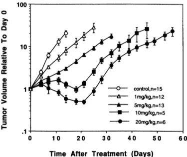
Fig. I. The effects of FMdC given as once daily i.p. administrations (10 times over 12 days) on (he growth of C33 carcinoma xenografts in nude mice. The mean tumor volume relative to the initial tumor volume at day 0 is plotted versus time for each group (±SE). The tumor volumes at day 0 were 82.7 ±38.1, 81.1 ±32.9. 102.7 ±19.7. 118.5 ±37.5, and 128.8 ±39.9 mm' for groups of control. 1. 5. 10. and 20 mg/kg FMdC, respectively.
regressions were assessed once/week. The tumor volume was calculated using the formula: V = length X width X thickness/2. The relative tumor volume (V/Vo) was calculated by dividing the measured tumor volume (V) by the initial tumor volume (vo) at day 0.
Tumor responses were quantified by tumor regrowth delay, local control, and long-term remission. Tumor regrowth delay was expressed as AGD and NGD. AGD was defined as the time for tumor volume in the treated groups to increase by five times the initial treatment size minus the time in the untreated control group to reach the same size. NGD was defined as the time for tumor volume to increase by five times the initial treatment size in the mice treated by the combination of FMdC and radiation minus the time for tumors to reach the same size in the mice treated with FMdC alone. Local control for the C33-A tumor is defined as absence of palpable tumor mass at 120 days after the end of the treatment. All regrowing tumors were recorded as relapse in the analysis whether or not the tumor diameter was smaller than that of the first day of irradiation. The percentage of controlled tumors at 120 days was plotted for each group, and the data were fitted by logit analysis. The effect of graded doses of radiation given alone or in combined regimens was evaluated as the TCDM1. The ER for C33-A tumor was calculated as the ratio of TCD50 produced by radiation alone to TCDSO produced by combined FMdC and radiation. Because U-87 MG glioblastoma lacks an apoptotic cell death path way (16) and is a very radioresistant tumor that has a TCD50 of 75.2 Gy under clamp hypoxia using single-dose irradiation (17), the local control rate was not available. Therefore, we used long-term remission for the U-87 MG tumor, which was defined as absence of palpable or residual tumor mass <2 times of initial volume 180 days after the end of treatment. To calculate the ER, we also included these long-term remission tumors and arbitrarily took 180 days as the time for these tumors to grow to five times the initial treatment size. The ER for the U-87 MG glioblastoma was calculated as the ratio of the NGD produced by combined FMdC and radiation to the AGD produced by radiation alone.
Toxicity Evaluation. Local skin toxicity of RT was evaluated by inspec
tion three times/week for the first 5 weeks, and then twice/week. The skin toxicity in the radiation field was scored as follows: I. local erythema: II. moist desquamation; and III. complete ulcération.Toxicity after injections of FMdC alone or combined with RT was evaluated by body weight measurements and peripheral WBC counting. Body weight was measured three times weekly from the first injection of FMdC until 4 weeks after the end of the treatment, and the weight loss was expressed as a percentage of the initial weight (<initial weight - lowest weight>/initial weight x 100%). Peripheral WBCs were monitored 1 day after the end of FMdC treatment and 3 days after finishing radiation treatment alone or combined with FMdC (correlated with
the nadir of WBC at those moments). WBCs were counted in 15 p.\ of blood (obtained from the tail vein) diluted 1:10 in Turck solution and manually counted. Lower limb paralysis was recorded after treatment.
Histopathological Studies. The s.c. tumors that were untreated or treated
with 10 daily administrations of 10 mg/kg FMdC (once daily, 5 days/week) were removed from nude mice 24 h after the last treatment and fixed in 4% buffered formalin. The residual tumor masses (which were defined as long-term remission) were also fixed in 4% buffered formalin, 6 months after the end of treatment. The specimens were embedded in paraffin, and 4-/im thick sections were stained sequentially with H&E for microscopic examination.
Potential Doubling Time and Volume Doubling Time. The potential
doubling times of C33-A and U-87 MG tumors were measured by iodode-oxyuridine labeling and flow cytometry. The mice bearing C33-A or U-87 MG tumors were given i.p. injections of iododeoxyuridine 6 h before the tumors were removed. The dose administrated was 150 mg/kg. The s.c. tumors were fixed in 70% ethanol and stored at 4°Cin the dark until analysis. Preparation of samples for flow cytometry was according to the method of Begg (18). Briefly, the tumor samples were cut into small pieces in Petri dishes and digested with 0.4 mg/ml pepsin for l h at 37°C,which produced a suspension of nuclei. DNA of the nuclei was denatured with 2N HC1 for 20 min at 37°C. Antibody staining consisted of the mouse anti-iododeoxyuridine antibody (1:100 dilution; Parte« AG, Basel, Switzerland) followed by an FlTC-conju-gated goat antimouse IgG antibody (1:50 dilution; Parted AC). Total DNA was stained with 50 /ig/ml propidium iodide. Flow cytometric analysis was carried out using FACScan flow cytometry (Becton Dickinson). The methods for calculating potential doubling time has been described previously (19). The volume doubling times were individually estimated from the growth curves of the tumors xenografted in the mice.
Statistical Analysis. Tumor AGD, NGD, and hematological toxicity in the
various conditions of therapy have been compared using the Student's / test (two-tailed). For comparison of long-term remission rates, the )C test was used. TCD50 was calculated according to the logit analysis.
RESULTS
Antitumor Effects of FMdC on C33-A and U-87 MG Tumor Xenografts. The response of C33-A xenografts to FMdC was dose-dependent (Fig. 1). Even at the low dose (i.e., 1 mg/kg), we observed a significant antitumor effect as compared with untreated control (P <0.0001). Significant higher tumor shrinkage was observed at 20 mg/kg FMdC. No significant body weight loss (S4.0%) was observed at the dose <5 mg/kg, whereas a 7.0% and 13.0% weight loss were observed at the dose of 10 and 20 mg/kg FMdC, respectively (P
m
E
o
o
(O 0)-*->.
o
o
JÉ 3o»
O.o
E
3Dose of FMdC (mg/kg)
Fig. 2. Peripheral blood leukocyte analysis of mice bearing C33-A and U-87 MG lumors 1 day after the end of FMdC treatment ( ±SE). The leukocyte counts of normal mice without lumors were 10,900 ±680/mm' (n = 12; mean ±SE).
100
n
OI
~ -5 »ce
o
"5
o
3 control, n=10 1mg/kg,n=8 5mg/kg,n=6 10mg/kg,n=13 20mg/kg,n=10 1 O 50 200Time After Treatment (Days)
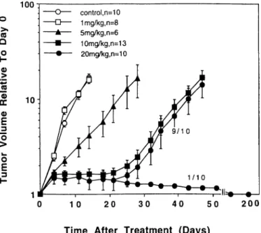
Fig. 3. The effects of FMdC given as once daily i.p. administrations ( 10 times over 12 days) on growth of U87 MG xenografts in nude mice (±SE). In the 20-mg/kg group. I of 11 mice died of acute toxicity (data not included).
<0.05). Even at a dose as high as 20 mg/kg, we did not observe toxic death. As far as hematological toxicity was concerned, we did observe a significant reduction of leukocyte counts from doses >5 mg/kg as compared with untreated controls (P <0.001), but no further decrease at higher doses (Fig. 2). Petechiae did not appear.
For U-87 MG xenografts, the antitumor effect became significant at doses >5 mg/kg (Fig. 3). However, no further increase in AGO was observed at a higher dose (i.e., 20 mg/kg), as compared with 10 mg/kg
(P = 0.18). There was also a moderate weight loss (9.5%) at a dose
of 20 mg/kg (P <0.05). In contrast to the mice bearing C33-A xenografts, we had to face toxic death in 1 of 11 mice (9.1%) treated at the higher dose level of 20 mg/kg. Tumor shrinkage did not appear, even up to a dose of 20 mg/kg, but significant prolonged AGDs were observed at doses of 5-20 mg/kg as compared with the untreated control (P <0.0001). In contrast to the C33-A model, a rise of leukocyte counts was observed at lower doses of 1 and 5 mg/kg in the U-87 MG model (P <0.0001). However, at the higher dose level, the leukocyte counts were significantly decreased compared with un treated control mice (P <0.0001; Fig. 2).
Mice surviving a higher dose of FMdC (i.e., 20 mg/kg) recovered their body weight within 7-10 days and their leukocyte counts com pletely within 10-14 days after the end of treatment (data not shown).
Radiosensitizing Effect of FMdC. The responses of C33-A tu mors to radiotherapy were dose-dependent, and the TCD50 for RT alone was 51.0 ±1.1 Gy (±95% confidence limits; Fig. 4). When 5 mg/kg FMdC was given i.p. l h before each irradiation, the dose effect curve was shifted to the left and the TCD5() dropped to 41.4 ±1.1 Gy (Fig. 4). When 10 mg/kg FMdC was used, the TCD50 was reduced to 38.2 ±1.1 Gy (Fig. 4). The ERs at the TCD50 level were 1.2 and 1.3 for 5 mg/kg and 10 mg/kg FMdC, respectively. The irradiated mice showed neither significant weight loss (data not shown) nor enhanced hematological toxicity (Fig. 5A), as compared with FMdC treatment alone. There was no increased skin toxicity with combined FMdC and RT as compared with RT alone (data not shown).
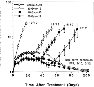
The response of U-87 MG tumors to RT were also dose-dependent (Fig. 6). When 5, 10, or 20 mg/kg FMdC were combined with 30 Gy of RT (Fig. 7), the tumor AGDs were significantly increased as compared with RT alone (P <0.002, P <0.0007, and P 5=0.0001 for
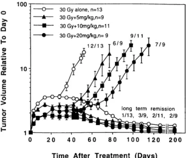
5, 10, and 20 mg/kg FMdC, respectively). If the AGDs produced by combined FMdC and RT were corrected for AGDs produced by FMdC alone, the corresponding ERs at the doses of 5, 10, and 20 mg/kg FMdC were 2.0, 1.4, and 1.8, respectively (Table 1). The results were confirmed in a second experiment, in which a RT of 40 Gy was combined with 10 or 20 mg/kg FMdC (Fig. 8). Again, the AGDs produced by combined RT and FMdC were significantly prolonged as compared with RT alone (P <0.002). The corresponding ERs at the doses of 10 and 20 mg/kg FMdC were 1.4 and 1.7, respectively (Table 1). However, as mentioned earlier, the toxic death was observed at the dose of 20 mg/kg. The long-term remission rate was also increased from 20% in cases of RT alone to 50% in cases of RT combined with 20 mg/kg FMdC (Fig. 8), but this difference was not significant (P >0.05). When FMdC was combined to 50 Gy of RT, a significantly increased long-term remission rate was observed as compared with RT alone (P <0.05; Table 2). However, the long-term remission rate was similar at the dose of 5 mg/kg as compared with 20 mg/kg.
Side effects of combined RT and FMdC in the mice bearing U-87 MG tumors were not the same as in the mice bearing C33-A tumors. At the dose of 5 mg/kg, the irradiated mice showed decreased leuko cyte counts as compared with FMdC treatment alone (P <0.05); however, the leukocyte counts were still higher than or similar to that of untreated control, even at a dose of 50 Gy (Fig. 5ß).At the doses of 10 mg or 20 mg/kg, the irradiated mice showed no enhanced hematological toxicity as compared with FMdC alone (Fig. 5fi). Increased toxicity, however, was observed at a higher radiation dose level (50 Gy) when 10 mg/kg FMdC was combined with RT (P <0.001). No significant weight loss was observed in all groups as compared with FMdC alone (data not shown). When 20 mg/kg FMdC was combined with RT, 9-18% of the treated mice died of toxicity 10-14 days after treatment (see Table 2 and legends to Figs. 7 and 8). The hematological toxicity might not be the main reason of death, because bone marrow transplantation with blood tranfusion did not reduce the death rate (7). When FMdC was combined with RT, skin toxicity was slightly increased as compared with RT alone, especially
1 00
i T + 10 mg/kg B RT + 5 mg/kg O RT alone
10 20 30 40 50 60 70
Dose of Radiotherapy
(Gy)
Fig. 4. Local tumor control of C33 xenografts to RT combined to daily 5 or 10 mg/kg FMdC. RT was given in 10 fractions over 12 days, and FMdC was given i.p. l h hefore each irradiation (±95% confidence limits).
_ 100 ro E E o o U) <u D O) .a E 300 200-100 30 40 50
Dose of Radiotherapy (Gy)
Fig. 5. Peripheral blood leukocyte analysis of mice bearing C33-A (A) and U-87 MO (fi) xenografts 3 days after the end of treatment with combined FMdC and RT (±SE).
30 40 50
Dose of Radiotherapy (Gy)
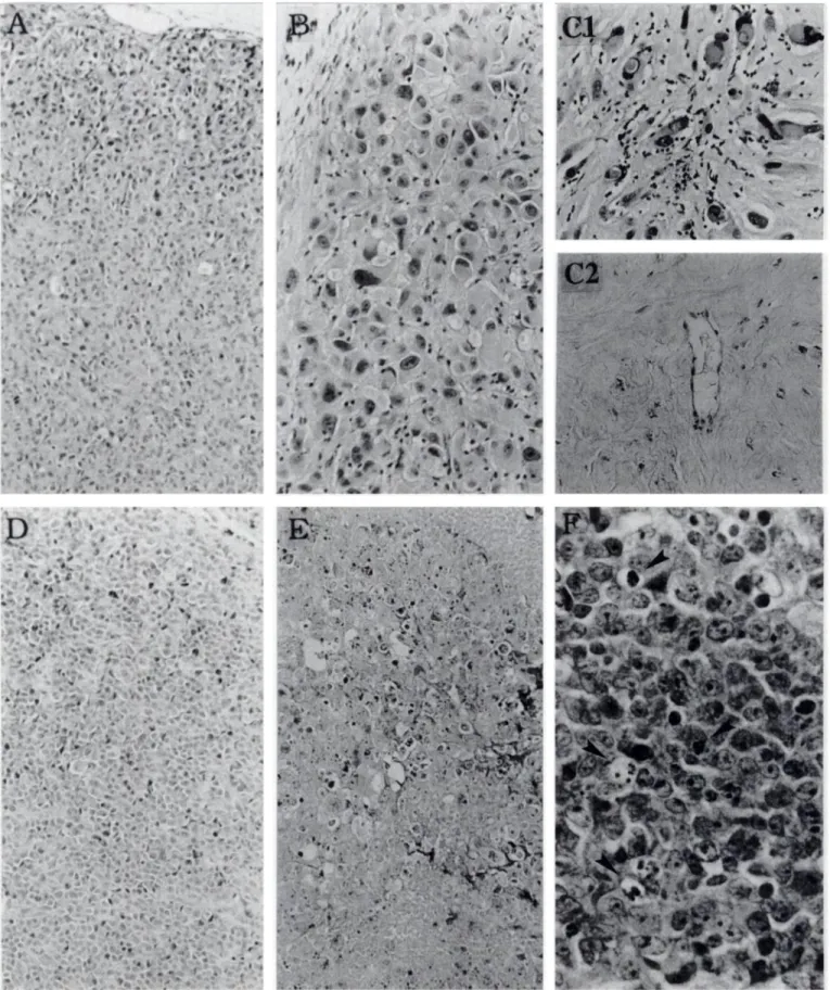
at the 40-Gy level (see Fig. 8 legend). Local acute skin reactions appeared about 12 days after treatment initiation, and the healing of radiation-damaged skin generally occurred 2-3 weeks after the end of treatment. One of 10 mice died of lower limb paralysis 3 months after the end of treatment in the group of 20 mg/kg FMdC + RT of 50 Gy. Histopathological Studies. No significant necrotic areas were ob served in the untreated s.c U-87 MG tumors (Fig. 9A), unless in the center of some tumor nodules (data not shown). Swollen and/or necrotic tumor cells, as well as infiltrated inflammatory cells, were observed after 10 daily i.p. treatments of 10 mg/kg FMdC (Fig. 9ß). Mitotic figures were evident in untreated control tumors, but not in the tumors treated with FMdC (data not shown). Significant interstitial fibrosis and concurrent inflammatory cell infiltration were observed in the residual U-87 MG tumor mass 6 months after the end of treatment with combined RT (40 Gy) and FMdC (20 mg/kg; Fig. 9C, top). Enlarged or swollen tumor cells with enlarged irregular nuclei con taining chromatin clumps were observed in some residual tumor masses (Fig. 9C, top). In those samples containing no residual tumor cells, only fibrosis without inflammatory cells was observed (Fig. 9C,
bottom). This may indicate that inflammatory cells are involved in the
process of removing dead tumor cells, because apoptotic cell death is not apparent in U-87 MG glioblastoma (16).
Swollen and/or necrotic tumor cells were observed in C33-A tu mors treated with 10 daily i.p. treatments of 10 mg/kg FMdC, how ever, no infiltrated inflammatory cells were visible (Fig. 9, E and F). Apoptotic cells were visible after FMdC treatment (Fig. 9F).
Potential Doubling Time and Volume Doubling Time. Under our experimental conditions, the potential doubling times of untreated C33-A and U-87 MG tumors were 2.1 ±0.6 (n = 4) and 1.6 ±0.4
(n = 4) days, and the volume doubling times were 3.4 ±0.9 (n = 15)
and 3.3 ±0.7 (n = 10) days, respectively.
DISCUSSION
The results of these in vivo studies provide experimental evidence that FMdC may be an effective anticancer agent in the treatment of human cervix cancer and glioblastoma. This compound induces re gression of s.c. tumors in a dose-dependent manner. Progressive tumor regrowth after the end of FMdC treatment alone, indicates that the daily administrations of FMdC are not curative for s.c. tumors. As
far as toxicity is concerned, we demonstrated that 10 daily adminis trations of 10 mg/kg FMdC over 12 days was the maximum tolerated dose in the mice. Daily doses >10 mg/kg for 10 times failed to produce more antitumor effects in the U-87 tumor, but resulted in a moderate weight loss and increased toxic death. As compared with untreated controls, the leukocyte counts in the mice bearing C33-A tumor were significantly reduced after treatment with a dose >5 mg/kg, whereas the leukocyte counts were significantly increased after treatment with 1-5 mg/kg FMdC in the mice bearing U-87 glioblastoma. However, at a higher dose (i.e., 20 mg/kg), the reduced leukocyte counts were also observed in the U-87 MG model. The reduced leukocyte counts in the mice bearing C33-A tumors could be explained by direct cytotoxicity of FMdC to bone marrow. However, the exact mechanism by which leukocytosis occurs in the mice bear ing U-87 MG tumor treated with a low dose of FMdC is not clear (20, 21). Our results correlate with prior xenograft studies of FMdC in
100 eo Q O <D •»-•
to
o>E
OE
3 J-control, n=10 30Gy,n=13 40Gy,n=10 50Gy,n=12long term remission 1/13. 2/10. 3/12
60 80 200
Time
After
Treatment
(Days)
Fig. 6. The effects of RT at different total doses on the growth of U-87 MG xenografts. RT was given in 10 fractions over I2 days (±SE).
100 o >.
£
<D jcç 0)rr
v
_3 O O E 3 30 Gy alone, n=13 30 Gy+5mg/kg,n=9 30Gy+10mg/kg,n=11 30 Gy+20mg/kg,n= 9 12/13 9/1 1 7/9long term remission 1/13, 3/9, 2/11, 2/9
20 40 60 80 100 120 200
Time
After
Treatment
(Days)
Fig. 7. The effects of 30 Gy of RT alone or combined with FMdC on the growth of U-87 MG xenografts ( ±SE). In the group of 30 Gy + 20 mg/kg FMdC, 2 of 11 mice died of acute toxicity 1-2 weeks after treatment (data not included).
Table 1 Effect of FMdC <m radiare sponse of U-87MG xenografîs
Treatment groupsUntreated eontrol5 mg/kg alone10 mg/kg alone20 mg/kg alone30 Gy'' alone40 Gy alone30 Gy + 5 mg/kg30 Gy + 10 mg/kg30 Gy + 20 mg/kg40 Gy + 10 mg/kg40 Gy 4- 20 mg/kgTime for tumor to grow to 5 times initialtreatmentsize (days)"6.3 ± 1.320.1 ± 1.435.7 ±39.8 ±41. 0±64.8 ±91.2 ±84.2 ±103.1 ±118.6 ±140.6 ±_2.3.6.7.8.5.4.5.4Tumor growthdelay (days)13.8'29.4'33.5'34.7'58.4'71.1'48.5''63.3"82.9''100.8''ER2.01.41.81.41.7 " Geometric mean ±SD.
FMdC was given as once daily i.p. injections, 10 times over 12 days. ' AGO.
RT was given in 10 fractions over 12 days. '' NGD.
human malignant breast, colon, prostate, and brain tumors. In these tumors, a dramatic tumor regression of s.c.-implanted tumors was obtained and a significant prolongation of survival was observed in intracerebral implants and liver métastases,as well as a reduction of the number of lung métastases(3-5, 7).
More importantly, FMdC significantly increased radiation response of human cervix cancer and gliobla.stoma xenografts. The ERs for C33-A tumor at TCD50 levels were 1.2 and 1.3 for 5 and 10 mg/kg FMdC applied daily, respectively. However, the ERs for C33-A tumor are less important as compared with WiDr tumors in which ERs of 1.9 and 2.4 were observed (7). One of the reasons for the differences may be that C33-A is a more aggressive tumor than the WiDr tumor. The potential doubling time and volume doubling time were 2.1 and 3.4 days for the C33-A tumor and 3.0 and 6.4 days for the WiDr tumor, respectively. Significantly increased AGDs and long-term remission rates were observed in U-87 MG tumors when 5-20 mg/kg FMdC were combined with RT. Combining FMdC (5-20 mg/kg) with RT (30 or 40 Gy ) produces a better therapeutic effect, with an ER ranging from 1.4-2.0. When 5-20 mg/kg FMdC were combined with 50 Gy of RT, significantly increased long-term remission rates of the treated tumors (80-88.9%) were observed, as compared with RT alone (25%). However, the high dose of FMdC (i.e., 20 mg/kg) is not able
to further increase a long-term remission rate as compared with 5 mg/kg.
The addition of FMdC to irradiation did not increase skin toxicity, weight loss, or hematological toxicity in the mice bearing C33-A tumors. A moderate increase of hcmatological toxicity was observed in the mice bearing U-87 MG tumors, when a higher dose of RT (50 Gy) was combined with 10 mg/kg FMdC. However, the hcmatolog ical toxicity was well-tolerated and was reversible. We do not know why the slightly increased skin toxicity was observed in the mice bearing U-87 MG tumors and not in the mice bearing C33-A and WiDr tumors (7).
Because of direct measurements of RR. dNTP pools and DNA repair were not performed in our studies, the precise mechanism of FMdC can only be inferred from prior studies. FMdC is a potent member of a class of mechanism-based inhibitors of RR, the enzyme responsible for de novo production of dNTPs by reduction of ribonu-cleotide at the level of diphosphates. The drug acts in a manner similar to other RR inhibitors such as hydroxyurea and gemcitabinc, which has been shown to cause inhibition of DNA synthesis specifically, without significantly inhibiting either protein or RNA synthesis. In hibition of DNA synthesis with these drugs is most probably due to a decrease in one or more of dNTP pools (6, 22-25) or chain termina tion after being converted to the triphosphate, as shown for
gemcit-100
ra Q o OCo
E
_o
E
40 Gy alone,n=10 40Gy+10mg/kg,n=10 40Gy+20mg/kg,n=10 6/1 O 5/1 Olong term remission 2/10, 4/10, 5/10
20 40 60 80 100120140 200
Time After
Treatment
(Days)
Fig. 8. The effects of 40 Gy of RT alone or combined with FMdC on the growth of U-87 MG xenografts ( ±SE). In the group of 40 Gy + 20 mg/kg FMdC. I of 11 mice died of acute toxicity 2 weeks after treatment (data not included). In the group of RT alone, i of 10 mice had skin toxicily grade 1. In the group of RT + 10 mg/kg FMdC. I of 10 mice had skin toxicity grade II. and 4 mice had grade 1. In the group of RT + 20 mg/kg FMdC. I of 10 survived mice had skin toxicity grade III. 3 mice had grade II, and 6 mice had grade I.
Table 2 Anlilumor and toxic effects of 50 Gy RT combined with FMdC on U-/Õ7MG xenografts GroupsRT aloneRT + 5 mg/kg"RT + 10 mg/kgRT + 20 mg/kgNo. ofmice12101010No.ofremission (%)3 (25)8 (80)''8 (80)''8 <88.9)'-''No. of micethat died (%)0001 (10)' " RT was given in 10 fractions over 12 days, and FMdC was given I h before each irradiation.
* P <0.05 as compared with RT alone.
' No. of (long-term) remission from 9 of 10 mice that survived. J P <0.001 as compared with RT alone.
' One mouse died of lower limb paralysis 3 months after treatment.
•
•-/-> .
è
/ ,•'
.
•:
'•.'•••••«••
- ¡
:.-
• •
: •••
•/,-•;
:«,.,•?••*
;^
•—»•••.i
//./
•/.',-••.
•
»•
••
* .•..:'/„,'»-.»
•
• /
* *
.
•
i "
r
V* "*'>*•- -
-'-
••.-.' —
. «
.•-' - ' • • ^'^•' ' " 4^*" • 3. »S%
' • '
'/
^- •
.. * »M
*
.•>y 1
* 0 »> W•*.
ci
I ! ' ft >*•- « F f •'i'^y-,
if * . ^ 'M . •E
-v,:%.
<
Vu.
itÃ-x.^ : ^
^•,-:'
a2î**:
ifêii^fe
Ha
\X5>Ã ff*
• *•
A<
: ' : ^-^^T
~yC u^^
Fig. 9. Histology of human gHobIasióma U-87 MG (A-D and human cervix cancer C33-A (D-/7)- The s.c. tumors treated with 10 daily administrations of 10 mg/kg FMdC were removed for analysis 24 h after the last treatment (/?, £.and /•*).The residual tumor masses were removed for analysis 6 months after the last treatment of combined FMdC of 20 mg/kg with RT of 40 Gy (CI. C2), In untreated tumors (A and D), tumor cell morphology is well preserved. In FMdC-trealed tumors, swollen and/or neerotic tumor cells are observed (B and £").CI. residual abnormal tumor cells with a significant amount of interstitial fibrosis are observed. C2, only fibrosis (without residual tumor cells) is observed. B and CI. infiltrating inflammatory cells are detectable. F, apoptotic cells are visible (arrow head). Images of A-E were captured through a x 10 objective; F was captured through a X40 objective.
abine (26). Although not proven in the present study, it is likely that FMdC exerts its radiosensitizing effects through inhibition of irradi ation-induced DNA repair processes. The reduced availability of DNA precursors or perturbation of dNTP pools (6) may result in an impairment of radiation-induced DNA damage repair and may be an important determinant of radiosensitization with FMdC (8). This has been shown in other RR inhibitors such as hydroxyurea (13) and gemcitabine (11, 12). Other drugs active on the dNTP pools have been shown to be potent radiosensitizers, such as fluorodeoxyuridine (in hibition of thymidylate synthase and depletion of dTTP pools; Refs. 27-31 ) and the thymidine analogues, bromodeoxyuridine and iodode-oxyuridine (depletion of dCTP and TTP pools; Refs. 10 and 32). Thus, the antitumor and radiosensitizing effects of FMdC may be associated with the depletion and intracellular imbalances of dNTP pools. The reduced availability of DNA precursors or perturbation of dNTP pools may also induce apoptotic or programmed cell death (33) and induce p53-mutated cells to progress into S phase (27, 30, 31, 34, 35). Lawrence hypothesizes that S phase progression seems to be a key factor in the cytotoxicity and radiosensitization of a variety of S phase-active agents, because if tumor cells were treated with aphidi-colin, S phase progress is arrested and the cytotoxicity and radiosen sitization does not occur (10, 30, 31). Regardless of the precise mechanism, the above studies show compelling evidence that FMdC may be an effective antitumor and radiosensitizing agent for treatment of human cervix cancer and glioblastoma and probably also for other tumors.
ACKNOWLEDGMENTS
We are grateful to Prof. René-OlivierMirimanoff for continuous support and Prof. Jean-Pierre Mach for kind help.
REFERENCES
1. McCarthy. J. R., Matthews, D. P., Stemerick, D. M.. Huber. E. W„Bey. P.. Lipper. B. J.. Snyder, R. D., and Sunkara. P. S. Stereospec ifie method to E and Z terminal fluoro olefms and ils application to the synthesis of 2'-deoxy-2'-fluoromethylene nucleoside as potential inhibitors of ribonucleoside diphosphate reducÃ-ase. J. Am. Chem. Soc., 113: 7439-7440, 1991.
2. Baker, C. H.. Banzon, J.. Bellinger, J. M., Stubbe, J.. Samano. V.. Robins, M. J., Lippert, B., Jam, E.. and Resvick, R. 2'-Deoxy-2'-methylenecytidine and 2'-deoxy-2',2'-difluorocytidine 5'-diphosphates: potent mechanism-based inhibitors of ribonu-cleotide reducÃ-ase.J. Med. Chem.. 34: 1879-1884. 1991.
3. Bitonti. A. J., Dumont, J. A.. Bush, T. L.. Cashman. E. A., Cross-Doersen, D. E., Wright. P. S., Matthews, D. P.. McCarthy. J. R.. and Kaplan. D. A. Regression of human breast tumor xenografts in response to (E)-2'-deoxy-2'-(fluoromethylene)-cytidine, an inhibitor of ribonucleoside diphosphate reducÃ-ase. Cancer Res., 54: 1485-1490, 1994.
4. Bitonti, A. J., Bush, T. L., Lewis, M. T., and Sunkara, P. S. Response of human colon and prostate tumor xenografts to (E)-2'-deoxy-2'-(fluoromethylene) cytidine, an inhibitor of ribonucleoside reducÃ-ase. Anticancer Res., /5: 1179-1182, 1995. 5. Piepmeier, J. M., Rabidou, N., Schold, S. C., Bitonti, A. J., Prakash, N. J.. and Bush.
T. L. In vitro and in vivo inhibition of glioblastoma and neuroblastoma with MDL10I731, a ribonucleoside diphosphate reductase inhibitor. Cancer Res., 56; 359-361, 1996.
6. Takahashi. T., Nakashima. A., Kanazawa, J., Yamaguchi, K., Akinaga, S.. Tamaoki, T., and Okabe. M. Metabolism and ribonucleotide reductase inhibition of (E)-2'-deoxy-2'-(fluoromethylene)cytidine. MDL 101,731, in human cervical carcinoma Heia S, cells. Cancer Chemother. Pharmacol., 41: 268-274, 1998.
7. Sun. L-Q., Li, Y-X.. Guillou, L.. Mirimanoff, R-O., and Coucke. P. A. Antitumor and radiosensitizing effects of (E)-2'-deoxy-2'-(fluoromethylene) cytidine. a novel inhib itor of ribonucleoside diphosphate reductase, on human colon carcinoma xenografts in nude mice. Cancer Res.. 57: 4023-4028. 1997.
8. Snyder, R. D. Effect of 2'-deoxy-2'-(fluoromethylene) cytidine on the ultraviolet and X-ray sensitivity of HeLa cells. Oncol. Res., 6: 177-182, 1994.
9. Elford, H. L., Freese, M., Passamani. E., and Morris. H. P. Ribonucleotide reductase and cell proliferation. J. Biol. Chem., 245: 5228-5233, 1970.
10. McGinn, C. J., Shewach, D. S., and Lawrence, T. S. Radiosensitizing nucleosides. J. Nati. Cancer Inst., 88: 1193-2003, 1996.
11. Lawrence, T. S., Chang, E. Y.. Hahn, T. M., Hertel, L. W.. and Shewach. D. S. Radiosensitization of pancreatic cancer cells by 2'.2'-difluoro-2'-deoxycytidinc. Int. J. Radiât.Oncol. Biol. Phys.. 34: 867-872, 1996.
12. Shewach, D. S., Hahn, T. M., Chang, E., Hertel, L. W., and Lawrence. T. S. Metabolism of 2',2'-difluoro-2'-deoxycytidine and radiation sensitization of human colon carcinoma cells. Cancer Res., 54: 3218-3223. 1994.
13. Collins, A., and Dates, D. Hydroxyurea: effects on deoxyribonucleotide pool sizes correlated with effects on DNA repair in mammalian cells. Eur. J. Biochem., 169: 299-305, 1987.
14. Butz, K., Shahabedin, L., Geisen, C., Spitkovsky, D., Ullmann, A., and Hoppe-Seyler, F. Funtional p53 protein in human papilloma virus-positive cancer cells. Oncogene, 10: 927-936, 1995.
15. Van Meir, E. G., Kikuchi, T., Tada. M.. Li. H., Diserens, A-C, Wojcik. B. E.. Huang. H-J. S., Friedman, T., De Tribolel. N., and Cavenee. W. K. Analysis of the p53 gene and its expression in human glioblastoma cells. Cancer Res., 54: 649-652. 1994. 16. Yount, G. L., Haas-Kogan. D. A.. Vidair, C. A., Haas, M.. Dewey. W. C.. and Israel.
M. A. Cell cycle synchrony unmasks the influence of p53 function on radiosensitiviy of human glioblastoma cells. Cancer Res.. 56: 500-506. 1996.
17. Taghian. A., DuBois, W.. Budach, W.. Baumann. M., Freeman. J.. and Suit. H. In vivo radiation sensitivity of glioblastoma multiforme. Int. J. Radial. Oncol. Biol. Phys., 32: 99-104, 1995.
18. Begg. A. C.. Hofland. I., Moonen, L., Bartelink, H., Schraub, S., Bontemps, P., Le Fur, R., Van Den Bogaert, W. Caspers. R., Van Glabbeke, M.. and Horiot. J. C. The predictive value of cell kinetic measurements in a European trial of accelerated fractionation in advanced head and neck tumors: an interim report. Int. J. Radial. Oncol. Biol. Phys., 19: 1449-1453, 1990.
19. Begg, A. C., McNally, N. J.. Shrieve. D. C.. and Kärcher.H. A method to measure the duration of DNA synthesis and the potential doubling time from a single sample. Cytometry. 6: 620-626. 1985.
20. Opdenakker. G., Fibbe, W. E., and Damme. J. V. The molecular basis of leukocytosis. Immunol. Today 19: 182-189. 1998.
21. Desbaillets, I., Diserens, A. C., De Tribolet, N., Hamou. M. F., and Van Meir, E. G. Upregulation of interleukin 8 by oxygen-deprived cells in glioblastoma suggests a role in leukocyte activation, chemotaxis. and angiogenesis. J. Exp. Med.. 186: 1201-1212. 1997.
22. Elford, H. L. Effect of hydroxyurea on ribonucleotide reductase. Biochem. Biophys. Res. Commun., 33: 129-135, 1968.
23. Yarbro, J. W. Mechanism of action of hydroxyurea. Semin. Oncol.. /°(Suppl. 9): 1-10, 1992.
24. Heinemann, V., Xu, Y-Z., Chubb, S.. Sen. A.. Hertel, L. W., Grindey. B. G., and Plunkett, W. Inhibition of ribonucleotide reduction in CCRF-CEM cells by 2',2'-difluorodeoxycytidine. Mol. Pharmacol., 38: 567-572, 1990.
25. Gandhi, V., and Plunkett, W. Modulatory activity of 2',2'-difluorodeoxycytidine on the phosphorylalion and cytotoxicity of arabinosyl nucleosides. Cancer Res., 50: 3675-3680. 1990.
26. Plunkett. W., Gandhi. V.. Chubb, S.. Nowak, B., Heinemann. V., Mineishi, S., Sen. A.. Hertel, L. W., and Grindey. G. B. 2'.2'-Difluorodeoxycytidine metabolism and mechanism of action in human leukemia cells. Nucleosides Nucleotides, 8: 775-785, 1989.
27. Miller, E. M., and Kinsclla. T. J. Radiosensitization of fluorodeoxyuridine: effects of thymidylate synthase inhibition and cell synchronization. Cancer Res.. 52: 1687-1694, 1992.
28. Lawrence. T. S.. Davis, M. A., McKeever, P. E. Maybaum, J., Stetson. P. L.. Normolle, D. P., and Ensminger, W. D. Fluorodeoxyuridine-mediated modulation of iododeoxyuridine incorporation and radiosensitization in human colon cancer cells in vitro and in vivo. Cancer Res., 5/: 3900-3905, 1991.
29. Heimburger, D. K., Shewach, D. S., and Lawrence, T. S. The effect of fluorode oxyuridine on sublethal damage repair in human colon cancer cells. Int. J. Radial. Oncol. Biol. Phys.. 51: 983-987, 1991.
30. Davis, M. A., Tang, H. Y., Maybaum, J., and Lawrence, T. S. Dependence of fluorodeoxyuridine-mediated radiosensitization on S phase progression. Int. J. Radial. Biol.. 67: 509-517. 1995.
31. Lawrence, T. S.. Davis. M. A.. Tang. H. Y.. and Maybaum, J. Fluorodeoxyuridine-mediated cytotoxicity and radiosensitization require S phase progression. Int. J. Radial. Biol.. 70: 273-280. 1996.
32. Shewach, D. S.. Ellero. J., Mancini. W. R., and Ensminger, W. D. Decrease in TTP pools mediated by 5-bromo-2'-deoxyuridine exposure in a human glioblastoma cell line. Biochem. Pharmacol., 43: 1579-1585, 1992.
33. Wright, P. S., Cross-Doersen. D. E.. Th'ng, J. P. H.. Guo. X-W.. Crisman, H. A., Bradbury. E. M.. Montgomery. L. R.. Thompson. F. Y., Loudy, D. E.. Johnston, J. O'N., and Bitonti, A. J. A ribonucleoside reductase inhibitor. MDL 101,731. induces apoplosis and elevates TRPM-2 mRNA levels in human prostate tumor xenografts. Exp. Cell Res., 222: 54-60, 1996.
34. Lawrence. T. S.. Davis, M. A., and Loney, T. L. Fluoropyrimidinc-mediatcd radio sensitization depends on cyclin E-dependent kinase activation. Cancer Res., 56: 3203-3206. 1996.
35. Lawrence, T. S., Eisbruch, A., and Shewach, D. S. Gemcitabine-medialed radiosen sitization. Semin. Oncol., 24(Suppl. 7): 24-28. 1997.