2005;65;1339-1340
Neurology
Corbo, R.B. Lipton, P.E. Bijur, D. Esses and E.J. Gallagher
Steven R. Brenner, Marta Allena, Delphine Magis, Jean Schoenen, B.W. Friedman, J.
of migraines
A trial of metoclopramide vs sumatriptan for the emergency department treatment
This information is current as of September 25, 2009
http://www.neurology.org/cgi/content/full/65/8/1339
located on the World Wide Web at:
The online version of this article, along with updated information and services, is
All rights reserved. Print ISSN: 0028-3878. Online ISSN: 1526-632X.
since 1951, it is now a weekly with 48 issues per year. Copyright © 2005 by AAN Enterprises, Inc.
® is the official journal of the American Academy of Neurology. Published continuously
Correspondence
A trial of metoclopramide vs sumatriptan for theemergency department treatment of migraines
To the Editor: Friedman et al.1 compared metoclopramide vs
sumatriptan for the emergency department (ED) treatment of mi-graine with interest. Utilizing diphenhydramine in combination with metoclopramide may have affected the results of the compar-ison since diphenhydramine has been used independently as a treatment for migraine. The suggested treatment is one to three doses daily (25 to 50 mg) either intramuscularly or intravenously and is used essentially as an abortive agent.2Diphenhydramine
has also been recommended for severe attacks of migraine during pregnancy, with metoclopramide being restricted to the third trimester.3
There has been a recent study comparing IV diphenhydramine vs IV dihydroergotamine (DHE)-45 in the treatment of severe migraine headache.4Combination treatment may provide benefit
for patients who don’t respond to individual agents, such as com-bining metoclopramide with a triptan in triptan-nonresponsive migraineurs.5
Used alone, diphenhydramine may have therapeutic effective-ness for headaches in addition to preventing akathisias and other dystonic reactions for which it was utilized in the present study. It has been used independently as a treatment for migraine and could have some potential for enhancing the effect of triptans in triptan nonresponders if used in combination therapy.
However, the combination of metoclopramide and diphenhy-dramine appears to be a reasonable treatment based on the favor-able outcome on headache noted in the comparison with sumatriptan.
Steven R. Brenner, MD, Saint Louis, MO
Reply from the Authors: We thank Dr. Brenner for his relevant
and informative summary of the role of diphenhydramine in migraines.
We agree that we tested the efficacy of metoclopramide com-bined with diphenhydramine in our study.1We recommend using
the combination of metoclopramide and diphenhydramine for ED patients with acute migraines. Although some data exist support-ing a role for diphenhydramine alone as migraine treatment,4this
is not yet established.
We agree that there might be a role for combination therapy in ED patients with severe migraines. As yet, there is no treatment paradigm for ED care comparable to the stratified care plan devel-oped for outpatient migraine management.6Thus, we do not know
which ED patients with acute migraines require multidrug ther-apy initially and which patients will be satisfactorily treated with a single agent.
To the Editor: In their recent article, Friedman et al.1conclude
that metoclopramide 20 mg IV may be preferable to sumatriptan 6
mg subcutaneous for the acute treatment of migraine attacks in the ED.
In the protocol of this study it appears that, in the metoclopra-mide arm, patients received 20 mg IV infusions every 30 minutes (average 2.2 infusions) of which the first and third contained 25 mg diphenhydramine, while the infusions in the sumatriptan arm only contained saline. If this is correct, it introduces considerable bias because diphenhydramine may have antimigraine properties. Diphenhydramine is commonly used IV to treat migraine attacks alone4 or combined with analgesics.7 Histamine may trigger a
migraine attack by increasing NO via H1 receptors.8
At best, the authors can conclude that the association of re-peated high dose IV metoclopramide and diphenhydramine has (at 2 hours) comparable efficacy to a single subcutaneous sumatriptan injection in severe migraine attacks. Another recent study9suggested that metoclopramide alone may not be sufficient
to interrupt a migraine attack showing that it was not better than placebo. However, in contrast to Friedman et al.’s study,1
metoclo-pramide was given as a single 10 mg injection and the primary outcome measure was pain relief at 30 minutes.
Marta Allena, Delphine Magis, Jean Schoenen, Liege, Belgium
Reply from the Authors: We thank Allena et al. for their review
of the role of metoclopramide and diphenhydramine in the treat-ment of acute migraines. We agree that we tested the efficacy of metoclopramide combined with diphenhydramine in our study.1
We recommend using the combination of metoclopramide and di-phenhydramine for ED patients with acute migraines.
Allena et al. hypothesize that the reason our antimigraine regimen was effective was the unrecognized benefit of diphenhy-dramine. Although some data suggest efficacy of diphenhydra-mine alone as migraine treatment,4we believe this is still unclear.
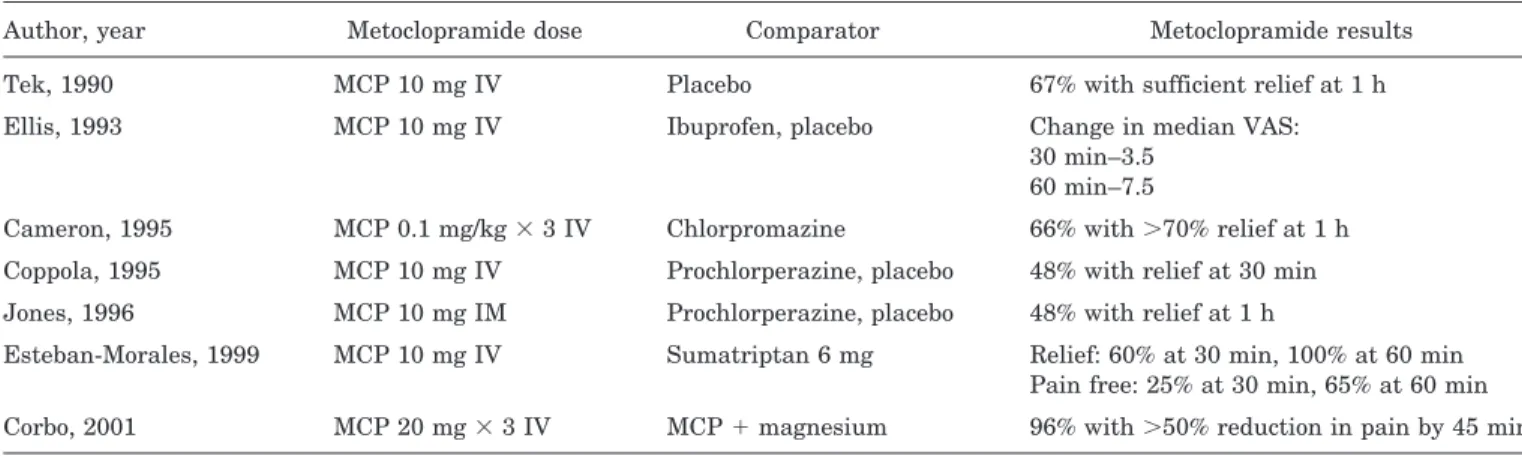
However, metoclopramide has been demonstrated to be more effective than placebo and other comparators in multiple studies (table). A recent metaanalysis similarly concluded that metoclo-pramide was an effective antimigraine treatment.10
We disagree with Allena et al.’s interpretation of the study by Cete et al.9In this study, 65% of subjects randomized to placebo
required rescue medication at 30 minutes, while only 38% of sub-jects randomized to metoclopramide required rescue medication. At 30 minutes, placebo patients had improved on the VAS by 25 while metoclopramide patients had improved by 40. This differ-ence of 15 in the VAS point estimates suggests a clinically rele-vant difference,11even if the study was not sufficiently powered to
achieve statistical significance for this finding.
Perhaps the dose of metoclopramide is relevant. Of the trials listed in the table, the two that used more aggressive dosing of metoclopramide (similar to our design) had excellent results. Dose-finding studies are needed to evaluate this hypothesis. B.W. Friedman, MD, MS, J. Corbo, MD, R.B. Lipton, MD, P.E. Bijur, PhD, D. Esses, MD, E.J. Gallagher, MD, Bronx, NY
Table Dosing of previous metoclopramide studies
Author, year Metoclopramide dose Comparator Metoclopramide results
Tek, 1990 MCP 10 mg IV Placebo 67% with sufficient relief at 1 h
Ellis, 1993 MCP 10 mg IV Ibuprofen, placebo Change in median VAS:
30 min–3.5 60 min–7.5
Cameron, 1995 MCP 0.1 mg/kg⫻ 3 IV Chlorpromazine 66% with⬎70% relief at 1 h Coppola, 1995 MCP 10 mg IV Prochlorperazine, placebo 48% with relief at 30 min Jones, 1996 MCP 10 mg IM Prochlorperazine, placebo 48% with relief at 1 h
Esteban-Morales, 1999 MCP 10 mg IV Sumatriptan 6 mg Relief: 60% at 30 min, 100% at 60 min Pain free: 25% at 30 min, 65% at 60 min Corbo, 2001 MCP 20 mg⫻ 3 IV MCP⫹ magnesium 96% with⬎50% reduction in pain by 45 min
October (2 of 2) 2005 NEUROLOGY 65 1339
at UNIV MEDECINE LEIG 22827242 on September 25, 2009
www.neurology.org
Copyright © 2005 by AAN Enterprises, Inc.
References
1. Friedman A, Corbo J, Lipton R, et al. A trial of metoclopramide vs sumatriptan for the emergency department treatment of migraines. Neurology 2005;64:463– 468.
2. Saper J, Silberstein SD, Gordon CD, Hamel RL, Swidan S. Table 9. Selected drugs used in the pharmacotherapy of head, neck and face pain. (Modified with permission from Saper, et al.) In, Handbook of Headache Management (2nd edition), Philadelphia: Lippincott Williams & Wilkins, 1999)
3. Aube M. Migraine in pregnancy. Neurology 1999;53(suppl 1):S26 –S28. 4. Swidan S, Lake A, Saper J. Efficacy of intravenous diphenhydramine
versus intravenous DHE-45 in the treatment of severe migraine head-ache. Cur Pain Headache Rep. 2005;9:65–70.
5. Schulman EA, Dermott KF. Sumatriptan plus metoclopramide in triptan-nonresponsive migraineurs. Headache 2003;43:446 – 447. 6. Lipton RB, Stewart WF, Stone AM, Lainez MJ, Sawyer JP. Stratified
care vs step care strategies for migraine: the Disability in Strategies of Care (DISC) Study: A randomized trial. Jama 2000;284:2599 –2605. 7. Vinson DR, Hurtado TR, Vandenberg JT, et al. Variations among
emer-gency departments in the treatment of benign headache. Ann Emerg Med 2003;41:90 –97.
8. Lassen LH, Thomsen LL, Olesen J. Histamine induces migraine via the H1-receptor. Support for NO hypothesis of migraine. Neuroreport 1995; 6:1475–1479.
9. Cete Y, Dora B, Ertan C, et al. A randomized prospective placebo-controlled study of intravenous magnesium sulphate vs metoclopramide in the management of acute migraine attacks in the Emergency Depart-ment. Cephalalgia 2005;25:199 –204.
10. Colman I, Brown MD, Innes GD, Grafstein E, Roberts TE, Rowe BH. Parenteral metoclopramide for acute migraine: meta-analysis of ran-domised controlled trials. BMJ 2004;329:1369 –1373.
11. Todd KH, Funk JP. The minimum clinically important difference in physician-assigned visual analog pain scores. Acad Emerg Med 1996;3:142–146.
Chronic inflammatory demyelinating
polyradiculoneuropathy: MRI study of brain and spinal cord
To the Editor: We read with great interest the recent article by
Laura et al.1 concerning cervical spinal cord atrophy in chronic
inflammatory demyelinating polyradiculoneuropathy (CIDP). They reported that the mean cervical cord area was significantly smaller in patients with CIDP compared to controls. We would like to report our experience regarding spinal cord findings in CIDP.
We had five cases with definite CIDP2 and evaluated other
demyelinating lesions than peripheral nerves. In our experience, no lesions were found in the intracranial, cervical, thoracic, and lumbar spinal cords. No spinal cord atrophy was seen.
We would be interested to find out the authors’ responses to some issues:
First, they reported that cord atrophy was confirmed in the cervical lesion. Were the thoracic cords normal?
Second, they demonstrated cervical and thoracic cords which seem to show cervical spondylotic lesions and seven cases were more than 50 years old in their series. In our cases, all of the cases were more than 50 years old, and all of our cases had spondylotic lesions in the cervical or lumbar cord. It is possible that spondy-lotic lesions may contribute to cord atrophy.
Third, no associations were found between cord area and dis-ease duration. What factor(s) determined cord atrophy in CIDP? In addition to degeneration secondary to axonal loss,3spondylotic
le-sions may contribute to cord atrophy in CIDP. Their finding of cord atrophy in CIDP is interesting and warrants further investigation. Y. Iwasaki, MD, O. Igarashi, MD, PhD, J. Aoyagi, MD,
K. Iwamoto, MD, K. Ikeda, MD, PhD, Tokyo, Japan
Reply from the Authors: We thank Iwasaki et al. for sharing
their experience of MRI in CIDP and for raising the queries about our study.1We note that they also did not find central
demyelinat-ing lesions in their patients with CIDP but unlike our study they also did not find cord atrophy. They raised three questions about our findings.
Firstly, they inquired as to whether we looked for thoracic cord atrophy. We used the method of Losseff et al.4, who had developed
a highly reproducible technique to measure atrophy of the spinal cord related to anatomical, imaging, and measurement factors. We
specifically chose the C2/3 intervertebral disc as a caudal land-mark since it is an uncommon site for disc protrusion5and we did
not measure spinal cord area in the thoracic cord since the tech-nique used at this level showed poor reproducibility.6
Secondly, they asked whether cervical spondylotic disease could have contributed to the cord atrophy we demonstrated. We only showed minor degenerative changes in four patients and did not demonstrate any cord compression. We therefore do not feel that the cervical degenerative disease contributed to the cord at-rophy. We also specifically chose the C2/3 level to measure cord atrophy as cord damage due to degenerative is relatively uncom-mon at this site.
Thirdly, they wondered what factors contributed to the cord atrophy in our series. We were not able to identify any specific contributing factors which may in part reflect our small sample size but we speculated that a dying back mechanism may be important as has been suggested for diabetic neuropathy.7
We agree that further study of this finding is needed. Mary M. Reilly, MD, Matilde Laura´, MD, London, UK
Copyright © 2005 by AAN Enterprises, Inc.
References
1. Laura M, Leong W, Murray NMF, et al. Chronic inflammatory demyeli-nating polyradiculoneuropathy: MRI study of brain and spinal cord. Neu-rology 2005;64:914 –916.
2. Saperstein DS, Katz JS, Amato AA, Barohn RJ. Clinical spectrum of chronic acquired demyelinating polyneuropathies. Muscle Nerve 2001; 24:311–324.
3. Nagamatsu N, Terao S, Misu K, et al. Axonal and peripheral involve-ment in chronic inflammatory demyelinating polyneuropathy. J Neurol Neurosurg Psychiatry 1999;66:727–734.
4. Losseff NA, Webb SL, O’Riordan JI, et al. Spinal cord atrophy and disability in multiple sclerosis. A new reproducible and sensitive MRI method with potential to monitor disease progression. Brain 1996;119: 701–708.
5. Thorpe JW, Kidd D, Kendall BE, et al. Spinal cord MRI using multi-array coils and fast spin echo. I. Technical aspects and findings in healthy adults. Neurology 1993;43:2625–2631.
6. Losseff N, Lai M, Miller DH, McDonald WI, Thompson AJ. The prognos-tic value of serial axial cord area measurement by magneprognos-tic resonance imaging (MRI) in multiple sclerosis (MS) [abstract]. J Neurol 1995; 242(suppl 2):S110.
7. Eaton SE, Harris ND, Rajbhandari SM, et al. Spinal-cord involvement in diabetic peripheral neuropathy. Lancet 2001;358(9275):35–36.
Long-term outcome of endovascular stenting for symptomatic basilar artery stenosis
To the Editor: We read with great interest the article by Yu et
al.1demonstrating the effectiveness of stenting symptomatic
basi-lar artery (BA) stenosis for reducing the risk of recurrent stroke and death. We would like to request clarification of specific issues so that readers, particularly those skilled in neurointerventional procedures, could be aided in decision-making.
The method section inaccurately described the type of wire used to cross the lesion. It is unlikely that high-grade stenosis
would be crossed using 0.035-inch wire instead of the standard 0.014-inch microwire. The balloon types that are used for predila-tation and the specific coronary stent types are known to affect procedural outcome but were not mentioned. Under, over, or nom-inal inflation of the stent-mounted balloon were not described. It is unclear how long heparin was administered or the combination of clopidogrel and aspirin following the procedure.
Prior to stenting, there was no description of the presenting TIAs. Two patients were not treated with any type of medical therapy but there are no details about the type of antiplatelet treatment in the others. After the procedure, there is no mention
of in-hospital length-of-stay after the procedures or worsening of pre-existing deficits and TIAs. Some patients may require neuro-critical care for several days with blood pressure and fluid aug-mentation to maintain adequate cerebral perfusion.
In the legends of the selected images, there was no mention of jailing perforating arteries; including loss of the left anterior inferior cerebellar artery in case B. In the long-term outcome, five cases with “dizzy spells” were not counted as possible vertigo or TIAs.
In the Discussion section, the authors conclude that the BA stenting appeared to be safe and effective. This study has many limitations to validate this conclusion. The cases where BA stent-ing failed are not mentioned which makes it difficult to interpret the success of current techniques.
We agree with the authors that further trials are needed to better evaluate this controversial subject. However, prior to fur-ther studies (involving the BA in particular), newer stents and delivery systems designed for intracranial arteries are needed. This is vital to avoid future setbacks in neurointerventional proce-dures. Given the current available data and techniques, BA stent-ing may be reserved for those patients with high-grade symptomatic stenosis who did not respond to combined aspirin and clopidogrel therapy.
Osama O. Zaidat, MD, MSc, Tony P. Smith, MD, Michael J. Alexander, MD, Durham, NC
Reply from the Authors: We thank Zaidat et al. for their
inter-est in our work.1 The Discussion section mentioned the major
periprocedural complications that occurred in patients with acute stroke and tandem stenosis.
Although different types of coronary stents and balloon angio-plasty with a nominal pressure of 8 to 14 atm were used in this case series, there was no correlation between types of balloon or stents and periprocedural complications. Fifteen patients received IV heparin for 24 hours followed by combination therapy with clopidogrel and aspirin. Three patients were placed on oral antico-agulants for 6 months and long-term clopidogrel therapy. Lesions were crossed using 0.014-in microwire instead of 0.035-in micro-wire as Zaidat noted. We appreciate the identification of this error in our article.
Presenting TIAs, individual patient medications, length-of-stay and neurointensive care management after the procedures were
not found to be associated with long-term outcome and therefore not detailed in our article. Our figure was used to show the feasi-bility of stenting for different types of stenotic lesions. The left anterior inferior cerebellar artery (AICA) in case B was visible distal to the vertebrobasilar junction in the post-stenting image.
Zaidat et al might have misidentified the loop between basilar artery and left AICA in the prestenting image as AICA. The loop was actually part of the left posterior inferior cerebellar artery. Consistent with other report,2our study also demonstrated that
symptomatic occlusion of pontine perforating arteries were very uncommon. During follow-up, five patients reported transient symptoms, including dizziness (2), sensation of head congestion (1), neck pain (1), and hand incoordination (1).
It is well known that patients often experience nonspecific symptoms after endovascular procedures that are not TIAs. We identified no basilar artery stenting failure in our study, confirm-ing other series reportconfirm-ing more than 95% success rate of stentconfirm-ing for basilar artery stenosis and other intracranial atherosclerotic lesions.2-4
Despite the limitations in retrospective study, our data showed that with a mean 26.7⫾ 12.1-month follow-up, 83.3% of patients had an excellent long-term outcome without vascular death. Therefore, endovascular stenting for symptomatic basilar artery stenosis ap-peared to be safe and effective in reducing stroke risk and death, and should be further evaluated by randomized clinical trial.
W. Yu, MD, PhD, W.S. Smith, MD, PhD, V. Singh, MD,
N.U. Ko, MD, S.P. Cullen, MD, C.F. Dowd, MD, V.V. Halbach, MD, R.T. Higashida, MD, San Francisco, CA
Copyright © 2005 by AAN Enterprises, Inc.
References
1. Yu W, Smith WS, Singh V, et al. Long-term outcome of endovascular stenting for symptomatic basilar artery stenosis. Neurology 2005;64: 1055–1057.
2. Gomez CR, Misra VK, Liu MW, et al. Elective stenting of symptomatic basilar artery stenosis. Stroke 2000;31:95–99.
3. SSYLVIA Study Investigators. Stenting of Symptomatic Atherosclerotic Lesions in the Vertebral or Intracranial Arteries (SSYLVIA): study re-sults. Stroke 2004;35:1388 –1392.
4. Jiang WJ, Wang YJ, Du B, et al. Stenting of symptomatic M1 stenosis of middle cerebral artery: An initial experience of 40 patients. Stroke 2004;35:1375–1380.
NINDS AIREN neuroimaging criteria do not distinguish stroke patients with and without dementia
To the Editor: Ballard et al.1 report that in a group of stroke
patients, the neuroimaging component within the National Insti-tute of Neurological Disorders and Stroke (NINDS) Internationale pour la Recherche et l’Enseignement en Neurosciences (AIREN) criteria2 for vascular dementia did not distinguish between
pa-tients with and without post-stroke dementia. In addition, groups did not differ in number or size of infarcts. However, patients with dementia had greater hippocampal atrophy. The authors conclude that the NINDS AIREN neuroimaging criteria may need to be revised, because they do not distinguish between stroke patients with and without dementia. Although we agree with the authors that the criteria may need revision, we would like to make some critical comments concerning the research question of their study. The NINDS AIREN criteria describe the clinical syndrome of vascular dementia (VaD). Like in any other subtype of dementia, clinical characteristics define whether or not a patient is de-mented. The neuroimaging component of the criteria only serves to determine the probability that the observed dementia is of vascular origin. In the present sample of patients who all suffered a cerebrovascular event, it was to be expected that the neuroimag-ing criteria did not reveal any difference between dementia-groups in terms of vascular burden. The authors merely show that a patient may fulfill the radiologic criteria for VaD as defined by the NINDS AIREN, and still not be demented. This is a reversal of the diagnostic process, and reminds us that an MRI scan may never be used in isolation to diagnose VaD. Furthermore, exclud-ing patients who were demented prior to the stroke may have
introduced a selection bias, while this group has the highest prob-ability of suffering VaD.
Moreover, the greater hippocampal atrophy of the demented subgroup suggests that these patients may suffer from Alzheimer disease (AD) rather than VaD, or have combined pathology. As the authors propose, vascular pathology such as a stroke may interact with preexisting subclinical Alzheimer pathology, resulting in clinical dementia of the Alzheimer type. As the NINDS AIREN criteria were not developed to detect AD, there is no reason to expect that these criteria would be sensitive to detect patients with AD.
Finally, it has been shown that the neuroimaging component of the NINDS AIREN criteria has insufficient reliability,3and this
might partly account for the inability to discriminate between groups. Operational criteria have been put forward and tested, resulting in a considerably improved reliability in experienced readers.
W.M. van der Flier, PhD, E.C.W. van Straaten, MD,
F. Barkhof, MD, PhD, P. Scheltens, MD, PhD, Amsterdam, The
Netherlands
Copyright © 2005 by AAN Enterprises, Inc.
References
1. Ballard CG, Burton EJ, Barber R, et al. NINDS AIREN neuroimaging criteria do not distinguish stroke patients with and without dementia. Neurology 2004;63:983–988.
2. Roma´n GC, Tatemichi TK, Erkinjuntti T, et al. Vascular dementia: diag-nostic criteria for research studies. Report of the NINDS-AIREN Inter-national Workshop. Neurology 1993;43:250 –260.
3. Van Straaten EC, Scheltens P, Knol DL, et al. Operational Definitions for the NINDS-AIREN Criteria for Vascular Dementia. An Interobserver Study. Stroke 2003;34:1907–1912.
October (2 of 2) 2005 NEUROLOGY 65 1341
at UNIV MEDECINE LEIG 22827242 on September 25, 2009
www.neurology.org