ATP Augments von Willebrand Factor-dependent Shear-induced
Platelet Aggregation through Ca
2ⴙ-Calmodulin and Myosin Light
Chain Kinase Activation*
Received for publication, February 24, 2004, and in revised form, April 14, 2004 Published, JBC Papers in Press, April 15, 2004, DOI 10.1074/jbc.M402032200 Ce´cile Oury‡§, Elsie Sticker‡, Heidi Cornelissen‡, Rita De Vos¶, Jos Vermylen‡,
and Marc F. Hoylaerts‡储
From the ‡Center for Molecular and Vascular Biology and the¶Laboratory of Morphology and Molecular Pathology,
University of Leuven, 3000 Leuven, Belgium
Shear stress triggers von Willebrand factor (VWF) binding to platelet glycoprotein Ib␣ and subsequent integrin ␣IIb3-dependent platelet aggregation.
Con-comitantly, nucleotides are released from platelet-dense granules, and ADP is known to contribute to shear-induced platelet aggregation (SIPA). We found that the impaired SIPA of platelets from a Hermansky-Pudlak patient lacking dense granules was restored by exogenousL-,␥-methylene ATP, a stable P2X1agonist, as well as by ADP, confirming that in addition to ADP (via P2Y1and P2Y12), ATP (via P2X1) also contributes
to SIPA. Likewise, SIPA of apyrase-treated platelets was restored upon P2X1activation withL-
,␥-methyl-ene ATP, which promoted granule centralization within platelets and stimulated P-selectin expression, which is a marker of ␣-granule release. In addition, during SIPA, platelet degranulation required both ex-tracellular Ca2ⴙ and VWF-glycoprotein Ib␣
interac-tions without involving␣IIb3. Neither platelet release
nor SIPA was affected by protein kinase C inactiva-tion, even though protein kinase C blockade inhibits platelet responses to collagen and thrombin in stirring conditions. In contrast, inhibiting myosin light chain (MLC) kinase with ML-7 reduced platelet release and SIPA by 30%. Accordingly, the potentiating effect of P2X1 stimulation on the aggregation of
apyrase-treated platelets coincided with intensified phospho-rylation of MLC and was abrogated by ML-7. SIPA-induced MLC phosphorylation occurred exclusively through released nucleotides and selective antago-nism of P2X1 with MRS2159-reduced SIPA, ATP
re-lease, and potently inhibited MLC phosphorylation. We conclude that the P2X1ion channel induces
MLC-mediated cytoskeletal rearrangements, thus contrib-uting to SIPA and degranulation during VWF-trig-gered platelet activation.
Blood platelets are constantly subjected to hemodynamic forces imposed by the blood flow, including fluid shear stress.
High shear stress is generated at sites of arterial injury where laminar blood flow is forced through a stenosis (1, 2). Shear stress-triggered platelet activation and subsequent aggrega-tion, termed SIPA1(3), may thus contribute to the pathogenesis
of vascular diseases. In addition, platelets from patients with acute myocardial infarction (4) or cerebral ischemia (5) display enhanced SIPA in vitro.
High shear stress is required for the interaction between von Willebrand factor (VWF) and platelet glycoprotein Ib␣ (GPIb␣) (for review, see Ref. 6), but the effects of shear forces on GPIb␣ signaling are only just beginning to be defined. Downstream effectors that have been implicated in the VWF-dependent activation of ␣IIb3 include phosphatidylinositol 3-kinase
(PI3K) (7), protein kinase C (PKC) (8), Syk, and Src, as well as co-associated immunoreceptor tyrosine-activated motif-con-taining transmembrane proteins and adaptor proteins (2). A recent study of platelet adhesion to dimeric VWF A1 domain, which recognizes only GPIb␣, showed that GPIb␣ itself can signal to activate␣IIb3through sequential actions of Src
ki-nases, Ca2⫹ oscillations, and PI3K/PKC (9). It has also been
proposed that GPIb signals directly as a consequence of its association with structural cytoskeletal proteins (10), among which GPIb␣-bound filamin A, filamentous actin cross-linked by␣-actinin, vinculin, and talin would directly link the cyto-plasmic domain of GPIb␣ with the 3tail of␣IIb3(11).
Studies examining SIPA have suggested that VWF primarily stimulates platelet activation through an indirect pathway linked to ADP release (12, 13). These studies have also shown that platelet activation initiated by VWF-GPIb␣ interaction requires a transmembrane Ca2⫹influx independent of released ADP and VWF binding to␣IIb3(14, 15), which promotes dense
granule secretion of ADP and activates integrin␣IIb3through engagement of the P2 receptors for ADP. A role for the platelet ADP receptors P2Y1 and P2Y12(reviewed in Ref. 16) in SIPA
has subsequently been confirmed (17, 18). Rese´ndiz et al. (19) recently reported that the selective P2Y12 antagonist AR-C69931MX inhibits the shear-induced aggregation of washed platelets and showed that P2Y12mediates shear-induced PI3K
activation, a process coupled to phosphorylation of PI3K-asso-ciated Syk tyrosine kinase.
In blood vessels, shear stress causes the release of high levels of nucleotides, including ATP and ADP, into the extracellular environment; these nucleotides mainly originate from endothe-lial cells (20) and platelet-dense granules (21). Although the
* This work was supported by Fonds voor Wetenschappelijk Onder-zoek-Vlaanderen (FWO) Project G.0227.03 and by Geconcerteerde Onderzoeksactie/2004/09. The costs of publication of this article were defrayed in part by the payment of page charges. This article must therefore be hereby marked “advertisement” in accordance with 18 U.S.C. Section 1734 solely to indicate this fact.
§ Recipient of a postdoctoral research mandate from the FWO.
储To whom correspondence should be addressed: University of Leu-ven, Center for Molecular and Vascular Biology, Herestraat 49, 3000 Leuven, Belgium. Tel.: 32-16-346145; Fax: 32-16-345990; E-mail: [email protected].
1The abbreviations used are: SIPA, shear-induced platelet aggrega-tion; meATP, methylene ATP; CaM, calmodulin; MLC, myosin light chain; PI3K, phosphatidylinositol 3-kinase; PKC, protein kinase C; VWF, von Willebrand factor; GP, glycoprotein; PAR, protease-activated receptor; TRAP1– 6, thrombin receptor activating peptide SFFLRN.
© 2004 by The American Society for Biochemistry and Molecular Biology, Inc. Printed in U.S.A.
This paper is available on line at http://www.jbc.org
26266
at UNIV DE LIEGE-MME F PASLE on September 5, 2008
www.jbc.org
two processes are related (21). In addition, Ca2⫹ influx-depend-ent phosphorylation of myosin light chain, preceding the myo-sin-actin interactions, has been proposed to be an initial step during SIPA (21).
ATP is the physiological agonist at P2X1, but its role in
platelet activation is only starting to be unraveled. In the aggregometer, the selective P2X1 agonists ␣,-meATP and
L-,␥-meATP evoke a transient Ca2⫹ influx accompanied by rapid and reversible platelet shape change and myosin light chain (MLC) phosphorylation, provided that measures are taken to avoid desensitization of P2X1 by ATP spontaneously
released during platelet preparation (23–25). Recent studies, using P2X1knock-out mice (26) or transgenic mice overexpress-ing this ion channel selectively in the megakaryocytic cell lin-eage (27), have reported a prominent role for P2X1in platelet
aggregation and thrombus formation in shear stress condi-tions. Notably, P2X1overexpressing platelets displayed potent SIPA in conditions where wild-type platelets hardly aggregated (27). In the present study, we have investigated the role of the P2X1-mediated Ca2⫹ influx and the associated downstream
signaling pathways in VWF-dependent shear-induced granule release and aggregation of washed human platelets.
EXPERIMENTAL PROCEDURES
Materials—ADP, L-,␥-meATP, the P2X1antagonist MRS2159, the P2Y1antagonist MRS2179, and the ectonucleotidase apyrase (grade I) were purchased from Sigma. AR-C69931MX was a gift from AstraZeneca R&D, Charnwood, UK. The P2X1antagonist MRS2159 at the concen-tration used in this study was verified to selectively inhibit a P2X1 -mediated platelet shape change without affecting platelet shape change and aggregation induced by ADP. The calmodulin inhibitor W-7, the MLC kinase inhibitor ML-7, the Rho kinase inhibitor Y-27632, and the PKC inhibitors GF109203X, Go6976, and Go6983 were purchased from Calbiochem. Human VWF was purified from plasma cryoprecipitate by gel filtration on a Sepharose 4B-CL column. Tirofiban (Aggrastat) was obtained from Merck Sharp and Dohme. The murine-neutralizing anti-VWF monoclonal antibody AJvW-2 was from Ajinomoto Co., Inc. (Ka-wasaki, Japan) (28), and the neutralizing monoclonal GPIb␣ anti-body G19H10 was raised in our laboratory. ADP andL-,␥-meATP were purified by high pressure liquid chromatography on an Adsorbosphere HS C18, 7m, 250 ⫻ 4.6-mm column (Alltech) as described by Oury et
al. (24).
Preparation of Washed Human Platelets—Washed human platelets
(2.5–3.5⫻ 105platelets/l) were prepared as described previously (29). Apyrase (0.5 units/ml) was added to the blood sample and platelet resuspension buffer when indicated. Platelet preparations were free of red blood cells, as validated via microscopy and blood cell counting (CellDyn 1300, Abbott, Abbott Park, IL).
Shear-induced Platelet Aggregation, ATP Secretion, and P-selectin Expression—Shear-induced aggregations of washed human platelets
were performed in an annular ring-shaped viscometer generating lam-inar flow (Ravenfield viscometer; Heywood, Lancashire, UK) at 37 °C in the presence of 2 mMCaCl2and 10g/ml human soluble VWF unless otherwise indicated. A shear rate of 9000 s⫺1was used, which corre-sponds to a shear stress of 80 dynes/cm2. The threshold shear rate causing platelet aggregation ranged between 3000 s⫺1and 5000 s⫺1. At defined time points, platelet samples were collected and fixed in 1% paraformaldehyde; the percentage of platelet aggregation was calcu-lated by comparing single platelet counts before and after shearing. Shear-induced P-selectin surface translocation on tirofiban-treated washed platelets was detected by flow cytometry with the phyco-erythrin-conjugated anti-P-selectin CD62P antibody (BD Biosciences). For ATP secretion assays, the reactions were stopped with an ice-cold Hepes buffer (20 mMHepes, pH 7.4, 150 mMNaCl, 5 mMEDTA). After immediate centrifugation at 4 °C, ATP was measured in the superna-tants by the addition of a luciferase/luciferin reagent (Chrono-Lume, Kordia) in a microplate luminometer LB96V (Berthold Technologies, Vilvoorde, Belgium). At least five independent experiments were
per-aliquots were loaded on SDS-PAGE (12.5%) and subjected to Western blotting. Thr-18/Ser-19 MLC phosphorylation was detected with the anti-phospho-MLC polyclonal antibody (Santa Cruz Biotechnology, Santa Cruz, CA) according to the instructions of the manufacturers.
Electron Microscopy—Washed platelets were immediately fixed
over-night at 4 °C in 2.5% (w/v) glutaraldehyde, 0.1Mphosphate buffer, pH 7.2. After centrifugation at 800⫻ g for 10 min, a condensed pellet of platelets was formed. After fixation in 1% OsO4(w/v), 0.1Mphosphate buffer, pH 7.2, and dehydration in a graded series of ethanol, the pellets were embedded in epoxy resin. Ultrathin sections were cut and stained with uranyl acetate and lead citrate before examination with a Zeiss EM 10 electron microscope (Oberkochen, Germany).
RESULTS
VWF-GPIb␣ Interactions and Extracellular Ca2⫹in SIPA—
When suspensions of washed human platelets were subjected to uniform high shear stress (80 dynes/cm2⫽ 9000 s⫺1shear
rate) in the presence of soluble human VWF in an annular ring-shaped viscometer, aggregation rapidly occurred, reaching a plateau after 3 min (Fig. 1A). In contrast, a shear rate of 1000 s⫺1did not cause platelet aggregation (Fig. 1A). The inclusion of the VWF A1 domain-blocking antibody AJvW-2, neutralizing anti-GPIb␣ antibody G19H10, or the ␣IIb3antagonist
tirofi-ban (Fig. 1B) confirmed the findings by others (30) that SIPA requires VWF interactions with GPIb␣ and the activation of ␣IIb3. We also confirmed that SIPA does not occur in the
absence of added extracellular Ca2⫹(14, 15) (Fig. 1B). Platelet aggregation was accompanied by gradually increas-ing shear stress-dependent ATP release (Fig. 1A). ATP release was initiated as early as 10 s after exposure to shear stress. When measured at the maximal aggregation time point, this release clearly relied on VWF-GPIb␣ signaling, as it was at least partly inhibited by AJvW-2 and G19H10 (Fig. 1B). Omit-ting the addition of extracellular Ca2⫹also reduced the amount of released ATP, although only partly (Fig. 1B). The combined absence of VWF and extracellular Ca2⫹almost abolished plate-let degranulation (Fig. 1B). In contrast,␣IIb3antagonism had no effect on the degree of platelet degranulation (Fig. 1B). Therefore, both VWF-GPIb␣ signaling and Ca2⫹contribute to platelet release under shear stress, a process that is independ-ent of␣IIb3outside-in signals.
MLC Kinase (but Not PKCs) Controls the SIPA-induced Platelet Degranulation—Platelet granule release that is induced by most agonists requires an increase of intracellular Ca2⫹and PKC activation (reviewed in Ref. 31). Whether or not PKC plays a role in SIPA is controversial (8, 32). In our experimental conditions, the nonselective PKC inhibitor GF109203X, which blocks both conventional (␣, I, II, ␥) and novel (␦, , , ⑀) PKC isoforms, failed to inhibit either platelet aggregation or release produced by shear stress (Fig. 2A), although it prevented such responses to collagen (2g/ml) or to the PAR-1-activating pep-tide TRAP1– 6(1M) under stirring conditions (Fig. 2B).
Simi-larly, the PKC␣/-specific inhibitor Go6976 did not affect SIPA and associated platelet release (Fig. 2A), although shear stress-independent collagen-induced release and aggregation were fully blocked (Fig. 2B). In agreement with previous reports that PKC␣/ isoforms do not play a major role downstream of PAR-1 (33), Go6976 had a minimal inhibitory effect on TRAP1– 6
-induced granule release (Fig. 2B). Finally, to determine whether other PKC isoforms could be involved in platelet re-lease accompanying SIPA, we used Go6983, which blocks the platelet-expressed atypical PKC isoform in addition to PKC ␣, I, II, ␥, and ␦. This inhibitor had no effect on shear-induced
at UNIV DE LIEGE-MME F PASLE on September 5, 2008
www.jbc.org
platelet release or aggregation, although it diminished TRAP1– 6-induced ATP release by about 50% without affecting platelet
aggregation (Fig. 2B). These results indicate that shear-in-duced platelet dense granule release and aggregation are de-pendent on signaling pathways distinct from those activated downstream of the collagen receptor GPVI or PAR-1. They also suggest that the known conventional, novel, and atypical PKC isoforms do not play a major role in platelet degranulation during SIPA.
In addition to PKC, GPVI-mediated platelet dense granule release also depends on Ca2⫹-sensitive MLC kinase activa-tion (25). Inhibiting this kinase with ML-7 abolished both the ATP release and platelet aggregation induced by collagen (2 g/ml) (Fig. 2B). As shown in Fig. 2A, ML-7 reduced the shear-induced ATP release and platelet aggregation by about
30%, indicating a role for MLC kinase in SIPA and associated platelet degranulation.
Secreted ADP and ATP in VWF-dependent Shear-induced Platelet Release and Aggregation—Under stirring conditions in an aggregometer, MLC kinase is activated downstream of the P2X1-mediated Ca2⫹influx, playing an essential role during
cy-toskeletal rearrangements evoked by selective agonists ( ␣,-meATP andL-,␥-meATP) of this ion channel (25). The activation of MLC kinase can also result from phospholipase C-dependent Ca2⫹ mobilization from internal stores downstream of the G
q
protein-coupled P2Y1receptor or of GPVI (34, 35). In agreement
with earlier reports (17–19), the selective antagonists of P2Y1
(MRS2179, 10M) and P2Y12(AR-C69931MX, 1M) inhibited
SIPA by more than 50% (Fig. 3). In previous studies of platelet aggregation performed under stirring conditions in an
aggre-FIG. 1. Role of VWF and Ca2ⴙ in
shear-induced platelet aggregation and ATP release. A, time course of
washed human platelet aggregation and ATP release induced at a shear rate of 9000 s⫺1(black squares). The absence of platelet responses at a shear rate of 1000 s⫺1is also shown (lower lines with open
squares). B, effects of preincubation with
the neutralizing anti-VWF monoclonal antibody AJvW-2 (20g/ml, 1 min), the neutralizing anti-GPIb␣ antibody G19H10 (20g/ml, 1 min), or the ␣IIb3antagonist tirofiban (1g/ml, 1 min), as well as the absence of added extracellular Ca2⫹on aggregation induced for 3 min at 9000 s⫺1. *, p⬍ 0.0001; #, p ⬍ 0.0001 versus control; §, p⬍ 0.0002.
1
at UNIV DE LIEGE-MME F PASLE on September 5, 2008
www.jbc.org
gometer, a high concentration of the ATP/ADP scavenger apyrase always was required to demonstrate P2X1function, as this ion
channel is rapidly desensitized by ATP released during platelet preparation (23–25). In sharp contrast, the P2X1 antagonist
MRS2159 (10 M) reduced SIPA by about 40% even when apyrase was omitted (Fig. 3). Thus, the three platelet P2 recep-tors contribute to SIPA through secreted ADP and ATP. The effect of combined P2X1and P2Y1antagonism on SIPA was not
significantly different from that of P2Y1antagonism only (Fig. 3),
suggesting that these receptors share common signaling path-ways. Neither the antagonism of P2X1, P2Y1, nor their combina-tion could inhibit the remaining SIPA observed in the presence of the P2Y12antagonist (data not shown).
ATP secretion assays performed in the presence of the selec-tive P2 receptor antagonists revealed a contribution of P2Y1, P2Y12, and P2X1 to VWF-dependent platelet degranulation
(measured after 3 min) (Fig. 3). With the exception of P2X1
antagonism, the effects of P2Y1and P2Y12antagonism on
se-cretion did not fully correlate with their ability to inhibit plate-let aggregation (Fig. 3), which is in agreement with the
inter-pretation that these receptors participate in other processes leading to SIPA besides secretion regulation.
P2X1Enhances Degranulation and SIPA via
Calmodulin-de-pendent MLC Kinase Activation—To further examine the con-tribution of individual nucleotides to SIPA, we have used plate-lets of a Hermansky-Pudlak patient lacking dense granules and secreting almost no ATP in response to supraoptimal doses of collagen (36). These platelets displayed severely impaired SIPA comparable with SIPA observed in the presence of P2Y1
or P2Y12receptor antagonists (Fig. 4A). Interestingly, platelet
aggregation could be largely restored by the addition of not only ADP but also of the selective P2X1agonistL-,␥-meATP imme-diately prior to shearing, which supports the central roles for both ATP and ADP secreted during SIPA.
We pursued this aspect of SIPA using normal platelets but in the presence of the ectonucleotidase apyrase, which degrades secreted ATP and ADP. The addition of apyrase (0.5 units/ml) to the platelet resuspension buffer reduced the level of SIPA to 70% of the control (Fig. 4B). In this condition, the selective P2Y1and P2X1receptor antagonists MRS2179 and MRS2159 no longer inhibited the remaining SIPA (not shown), indicating that the reduction of aggregation by apyrase is due to the absent activation of P2Y1 and P2X1 signaling (Fig. 4A). In
contrast, the P2Y12antagonist still reduced the aggregation of apyrase-treated platelets from 26.3⫾ 9.5% to 17.1 ⫾ 3.2%, in-dicating the presence of sufficient remaining secreted ADP to activate this receptor.
The addition of the selective apyrase-resistant P2X1agonist L-,␥-meATP to this set-up just prior to shearing fully restored the aggregation of apyrase-treated platelets (Fig. 4B). The re-sulting aggregation still depended on VWF and␣IIb3
activa-tion, as shown by its full inhibition by AJvW-2 and tirofiban (Fig. 4B). Therefore, this experimental approach enabled us to study the P2X1-driven signaling pathways in more detail
dur-ing SIPA.
We found that the enhanced SIPA was the result of CaM and MLC kinase activation, as W-7 and ML-7 inhibited theL-
,␥-FIG. 2. MLC kinase versus PKC in SIPA and platelet ATP
re-lease. A, aggregation of washed human platelets was induced for 3 min
at a shear rate of 9000 s⫺1following preincubation with the protein kinase C inhibitors GF109203X (⫹GF, 10M, 5 min), Go6976 (1M, 5
min), or Go6983 (1M, 5 min) or the MLC kinase antagonist ML-7 (10 M, 10 min). The effect of these inhibitors on ATP secretion measured after 3 min of shearing is also shown. *, p⬍ 0.05. B, aggregation of washed human platelets produced in an aggregometer by collagen (2 g/ml) or TRAP1– 6(1MSFFLRN) in the absence (control) or presence (⫹) of the same inhibitors, as indicated. The corresponding concentra-tions (M) of released ATP are shown in brackets.
FIG. 3. P2X1, P2Y1, and P2Y12 in SIPA and ATP release. A,
aggregation of washed platelets was induced for 3 min at a shear rate of 9000 s⫺1 following a 1-min preincubation with the selective P2 receptor antagonists for P2Y12(AR-C69931MX, 1M), P2Y1(MRS2179, 10M), and P2X1(MRS2159, 10M) or with combined MRS2179⫹ MRS2159. *, p⬍ 0.001; #, p ⫽ 0.04 versus control. B, ATP secretion measured after 3 min of shearing in the presence or absence of the P2 receptor antagonists. *, p⬍ 0.05; NS, not significant.
at UNIV DE LIEGE-MME F PASLE on September 5, 2008
www.jbc.org
meATP amplification (Fig. 4B). Notably, these inhibitors had no effect on the SIPA of apyrase-treated platelets without ad-ditional stimulation byL-,␥-meATP (data not shown), indicat-ing that the activation of these pathways exclusively depends on P2X1 potentiation. The fact that W-7 inhibited the L- ,␥-meATP-driven SIPA to a larger extent than ML-7 may indicate the existence of an additional CaM-dependent pathway acti-vated downstream of P2X1. Thus, exogenous P2X1 activation
selectively triggers CaM-dependent pathways contributing to SIPA. Moreover, in the presence of the P2Y12receptor
antag-onist AR-C69931MX, the L-,␥-meATP-driven SIPA was re-duced to the same level (16.2⫾ 3.4%) as that achieved during inhibition of apyrase-treated platelets themselves, confirming a central role for ADP also in theL-,␥-meATP-driven SIPA.
P2X1 Activation Promotes Shear-induced Platelet Granule
Centralization and Release—We investigated whether P2X1
-mediated MLC kinase activation would be instrumental in platelet degranulation to explain the enhanced SIPA of Her-mansky-Pudlak patient platelets or of apyrase-treated plate-lets in the presence ofL-,␥-meATP. Flow cytometry analysis of P-selectin expression (as a marker of␣-granule release) was done on the surface of apyrase-treated (0.5 units/ml) and tiro-fiban-treated platelets exposed to VWF and shear for 5 min in the presence or absence ofL-,␥-meATP. Fig. 5 shows a signif-icant increase of the percentage of platelets expressing P-selec-tin following their pretreatment withL-,␥-meATP. P-selectin expression was, in turn, reduced to the initial level in the presence of the MLC kinase inhibitor ML-7 (Fig. 5). Therefore, during SIPA, the P2X1-mediated MLC kinase activation can reinforce the shear-induced platelet degranulation.
The ability of P2X1to potentiate platelet aggregation during SIPA was further investigated by electron microscopy of plate-let aggregates (Fig. 5). Following 3 min of shearing, the apyrase (0.5 units/ml)-treated platelets formed small aggregates and displayed granule centralization and pseudopodia, but most of the granules were intact (Fig. 5c). On the contrary, in the presence ofL-,␥-meATP, larger platelet aggregates were ob-served in which more platelets were degranulated; in the plate-lets still showing intact granules, their centralization appeared more advanced than in the absence ofL-,␥-meATP, indicative of more pronounced cytoskeletal rearrangements (Fig. 5d). Platelets subject to low shear stress (1000 s⫺1shear rate) did not show any morphological changes (Fig. 5b) as compared with resting platelets (Fig. 5a). Thus, P2X1-driven MLC kinase
ac-tivation in SIPA contributes both to cytoskeletal rearrange-ments in platelets and to degranulation.
Shear-induced MLC Phosphorylation Depends on P2X1,
P2Y1, and P2Y12—Activation of CaM-regulated MLC kinase
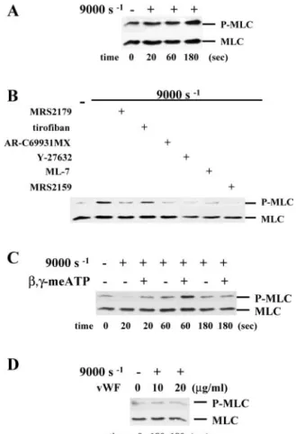
and Rho kinase results in Ca2⫹-dependent and -independent phosphorylation of MLC (34), which is a key step in platelet activation. Shear stress caused phosphorylation of MLC (de-tected as early as 20 s and reaching its maximum after 3 min) (Fig. 6A), which was inhibited both by ML-7 and the Rho kinase inhibitor Y-27632 (Fig. 6B). Such phosphorylation was not af-fected by the␣IIb3 antagonist tirofiban (Fig. 6B), indicating that it occurred independently of␣IIb3outside-in signals. In
agreement with a role for Rho kinase in SIPA, platelet aggre-gation was reduced from 43.9⫾ 9.5% to 23.6 ⫾ 8.7% by adding Y-27632 (5M) during SIPA (37).
We found that antagonizing P2X1 (MRS2159), P2Y1
FIG. 4. P2X1activation potentiates SIPA through CaM and MLC kinase. A, SIPA with platelets from a
Herman-sky-Pudlak patient (HPS) compared with platelets from healthy individuals
(con-trol).L-,␥-meATP (10 M) or ADP (1M) was added to the Hermansky-Pudlak pa-tient platelets prior to shearing, as indi-cated. B, aggregation of washed platelets and of apyrase (0.5 units/ml)-treated platelets was induced for 3 min at a shear rate of 9000 s⫺1(control) in the presence or in the absence of the selective P2X1 agonist L-,␥-meATP (10 M) following preincubation with the neutralizing anti-VWF monoclonal antibody AJvW-2 (20 g/ml), the ␣IIb3 antagonist tirofiban (⫹tir, 1g/ml), the CaM antagonist W-7 (100 M), the MLC kinase antagonist ML-7 (10 M), or the P2Y12 antagonist AR-C69931MX, as indicated. $, p⫽ 0.004
versus control; *, p⫽ 0.001 versus
apyrase⫺L-,␥-meATP; **, p ⬍ 0.005; #,
p⬍ 0.01 versus apyrase ⫹L-,␥-meATP;
¶, p⫽ 0.02 versus presence of W-7). The presence of apyrase andL-,␥-meATP are
represented by solid black lines under-scoring the graph.
1
at UNIV DE LIEGE-MME F PASLE on September 5, 2008
www.jbc.org
(MRS2179), or P2Y12(AR-C69931MX) inhibited
phosphoryla-tion of MLC to a variable degree (Fig. 6B). Phosphorylaphosphoryla-tion was inhibited potently by P2X1 neutralization. Correspondingly,
the addition of 0.5 units/ml apyrase (i.e. abolishing platelet activation via P2Y1 and P2X1) almost abolished MLC
phospho-rylation (Fig. 6C); moreover, shearing for 20 s resulted in a phosphorylation state below that of resting platelets (Fig. 6C). The addition of 5 units/ml apyrase abrogated the overall re-sponse (Fig. 6D), which could not be restored by doubling the concentration of exogenous VWF (20 g/ml) (Fig. 6D), even though this led to an ⬃20% increase of aggregation (not shown). Thus, VWF does not trigger MLC phosphorylation without involving secreted nucleotides.
Fig. 6C also shows that, in the presence of 0.5 units/ml apyrase, the addition ofL-,␥-meATP prior to shear potently enhanced MLC phosphorylation induced after 20 and 60 s; such L-,␥-meATP-elicited phosphorylation was quickly reversible as it was no longer detected after 3 min. Therefore, the ability ofL-,␥-meATP to cause MLC phosphorylation coincides with its ability to potentiate the shear-induced aggregation of apyrase-treated platelets.
DISCUSSION
In the present study, we describe a role for the P2X1
-medi-ated CaM and MLC kinase activation in VWF-dependent SIPA through secreted ATP. This pathway contributes to the extra-cellular Ca2⫹-dependent platelet degranulation induced by shear stress and acts in conjunction with VWF-GPIb␣ signal-ing. As schematically represented in Fig. 7, platelet activation initiated by the VWF-GPIb␣ interaction required a
transmem-brane Ca2⫹influx independent of both ADP and VWF binding
to␣IIb3(14, 15). Our observation that the P2X1receptor
an-tagonist MRS2159 only partly inhibits platelet degranulation and aggregation (to a maximum of 60%), whereas omitting extracellular Ca2⫹or neutralizing VWF abolishes it, suggests
that the VWF/GPIb-associated transmembrane Ca2⫹ influx only partly relies on P2X1-mediated Ca2⫹influx.
In contrast to the effects on aggregation, the removal of VWF or antagonizing its binding to GPIb␣ only partially reduced the shear-induced platelet ATP release unless extracellular Ca2⫹ was omitted at the same time. The antagonist of␣IIb3,
tirofi-ban, had no effect on shear-induced platelet release, excluding contributions by VWF-␣IIb3outside-in signaling to secretion regulation. Antagonizing P2X1with MRS2159 diminished the
ATP release, which was also seen with the antagonism of P2Y1
and P2Y12, indicating a role for all of these receptors in the
amplification of the VWF/GPIb␣-triggered platelet degranula-tion (Fig. 7).
The ATP-gated P2X1ion channel only operates in the presence
of physiological extracellular Ca2⫹concentrations (23, 24). Ca2⫹ is a key element in granule secretion (reviewed in Ref. 31). The proteins that mediate the effects of Ca2⫹in secretion fall into two categories: the EF hand proteins and the Ca2⫹ /phospholipid-binding proteins. EF hand proteins found in platelets include calmodulin and calcyclin. Calmodulin binds to platelet ␣-gran-ules. Ca2⫹/calmodulin-dependent phosphorylation of MLC
ki-FIG. 5. P2X1activation enhances platelet degranulation. Upper panel, flow cytometry analyses of P-selectin surface expression on
tiro-fiban- and apyrase (0.5 units/ml)-treated washed platelets exposed to VWF at a shear rate of 9000 s⫺1for 5 min or not (control) in the presence or absence ofL-,␥-meATP (10 M) and the MLC kinase inhibitor ML-7 (10 M) as indicated. #, p⫽ 0.01 versus control; *, p ⫽ 0.02 versus absence ofL-,␥-meATP; **, p ⫽ 0.04 versus presence ofL-,␥-meATP.
Lower panels (a– d), transmission electron microscopy of SIPA. Apyrase
(0.5 unit/ml)-treated washed human platelets were subjected or not (a) to a shear rate of 1000 s⫺1(b) or 9000 s⫺1(c) in the absence or presence ofL-,␥-meATP (10 M) (d) for 3 min before being analyzed by electron microscopy. Scale bars⫽ 1m.
FIG. 6. Shear-induced MLC phosphorylation depends
exclu-sively on secreted ATP and ADP. A, Western blotting of MLC
phosphorylation (P-MLC) at a shear rate of 9000 s⫺1. The time course of MLC phosphorylation in the absence of apyrase is shown. B, MLC-phosphorylation produced after 3 min of shear following preincubation with the selective P2 receptor antagonists (as described in the legend for Fig. 3), ML-7 (in Fig. 2), Y-27632 (5M), or tirofiban (in Fig. 1B), as indicated. C, same experiment as in A performed on platelets treated with 0.5 units/ml apyrase in the absence or presence ofL-,␥-meATP (10
M). D, platelets were treated with 5 units/ml apyrase, and shear was applied for 3 min in the presence of 10 or 20g/ml VWF, as indicated.
at UNIV DE LIEGE-MME F PASLE on September 5, 2008
www.jbc.org
nase mediates secretion via activation of MLC with subsequent contraction of the actomyosin; therefore, the concurrent central-ization of granules is thought to facilitate granule secretion (38). Calmodulin may also bind specifically to the vesicle-associated membrane protein (39), contributing to granule release by di-rectly affecting the exocytotic core complex.
Here, we found that shear-induced MLC phosphorylation involves both Ca2⫹-dependent MLC kinase and Ca2⫹ -inde-pendent Rho kinase signaling, as it was inhibited by the MLC kinase inhibitor ML-7 as well as by the Rho kinase inhibitor Y-27632. The phosphorylation of MLC was abolished by the ATP/ADP scavenger apyrase and was potently reduced by the selective antagonism of P2X1, P2Y1, or P2Y12, indicating a central role for secreted ATP and ADP in this event.
Compatible with a prominent role for the P2X1ion channel
under high shear stress (26, 27), apyrase was not needed to demonstrate P2X1function during SIPA. On the other hand, apyrase could be used under these conditions to control P2X1
function via the addition of stable selective P2X1agonists and
to drive platelet activation through P2X1-dependent pathways
of signaling. The ability of the selective P2X1 agonist L- ,␥-meATP to restore the aggregation of apyrase-treated platelets coincided with its potency in causing early phosphorylation of MLC and increased platelet P-selectin expression, a marker of ␣-granule release. The potentiating effect ofL-,␥-meATP on SIPA was prevented by inhibitors of CaM (W-7) and of MLC kinase (ML-7). These findings were substantiated by electron microscopy analyses of the platelet aggregates, depicting more pronounced granule centralization as well as degranulation in the presence ofL-,␥-meATP. Our observation that exogenous
P2X1 activation can restore the severely defective SIPA of
platelets lacking dense granules (Hermansky-Pudlak patient) corroborates a role for P2X1-mediated cytoskeletal
rearrange-ments in facilitating␣-granule release.
Thus, our findings suggest that P2X1 participates in the
extracellular Ca2⫹-dependent shear-induced platelet granule release through CaM and MLC kinase-triggered MLC phospho-rylation and concurrent cytoskeletal rearrangements, facilitat-ing SIPA and leadfacilitat-ing to granule centralization as an onset to degranulation (Fig. 7). Because P2Y1antagonism also inhibited
shear-induced MLC phosphorylation (although weakly), a role for Ca2⫹mobilized from internal stores in MLC kinase activa-tion is possible (34, 35). Rapid P2X1and slower P2Y1signals
(activated by co-released ATP and ADP, respectively) converge to a common effector (MLC kinase) contributing to cytoskeleton remodeling and platelet release (Fig. 7). Accordingly, combined P2X1 and P2Y1 antagonism leads to the same inhibition of
SIPA at 3 min as P2Y1antagonism only.
The study by Schoenwaelder et al. (37) defines an important role for RhoA and Rho kinase in SIPA but not in platelet release. The inhibition of shear-induced MLC phosphorylation by selective P2Y1 or P2Y12 antagonism suggests that ADP activates Rho kinase through Gq(40) and Gi␣2(41) also under shear stress (Fig. 7). Further investigations are required to determine the function of these pathways during SIPA. The failure of Rho kinase inhibitors to affect platelet release (Ref. 37 and the present study) may be explained by the existence of two pools of myosin present in different cell compartments (42) and emphasizes the role of MLC kinase activation in nucleo-tide-mediated granule release. In agreement with findings by Rese´ndez et al. (19), we further found that P2Y12 mediated
PI3K activation during SIPA. The resulting Akt (protein kinase B) phosphorylation was inhibited by P2Y12 neutralization.
PI3K inhibition partially inhibited SIPA but had no effect on ATP release (not shown); i.e. P2Y12-recruited PI3K activates
platelets via a separate pathway (Fig. 7) potentially by regu-lating the function of ␣IIb3 via Akt and the integrin-linked
kinase ILK (43).
Platelet granule release induced by most platelet agonists depends on PKC activation (reviewed in Ref. 31). However, none of the PKC inhibitors used in this study affected the shear-induced platelet release or aggregation. The nonselective PKC inhibitor GF109203X (which blocks both diacylglycerol-and Ca2⫹-sensitive conventional PKC isoforms and the diacyl-glycerol-sensitive Ca2⫹-insensitive novel isoforms), the more potent PKC␣/-specific inhibitor Go6976, and Go6983 (which inhibits the atypical isoform in addition to ␣, , ␥, and ␦) were all without effect, even though they inhibited platelet release in response to collagen or TRAP1– 6. This finding is consistent
with a previous study in which the nonselective protein kinase inhibitor staurosporine failed to inhibit SIPA (32). This is in contrast to the study by Kroll et al. (8) showing inhibition of the full aggregation response by the staurosporine analogue Ro 31-7549. The effect of this inhibitor on platelet release was not reported in their study. Ro 31-7549, similar to Ro 31-8220 and GF109203X, belongs to a category of nonselective PKC inhibi-tors showing similar potency in inhibiting collagen-, thrombox-ane A2 analogue-, and thrombin-induced platelet release and aggregation (29, 33, 44). These authors show that SIPA is associated with the phosphorylation of pleckstrin, a PKC sub-strate that occurs in the absence of diacylglycerol or hydrolysis of phosphatidylinositol 4,5-bisphosphate in a Ca2⫹-dependent
manner. It has to be noticed that EGTA, a chelator of extracel-lular Ca2⫹, had minimal effect on such phosphorylation, ex-cluding a role for transmembrane Ca2⫹influx. They conclude that a diacylglycerol-independent pathway of PKC activation
FIG. 7. Schematic representation of the central role of P2
re-ceptor signaling in SIPA. In addition to the shear-induced
interac-tion of VWF with GPIb␣, a role for all three P2 receptors is depicted in SIPA. Extracellular Ca2⫹-dependent GPIb␣ signaling triggers cytoskel-etal rearrangements and␣IIb3activation but also early degranulation leading to subsequent activation of P2 receptors via released ADP and ATP. Downstream P2 receptor pathways include P2Y1-mediated Ca
2⫹ mobilization and P2X1-mediated Ca2⫹influx, as well as P2Y12 -depend-ent PI3K and Akt activation and RhoA/Rho kinase activation. The three P2 receptors are needed to cause MLC phosphorylation through CaM/ MLC kinase and/or RhoA/Rho kinase pathways, leading to shear-asso-ciated cytoskeletal reorganization, facilitating platelet aggregation and enhancing platelet secretion. The fast ATP-activated Ca2⫹ influx through P2X1triggers rapid CaM activation, which is critical for a process requiring a prompt platelet-activating signal. eCa2⫹and iCa2⫹, extracellular and intracellular Ca2⫹, respectively.
1
at UNIV DE LIEGE-MME F PASLE on September 5, 2008
www.jbc.org
cytoskeleton translocation of actin, actin-binding protein, and myosin as a rapid and transient movement of MLC kinase wherein the amount of cytoskeletal PKC did not change, unlike what is seen with thrombin. This observation further suggests distinct roles of PKC (if any) and MLC kinase during platelet responses to shear stress. The transient association of MLC kinase with the cytoskeleton may coincide with the quickly reversible MLC phosphorylation produced via P2X1activation.
In conclusion, P2X1contributes to SIPA through its ability to
rapidly elicit Ca2⫹influx-triggered signals leading to prompt CaM-dependent signaling, platelet activation, and platelet granule release. Further studies are required to elucidate the mechanisms of VWF-GPIb␣-dependent platelet granule re-lease. It also remains to be determined whether the presently identified P2X1/CaM/MLC kinase pathway plays a role during platelet activation processes accompanying platelet recruit-ment and adhesion on immobilized VWF and, most impor-tantly, during thrombosis.
Acknowledgment—We gratefully acknowledge critical comments by
Dr. J. W. M. Heemskerk, Maastricht, The Netherlands.
REFERENCES
1. Kroll, M. H., Hellums, J. D., McIntire, L. V., Schafer, A. I., and Moake, J. L. (1996) Blood 88, 1525–1541
2. Berndt, M. C., Shen, Y., Dopheide, S. M., Gardiner, E. E., and Andrews, R. K. (2001) Thromb. Haemostasis 86, 178 –188
3. Ikeda, Y., Murata, M., and Goto, S. (1997) Ann. N. Y. Acad. Sci. 811, 325–336 4. Goto, S., Sakai, H., Goto, M., Ono, M., Ikeda, Y., Handa, S., and Ruggeri, Z. M.
(1999) Circulation 99, 608 – 613
5. Konstantopoulos, K., Grotta, J. C., Sills, C., Wu, K. K., and Hellums, J. D. (1995) Thromb. Haemostasis 74, 1329 –1334
6. Ruggeri, Z. M. (2003) Curr. Opin. Hematol. 10, 142–149
7. Yap, C. L., Anderson, K. E., Hughan, S. C., Dopheide, S. M., Salem, H. H., and Jackson, S. P. (2002) Blood 99, 151–158
8. Kroll, M. H., Hellums, J. D., Guo, Z., Durante, W., Razdan, K., Hrbolich, J. K., and Schafer, A. I. (1993) J. Biol. Chem. 268, 3520 –3524
9. Kasirer-Friede, A., Cozzi, M. R., Mazzucato, M., De Marco, L., Ruggeri, Z. M., and Shattil, S. J. (2004) Blood 103, 3403–3411
10. Christodoulides, N., Feng, S., Resendiz, J. C., Berndt, M. C., and Kroll, M. H. (2001) Thromb. Res. 102, 133–142
11. Feng, S., Rese´ndiz, J. C., Lu, X., and Kroll, M. H. (2003) Blood 102, 2122–2129 12. Moritz, M. W., Reimers, R. C., Baker, R. K., Sutera, S. P., and Joist, J. H.
(1983) J. Lab. Clin. Med. 101, 537–544
13. Moake, J. L., Turner, N. A., Stathopoulos, N. A., Nolasco, L., and Hellums, J. D. (1988) Blood 71, 1366 –1374
14. Chow, T. W., Hellums, J. D., Moake, J. L., and Kroll, M. H. (1992) Blood 80, 113–120
15. Ikeda, Y., Handa, M., Kamata, T., Kawano, K., Kawai, Y., Watanabe, K.
19. Rese´ndiz, J. C., Feng, S., Ji, G. A., Francis, K. A., Berndt, M. C., and Kroll, M. H. (2003) Mol. Pharmacol. 63, 639 – 645
20. Bodin, P., Bailey, D., and Burnstock, G. (1991) Br. J. Pharmacol. 103, 1203–1205
21. Nakai, K., Hayashi, T., Nagaya, S., Toyoda, H., Yamamoto, M., Shiku, H., Ikeda, Y., and Nishikawa, M. (1997) Life Sci. 60, 181–191
22. Bodin, P., and Burnstock, G. (2001) J. Cardiovasc. Pharmacol. 38, 900 –908 23. Rolf, M. G., Brearley, C. A., and Mahaut-Smith, M. P. (2001) Thromb.
Hae-mostasis 85, 303–308
24. Oury, C., Toth-Zsamboki, E., Thys, C., Tytgat, J., Vermylen, J., and Hoylaerts, M. F. (2001) Thromb. Haemostasis 86, 1264 –1271
25. Toth-Zsamboki, E., Oury, C., De Vos, R., Vermylen, J., and Hoylaerts, M. F. (2003) J. Biol. Chem. 278, 46661– 46667
26. Hechler, B., Lenain, N., Marchese, P., Vial, C., Heim, V., Freund, M., Ca-zenave, J. P., Cattaneo, M., Ruggeri, Z. M., Evans, R., and Gachet, C. (2003) J. Exp. Med. 198, 661– 667
27. Oury, C., Kuijpers, M. J. E., Toth-Zsamboki, E., Bonnefoy, A., Danloy, S., Vreys, I., Feijge, M. A., De Vos, R., Vermylen, J., Heemskerk, J. W. M., and Hoylaerts, M. F. (2003) Blood 101, 3969 –3976
28. Kageyama, S., Yamamoto, H., Nagano, M., Arisaka, H., Kayahara, T., and Yoshimoto, R. (1997) Br. J. Pharmacol. 122, 165–171
29. Oury, C., Toth-Zsamboki, E., Vermylen, J., and Hoylaerts, M. F. (2002) Blood 100, 2499 –2505
30. Ikeda, Y., Handa, M., Kawano, K., Kamata, T., Murata, M., Araki, Y., Anbo, H., Kawai, Y., Watanabe, K., Itagaki, I., Sakai, K., and Ruggeri, Z. M. (1991) J. Clin. Investig. 87, 1234 –1240
31. Flaumenhaft, R. (2003) Arterioscler. Thromb. Vasc. Biol. 23, 1152–1160 32. Oda, A., Yokoyama, K., Murata, M., Tokuhira, M., Nakamura, K., Handa, M.,
Watanabe, K., and Ikeda, Y. (1995) Thromb. Haemostasis 74, 736 –742 33. Murugappan, S., Tuluc, F., Dorsam, R. T., Shankar, H., and Kunapuli, S. P.
(2004) J. Biol. Chem. 279, 2360 –2367
34. Bauer, M., Retzer, M., Wilde, J. I., Maschberger, P., Essler, M., Aepfelbacher, M., Watson, S. P., and Siess, W. (1999) Blood 94, 1665–1672
35. Paul, B. Z., Daniel, J. L., and Kunapuli, S. P. (1999) J. Biol. Chem. 274, 28293–28300
36. Zhang, Q., Zhao, B., Li, W., Oiso, N., Novak, E. K., Rusiniak, M. E., Gautam, R., Chintala, S., O’Brien, E. P., Zhang, Y., Roe, B. A., Elliott, R. W., Eicher, E. M., Liang, P., Kratz, C., Legius, E., Spritz, R. A., O’Sullivan, T. N., Copeland, N. G., Jenkins, N. A., and Swank, R. T. (2003) Nat. Genet. 33, 145–153
37. Schoenwaelder, S. M., Hughan, S. C., Boniface, K., Fernando, S., Holdsworth, M., Thompson, P. E., Salem, H. H., and Jackson, S. P. (2002) J. Biol. Chem. 277, 14738 –14746
38. Painter, R. G., and Ginsberg, M. H. (1984) Exp. Cell Res. 155, 198 –212 39. Quetglas, S., Leveque, C., Miquelis, R., Sato, K., and Seagar, M. (2000) Proc.
Natl. Acad. Sci. U. S. A. 97, 9695–9700
40. Vogt, S., Grosse, R., Schultz, G., and Offermans, S. (2003) J. Biol. Chem. 278, 28743–28749
41. Liu, F., Verin, A. D., Wang, P., Day, R., Wersto, R. P., Chrest, F. J., English, D. K., and Garcia, J. G. (2001) Am. J. Respir. Cell Mol. Biol. 24, 711–719 42. Burridge, K., Chrzanowska-Wodnicka, M., and Zhong, C. (1997) Trends Cell
Biol. 7, 342–347
43. Pasquet, J. M., Noury, M., and Nurden, A. T. (2002) Thromb. Haemostasis 88, 115–122
44. Paul, B. Z., Jin, J., and Kunapuli, S. P. (1999) J. Biol. Chem. 274, 29108 –29114
at UNIV DE LIEGE-MME F PASLE on September 5, 2008
www.jbc.org