TIMP-2 and PAI-1 mRNA levels are lower in aneurysmal as compared to
athero-occlusive abdominal aortas
Olivier D. Defawea,b, Alain Coligea, Charles A. Lamberta, Carine Munautc, Philippe Delvenneb, Charles M.
Lapièrea, Raymond Limetb, Betty V. Nusgensa, Natzi Sakalihasanb
aLaboratory of Connective Tissues Biology, Tour de Pathologie B23/3, CHU Sart-Tilman, University of Liège, Liège 4000, Belgium bDepartment of Cardiovascular Surgery, University of Liège, Liège, Belgium
cLaboratory of Tumor and Developmental Biology, University of Liège, Liège, Belgium dDepartment of Anatomopathology, University of Liège, Liège, Belgium
Abstract
Objective: Significant alterations of the vascular wall occurs in abdominal aortic aneurysm (AAA) and
atherosclerotic occlusive disease (AOD) that ultimately may lead to either vascular rupture or obstruction. These modifications have been ascribed to one or a group of proteases, their inhibitors or to the matrix macromolecules involved in the repair process without considering the extent of the observed variations.
Methods: The mRNA steady-state level of a large spectrum of proteolytic enzymes (matrix metalloproteinases: MMP-1, -2, -3, -8, -9, -11, -12, -13, -14; urokinase plasminogen activator: u-PA), their physiological inhibitors (tissue inhibitors of MMPs: TIMP-1, -2, -3; plasminogen activator inhibitor: PAI-1) and that of structural matrix proteins (collagens type I and III, decorin, elastin, fibrillins 1 and 2) was determined by RT-PCR made
quantitative by using a synthetic RNA as internal standard in each reaction mixture. The profile of expression was evaluated in AAA (n = 1) and AOD (n = 5) and compared to non-diseased abdominal (CAA, n = 1) and thoracic aorta (CTA, n = 5).
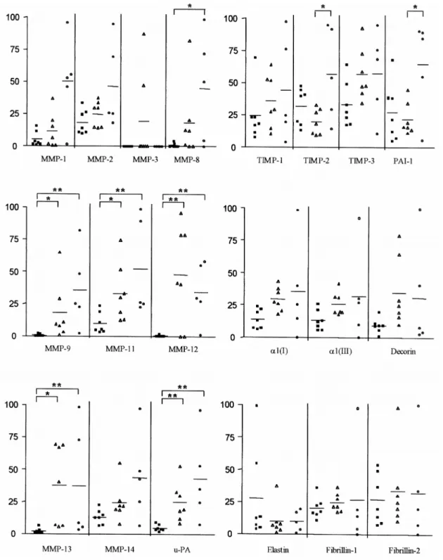
Results: The MMPs -8, -9, -12 and -13 mostly associated with inflammatory cells were not or barely detected in CAA and CTA while they were largely and similarly expressed in AAA and AOD. Expression of protease inhibitors or structural proteins were only slightly increased in both pathological conditions with the exception of elastin which was reduced. The main significant difference between AAA and AOD was a lower expression of TIMP-2 and PAI-1 in the aneurysmal lesions.
Conclusions: The remodeling of the aortic wall in AAA and AOD involves gene activation of a large and similar spectrum of proteolytic enzymes while the expression of two physiological inhibitors, TIMP-2 and PAI-1, is significantly lower in AAA compared to AOD. The repair process in the aneurysmal disease seems similar to that of the occlusive disease.
Keywords : Arteries ; Atherosclerosis ; Extracellular matrix ; Gene expression ; Inflammation
1. Introduction
Abdominal aortic aneurysms (AAA) and atherosclerotic occlusive diseases (AOD) arise from a common basic mechanism, atheroma, but their outcome is different. In AOD, the outer diameter of the vessel is preserved and the medial layer remains largely intact while a progressive narrowing of the lumen, resulting from intimal accumulation of lipids, matrix proteins and cells, ultimately interrupts the blood flow [1]. In contrast, AAA is characterized by a preserved blood flow, enlargement of the blood vessel and, eventually, its rupture which is related to a fragmentation and rarefaction of the elastic lamellae [1]. We previously showed that the elastin loss is an early event in the course of the aneurysmal disease while the collagen concentration remained unchanged reflecting a compensatory adventitial fibrosis [2]. The AAA is also characterized by a medial and adventitial infiltration by inflammatory cells while this infiltration occurs mainly in the intima of the AOD [3]. Matrix metalloproteinases (MMPs) can collegially degrade all the components of the extracellular matrix. Secreted as inactive zymogens, MMPs are processed into their active forms by other MMPs or serine proteases such as plasmin. Plasmin itself results from the activation of plasminogen by plasminogen activators (urokinase-type: u-PA; and tissue-type: t-PA), a reaction controlled by the plasminogen activator inhibitor (PAI-1). The MMPs activity is further controlled by physiological inhibitors, the tissue inhibitors of metalloproteinases (TIMPs) [4,5]. The involvement of proteases/antiproteases in AAA and AOD is well documented in aneurysmal progression and plaque instability [6]. Data are however sometimes conflicting and most often restricted to one or a few of the potential actors in the pathomechanism of the lesions.
The extensive remodelling of the aortic wall seen in AAA and AOD involves both degradation and synthesis of structural matrix proteins. The biomechanical properties of the vessel largely depend on the adequate proportion of collagen, responsible for the tensile strength of the wall, and elastic fibers forming its extensile network [7]. A compensatory repair process has been documented in human AAA and AOD [8] as well as in animal models [9]. In order to evaluate the respective implication of proteolysis, antiproteolysis and repair processes in normal aorta and in AAA and AOD, we devised a sensitive and quantitative reverse transcription-PCR (RT-PCR) procedure to compare in the same series of samples the mRNA steady-state level of a panel of the most representative proteolytic enzymes, their inhibitors and the main structural matrix macromolecules. Large differences occurred in the pathological samples as compared to control tissues. Besides a decreased level in elastin mRNA in AAA and AOD, a similar increase in the expression of all the tested genes is observed in both diseased aorta except for a significantly decreased expression of two proteases inhibitors, PAI-1 and TIMP2, in AAA and a complete absence of active or latent MMP-9 observed by zymography in AOD.
2. Methods
2.1. Patients characteristics
Full-thickness infrarenal aortic tissue was obtained during elective surgery for degenerative AAA from seven patients (mean age, 67 years; range 55-82 years). All patients were asymptomatic at the time of surgery. The mean size of the aneurysms was 58 mm (range 39-80 mm). Atherosclerotic occlusive aortas were harvested during surgery for infrarenal occlusive disease in five patients (mean age, 63 years; range 50-70 years). Thoracic aorta presenting weak or not clinically observable atherosclerosis was obtained during coronary bypass surgery in seven patients (mean age, 65 years; range 54-74 years). Clinically normal abdominal aortas were collected from seven patients deceased of non-vascular disease (mean age, 60 years; range 51-74 years). The study was approved by the local Ethic Committee and conforms with the principles outlined in the Declaration of Helsinki.
2.2. RNA isolation and quantitative RT-PCR procedure
After isolation [10], RNA concentration was measured by a fluorimetric assay (SpectraMax, Gemini-XS). Pairs of RT-PCR primers (see Table 1) were selected according with the following criteria: (i) a high and similar annealing temperature; (ii) minimum complementarity between primer sequences; and (iii) localization on different exons. For each investigated mRNA, a synthetic RNA (sRNA) was generated, according to previously published works [11-13], in order to monitor in each tube the efficiency of both the reverse transcription and the amplification reactions. The sRNAs give rise to products of a size slightly different from that of the endogenous mRNA (Table 1 and Fig. 1). RT-PCR was performed under non-competitive conditions in an automated system (GeneAmp PCR System 9600, Perkin-Elmer) using the GeneAmp Thermostable rTth Reverse Transcriptase RNA PCR kit (Perkin-Elmer), specific pairs of primers (5 pmol each), 5 ng of total cellular RNA and a known copy number of sRNA per 25 µl reaction mixture. The RT step (70 °C for 15 min) using the antisense primer in presence of Mn2+ was followed by addition of the sense primer in presence of Mg2+ (i) 2-min incubation at 95 °C,
(ii) PCR amplification for the adequate number of cycles and (iii) a final elongation step of 2 min at 72 °C. The PCR conditions for the amplification of most cDNA were: 94 °C for 15 s; 66 °C for 20 s; and 72 °C for 10 s. For MMP-3, -11 and -13, conditions were: 94 °C for 15 s, 63 °C for 30 s and 72 °C for 30 s. The RT-PCR products were resolved on 10% polyacrylamide gels and quantified (Fluor-S-Multilmager, BioRad) after staining (GelStar dye, FMC BioProducts). Each sample was analysed in duplicate. The optical density of the endogenous RNA was normalized by the value of the sRNA (see Fig. 1) and expressed in arbitrary units per unit of 28S ribosomal RNA.
Table 1 Sequence of forward and reverse primers used for RT-PCR amplification of the target RNA and length
of the RT-PCR products
RNA species
Forward (5'-3') Reverse (5'-3') Length (bp) of
the product from
RT-PCR Endogenous RNA sRNA
MMP-1 GAGCAAACACATCTGAGGTA
CAGGA TTGTCCCGATGATCTCCCCTGACA 185 267
MMP-2 AGATCTTCTTCTTCAAGGAC CGGTT GGCTGGTCAGTGGCTTGGGGTA 225 271 MMP-3 GATCTCTTCATTTTGGCCATC TCTTC CTTCCAGTATTTGTCCTCTA CAAAGAA 246 272 MMP-8 CCAAGTGGGAACGCACTAAC TTGA TGGAGAATTGTCACCGTGATCTCTT 200 267 MMP-9 GCGGAGATTGGGAACCAGCT GTA GACGCGCCTGTGTACACCCACA 208 266 MMP-11 ATTTGGTTCTTCCAAGGTGCT CAGT CCTCGGAAGAAGTAGATCTTGTTCT 155 268 MMP-12 ACATTTCGCCTCTCTGCTGAT
GAC CAGAAACCTTCAGCCAGAAGAACC 196 245
MMP-13 ATGATCTTTAAAGACAGATT
CTTCTGG TGGGATAACCTTCCAGAATGTCATAA 203 270
MMP-14 GGATACCCAATGCCCATTGG
CCA CCATTGGGCATCCAGAAGAGAGC 221 269
TIMP-1 CATCCTGTTGTTGCTGTGGCT GAT GTCATCTTGATCTCATAACGCTGG 170 271 TIMP-2 CTCGCTGGACGTTGGAGGAA AGAA AGCCCATCTGGTACCTGTGG TTCA 155 269 TIMP-3 CTTCTGCAACTCCGACATCG
TGAT CAGCAGGTACTGGTACTTGTTGAC 210 269
uPA ACTACTACGGCTCTGAAGTC
ACCA GAAGTGTGAGACTCTCGTGTAGAC 199 245
PAI-1 AGGGCTTCATGCCCCACTTC TTCA AGTAGAGGGCATTCACCAG CACCA 191 269 α1(I) CCCACCAATCACCTGCGTAC
AGA TTCTTGGTCGGTGGGTGACTCTGA 214 267
α1(III) GAGATGTCTGGAAGCCAGAA CCAT GATCTCCCTTGGGGCCTTGAGGT 207 265 Decorin CCTGAAAGGACTGAATAATT TGGCTA GTTGCTGAAAAGACTCACAC CCGAA 277 201 Fibrillin 1 GGTGAATGTACAAACACAGT
CAGCA ATAGGAACAGAGCACAGCTTGTTGA 275 210
Fibrillin 2 ATGGCTCTCGATGCATCGAT
CAGA CATTGCCACTTGGGGCAAAGCCA 282 199
Elastin CCGCTAAGGCAGCCAAGTAT GGA AGCTCCAACCCCGTAAGTAG GAAT 275 189 28S GTTCACCCACTAATAGGGAA
CGTGA GGATTCTGACTTAGAGGCGTTCAGT 212 269
MMP, matrix metalloproteinase; TIMP, tissue inhibitor of matrix metalloproteinase; u-PA, urokinase plasminogen activator; PAI, plasminogen activator inhibitor; α1(I), α1 chain of type I collagen; α1(ΠI), α1 chain of type III collagen; 28S, 28S ribosomal RNA; sRNA, synthetic RNA used as internal standard for RT and PCR reactions.
mRNA (32 cycles), 28S ribosomal RNA (28S) (18 cycles) and their respective synthetic RNA (sRNA). The assay was performed in duplicate using RNA from normal abdominal (CAA), thoracic (CTA) or aneurysmal (AAA) and occlusive (AOD) aorta. The sample migrated in the last right lane contained all the reagents including the sRNA but no cellular RNA.
2.3. Zymographic analysis of the gelatinases MMP-2 and MMP-9
The analysis of tissue homogenates of CAA, AAA and AOD was performed by the procedure described earlier [14].
2.4. Statistics
Statistical differences among groups were tested by one-way ANOVA after normalization using neperian Log. A probability value ≤0.05 was considered as significant.
3. Results
3.1. RT-PCR assay
The mRNA levels of the selected genes were determined using total RNA purified from seven control thoracic aorta (CTA), seven control abdominal aorta (CAA), seven abdominal aortic aneurysms (AAA) and five aortic occlusive diseases (AOD) in the same run of RT-PCR. The electrophoretic pattern of the RT-PCR products of MMP-9 mRNA, that of ribosomal 28S rRNA and their respective sRNA is illustrated in Fig. 1 as a
representative example. Each sample was analyzed in duplicate. A large increase of MMP-9 mRNA in the diseased vessels (AAA, AOD) compared to control aorta (CTA, CAA) was observed while the signals for sRNA and cellular 28S rRNA were almost constant. This assay allows a quantitative and comparative titration of specific mRNA in specimens displaying an extended range in level of expression. The accuracy and reliability of this procedure has been reported previously [11-13].
3.2. Comparative analysis of the mRNA steady-state levels in normal thoracic and abdominal aorta
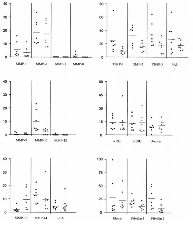
The three groups of mRNA, coding, respectively, for proteolytic enzymes, their physiologic inhibitors and selected structural matrix macromolecules, were measured in clinically normal segments of aorta at two different locations, thoracic (CTA) and abdominal (CAA). No significant difference was observed between the two types of samples (Table 2). It is worth noting that MMP-3, as well as MMP-8, -9 and -12, most often associated with inflammatory cells, were generally not or barely detected as seen from the individual values illustrated in Fig. 2. Since the efficiency of the amplification process lies within the same order of magnitude for each individual primer pair, as judged from results obtained from a known copy number of the various sRNA (data not shown), the relative abundance of each mRNA can be roughly estimated from the number of cycles of PCR required to obtain measurable amplification products (Table 2). This indicates that the mRNAs for structural proteins and for TIMPs, requiring less amplification cycles, are more expressed than most of the MMPs. The values reported in Table 2 can be considered as representative of the phenotype of the resident cells in non-diseased late adult aortic wall. Even though the pathological aortic samples will only be compared to CAA, the data concerning CTA are provided to support the reliability of the procedure and as information that might be useful to others.
Table 2 Steady-state levels of mRNA in control aorta
Cycles
(n) CAA CTA CAA/CTA
Proteinases MMP-1 37 5±6 3±4 1.7 MMP-2 31 18±9 17±10 1.1 MMP-3 37 0 0 n.a MMP-8 37 <1 <1 n.a MMP-9 32 <1 <1 n.a MMP-11 37 9±8 2±2 5.0 MMP-12 31 <1 <1 n.a MMP-13 37 1±2 9±8 0.2 MMP-14 29 12±6 9±10 1.4 u-PA 31 4±3 5±6 0.8 Inhibitors TIMP-1 29 24±21 8±4 2.8 TIMP-2 28 32±14 15±6 2.0 TIMP-3 27 28±15 14±10 2.0 PAI-1 31 27±22 17±7 1.7
Structural proteins a1(I) 28 13±7 13±14 1.0
A1(III) 27 13±8 13±10 1.0
Decorin 31 9±6 11±6 0.8
Elastin 27 27±36 23±19 1.3
Fibrillin 1 31 20±8 11±6 1.7
Fibrillin2 36 26±19 7±9 3.3
CAA, control abdominal aorta; CTA, control thoracic aorta. *Mean ratio between the mRNA levels. Number (n) of amplification cycle. The values expressed in arbitrary units are means+S.D. n.a., not applicable.
3.3. Comparative mRNA expression in control, aneurysmal and occlusive abdominal aorta
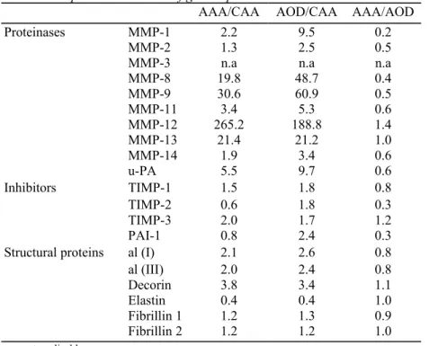
The mRNAs from the diseased aorta (AAA and AOD) were reverse-transcribed, amplified and measured simultaneously with the control tissues (CAA and CTA) as illustrated in Fig. 1 for MMP9. Amplification products of the investigated mRNAs were detected in most samples of the pathological vessels. The exceptions were MMP-3 found in only two AAA while other MMPs were sporadically absent (Fig. 3). The mean mRNA level of all the MMPs and that of the inhibitors were always higher in AAA and AOD than in the CAA (ratio >1), except TIMP2 and PAI-1 in AAA (Table 3). The main and most significant differences, when compared to control tissue, were observed for MMP-8, -9, -11, -12, -13 and u-PA. In our groups of samples, MMP-3 is regularly undetectable, except in two AAA. MMP-8 is expressed in several pathological specimens and only in two controls. A nonsignificant increase of mRNA coding for structural proteins was observed similarly in both pathological (AAA and AOD) aorta by comparison with normal aorta except for elastin mRNA, which was reduced in both pathologies although to a non-significant level (Fig. 3). MMP2 and its activator MMP14 were also slightly increased (not significant) by a factor of two. The only differences that reached statistical significance between the expression profile of the mRNAs of AAA and AOD was a reduced level of TIMP-2 and PAI-1 in AAA.
3.4. Gelatinolytic activity
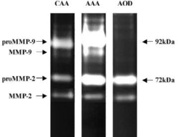
As illustrated in a representative zymogram (Fig. 4), a large amount of proMMP-9 is observed in AAA, largely more intense than in CAA. In AAA, a band of activated MMP-9 is also visible while absent in CAA. No proMMP-9 or activated MMP-9 is present in AOD. The amount of proMMP-2 and MMP-2 are similar in AAA and AOD and much larger than in CAA.
Fig. 2. Expression of mRNA of proteolytic enzymes (MMPs and u-PA), their physiologic inhibitors (TIMPs and
PAI-1), and structural proteins (type I and type III collagen, decorin, elastin, fibrillin 1 and 2) in samples of control abdominal aorta (CAA, solid squares), and control thoracic aorta (CTA, open squares). The results are expressed in arbitrary units per unit of 28S rRNA allowing a comparative analysis between samples for each mRNA. No statistical difference between CAA and CTA was observed.
Table 3 Comparative variation of gene expression between AAA or AOD and CAA
AAA/CAA AOD/CAA AAA/AOD
Proteinases MMP-1 2.2 9.5 0.2
MMP-2 1.3 2.5 0.5
MMP-3 n.a n.a n.a
MMP-8 19.8 48.7 0.4 MMP-9 30.6 60.9 0.5 MMP-11 3.4 5.3 0.6 MMP-12 265.2 188.8 1.4 MMP-13 21.4 21.2 1.0 MMP-14 1.9 3.4 0.6 u-PA 5.5 9.7 0.6 Inhibitors TIMP-1 1.5 1.8 0.8 TIMP-2 0.6 1.8 0.3 TIMP-3 2.0 1.7 1.2 PAI-1 0.8 2.4 0.3
Structural proteins al (I) 2.1 2.6 0.8
al (III) 2.0 2.4 0.8
Decorin 3.8 3.4 1.1
Elastin 0.4 0.4 1.0
Fibrillin 1 1.2 1.3 0.9
Fibrillin 2 1.2 1.2 1.0
Fig. 3. Expression of mRNA of MMPs, u-PA, TIMPs, PAI-1, type I and type III collagen, decorin, elastin,
fibrillin 1 and 2 in samples of control abdominal aorta (CAA, solid squares), abdominal aortic aneurysms (AAA, open triangles) and aortic occlusive disease (AOD, open circles). The results are expressed in arbitrary units per unit of 28S rRNA allowing a comparative analysis between samples for each mRNA. **P≤0.01, *P≤0.05.
Fig. 4. Representative example of MMP-2 and MMP-9 activity under latent or activated forms measured by
gelatin zymography in extracts of normal (CAA) and diseased aortic wall (AAA and AOD).
4. Discussion
4.1. Technological considerations
In order to reconcile some apparently conflicting data of the literature and to obtain a better and quantitative overview of the pathophysiological processes implicated in AAA and AOD, we measured the expression of three series of genes in clinically normal thoracic (CTA) and abdominal (CAA) aorta compared to samples of AAA or AOD. The mRNA level of proteolytic enzymes (MMPs, u-PA), their inhibitors (TIMPs, PAI-1) and that of the most abundant structural proteins found in the aortic wall was measured by RT-PCR. This procedure was made quantitative by adding in each sample a known copy number of specific synthetic RNAs as internal standards [11-13]. Such standards, co-reverse transcribed and co-amplified with the cellular mRNA using the same primers, allow to monitor the efficiency of both the reverse-transcription and the amplification steps, which is not the case with the cDNA standards such as those used by Carrell et al. [15].
Our sensitive procedure provided quantitative and comparative data for samples expressing an extended range in mRNA levels and permitted to estimate differences in expression more precisely than other assays [16].
4.2. Definition of the biosynthetic phenotype of normal aorta
The thoracic (CTA) and abdominal (CAA) aorta display a very similar biosynthetic phenotype with a significant expression of the structural proteins, including elastin and fibrillins which are involved in the maintenance of the matrix in the vessel, in agreement with previous observations [17]. The mRNA of the MMPs known to be induced in the context of inflammation (MMP-8, -9 and -12) and that of MMP-3 are not expressed to a
significant level in the control specimens. By contrast, the mRNAs of the MMP inhibitors (TIMPs) are produced as well as the plasminogen activator u-PA and its inhibitor, PAI-1.
4.3. Expression of the MMPs in AAA and AOD
The genes that are expressed at a significant level (≤31 cycles) in the control tissues (CAA and CTA) tend to be higher in AAA or AOD as are MMP-2 and -14, the TIMP-1 and -3 and the structural proteins. These genes participate in the biosynthetic phenotype of the resident cells in the normal tissue. Their tendency to increase in the diseased vessels might be related to an activation of the biosynthetic activity of the SMC. This activation is likely to be underestimated in the lesional tissues by dilution with RNA coming from blood born cells and by the rarefaction of SMC, at least in AAA. Disruption and degradation of elastic lamellae in the media is a prominent feature of AAA that occurs early in the course of the disease [2] and the implication of MMP-9 in its
development is clearly supported by animal experimentation. Using mice with targeted gene deletion of MMP-9 or MMP-12 and a double MMP-9/MMP-12 KO only the animals with a deletion of the MMP-9 gene are protected against aneurysmal development [18]. The similarly increased [19,20] or an even higher expression of
MMP-9 mRNA in AOD than in AAA as shown here, questions the specific role of this enzyme in aneurysmal dilatation. MMP-9 is produced by the macrophages infiltrating the aortic wall [20]. It is also produced by SMC at least in culture [21]. The larger expression of this mRNA in AOD while the density of the macrophages is lower compared to AAA [2] might indicate that the SMC might participate in the overexpression perhaps upon stimulation by the cytokines released from the inflammatory infiltrate.
The tensile strength of the vessel is conferred by the cross-linked polymeric collagens, mainly the fibers of type I and III. Aortic dilatation in aneurysmal progression is linked to a profound remodelling of the fibrillar network of the vessel wall that requires collagenolytic activity. While collagenase-1 (MMP-1) was not much increased as compared to control tissue, the collagenase-3 (MMP-13) was elevated in AAA and AOD in agreement with a previous study [22] immunolocalizing MMP-13 in the SMC within the outer wall in AAA and in the intimal plaque in AOD. Besides its collagenolytic activity, MMP-13 can degrade a broad spectrum of matrix proteins and activate other MMPs [23] suggesting its central role during the evolution of these diseases. Collagenase-2 (MMP-8), a most effective collagenase to initiate type I collagen degradation not detected in the study of Carrell et al. [15], was present in eight out of our 12 patients, while barely detected in the controls. As
polymorphonuclear neutrophils are almost absent in the AAA [14] and AOD [3], MMP-8 could be produced by the endothelial cells, the SMC or the macrophages, as supported by its recent detection in vulnerable
atherosclerotic plaques and SMC in culture [24]. An intriguing observation in our samples is the very low amount of MMP-3, requiring the highest acceptable number of amplification cycles for detection, expressed in only two AAA samples, and absent in all control and AOD specimens. Although we basically agree with the conclusion of Carrell et al. [15] that MMPs are involved in the pathomechanisms of AAA development, our results do not support a specific role of MMP-3 in this process.
4.4. Imbalanced expression of the inhibitors
When compared to the strikingly similar overexpression of most genes in AAA and AOD, two gene products make the exception, TIMP2 and PAI-1. For both mRNAs, the statistically significant difference that we observed results of a non significant reduction in AAA compared to CAA and a non significant increase in AOD. Since u-PA and MMP-8, -9, -12 and -13 mRNAs are largely increased in both pathologies, whereas their inhibitors, u- PAI-1 and TIMP2 are lower in AAA as compared to AOD, it could be suspected that the functional balance of proteolysis/ antiproteolysis is altered in AAA. This consideration is supported by observations in the apoE-/-,
plau-/- (u-PA invalidated) double KO mice in which u-PA deficiency protects against media destruction and
aneurysm formation [25]. Moreover, local overexpression of PAI-1 demonstrates the same type of protective potential and a decreased MMP9 activity [26]. The reduction of TIMP2 may also participate in the increased proteolysis in AAA. A significantly different polymorphic allele frequency in male AAA found in the coding region of TIMP2 is intriguing and deserves further investigations [27]. Furthermore, TIMP2 at low concentration participates in association with MMP14 to the activation of MMP2 [28]. We previously showed that a significant part of the MMP2 was present in the AAA wall under an activated form [14]. The role of stromelysin-3 (MMP-11), moderately but significantly increased in AAA and AOD, is putative since the only known substrate of this MMP is the α1-proteinase inhibitor (α1-AP) [29], a serine elastase inhibitor also known for its antiapoptotic activity [30]. Since patients with α1-AP deficiency present multiple aneurysms and emphysema by elastolysis in the lung [31], its potential significance in the aneurysmal disease also merits further investigation.
It has been observed that MMP-9 is found in its precursor and activated form in AAA [14] while neither the proMMP-9 nor the activated MMP-9 are detected by zymography in AOD ([32,33] and our data). Vine and Powell [34] have observed that the elastolytic activity of the homogenates of AAA and AOD increases largely upon treatment of the extracts by KSCN to release the proteases from their complex with α2-macroglobulin
(α2M). The lack of MMP-9 (pro- and activated) in our samples of AOD might be related to a similar process.
Beside its large amount of liver origin in the plasma, α2M is also produced by macrophages mainly upon
cytokine stimulation [35]. As observed in vitro the secreted proteinases might react with the secreted α2M to
form complex catabolized by the macrophages. If such a process occurs in AOD and not in AAA as suggested in our zymograms, it might explain the divergent evolution of the blood vessel wall in these diseases. Further work is progressing to clarify this issue.
4.5. Extracellular matrix proteins
The mRNA level of the structural proteins involved in the formation and maintenance of the resistant collagen framework of the wall was slightly increased in AAA and AOD by comparison to control aorta. It could represent the expression of a repair mechanism performed by the resident cells, fibroblasts and SMC [8]. The lower than normal expression of the elastin gene associated with an increased collagen and decorin expression
suggests in both aortic diseases that the composition of the repair connective tissue may be inappropriate and perhaps responsible for alterations of the biomechanical properties of the vessel. Again, the lack of difference between AAA and AOD is striking.
The transcriptional profiling for the three groups of genes reported in this study has been performed under conditions of amplification that allow a direct comparison between control aortic samples and the two pathologies. In our procedure the range of variations, although quite extensive (in the order of a few hundred times between the lowest and the highest value for each mRNA), is much smaller than those observed by Carrel et al. (up to 10 000 times) [15]. This variability within each group of samples is multifactorial and potentially related to the genetic heterogeneity of the human race, additional pathologies or their therapy, and possible minor clinically non-apparent defects related to aging in the control samples. In the pathological specimens, the extent of heterogeneity is caused by similar factors probably enhanced by genetic differences in the
transcriptional efficiency of some of the tested genes [36,37]. Furthermore, AAA and problably AOD are heterogeneous not only in terms of size but also in metabolic activity as we recently demonstrated by PET scan imaging [38]. Additionally, each of the specimens is not uniform but made of a mosaic of zones in which the pathological process displays different levels of activity. Our recent results (unpublished data) in one ruptured AAA show that the level of MMPs (-1, -2, -9, -12 and -14) decreases as a function of the distance in samples collected progressively away from the site of rupture while the m-RNA of MMP-3, -8 and -11 was either absent or significantly high in the same series of samples. This could also explain some of the differences between the published data and our work.
Our study investigated the simultaneous implication of three biological processes, proteolysis, anti-proteolysis and repair, in the AAA and AOD. Altogether, our results obtained by measuring mRNA to evaluate gene expression show that, when compared to the basal physiological phenotype of clinically normal age-matched aortas, both the aneurysmal and the atherosclerotic lesions are similarly characterized by a largely increased proteolytic repertoire which is not compensated by a similar increased level of proteases inhibitors and structural proteins involved in the repair process. The profile of mRNA expression of 18 genes is quite comparable in AAA and AOD, while only two genes, TIMP-2 and PAI-1, significantly differ between the two pathologies. These observations suggest a significant role for proteinase inhibitors during the divergent evolution of AAA and AOD.
Acknowledgements
We gratefully thank Marie-Jeanne Nix and Antoine Heyeres for their expert technical assistance. This work was supported by the Belgian 'Fonds Scientifique de la Recherche Médicale' and the 'Fonds de Recherche
Scientifique' of the Medical School at the University of Liège References
[1] Ghorpade A, Baxter BT. Biochemistry and molecular regulation of matrix macromolecules in abdominal aortic aneurysms. Ann NY Acad Sci 1996;800:138-150.
[2] Sakalihasan N, Heyeres A, Nusgens BV et al. Modifications of the extracellular matrix of aneurysmal abdominal aortas as a function of their size. Eur J Vasc Surg 1993;7:633-637.
[3] Koch AE, Haines GK, Rizzo RJ et al. Human abdominal aortic aneurysms: Immunophenotypic analysis suggesting an immune-mediated response. Am J Pathol 1990;137:1199-1213.
[4] Sternlicht MD, Werb Z. How matrix metalloproteinases regulate cell behavior. Annu Rev Cell Dev Biol 2001;17:463-516. [5] Wojowicz-Praga SM, Dickson RB, Hawkins MJ. Matrix metalloproteinase inhibitors. Invest New Drugs 1997;15:61-75.
[6] Knox JB, Sukhova GK, Whittemore AD et al. Evidence for altered balance between matrix metalloproteinases and their inhibitors in human aortic diseases. Circulation 1997;95:205-212.
[7] Mesh Ch L, Baxter BT, Pearce WH et al. Collagen and elastin gene expression in aortic aneurysms. Surgery 1992;112:256-262. [8] Baxter BT, McGee GS, Shively VP et al. Elastin content, crosslinks, and mRNA in normal and aneurysmal human aorta. J Vasc Surg 1992;16:192-200.
[9] Huffman MD, Curci JA, Moore G et al. Functional importance of connective tissue repair during the development of experimental abdominal aortic aneurysms. Surgery 2000;128:429-438.
[10] Chirgwin JM, Przybyla AE, MacDonald RJ et al. Isolation of biologically active ribonucleic acid from sources enriched in ribonuclease. Biochemistry 1979;18:5294-5299.
[11] Lambert ChA, Colige AC, Munaut C et al. Distinct pathways in the overexpression of matrix metalloproteinases in human fibroblasts by relaxation of mechanical tension. Matrix Biol 2001;20:397-408.
[12] Lambert ChA, Colige AC, Lapière ChM et al. Coordinated regulation of procollagens I and III and their post-translational enzymes by dissipation of mechanical tension in human dermal fibroblasts. Eur J Cell Biol 2001;80:479-485.
[13] Nusgens B, Humbert P, Rougier A et al. Topically applied vitamin C enhances the mRNA level of collagens I and III, their processing enzymes and TIMP1 in the human dermis. J Invest Dermatol 2001;116:853-859.
[14] Sakalihasan N, Delvenne Ph, Nusgens BVet al. Activated forms of MMP2 and MMP9 in abdominal aortic aneurysms. J Vasc Surg 1996;24:127-133.
[15] Carrell TWG, Burnand KG, Wells GMA et al. Stromelysin-1 (matrix metalloproteinase-3) and tissue inhibitor of metalloproteinase-3 are overexpressed in the wall of abdominal aortic aneurysms. Circulation 2002;105:477-482.
[16] Freeman WM, Walker SJ, Vrana KE. Quantitative RT-PCR: Pitfalls and potential. Biotechniques 1999;26:112-125.
[17] Godfrey M, Nejezchleb PA, Schaefer GB et al. Elastin and fibrillin mRNA and protein levels in the ontogeny of normal human aorta. Connect Tissue Res 1993;29:61-69.
[18] Pyo R, Lee JK, Shipley JM et al. Targeted gene disruption of matrix metalloproteinase-9 (gelatinase B) suppresses development of experimental abdominal aortic aneurysms. J Clin Invest 2000;105:1641-1649.
[19] Armstrong PJ, Johanning JM, Calton WC et al. Differential gene expression in human abdominal aorta: aneurysmal versus occlusive disease. J Vasc Surg 2002;35:346-355.
[20] McMillan WD, Patterson BK, Keen RR et al. In situ localization and quantification of mRNA for 92-kD type IV collagenase and its inhibitor in aneurysmal, occlusive, and normal aorta. Arterioscler Thromb Vasc Biol 1995;15:1139-1144.
[21] Patel MI, Melrose J, Ghosh P et al. Increased synthesis of matrix metalloproteinases by aortic smooth muscle cells is implicated in the etiopathogenesis of abdominal aortic aneurysms. J Vasc Surg 1996;24:82-92.
[22] Mao DL, Lee JK, Vanvickle SJ et al. Expression of collagenase-3 (MMP-13) in human abdominal aortic aneurysms and vascular smooth muscle cells in culture. Biochem Biophys Res Commun 1999;261:904-910.
[23] Knauper V Smith B, Lopez-Otin C et al. Activation of progelatinase B (proMMP-9) by active collagenase-3 (MMP-13). Eur J Biochem 1997;248:369-373.
[24] Herman MP, Sukhova GK, Libby P et al. Expression of neutrophil collagenase (matrix metalloproteinase-8) in human atheroma—A novel collagenolytic pathway suggested by transcriptional profiling. Circulation 2001;104:1899-1904.
[25] Carmeliet P, Moons L, Lijnen R et al. Urokinase-generated plasmin activates matrix metalloproteinases during aneurysm formation. Nat Genet 1997;17:439-444.
[26] Allaire E, Hasenstab D, Kenagy RD et al. Prevention of aneurysm development and rupture by local overexpression of plasminogen activator inhibitor-1. Circulation 1998;98:249-255.
[27] Wang X, Tromp G, Cole W et al. Analysis of coding sequences for tissue inhibitor of metalloproteinases 1 (TIMP1) and 2 (TIMP2) in patients with aneurysms. Matrix Biol 1999;18:121-124.
[28] Strongin AY, Collier I, Bannikov G et al. Mechanism of cell surface activation of 72-kDa type IV collagenase. J Biol Chem 1995;270:5331-5338.
[29] Noel A, Boulay A, Kebers F et al. Demonstration in vivo that stromelysin-3 functions through its proteolytic activity. Oncogene 2000;19:1605-1612.
[30] Ikari Y, Mulvihill E, Schwartz SM. Alpha 1-proteinase inhibitor, alpha 1-antichymotrypsin, and alpha 2-macroglobulin are the an- tiapoptotic factors of vascular smooth muscle cells. J Biol Chem 2001;276:11798-11803.
[31] Mtchell MB, McAnena OJ, Rutherford RB. Ruptured mesenteric artery aneurysm in a patient with alpha 1-antitrypsin deficiency: etiologic implications. J Vasc Surg 1993;17:420-424.
[32] Yamashita A, Noma T, Nakazawa A et al. Enhanced expression of matrix metalloproteinase-9 in abdominal aortic aneurysms. World J Surg 2001;25:259-265.
[33] Thompson RW, Holmes DR, Mertens RA et al. Production and localization of 92-kilodalton gelatinase in abdominal aortic aneurysms. J Clin Invest 1995;96:318-326.
[34] Vine N, Powell JT. Metalloproteinases in degenerative aortic disease. Clin Sci 1991;81:233-239.
[35] Lysiak JJ, Hussaini IM, Gonias SL. α2-Macroglobulin synthesis by the human monocytic cell line THP-1 is differentiation state-dependent. J Cell Biochem 1997;67:492-497.
[36] Ye S, Eriksson P, Hamsten A et al. Progression of coronary atherosclerosis is associated with a common genetic variant of the human stromelysin-1 promoter which results in reduced gene expression. J Biol Chem 1996;271:13055-13060.
[37] Zhang BP, Ye S, Herrmann SM et al. Functional polymorphism in the regulatory region of gelatinase B gene in relation to severity of coronary atherosclerosis. Circulation 1999;99:1788-1794.
[38] Sakalihasan N, Van Damme H, Gomez P et al. Positron emission tomography (PET) evaluation of abdominal aortic aneurysm. Eur J Vasc Endovasc Surg 2002;23:431-436.