ACCUMULATION AND SEQUESTRATION OF METALS IN DIFFERENT STRAINS OF CHLAMYDOMONAS THE SIS PRESENTED AS PARTIAL REQUIREMENT OF DOCTORA TE IN CHEMISTR Y BY MAHSHID SAMADANI AUGUST 2018
Avertissement
La diffusion de cette thèse se fait dans le respect des droits de son auteur, qui a signé le formulaire Autorisation de reproduire et de diffuser un travail de recherche de cycles supérieurs (SDU-522- Rév.0?-2011). Cette autorisation stipule que «conformément à l'article 11 du Règlement no 8 des études de cycles supérieurs, [l'auteur] concède à l'Université du Québec à Montréal une licence non exclusive d'utilisation et de publication de la totalité ou d'une partie importante de [son] travail de recherche pour des fins pédagogiques et non commerciales. Plus précisément, [l'auteur] autorise l'Université du Québec à Montréal à reproduire, diffuser, prêter, distribuer ou vendre des copies de [son] travail de recherche à des fins non commerciales sur quelque support que ce soit, y compris l'Internet. Cette licence et cette autorisation n'entraînent pas une renonciation de [la] part [de l'auteur] à [ses] droits moraux ni à [ses] droits de propriété intellectuelle. Sauf entente contraire, [l'auteur] conserve la liberté de diffuser et de commercialiser ou non ce travail dont [il] possède un exemplaire.»
L'ACCUMULATION ET LA SÉQUESTRATION DES MÉTAUX CHEZ DIFFÉRENTES SOUCHES DE CHLAMYDOMONAS
THÈSE PRÉSENTÉE
COMME EXIGENCE PARTIELLE DU DOCTORAT EN CHIMIE
PAR
MAHSHID SAMADANI
1 would like to express the deepest appreciation to my supervisor, Professor David Dewez, who offered numerous helpful insights, suggestions, guidance, and corrections without which this dissertation would not have been possible to progress and finish. I have been very fortunate to work in his laboratory and to collaborate with his research group, and I am very grateful for ali skills and knowledge which I have obtained working in his laboratory.
1 would like to acknowledge gratefully the valuable explanations and many useful comments and suggestions from the jury members: Pr. Huu V an Tra, Pr. Abdelkrim Azzouz, Pr. Philippe Juneau, and Pr. William Zerges, who helped and encouraged me to continue and finalize this work.
I would like to thank deeply for the expert assistance of Professor François Perreault who taught me laboratory techniques and without his scientific helps, patience, and guidance the work would have been much harder for me.
1 would like to appreciate greatly for the technical support of Denis Flipo at the confocal microscopy imaging facility of TOXEN (Department of Biological Sciences, UQAM).
1 am very grateful for ali supports and helps from my colleagues in laboratory, Dr. Laura Pirastru for her great knowledge of algal culture, who helped me to cultivate and to maintain microalgae in the laboratory, Dr. Abdallah Oukarroum for ali his encouragement, Émillie for giving me lots of positive energy, Vahid and Jonathan for their collaboration to do sorne parts of practical experiments.
1 have also special thanks to UQÀM's personnel those supported me kindly pruticularly my great appreciation to Dr. Gwenaël Chamoulaud and Mme Isabelle Rbeault for technical services and helpful advices to accomplish my experimental tests with ICP-AES and F AAS.
I would also have a special gratitude for my best friend Dr. Pejman Nekoovaght for all his supports ever since 1 have arrived in Canada. He encouraged, gave me insights and enthusiasms to continue my graduate studies at UQÀM. He also graciously assisted in editing the present dissertation.
Finally, my special acknowledgment goes to my parents and my family for their great spiritual support and their encomagement.
LISTE OF FIGURES ... vii LIST OF TABLES ... xii LIST OF ABBREVIATIONS ... xiii
LIST OF SYMBOLS AND UNITS ... xviii
RÉSUMÉ ... xx
ABSTRACT ... xxii INTRODUCTION ... 1
CHAPTER I ... ." ... 6
ENVIRONMENT AL IMPACT OF METALS AND GREEN ALGAE ... 6
1.1 Environmental impact of mercury and cadmiwn ... 6
1.2 Using green algae for the phycoremediation of metals ... 10
1.3 Using green algae as a mode! of cellular toxicity ... 12
1.3.1 Cellular defense mechanisms against metal toxicity ... 12
1.3.2 Wild-type and cel! wall-deficient mutants of Chlamydomonas ... 16
1.3.3 Toxicity impact of metals at the cellular level ... 17
CHAPTER II ... 21
ROLE OF INORGANIC POL YPHOSPHATE IN LIVING CELLS ... 21
2.1 Chemical structure of inorganic polyphosphate ... 21
2.2 Polyphosphate in living organisms and its localization ... 22
2.3 Polyphosphate metabolism ... 24
2.4 Physiologie functions of polyphosphate : ........... 25 2.4.1 Reservoir of phosphate (Pi) energy ... 25 2.4.2 Cation sequestration and storage ... 26 2.4.3 Protective role against environmental stresses ... 28 CHAPTER III ... 33
METHODOLOGICAL APPROACHES: PRINCIPLES OF METHODS ... 33
3.1 Cultivation and maintenance of Chlamydomonas strains in the laboratory ... 33
3.2 Experimental treatment of algal cells (general aspects) ... 36
3.3 UV-Vis spectrophotometer for the monitoring of cell density ... 3 8 3.4 Flow cytometer for the determination of the relative cell size, cellular granularity, and cell viability of al gal cells ... 39
3.5 Atomic absorption (FAAS) and ICP-OES for the determination of the accumulation of metals in al gal biomass ... .41
3.6 Chlorophyll a fluorescence measurement for the determination of the performance ofphotosynthetic activity in algae ... .43
3.7 TEM and EDX analysis for the cellular localization ofmetals in algal biomass
46
3.8 Confocal microscope for the measurement of fluorescence emission from extracytoplasmic polyphosphate ... 4 7 3. 9 Zeta potential measurement ... 48CHAPTER IV ... .' ... 50
EFFECT OF CADMIUM ACCUMULATION ON GREEN ALGAE CHLAMYDOMONAS REINHARDTII AND ACID-TOLERANT CHLAMYDOMONAS CPCC 121 ... 50
4.1 Results and discussion ... 50
4.1.1 Speciation and bioavailability of Cd ... 50
4.1.2 Inhibition of growth rate ... 51
4.1.3 Cd accumulation in al gal biomass ... 52
4.1.4 Alteration of cellular characteristics ... 55
4.1.5 Alteration of PSil activity ... 60
4.1.6 Cellular localization of Cd ... 63
4.2 Summary: algal tolerance for accumulated Cd effect.. ... 66
4.3 Conclusion: significance for bioremediation ... 67
CHAPTER V ... 69
CADMIUM ACCUMULATION AND TOXICITY AFFECT THE EXTRACYTOPLASMIC POL YPHOSPHATE LEVEL IN CHLAMYDOMONAS REINHARDT!! .................................................... 69
5.2 Conclusion: physiological relevance of extracytoplasmic polyP ... 80
CHAPTER VI. ... 81
EFFECT OF MERCURY ON THE POL YPHOSPHATE LEVEL OF ALGA CHLAMYDOMONAS REINHARDTII ... ...... 81
6.1 Results ... 81
6.1.1 Inhibition of growth rate ... : ... 81
6.1.2 Change of polyP lev el under Hg stress ... 82
6.1 .3 Lev el of polyP related to growth rate ... 86
6.1.4 Accumulation of Hg in algal biomass ... 87
6.1.5 Level ofpolyP related to the accumulation of Hg ... ,.89
6.1.6 Inhibition of Photosystem II photochemistry ... 89
6.2 Discussion ... 91
6.2.1 Effect of Hg on algal cells ... 91 6.2.2 Physiological role of polyP ... 93
6.3 Conclusion ... 95
CONCLUSION ... 97 REFERENCES ... 101
ANNEXE A ... 113
--Figure Page Figure 1.1 TEM image of algal cell ultrastructure for Chlamydomonas reinhardtii wild-type (CC-125) cultivated in High Salt Medium (HSM) ... 11 Figure 1.2 a) General mechanism involved in PCs synthesis b) Chemical structures of
phytochelatin and glutathione ... 14 Figure 1.3 General schema of metal detoxification mechanism by phytochelatins in microalgae (made from Howe and Merchant., 1992, Perales-Vela et al., 2006) ... 15 Figure 1.4 Protein complexes and cofactors involved in the linear transport of
electrons and protons of photosynthesis in higher plants. Abbreviations: (Mn4) Mn4ÜxCa where x2:. 4, (Yz) tyrosine-161, (Pheo) pheophytin, (QA) and (Qs) two plastoquinone molecules, (PQ) plastoquinone molecules, (FeS) iron-sulfur protein, (PC) plastocyanin, (Ao) a special chlorophyll a molecule, (At) vitamin K, (Fd ) ferredoxin, (FNR) ferredoxin-NADP reductase, (LHCI and LHCII) light-harvesting complexes of photosystems I and II, (NADP reductase and NADP+), nicotinamide-adenine dinucleotide phosphate (Whitmarsh et Govindjee., 1999). 19 Figure 2.1 Inorganic polyphosphate structure ... 21 Figure 2.2 Polyphosphate as a substitute for ATP, made from (Hsieh et al., 1993;
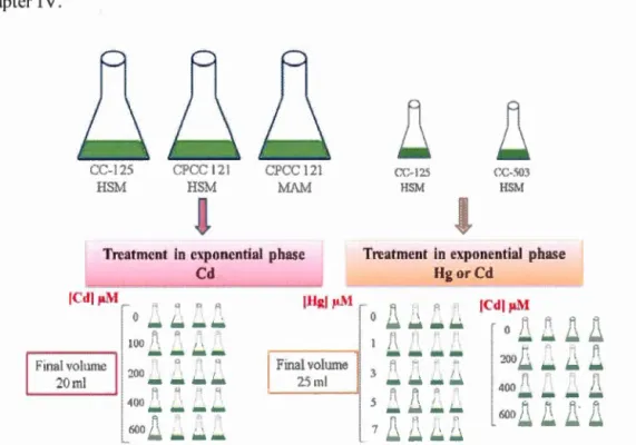
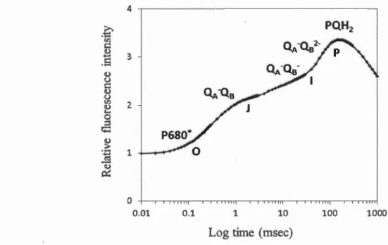
Komberg, 1995; Komberg et al., 1999) ... , ... 27 Figure 3.1 Cultivation and maintenance of Chlamydomonas strains ... 34 Figure 3.2 Standard (A) and growth (B) curves related to Chlamydomonas strains .. 36 Figure 3.3 Experimental treatment of algal cells with Cd or Hg (II) ... 37 Figure 3.4 Rapid chlorophyll a fluorescence induction on logarithmic time scale showing the transient 0-J-1-P; (QA) and (Qs) represent the primary and secondary acceptors of PSU respective! y , (P680*), excited electronic state of P680, (POH2), reduced plastoquinone (measured in the laboratory with Handy-PEA, Hansatech Ltd, Norfolk, UK, C. reinhardtii in HSM medium ... .44
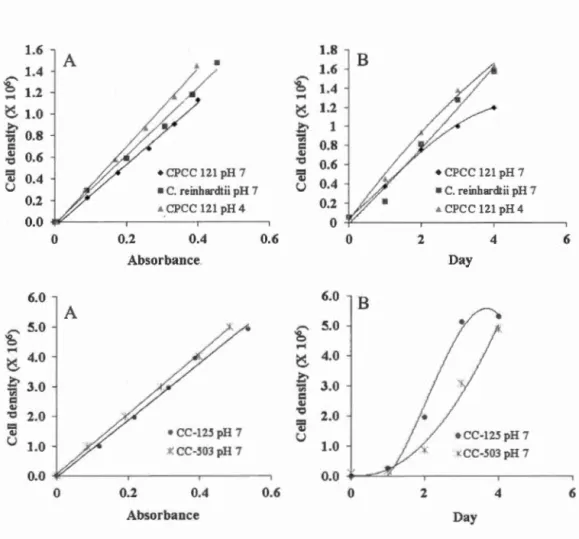
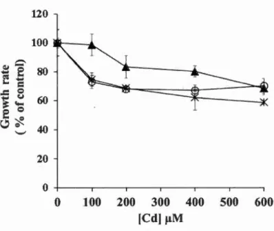
Figure 4.1 Change in the growth rate(% of control) under different concentrations of Cd (~-tM) and pH condition for a period of 48 h. Legend: x, C. reinhardtii at pH 7; o, CPCC 121 at pH 7; ~, CPCC 121 at pH 4. At pH 7, both control C. reinhardtii and CPCC 121 had a growth rate of 0.7 cell per day, and at pH 4, CPCC 121 had a growth rate of 0.6 cell per day. Each data indicated the average and coefficient of variation (CV %) of 4 replicates. Differences between treated-samples and control were ali significant at the leve! of0.05 (p < 0.05) with the exception ofCPCC 121 at pH 4 under 100 ~-tM of Cd versus control. ... 53
Figure 4.2 Accumulation of Cd in Chlamydomonas reinhardtii and CPCC 121 exposed during 48 h to different concentrations of Cd (~-tM) and two pH conditions. Legend: A, C. reinhardtii at pH 7; B, CPCC 121 at pH 7; C, CPCC 121 at pH 4. Each data indicated the average and standard deviation of 4 replicates. Significant differences relative to the control at the leve! of 0.05 (p < 0.05) were indicated by asterisk (*) ... 54
Figure 4.3 Change in the relative cell size (% of control) evaluated by the forward light scattered (FSC) of C. reinhardtii (A) and CPCC 121 (B) exposed at pH 7 during 48 h to different concentrations of Cd (~-tM). Each data indicated the average and coefficient of variation (CV%) of 8 replicates. Significant differences relative to the control at the leve! of 0.05 (p < 0.05) were indicated by asterisk (*) . ... 56
Figure 4.4 Change in the cell granularity (% of control) evaluated by the side light scattered (SSC) of C. reinhardtii at pH 7 (A), and CPCC 121 at pH 4 (B) exposed during 48 h to different concentrations of Cd (~-tM). Each data indicated the average and coefficient of variation (CV %) of 8 replicates. Significant differences relative to the control at the leve! of 0.05 (p < 0.05) were indicated by asterisk (*) . ... 58 Figure 4.5 Change in esterases enzymatic activity (%of control) of C. reinhardtii and CPCC 121 at pH 7 (A, B) and CPCC 121 at pH 4 (C) exposed during 48 h to different concentrations of Cd (~-tM). Each data indicated the average and coefficient of variation (CV %) of 8 replicates. Data showed no significant differences at the leve! of 0.05 (p < 0.05) between treated-samples and control. .. 60
Figure 4.6 Chl a fluorescence kinetics of Chlamydomonas strains exposed at pH 7 and 4 during 24 h to different concentrations of Cd in !J.M. Data represents the average of 4 replicates. Solid and dashed !ines represent respectively the fluorescence intensity of C. reinhardtii at pH 7 and CPCC 121 at pH 7 and 4 .... 61 Figure 4.7 Change in the performance index of Photosystem II activity (PI, % of
and pH condition. Legend: x, C. reinhardtii at pH 7; o, CPCC 121 at pH 7; .Â., CPCC 121 at pH 4. Each data indicated the average and coefficient of variation (CV %) of 4 replicates. Statistical analysis indicated that all treated samples (200-600 ~-tM of Cd) were significantly different to their respective control at the leve! of 0.05 (p < 0.05) ... 63
Figure 4.8 TEM images of algal cell ultrastructure for Chlamydomonas reinhardtii and CPCC 121. Control samples (a), and 24 h Cd-treated (600 ~-tM) samples (b). 64
Figure 4.9 Energy dispersive X-ray spectroscopy of localized intracellular Cd in the cytoplasm for 24 h Cd-treated (600 ~-tM) C. reinhardtii at pH 7 (a), 24 h Cd-treated (600 ~-tM) CPCC 121 at pH 7 (b), and 24 h Cd-treated (600 ~-tM) CPCC 121 at pH 4 (c). The EDX spectrum was obtained by collecting X-ray signais from the selected area of the sample, when it was radiated by the focused e-beam. Legend: B, selected area in algal cell of C. reinhardtii at pH 7; C, selected area in algal cell of CPCC 121 at pH 7; and A, selected area in al gal cell of CPCC 121 at pH 4 .... 65
Figure 4.10 EDX spectrum of Cd distribution within the cell wall ofCPCC 121. The electron probe was focused on (A) representing a selected area on the cell wall of CPCC 121 treated during 24 h to 600 ~-tM of Cd at pH 7 ... 66
Figure 4.11 Scheme representing algal cells of Chlamydomonas reinhardtii and CPCC 121 at 24 h of exposure to 600 ~-tM of Cd under two pH conditions. C, chloroplast; N, nucleus; P, pyrenoid. The stars illustra te the toxicity impact of Cd2+ in algal cells. As indicated by the results (see subsection 4.1.3), the toxicity impact is related to the accumulation of Cd2+ in the cells ... 68
Figure 5.1 Change in the cell density for both strains of Chlamydomonas exposed during 24-72 h to 200-600 ~-tM of Cd. Differences between the control and ali treated-samples were significant for p < 0.05 during 24-72 h ... 70
Figure· 5.2 Fluorescence emission of extracytoplasmic polyP-DAPI from cells of CC-125 (A) and CC-503 (B), deterrnined by confocal microscopy imaging. Green and red fluorescence from cells represent respectively polyP and chlorophylls in confocal microscopie images (scale bar of 10 ~-tm) for CC-125 (a1 and a2) and CC-503 (b1 and b2) ... 71
Figure 5.3 Fluorescence intensity of the extracytoplasmic polyP levet (% of control) for both Chlamydomonas strains exposed during 24-72 h to 200-600 ~-tM of Cd. Data represents the average of 10 cells captured for each microscopie image, and the bars indicate the coefficient of variation (CV %). The asterisk (*) indicates significant differences between treatments and the control condition (p < 0.05) .. 72
Figure 5.4 Confocal microscopie images of Chlamydomonas cells (scale bar of 10 11m) exposed during 72 h to 200-600 f.!M of Cd. These images show the green fluorescence of extracytoplasmic polyP-DAPI and the red· fluorescence of chlorophylls (from the chloroplast) ... 73
Figure 5.5 Image magnification ofCC-125 cells from confocal microscopy (scale bar of 10 11m). At the left, an individual control ce li. At the right, palmelloid colonies of 4 cells, under 400 11M of Cd during 72 h. The green fluorescence is emitted from the extracytoplasmic polyP-DAPI, and the red fluorescence by the Chlorophylls ... 74
Figure 5.6 Confocal microscopie images of CC-125 cells (scale bar of 10 f.!m) treated during 24 h to 0 and 200 11M of Cd. The formation of palmelloid colonies was noticed for Cd-treated algal cells. The green fluorescence: Extracytoplasmic polyP-DAPI; and the red fluorescence: Chlorophylls (from the chloroplast) ... 75
Figure 5.7 Confocal microscopie images ofCC-125 cells (scale bar of 10 11m) treated during 48 h to 0, 200 and 400 f..!M of Cd. For Cd-treated cells, microscopie images show cl earl y the formation of palmelloid colonies within a common cell wall. The green and red fluorescence indicate respectively the extracytoplasmic polyP-DAPI and chlorophylls ... 76
Figure 5.8 Accumulation of Cd in algal biomass of both Chlamydomonas strains exposed during 24-72 h to 200-600 11M of Cd. Ali treated-conditions showed significant differences to the control for p < 0.05 ... 77
Figure 5.9 The level of extracytoplasmic polyP (% of control) in relation to the accumulation of Cd for CC-125 and CC-503 exposed during 24-72 h to Cd concentrations (200-600 f.!M) ... 78
Figure 5.10 The level of extracytoplasmic polyP (% of control) in relation to the maximal PSU quantum yield (Fv/FM) for CC-125 and CC-503 exposed during 24-72 h to Cd concentrations (200-600 f.!M) ... 80
Figure 6.1 Change in the cell density for both strains CC-125 and CC-503 exposed during 24-72 h to different concentrations of Hg ( 1-7 f.!M). Differences between treated-samples and control were ali significant (p < 0.05), except for CC-503 exposed to 1 f.!M of Hg during 48-72 h ... 82
Figure 6.2 Fluorescence emission of polyP-DAPI in cells of CC-125 determined by confocal microscopy imaging. A, a, and az, non-treated cells; B, b, and bz, 24 h treated-cells to 5 11M of Hg. In confocal microscopie images, green and red fluorescence represent respectively polyP and chlorophylls (scale bar of 10 f.!m).84
Figure 6.3 Fluorescence emission of polyP-DAPI in cells of CC-503 determined by confocal microscopy imaging. A, a, and a2, non-treated cells; B, b, and b2, 24 h treated-cells to 5 J..I.M of Hg. In confocal microscopie images, green and red
fluorescence represent respectively polyP and chlorophylls (scale bar of 10 J..lm).85
Figure 6.4 Change in the polyP leve! of the cell wall (% of control) for CC-125 and CC-503 exposed during 24-72 h to 1 and 3 J..I.M of Hg. lt was not possible to detect
the fluorescence intensity of extracytoplasmic polyP-DAPI for treated-cells under
5 and 7 J..I.M of Hg. *significant differences relative to the control for p < 0.05 .... 86 Figure 6.5 Change in the fluorescence intensity of the polyP leve! in relation to the
change of the cell density, for CC-125 and CC-503 exposed du ring 24-72 h to
different concentrations of Hg (1-5 J..I.M) ... 87 Figure 6.6 Accumulation of Hg in al gal biomass (J..lg of Hg 1 mg of dry mass) for
CC-125 and CC-503 exposed during 24-72 h to 5 and 7 J..I.M of Hg. Data represents the average of 4 replicates, and the bars indicate the standard deviation. The accumulation of Hg in algal biomass for all treated conditions was significantly different to the control for p < 0.05. For treatments of 1 and 3 J..I.M of Hg, the accumulation of Hg in algal biomass was described in the results section ... 88 Figure 6.7 Change in the fluorescence intensity of the polyP leve! in relation to the accumulation of Hg in algal biomass, for CC-125 and CC-503 exposed during 24
-72 h to different concentrations of Hg (1-5 J..I.M). The results conceming the
treatment condition of 7 J..I.M of Hg were similar to the 5 J..I.M of Hg ... 89 Figure 6.8 Scheme representing the degradation of extracytoplasmic polyP, its
participation to sequester Hg, and the release of the Hg-phosphate complex out of
Table 3.1 Linear equations for each culture grown in used medium, where Y and X represent the cell density and the absorbance (750 nm) of cultures, respectively. Linear regression was started at Y = 0 and X = 0 when there was no cells in medium ... 35
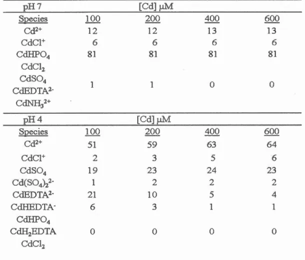
Table 4.1 Proportion of Cd species (%) in culture medium at pH 7 and 4 determined by the chemical equilibrium calculation (Visual MINTEQ 2.61) ... 51
Table 5.1 Change in the growth rate (in cell d-1) for both Chlamydomonas strains
exposed during 72 h to different concentrations of Cd ... 70
Table 5.2 Significant differences between treatments for accumulation of Cd in algal biomass of both Chlamydomonas strains for each strain at each time of exposure (p < 0.05) ... 78
Table 6.1 Change in the growth rate (cell d-1) for both strains CC-125 and CC-503 exposed during 72 h to different concentrations of Hg ... 81
Table 6.2 Change in the ratio of Fv/FM (%of control) and the fluorescence intensity of extracytoplasmic polyP (% of control), when CC-125 was exposed during 24-72 h to different concentrations of Hg. N.D., not determined due to high mortality. * significantly different to the control (p < 0.05) ... 90 Table 6.3 Change in the ratio of Fv/FM (% of control) and the fluorescence intensity of extracytoplasmic polyP (%of control), when CC-503 was exposed during 24-72 h to different concentrations of Hg. N.D., not determined due to high mortality. *significantly different to the control (p < 0.05) ... 91
AAS Atomic Absorption Spectrometry
ADP Adenosine Diphosphate
AEM Analytical Electron Microscopy
AMP Adenosine Monophosphate
APX Ascorbate Peroxidase
ATP Adenosine Triphosphate
CAT Catalases
CCD Charge-Coupled Deviee
Chi Chlorophyll
Chi* Excited Chlorophyll .
Cys Cysteine
DAPI 4' ,6-diamidino-2-phenylindole
ECso Half Maximal Effective Concentration
EDTA Ethylene Diamine Tetra Acetic Acid FAAS Flame Atomic Absorption Spectrometry
Fd FDA FEG FFP FNR FSC Fv Glu y-Glu-Cys y-Glu-Cys synt Gly GSH G synt HMW HSM ICP Ferredoxin Fluorescein Diacetate Field-Emission Gun Frorit-Focal Plane
Maximal Fluorescence Intensity
F erredoxin-NADP Reductase
Forward-Angle Light Scatter
Variable Fluorescence Intensity
Glutamine
Gamma Glutamylcysteine
Gamma Glutamylcysteine Synthetase
Glycine Glutathione
Glutathione Synthetase
5-(and-6)-Carboxy-2 , 7 -Dihydrodifluorofluorescein Di acetate
High Molecular Weight High Salt Medium
LHCI Light-Harvesting Complexes of Photosystems 1 LHCII Light-Harvesting Complexes ofPhotosystems II
LMW Low Molecular Weight
LSCM Laser Scanning Confocal Microscopy
Mn+ Metal Ions
MAM Modified Acid Medium
MOPS 3-[N-Morpholino] Propanesulfonic Acid
NADP Nicotinamide-Adenine Dinucleotide Phosphate
NMR Nuclear Magnetic Resonance
OD6oo Optical Density at 600 nrn
OES Optical Emission Spectrometry
PAH Polycyclic Aromatic Hydrocarbons
PCs Phytochelatins
PC Plastocyanin
PCs Phytochelatin Synthase
PC synt Phytochelatin Synthetase
PEA Plant Efficiency Analysis
Pi Ortho phosphate
PI Performance Index ofPSII Activity
Pit Inorganic Phosphate Transport System
PMT Photomultiplier Tube 31P-NMR Phosphorus-31 NMR PolyP Polyphosphate PPi Pyrophosphate PPK Polyphosphate Kinase PPX Exopolyphosphatase PSI Photosystem 1 PSII Photosystem II PQ Plastoquinone PQH2 Reduced Plastoquinone
RCDR Relative Cell Division Rate
ROS Reactive Oxygen Species
SOD Superoxide Dismutase
ssc
Side-Angle Light ScatterTAP TEM
Q
A
Qs XEDS X VIl Tris-Acetate-Phosphate MediumTransmission Electron Microscopy
Quinone A
Quinone B
o
c
Degree Celsiusd Day
eV Electron Volt
h Hour
M Mole per Liter
mM Millimole per Liter
!lM Micromole per Liter
llg Micro gram mg Milligram m2 Meter Square Il ffi Micrometer nm Nanometer L Liter mL Milliliter mm Minute
% PPM
rpm
s
Percentage
Parts per Million
Revolutions per Minute
Second
1
1Le cadmium et le mercure sont des contaminants dangereux pour l'être humain et les écosystèmes aquatiques, et leur rejet d'origine anthropique dans les eaux et rivières continuent malheureusement. En particulier, du point de vue du milieu aquatique, la forme ionique du Cd peut être biodisponible pour les organismes aquatiques, et la transformation du Hg2+ sous sa forme organique toxique, le méthylmercure peut être bioaccumulé et sa dose bioamplifiée le long de la chaîne alimentaire aquatique. Dans ce projet de recherche, l'algue verte Chlamydomonas a été utilisée comme un organisme modèle pour investiguer l'accumulation et la séquestration du cadmium et du mercure dans les cellules algales, leurs toxicités cellulaires et moléculaires, et les mécanismes de protection impliqués dans les cellules. Par conséquent, ce projet de recherche est structuré en trois études en adressant chacune un objectif spécifique. Dans la première étude, la capacité d'accumulation du cadmium a été étudiée chez deux souches d'algues, la souche sauvage Chlamydomonas reinhardtii et la souche acido-tolérante Chlamydomonas CPCC 121 exposées durant 48 h aux différentes concentrations de Cd (1 00 à 600 !J.M) sous deux conditions de pH ( 4 et 7). La souche C. reinhardtii a montré une accumulation plus élevée de Cd par rapport à la souche CPCC 121 en induisant un impact toxique plus fort. En comparaison, Chlamydomonas CPCC 121 a montré une tolérance pour le Cd qui était corrélée avec la diminution de l'accumulation intracellulaire de Cd. Cependant, la capacité d'accumulation de cadmium était la plus faible chez CPCC 121 à pH 4, limitant son utilisation pour les applications de biorestauration. Dans la seconde étude, l'effet de CdCh a été étudié sur le niveau de polyphosphate extracytoplasmique (polyP) en utilisant les souches CC-125 et CC-503 ayant une paroi cellulaire déficiente. Les souches de C. reinhardtii ont été exposées pendant 24-72 h aux différentes concentrations de Cd (200-600 !J.M). Les résultats ont montré la diminution de la synthèse ou la dégradation de polyP extracytoplasmique qui était corrélée avec l'accumulation de Cd, même chez la souche CC-503 avec un niveau inférieur de poly.P. En plus, le niveau du polyP a été.diminué en relation avec la diminution du ratio F viF M pour les deux souches pendant 48 h. En comparaison avec CC-125, le niveau de polyP n'a pas été récupéré après 72 h chez CC-503.
Cependant, les deux souches d'algues ont accumulé une quantité considérable de Cd dans leur biomasse et le polyP n'a pas participé à la séquestration de Cd. Par conséquent, cette étude a montré l'effet du Cd sur le niveau de polyP qui était dépendant de la concentration de Cd, tandis que le Cd a inhibé le taux de la croissance cellulaire indépendamment de la concentration de Cd. En utilisant C.
des métaux, tels que le Cd. Dans la troisième étude, l'accumulation et l'effet de la toxicité du mercure a été déterminé chez deux souches de C. reinhardtii, CC-125 et CC-503 ayant une paroi cellulaire déficiente, pendant 24-72 h d'exposition à 1-7 ~-tM de Hg. Le niveau de polyP extracytoplasmique a été déterminé afin de comprendre le rôle physiologique de polyP dans les cellules algales traitées au Hg. Lorsque les cellules d'algues ont été exposées à 1 et 3 ~-tM de Hg, l'accumulation du Hg était corrélée avec la dégradation de polyP pour les deux souches. Ces résultats suggèrent que la dégradation de polyP a participé dans la séquestration de Hg, même chez CC-503 avec un niveau inférieur de polyP. Ce mécanisme pourrait expliquer à 72 h la récupération du niveau de polyP, la performance de la photochimie du PSII, la faible inhibition du taux de la croissance cellulaire, et la faible accumulation de Hg dans la biomasse. Nos résultats montrent que le changement du niveau de polyP est corrélé avec 1' accumulation et 1' effet du mercure sur les cellules d'algues, permettant son utilisation comme un biomarqueur de la toxicité du Hg.
Mots clés : Cadmium; Mercure; Accumulation; Toxicité; Tolérance; Polyphosphate; polyP extracytoplasmique; Algue verte; Chlamydomonas reinhardtii; Chlamydomonas CPCC 121.
Cadmium and mercury represent dangerous contaminants for both aquatic ecosystems and human health, and their release in waters and rivers continue unfortunately from anthropogenic activities. Particularly, from the point of view of aquatic environment, the ionie form of Cd can be bioavailable to aquatic organisms, and the transformation of Hg2
+ into high toxic organic form, methylmercury, can be bioaccumulated and its dose bioamplified through the aquatic food chain. In this research project, the use of green alga Chlamydomonas was used as a mode! organism to investi gate the accumulation and the sequestration of Cd and Hg in al gal cells, their toxicity effects at cellular and molecular levels, and the protection mechanisms involved in algal cells. Therefore, this research project is divided into three studies addressing each a specifie objective. In the first study, the capacity of cadmium accumulation was investigated for two algal strains, Chlamydomonas reinhardtii and the acid-tolerant strain CPCC 121, during 48 h under 100-600 ~-tM of Cd and two pH conditions ( 4 and 7). C. reinhardtii demonstrated a higher accumulation of Cd compared to the strain CPCC 121, inducing a stronger cellular toxic impact. In comparison, Chlamydomonas CPCC 121 demonstrated a tolerance for Cd, which was correlated with the decrease of intracellular Cd accumulation. However, the capacity of cadmium accumulation was lower in CPCC 121 at pH 4 than pH 7, limiting its use for bioremediation applications. In the second study, the effect of CdCh on the leve! of extracytoplasmic polyphosphate (polyP) was investigated using CC-125 and the cell wall-deficient CC-503. Two strains of C.
reinhardtii were exposed under neutra! pH during 24-72 h to different concentrations
of Cd (200-600 ~-tM). The results demonstrated the decrease in synthesis and/or the degradation of extracytoplasmic polyP, which was correlated with the accumulation of Cd, even in CC-503 having a lower lev el of polyP. Furthermore, the lev el of polyP decreased in relation to the decrease of Fv/FM value for both strains during 48 h. In comparison with CC-125, the leve! of polyP could not be recovered at 72 h for
CC-503. Nevertheless, both algal strains were able to accumulate significant amount of
Cd in their biomass, and the polyP did not participate in the sequestration of Cd. Therefore, this study demonstrated the effect of Cd on the polyP level, which was dependent on the concentration of Cd, whereas Cd inhibited the growth rate regardless of Cd concentration. By using C. reinhardtii, the polyP leve! can be used as a biomarker of Cd toxicity. In the third study, the accumulation and toxicity effect of mercury was determined on two strains of C. reinhardtii, CC-125 and CC-503 as a cell wall-deficient strain, during 24-72 h of exposure to 1-7 ~-tM of Hg. The leve! of extracytoplasmic polyP was determined to understand the polyP physiological role in
Hg-treated cells. When the algal cells were exposed to 1 and 3 ~-tM of Hg, the
accumulation of Hg was correlated with the degradation of polyP for both strains.
These results suggested that the degradation of polyP participated in the sequestration
of Hg even in CC-503 with a lower level of pol y P. This mechanism might explain at
72 h the recovery of the polyP leve!, the efficiency of maximum PSII quantum yield, the low inhibition of growth rate, and the low accumulated Hg in algal biomass. Our results demonstrated that the change of polyP leve! was correlated with the
accumulation and effect of Hg on al gal cells, which can be used as a bio marker of Hg
toxicity.
Keywords: Cadmium; Mercury; Accumulation; Toxicity; Tolerance; Polyphosphate;
Extracytoplasmic polyP; Green algae; Chlamydomonas reinhardtii; Chlamydomonas
Having access to safe drinking water is one of the most important needs for humans society, considering that more than a third of the people in the world have been affected by unsafe drinking water (Schwarzenbach et al., 201 0). Aquatic environments have been subjected to the contamination by organic and inorganic chemicals since the beginning and the development of the industrial age (Adriano et al., 2005). From the environmental point of view, inorganic pollutants (for example: Cr, Ni, Cu, Zn, Cd, Pb, Hg) and metalloids (e.g., Se, As), are not degraded like organic pollutants, and their transport and bioavailability can be influenced by oxidation/reduction, complexation, adsorption, and precipitation/dissolution reactions (Schwarzenbach et al., 201 0). Among the metals mentioned above, sorne are considered as essential micronutrients, such as copper and zinc, while others including mercury, cadmium, and lead are not essential for biological activities to regulate physiological processes in plants (Clemens, 2001).
In addition, humans can be exposed to pollutants including toxic metals v1a contaminated water and the food chain by the consumption of vegetables high in Cd and seafood high in Hg (Adriano et al., 2005; Schwarzenbach et al., 201 0). Serious health disorders have been reported following public exposure to different metals. For example, mercury is considered as a neurotoxic material that can damage DNA and the kidneys (Tchounwou et al., 2003). Cadmium can also cause kidney and bone damage, and the increase of cancer risks has been reported even at low leve! of environmentally exposed populations (Jarup et Akesson, 2009).
Industrial and municipal wastewaters can contain many toxic metals that must be removed before discharge or reuse. Physicochemical methods, such as ion exchange, electrolysis, chemical precipitation, membrane filtration and adsorption have been typically used to decontaminate wastewaters (Fu et Wang, 2011). However, these methods can be ineffective or expensive for treating large volumes of water, since they consume high amount of chemicals and energy, making these conventional approaches less practical for low metal concentrations. Recently, the use of microalgae in wastewater bioremediation started to be a promising low-cost alternative to conventional methods. Indeed, it is weil known that microalgae have a high capacity for metal uptake via channel-mediated transport due to their unicellular morphology that provide a large contact surface area to aquatic pollutants (Monteiro
et al., 2012). Moreover, microalgae are able to use inorganic nitrogen and phosphorus
for growth, and the produced biomass can be used for various applications including the production of biogas such as methane from anaerobie digestion (Munoz et al., 2005).
Since one must consider that algal strains differ in their capacity to uptake metals, in-depth studies of metal uptake and sequestration on different algal strains are necessary for providing the knowledge in the development of wastewater bioremediation strategies. Furthermore, the selection of algal strains must consider the capacity of cells to tolerate intracellular metals, since deleterious effects may limit cellular accumulation and sequestration capacity. For the study of cellular functions related to metal accumulation and tolerance, strains of Chlamydomonas represent appropriate unicellular mode! organisms due to rapid life cycle and their widespread use in cellular physiological studies (Hanikenne, 2003; Mendez-Alvarez et al., 1999). Previously, the toxicity of Cd has been reported on Chlamydomonas strains, and deleterious effects were indicated by the inhibition of growth (Jamers et al., 2009; Prasad et al., 1998), ultrastructural cellular changes such as cytoplasmic vacuolization, starch accumulation and cytoplasmic electron-dense granules
(Aguilera et Amils, 2005; Nishikawa et al., 2003), the development of membranous organelles (Visviki et Rachlin, 1994), the increase of the relative cell size (Jamers et al., 2009; Visviki et Rachlin, 1994), and the inhibition of the photosynthetic activity (Falier et al., 2005; Perreault et al., 2011; Vega et al., 2006).
Since microalgae are prîmary producers in aquatic ecosystems, they are widely
used in laboratory bioassays for analyzing metal toxicity mechanisms and effects. lt
was found by Juneau et al. (2001) that Microcystis aeruginosa was the most sensitive
species among six algal species to mercury since they observed a fast inhibitory effect
of 37 nM Hg (HgCh) on the photosynthetic (PSII-PSI) activity only after 5 h (Juneau
et al., 2001). In another study, the maximum quantum efficiency of PSII photochemistry (Fv/FM) and PSII operating efficiency (f..F/Fm') were decreased in Chlamydomonas reinhardtii under treatment with MeHg at concentrations above 1 ~-tM. However, this inhibitory effect was not observed under treatment with 5 ~-tM
HgCh for 5 h on both ratios (Kukarskikh et al., 2003 ). Moreover, previous studies on
metal toxicity in microalgae showed the accumulation of metals (Cd2+, Cu2+ and Zn2+) in the cell wall (Macfie et al., 1994), the alteration of cellular ultrastructures
(Jamers et al., 2009; Visviki et Rachlin, 1994), and the increase in number and
volume of vacuoles containing metallic electron-dense deposits (Aguilera et Amils,
2005; Nishikawa et al., 2003). Others investigations emphasized on understanding metal tolerance mechanisms in microalgae. A previous study found that intracellular
Hg2+ was able to activate antioxidant enzymes (superoxide dismutase, catalase, and
ascorbate peroxidase) for the elimination of generated reactive oxygen species (Elbaz
et al., 201 0). Others studies demonstrated that the uptake of metals (Cu2+, Hg2+, Ag2+,
and Cd2+) in microalgae increased significantly the synthesis of chelating compounds
such as cysteine, glutathione, and phytochelatins permitting to regulate the
intracellular concentrations of rn etals (Howe et Merchant, 1992; La voie et al., 2009;
Perales-Vela et al., 2006; Stoiber et al., 2010). Another tolerance mechanism was
of Cd (20 f.!M) on the metabolism of polyphosphate (polyP) was investigated on
Chlamydomonas acidophila (KT-1) by in vivo 31P-nuclear magnetic resonance and by
energy-dispersive X-ray analysis (Nishikawa et al., 2003). Under Cd treatment for 3 days, the authors showed a complete degradation of polyP which was related to a strong increase of phosphate and the accumulation of Cd in vacuoles. They suggested that the vacuolar compartmentalization of Cd was as a cellular detoxification mechanism. In another study on C. acidophila KT -1, the same research group presented a decrease of 43 % in accumulated Cd, when Cd-treated algal cells were transferred into a Cd-free medium (Nishikawa et al., 2009). Therefore, they proposed that the polyP metabolism in C. acidophila IÇT -1 contributed to the cellular tolerance for Cd, by chelating Cd in the vacuole as a Cd-phosphate complex and releasing it out of the cell.
The main objective of this research project was to investigate the accumulation and the sequestration of cadmium and mercury in different algal strains of
Chlamydomonas. Therefore, this research project is presented in this thesis as three
specifie studies:
In the first study, the specifie objective was to investigate the effect of Cd accumulation on green algae Chlamydomonas reinhardtii and Chlamydomonas CPCC 121. Both algal strains were exposed during 48 h to different concentrations of Cd (1 00-600 f.!M). Since acidic waters contain higher amounts of dissolved metals, the capacity of Cd accumulation and cellular tolerance of CPCC 121 was determined under two different pH conditions ( 4 and 7). Indeed, the tolerance ability for metals ofCPCC 121 has been poorly studied. Under these experimental conditions, changes in cellular and biochemical parameters indicating the growth rate, cellular morphology, activity level of esterases, and PSII activity performance were used as indicators of Cd toxicity. The accumulation of Cd was confirmed by TEM images and energy-dispersive X-ray spectroscopy (EDX). Therefore, advantages and
limitations of both Chlamydomonas strains were discussed in the perspective of Cd bioremediation. The results are presented and discussed in Chapter IV.
In the second study, the specifie objective was to determine the accumulation and toxicity effect of Cd Ch on the level of extracytoplasmic polyphosphate (polyP). Two strains of Chlamydomonas reinhardtii, CC-125 and CC-503 that has an impaired cell wall, were exposed under neutra! pH during 24-72 h to different concentrations of Cd (200-600 !J.M). Under these conditions, the level of extracytoplasmic polyP was investigàted in relation to the presence or the deficiency of the cell wall barrier. In addition, the monitoring of polyP levet as a biomarker of Cd toxicity using alga C. reinhardtii was discussed. Therefore, our results contributed to understand the involvement of extracytoplasmic polyP on algal cells of C. reinhardtii under Cd stress effect at neutra! pH. The results are presented and discussed in Chapter V.
In the third study, the specifie objective was to investigate the accumulation and toxicity effect of HgCh on two strains of Chlamydomonas reinhardtii, CC-125 and CC-503 as a cell wall-deficient mutant, to evaluate the importance of the cell wall as a protective barrier. In this perspective, the level of polyphosphate (polyP) in the cell wall ( extracytoplasmic) was determined to understand the physiological involvement of polyP under Hg stress effect. Therefore, we discussed the role of polyP in the cellular tolerance for Hg and the use of polyP level as a toxicity biomarker of Hg in algal cells. The results are presented and discussed in chapter VI.
ENVIRONMENT AL IMPACT OF METALS AND GREEN ALGAE
1.1 Environmental impact of mercury and cadmium
From the point of view of ecotoxicology and depending on the source of contaminants, environmental contaminants can be divided initially into two major categories: organic (from living organisms) or inorganic (from mineral sources). However this distinction is not precise since for example carbon dioxide is classified as an inorganic gas, but it is produced by living organisms. lt should be noted that,
sorne important environmental organic contaminants were obtained from both natural
and anthropogenic sources, and as a good example polycyclic aromatic hydrocarbons (P AH) can be indicated. In the term of inorganic contaminants, metallic elements are considered as a major class of these contaminants. Metals are natural elements in crustal rocks and soils, as well it can be released into the water, air, and terrestrial
environment by hurnan activities from industrial manufacturing, mining, combustion
products, and agricultural pesticides (Newman et Unger, 2002).
Mercury (Hg) is one the most hazardous metals that is known as a global aquatic contaminants, and its distribution continues through the global cycle of mercury and anthropogenic sources in waters and rivers. In the global cycle of mercury, 99 % of mercury is in the form of mercury vapor, which is considerable. Mercury vapor is a
chemically stable monatomic gas, so its residence time in the atrnosphere is estimated to be of a year or more. Therefore, it is obvious that the global cycle of mercury begins with its evaporation from land and water surfaces (sea and rivers). However, its oxidation via the action of ozone leads to its conversion into water-soluble ionie mercury (Hg2+) that can be returned on the surface of earth as rain water. Hurnan can be exposed to mercury via the rnethylation of divalent inorganic mercury (Hg2+) by organisms (bacteria and phytoplankton) and its accumulation in the aquatic food chain (from plankton and algae to fish and then to hurnan). On the other hand, the atmosphere contains Jess than 1 % of methylrnercury compounds that their origin is not yet known and these compounds can be taken by the aquatic biota. The reduction of Hg2+ by aquatic microorganisrns leads to the formation of Hg0 in surface waters (Clarkson, 1998).
The rnethylation of inorganic mercury (Hg2+ as the main species) occurs in waters and sediments, which needs the presence of a sui table methyl donor molecule such as biotic or abiotic materials (Ullrich et al., 2001). However, many studies suggested a high potential of rnicrobial methylation in sediment sites under anaerobie conditions and sulfate-reducing bacteria as the principal methylators (Compeau et Bartha, 1985). In aquatic environrnent, the biotic methylation of inorganic mercury happens in microorganisms (aerobes and anaerobes) after the uptake of the soluble mercury (Uilrich et al., 2001). Abiotic methylation is possible in the presence of chemical methylating agents such as methylcobalarnin and methyltin compounds, and hurnic matter released to the environrnent by biotic processes. Although the origin of these compounds cornes from biotic processes, the abiotic terrn refers to any non-enzyrnatic methylation (Weber, 1993).
Methylmercury is the most abundant organomercury compound that exists m freshwaters (lakes and ri vers) and can be generated from Hg2+ by different mechanisms (Ullrich et al., 2001 ). Although the hydrolysis reaction of
methylmercury is thermodynamically feasible, kinetically it is not realizable, so it can be stable in an aqueous solution. It should be noted that, however, methylmercury can be degraded photochemically by microbial action. The Hg0 and dimethylmercury could not be simply accumulated by the aquatic biota because they are not reactive with cellular components, while the Hg2+ and methylmercury are able to be accumulated in the aquatic food chain. However, the bioaccumulation of methylmercury is more possible than inorganic mercury in the aquatic biota. One reason that can explain the difference between the bioaccumulation of inorganic (HgCb) and organic mercury (CH3HgCl) is the lipid solubility of CH3HgCl that permits to maintain it in the fatty tissues of animais (Morel et al., 1998). In fish, however, the study of Boudou and Ribeyre (1997) showed that methylmercury liposolubility could not be the only reason for more bioaccumulation of methylmercury in fish muscle. In fact, the high absorption of MeHg by the intestine wall was more important since inorganic mercury was adsorbed at the microvilli interface (Boudou et Ribeyre, 1997).
Besicles natural sources of mercury, human activities such as combustion of fossil fuels ( coal) and municipal waste incineration, metal smelting, mining and industrial waste discharges of mercury led to add the large quantities of mercury to the environment. Organic forms of mercury including methyl, ethyl, and phenyl types have been used as biocides, pesticides, and household antiseptics which can explain their presence into the industrial environment. Therefore, the wide mercury distribution in waters and ri vers is clear (Tchounwou et al., 2003). From the point of view of human exposure to mercury, two forms of mercury are more important, dental amalgam fillings and methylmercury compounds (Clarkson, 1998). Indeed, the use of mercury amalgam tooth fillings is very common since nineteenth century. Therefore, mercury vapor can be inhaled during chewing (Clarkson, 2002), while there is no serious restriction against using these compounds. Methylmercury compounds can be accumulated in fish tissues and its dose can be bioamplified
through the aquatic food chain (Boudou et Ribeyre, 1997; Ullrich et al., 2001).
Indeed, these compounds are considered toxic towards public health, causing brain,
kidneys and immune system damages (Bernard, 2011; Tchounwou et al., 2003).
Today, toxic effects of cadmium towards human, plants, and animais are
well-known. Cadmium can be released in atmospheric, aquatic and terrestrial environment
from natural and anthropogenic sources. Volcanoes, forest fires, and airborne soil
particles are the major natural sources of cadmium which can be transferred in
atmosphere by aerosol, and deposited in soils, aquatic systems, and sediments. Moreover, cadmium exists naturally in many minerais, rocks and soils in low
concentrations. Water soluble cadmium compow1ds can be mobile and bioavailable
for living cells in water and soils depending on their chemistry. The major anthropic
sources of Cd released to aquatic environment are non-ferrous metal smelting and
refining, manufacturing of ~hemicals and metals, mining, waste incineration, and
domestic wastewater. Nickel-cadmium batteries were considered as a major source of
cadmium disposed with municipal wastes (UNEP, 201 0).
Non-smoking human populations are exposed to cadmium by multiple sources
predominately from food (90 %). In general, all foods contain cadmium with different
concentrations depending on the level of contaminated areas and the tYpe of food. Cadmium can be found more in vegetables than other types of food including meat, egg, and milk (Jarup et Akesson, 2009; UNEP, 201 0). Kidneys are the first target organsin chronic cadmium poisoning (Bernard, 2011). Kidney tubular damage, renal
dysfunction, bone damage, and cancer have been shown as the result of
environmentally exposed populations to low-level concentrations of Cd (Jarup et Akesson, 2009; Satarug et al., 201 0).
Therefore, appropriate strategies and efficient and economie methods should be
applied to remove metals from freshwater aquatic reservoirs and to reduce their
was our interest to develop the knowledge of phycoremediation of contaminated water by metals using different strains of Chlamydomonas. Furthermore, it is important to notice that microalgae are the primary producers in aquatic environment which occupy the first place in the food chain with a high capacity for metal uptake. 1.2 U sing green algae for the phycoremediation of rn etals
Metal removal from aquatic environment has become an important challenge in wastewaters remediation, since population growth and industrialization. Even though various conventional methods including chemical precipitation, ion exchange, adsorption, ultrafiltration, reverse osmosis, electrodialysis, coagulation-flocculation, flotation and electrochemical methods have been used to remove metals from wastewaters, sorne disadvantages have limited their use in metal removal. For example, chemical precipitation is less practical for low metal concentrations and creates large amount of sludge. As weil, ion exchange can be expensive for large volumes of water since it needs the regeneration of resins by chemical reagents, and electrochemical methods consume high amount of electrical energy (Fu et Wang, 2011).
Bioremediation technologies became a promising low-cost and efficient alternative in an eco-friendly manner for contaminant removal from aqueous system. Phycoremediation as a new approach of biological remediation technologies represents the use of algae for the removal or biotransformation of contaminants including metals from polluted environmental matrices (Kumar et al., 2015). Unicellular morphology of microalgae provides a large contact surface area to aquatic pollutants therefore a high capacity for metal uptake (Monteiro et al., 2012). On the other hand, microalgae have shown efficient defense mechanisms to detoxify metal and to survive in metal-containing media (It will be discussed in Section 1.3). Both of these capacities, the uptake and detoxifying process, make them appropriate candidates for biological remediation systems to remove metals. The advantages of
using microalgae in the removal of metals from aquatic environment are a high and rapid capacity to accumulate metals, no toxic sludge generation, the elimination of azote and phosphorous preventing eutrophication, the inexpensive and easy growth using solar light and C02, saving time and energy, the rapid growth rate compared to higher plants, the potential for C02 capture and biofuel production (Kumar et al., 2015; Monteiro et al., 2012; Sivakumar et al., 2012).
Sorne species of Chlamydomonas are able to grow under both conditions: the phototrophic growth by using C02 as a sole carbon source and light energy involving photosynthesis process, and the heterotrophic growth in darkness with acetate as a carbon source. This heterotrophic property permits the isolation of viable mutants which are light-sensitive and unable to perform photosynthesis (Dent et al., 2001; Harris, 2009). The cell structure of C. reinhardtii (wild-type) includes the central nucleus with a prominent nucleolus, a single basal chloroplast surrounding the nucleus, pyrenoid within the chloroplast, and contractile vacuoles. The cell wall is located close to the plasma membrane and depending on the genus can vary in thickness (Harris, 2009). As Figure 1.1 shows most of the cellular volume is occupied by the chloroplast.
Figure 1.1 TEM image of alga1 cell ultrastructure for Chlamydomonas reinhardtii wild-type
Green alga Chlamydomonas is a unicellular eukaryote organism having a rapid
growth rate with a doubling time less than 10 h (Dent et al., 2001 ). Therefore, its manipulation can be performed easily for the study of detoxification mechanisms
associated with metallic stress. Chlamydomonas is an excellent genetic mode! (Dent
et al., 2001 ), so numero us genetically modified strains are available (Joint Genome Institute, JGI, vers 4.0). Chlamydomonas reinhardtii is an organism with capability of growth in the laboratory either in liquid culture or on agar in simple media (Harris,
2009). Cell population in the liquid cultures of Chlamydomonas are almost
homogenous (Dent et al., 2001 ). Therefore, it would be possible to analyse cellular
and biochemical changes induced by metal toxicity. Chlamydomonas species
represent an appropriate unicellular mode! organism for physiological and
toxicological studies (Dent et al., 2001; Hanikenne, 2003; Mendez-Alvarez et al.,
1999). ln addition, acid tolerant Chlamydomonas species were isolated from different
acid environment in Japan, Germany, Canada, and the United States of America.
These species were able to maintain the internai pH to neutra! (Harris, 2009). An acid
resistant Chlamydomonas species, C. acidophila, showed high tolerance to the
exposure of severa! metals in acidic conditions (Nishikawa et Tominaga, 2001;
Perreault et al., 201 0; Spijkerman et al., 2007).
1.3 Using green algae as a mode! of cellular toxicity 1.3.1 Cellular defense mechanisms against metal toxicity
In response to metal toxicity, different resistance mechanisms have been developed by microalgae to allow their survival in media when toxic metals are present. In this subsection, the tolerance mechanisms by microalgae for metals are
explained by the binding of metal ions to the cell wall, the chelation with
phytochelatins (PCs), the sequestration and compartmentalization in the vacuole, the
cell ultrastructural changes, the polyphosphate sequestration and metabolism, and the
The role of the cell wall to protect microalgal cells against taxie metal ions was studied by Macfie and co-workers by using two strains of C reinhardtii. The cell
density of the wild-type strain of C reinhardtii was significantly higher than the
strain having an impaired cell wall when exposed to Cd2+, Cu2+, Co2+, Ni2+, and more
clearly to Co2+ and Cu2+ at pH conditions 5 and 6.8. Therefore, they suggested the
presence of sensitive sites inside or at the surface of the cell wall that can prevent
metal uptake into the cellular cytoplasm through its binding (Mac fie et al., 1994 ).
However, in another study of this research group, the wild-type strain of C.
reinhardtii showed more capability of Cd accumulation per unit weight than the
wall-Jess cells, while bath strains showed a comparable amount of Cu uptake with more
sensitivity of the wall-Jess cells. Thus, they concluded both detoxification
mechanisms, external and internai, of metal ions in C. reinhardtii (Macfie et
Welbourn, 2000). As a consequence, although the microalgal cel! wall acts as the first
barrier to metal ions uptake inside the cells, the cell wall capacity of microalgae for
high metal-binding can be accounted for metal removal from the aquatic
environment.
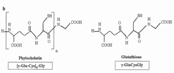
Chelation of metals with phytochelatins (PCs) is considered as an important cellular mechanism for metal detoxification. Phytochelatins consist of three amino
acids, glutamine (Glu), cysteine (Cys), and glycine (Gly), which are characterized by
the repetition of the dipeptide units (y-Glu-Cys) followed by a terminal Gly (Pal et
Rai, 201 0). Indeed, mono mer is provided by tri peptide glutathione (GSH;
y-GluCysGly). The enzyme, phytochelatin synthase (PCs), is a y-glutamylcysteine
dipeptidyl transpeptidase, which catalyzes the transpeptidation of y- Glu-Cys moiety
from glutathione to a second glutathione molecule. This process leads to produce (y-Glu-Cys)2-Gly or an n
+
1 oligomer, (y-Glu-Cys)n-Gly which is the general formula of phytochelatins (Grill et al., 1989) (Figure 1.2). In (y-Glu-Cys)n-Gly, "n" generally(PCs) is constitutively present in various plant species, and the Cd2+ has demonstrated
to be a strong activator of this enzyme (Grill et al., 1989).
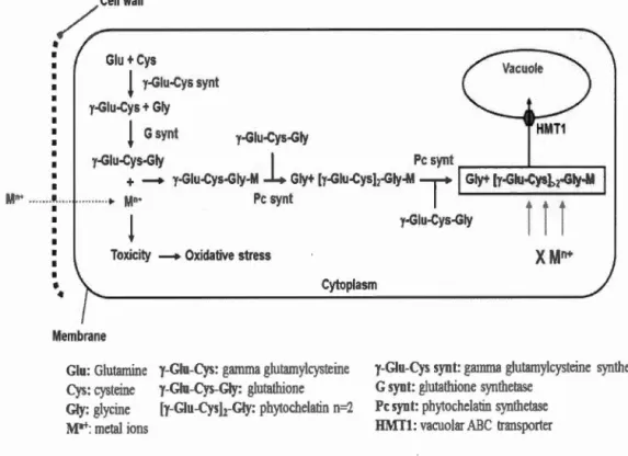
The mechanism of metal detoxification by phytochelatins m microalgae is
summarized in Figure 1.3. At first, in the presence of metal ions such as Cd2+, the activity of PCs is increased (Perales-Vela et al., 2006). Equally important, the enzymes involved m glutathione synthesis, y-glutamylcysteine synthetase and glutathione synthetase, play an essential role in the regulation of phytochelatin
biosynthetic pathway since glutathione is the primary peptide involved in binding
metal ions (Howe et Merchant, 1992). The second step is the chelation of metal ions
PCS
a: [y-Glu-Cys]-Gly
+
[y-Glu-Cys]"-Gly ~ [y-Glu-Cys]11+1-Gly+
Gly b Pbytochelatin [ [y-Giu-CysJn-Gly H NV COOH n SH Ho
(
/
~V
COO
H
H
-Y"J\J-(
COOH H Glutat:hione y-GluCysGlyFigure 1.2 a) General mechanism involved in PCs synthesis b) Chemical structures of phytochelatin and glutathione.
and the formation of metal-PCs complex by the synthetized phytochelatins. This pro cess prevents the circulation of free metal ions in cytoplasm, so toxic reactions are
reduced between metal ions and essential macromolecules such as enzymes, nucleic
acids, and lipids. Finally, the metai-PCs complexes will be transported to the vacuoles by vacuolar transporter (HMT1) and are stored as a final form (HMW). Indeed, metal-PCs complexes are divided into two categories: low molecular weight (LMW)
molecular weight are formed by the incorporation of sulfide inorganic ions (S2") that improve the stabilization of metal-PCs complexes (Perales-Vela et al., 2006).
Sequestration and compartmentalization of metal ions in the vacuole is also suggested as a detoxification mechanism in algae. Spherical vacuolar inclusions in marine diatom Skeletonema costatum, which contained either sulfur and cadmium or sulfur and copper, were detected when exposure to Cd (1 mg L-1
) or copper (0.05 mg L-1), respectively with a sulfur/metal ratio of 1.5 (Nassiri et al., 1997). The appearance of electron-dense granules was observed in the vacuoles of C. acidophila
and Chlamydomonas sp. exposed to Cd, which contributed in detoxification
mechanisms (Aguilera et Amils, 2005; Nishikawa et al., 2003).
Cell wall
~---Glu+ Cys!
y-Glu-Cys synt y-Glu-Cys + Gly!
G synt y-Giu-Cys-Giy HMT1 y-Giu-Cys-Giyl
Pc synt ..---...1---.M,.. ··· ... ~ •• - y-Giu-Cys-Giy-~c syntGiy+ [y-Giu-Cyslz-Giy-M
T
Gly+ [y-Giu-CysJ,2·Giy·Ml
y-Giu-Cys-Giyi
Î
i
Toxicity - Oxidative stress
1
•
•
CytoplasmMembrane
Glu: Glutamine y-Glu-Cys: gamma glutamylcysteine
Cys: cysteine y-Giu-Cys-G~·: glutathione Gly: glycine [y-Giu-Cys)z-Giy: phytochelatin n=2 M•+: metal ions
y-Glu-Cys synt: gamma glutamylcysteine synthetase
G syn t: glutathione synthetase Pc synt: phy1ochelatin synthetase HMTl: vacuolar i\BC transporter
Figure 1.3 General schema of metal detoxification mechanism by phytochelatins in
The accumulation of lipid was observed in alga Dunaliella sa/ina after a long term exposure to Cu2+ (4.9 x 10⁻⁴ J..LM) and Cd2+ (4.5 x 10⁻⁶ J..LM), which was suggested as a response to nutrient limitation induced by copper and cadmium (Visviki et Rachlin, 1994). The appearance of membranous organelles was detected in 45.5% and 60.6% of C. buziosa cells exposed to Cd2+ (0.025 J..LM) and Cu2+ (0.78 J..LM), respectively at 96 h. The authors suggested the development of these organelles as a detoxification mechanism, since their great surface area can bind metal ions to sulfhydryl groups (Visviki et Rachlin, 1994). The accumulation of starch granules was detected in C. acidophila when the cells were exposed to Cd (20 J..LM) for three days to compensate the deterioration of mitochondria (Nishikawa et al., 2003 ).
Metal excretion is another mechanism that algal cells used to protect against the toxicity of metals in the environment. For example, the excretion of Cd from C. acidophila KT -1 cells was reported by the study of Nishikawa et al. (2009). Finally, the role of polyphosphate bodies in metal detoxification in algae will be discussed in Chapter Il.
1.3.2 Wild-type and cell wall-deficient mutants of Chlamydomonas
As mentioned before (Section 1.2), Chlamydomonas represents an appropriate unicellular model organism for physiological and toxicological studies. The cell wall is considered as a barrier between the physical environment and the cytoplasm, which is able to bind metals (Kumar et al., 2015).
The cell wall of Chlamydomonas consists mainly of hydroxyproline-rich glycoproteins with galactose and arabinose as the predominant sugars in C. reinhardtii (Harris, 2009). Robert et al. (1972) defined seven wall layers in electron micrographs of C. reinhardtii, and Goodenough and Heuser (1985) described the structure of each layer (Goodenough et Heuser, 1985; Roberts et al., 1972). The deepest layer, W1, varies in thickness from 30 to 200 nm. The second layer, W2, is composed of a fibrous glycoprotein network, and with layers W4 (a granular layer)
and W6 ali together make the "central triplet". These layers are very constant in appearance and size, while the outer layer, W7, may be absent under sorne growth conditions. Layers W3 and WS were electron-transparent regions, so Goodenough and Heuser (1985) could not describe their structures. Chlamydomonas wall-deficient mutants were isolated and classified into three morphological groups. The cw92 mutant, used in our studies, is classified in class C in which the cell walls are absent or produced in a quantity less than wild-type cells (Harris, 2009).
1.3.3 Toxicity impact of metals at the cellular level
Toxicity impact of metals on algal cells can be related to the displacement of essential metal ions in biomolecules with toxic metals as well to the increase of the cellular concentration of reactive oxygen species (ROS). As it was shown by different studies, toxicity impact of metals can cause the alteration of physiological and biochemical processes such as cell growth and photosynthesis and the change of cellular morphology (Aguilera et Amils, 2005; Elbaz et al., 2010; Jan1ers et al., 2009;
Macfie et al., 1994; Nassiri et al., 1997; Nishikawa et Tominaga, 2001; Nishikawa et al., 2003; Perreault et al., 2011; Pinto et al., 2003; Szivak et al., 2009; Tripathi et al.,
2006; Visviki et Rachlin, 1994; Walsh et Hunter, 1992).
Cell growth as a toxicity indicator of metals to microalgae was used in many eco toxicological studies. However, growth inhibition in microalgae is dependent on the concentration and the chemical nature of metal ions as weil as algal strains. For example, a low concentration of Hg (1 J..LM) did not show growth inhibition in C.
reinhardtii during 96 h, while high mercury concentrations inhibited strongly its
growth (Elbaz et al., 201 0). In another study, effective metal concentrations limiting the growth by 50% (ECso) in C. acidophila varied among used metals (14.4 J..LM Cd2
+,
81.3 J..LM Co2+, 141 J..LM Cu2+, 1.16 J..LM mM Zn2+) during 72 h (Nishikawa et Tominaga, 2001). As mentioned before (Subsection 1.3.1), the wild-type and
wall-Jess mutants of C reinhardtii manifested different cell density against toxicity effects
of four metals (Macfie et al., 1994).
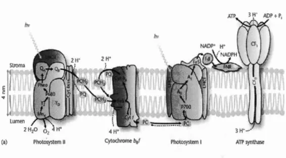
Oxygenic photosynthesis is a biological oxidation-reduction process by which ali
oxygen-evolving organisms including algae absorb light energy to produce carbohydrates from carbon dioxide and water. Oxygenic photosynthesis depends on two reaction center complexes, Photosystem I (PSI) and Photosystem II (PSII). Each
reaction center contains its own reaction center chlorophyll pair: P680 in PSII and P700 in PSI. PSII and PSI are connected by the cytochrome bf complex and a series of mobile electron carriers including plastoquinone and plastocyanin. PSII and PSI participate in the noncyclic transport of electrons from water to NADP+, and the PSII and PSI antenna systems capture light to provide the energy for this process.
Accompanying this electron transport, a proton gradient is created across the membrane, and the energy stored in this electrochemical proton gradient is used for
the synthesis of ATP by A TP synthase. NAD PH and A TP, the products of the light-driven electron and proton transport reactions, provide the necessary energy to reduce carbon dioxide to carbohydrate (Figure 1.4) (Govindjee et al., 2010; Malkin et
Niyogi, 2000; Whitmarsh et Govindjee., 1999). Both Photosystems I and II activities
can be altered by toxic metals. For example, chl a fluorescence intensity was
decreased gradually for ali PSII reduction steps (shown by the 0-J, J-I, and I-P transients) when cells of C. reinhardtii were exposed to different concentrations of Cd2+ from 0.15 to 4.62 ~-tM during 24 h, indicating a decrease of electron transport
from the water-splitting system to PSI. Such graduai inhibition was also observed for PSI activity. In fact, a reduced maximal (delta 1)/ Io 820nm showed the decrease in the
nurnber of photoactive PSI reaction centers that was of 50 % for cells treated with