Université de Montréal
fMRI exploration of the cerebral mechanisms of the
perception of pain in others via facial expression
par Lesley Budell
Département de Neurosciences Faculté de Médecine
Thèse présentée à la Faculté des études supérieures en vue de l’obtention du grade de Philosophiae Doctor (Ph.D.)
en sciences neurologiques
Juin, 2017
Université de Montréal
Faculté des études supérieures et postdoctorales
Cette thèse intitulée :
fMRI exploration of the cerebral mechanisms of the perception of pain in others via facial expression
Présentée par : Lesley Budell
a été évaluée par un jury composé des personnes suivantes :
John Kalaska, président-rapporteur Pierre Rainville, directeur de recherche
Résumé
La douleur ressentie entraine des réactions de différents ordres : physique, neurologique, comportemental. L’expression de la douleur sur un visage constitue une de ces réactions, d’ordre comportemental. Cette expression faciale intègre les éléments caractérisant la douleur ressentie et il est possible de l’analyser en tant qu’observateur extérieur. Les travaux d’imagerie cérébrale étudiant la réaction du cerveau à la perception d’une douleur chez autrui ont mis en lumière un chevauchement entre les régions du cerveau réagissant à une douleur personnellement ressentie et celles réagissant à l’observation d’une expression de douleur chez les autres. Dans la première étude présentée ici, la réaction du cerveau à l’expression de la douleur chez autrui a été analysée en établissant dans quelle mesure l’intensité plus ou moins forte de la douleur exprimée pouvait moduler cette réaction. Les résultats de cette étude indiquent que la perception de la douleur chez autrui ne concerne pas seulement certaines régions du cerveau réagissant à la douleur personnellement ressentie mais aussi des régions habituellement impliquées dans le système des neurones miroirs (MNS; « human mirror neuron system ») ainsi que dans des régions associées à la Théorie de l’esprit (‘Theory of Mind’, ToM; ou « mentalizing »). En outre, ce travail montre que l’implication relative de ces différentes régions varie selon que la personne évalue la signification affective de l’expression – la magnitude de la douleur – ou qu’elle discrimine les composantes motrices de l’expression – les mouvements faciaux. Une deuxième étude a donc été réalisée, s’appuyant sur un paradigme combinant l’observation et l’exécution pour vérifier et confirmer la « réponse miroir » observée dans la première étude et pour examiner plus en détail les différences apparentes entre la résonance émotionnelle et la résonance motrice. En confirmation de la première étude, il a été établi que ce sont différentes régions du cerveau qui sont impliquées dans les réactions à l’expression de la douleur selon qu’elles relèvent de la résonance émotionnelle ou de la résonance motrice. En somme, ces résultats montrent que la perception de la douleur chez autrui est un processus complexe qui met en jeu un chevauchement entre les régions réagissant à une douleur personnellement ressentie et à une douleur constatée chez autrui, ainsi que les phénomènes de résonance motrice (« mirroring ») et de « mentalizing », processus plus généraux de la cognition sociale.
Mots-clés : imagerie par résonance magnétique fonctionnelle (IRMf), empathie de la douleur, système de neurones miroir humain, mentalisation, cortex cingulaire antérieur, gyrus frontal inférieur, lobule pariétal inférieur
Abstract
The pain experience provokes several responses – physical, neural, behavioral. The facial expression of pain is one such behavioral response: it encodes the subjective experience of pain and, as observers, we can decode it. Neuroimaging studies looking at the brain response to the perception of pain in others have identified overlap between brain areas active for the experience of self-pain, and those active during the observation of pain in others. In the first study described below, the brain response to pain in others was investigated using a paradigm that investigated how the intensity of the perceived pain modulated the brain response. The results of this work indicate that the perception of pain in others involves not only certain brain regions involved in self-pain, but also regions previously implicated in the human mirror neuron system (MNS), as well as areas underlying Theory of Mind (i.e. mentalizing). Further, the relative contribution of these areas depended on whether the subject is evaluating the affective meaning of the expression – the pain magnitude – or if they are discriminating the motor components of the expression – the facial movements. A second study was thus designed, using a combined observation and execution paradigm, to confirm the mirroring response observed in the first study, as well as to further explore the hypothesized difference between emotional and motor mirroring. Similarly to the first study, it was found that different brain regions are responsible for mirroring for emotional, versus motor, content of the observed pain expressions. Taken together, these results reveal the perception of pain in others to be a complex process that involves overlap between self and other affective pain areas, as well as mirroring and mentalizing – more general processes of social cognition.
Keywords: functional magnetic resonance imaging, pain empathy, human mirror neuron system, mentalizing, anterior cingulate cortex, anterior insula, inferior frontal gyrus, inferior parietal lobule
Table of Contents
Résumé ... i
Abstract... iii
Table of Contents ... iv
List of Tables ... vi
List of Figures ... vii
List of Abbreviations ... viii
Acknowledgements ... x
Chapter 1: Introduction ... 1
1.1. A pain story ... 1
1.2. Objectives and general outline ... 3
1.3. Pain in the self (execution)... 4
1.3.1. Body to brain: pain perception via afferent pathways from periphery to brain ... 6
1.3.2. Brain to body: the response to pain, and pain behaviors ... 13
1.3.3. Encoding pain in the face ... 14
1.4. Perception of pain in others (observation) ... 16
1.4.1. Pain empathy in the brain ... 16
1.4.2. Faces as the channel: decoding pain from the face... 18
1.5. Hypotheses for studies in this work ... 22
Chapter 2: Articles ... 24
Article 1: Brain responses to facial expressions of pain: emotional or motor mirroring? .... 25
Article 2: Mirroring pain in the brain: emotional expression versus motor imitation. ... 58
3.3.1. Role of aINS during perception of facial expression of pain ... 110
3.3.2. Role of ACC during perception of facial expression of pain ... 111
3.3.3. Specificity for pain: is it pain or sensory salience? ... 113
3.4. Pain communication via facial expression engages brain systems implicated in motor mirroring and mentalizing ... 114
3.4.1. Motor mirroring ... 115
3.4.2. Mentalizing ... 121
3.4.3. Mirroring vs mentalizing in the perception of facial expression of pain ... 123
3.5. Limitations ... 125
3.6. Future directions ... 126
3.7. Conclusion ... 128
List of Tables
Chapter 2 – Article 1:
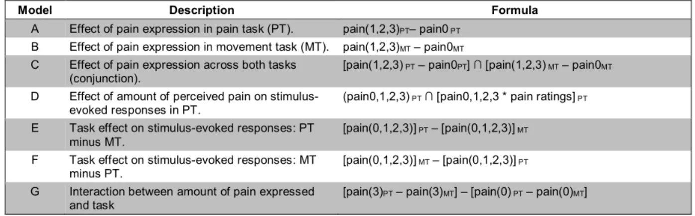
Table 1. Models used in the analysis of imaging data in this study. ... 36
Table 2. Effects of pain expression. ... 39
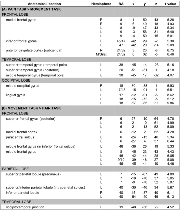
Table 3. Main effects of task: peak values for areas of significant BOLD response change during viewing of the facial expression stimuli in the pain evaluation task condition (A), versus the movement discrimination task condition (B). ... 42
Table S1. Effects of pain expression in the movement task (Contrast B). ... 50
Table S2. Conjunction analysis of pain and movement tasks. (Contrast C). ... 51
Chapter 2 – Article 2: Table 1. Contrast models used in the analysis of imaging data in this study. ... 70
Table 2. Effects of observation and execution of facial expressions in the pain expression task (including pain and neutral conditions). ... 75
Table 3. Effects of observation and execution of pain expressions (pain vs neutral). ... 78
Table 4. Main effects of task during both observation and execution (pain expressions only†): ... 81
Table S1. Effects of pain expression during observation in pain expression task. ... 89
Table S2. Effects of pain expression during observation in movement imitation task. ... 91
Table S3. Main effects of task during observation. ... 92
List of Figures
Chapter 2 – Article 1:
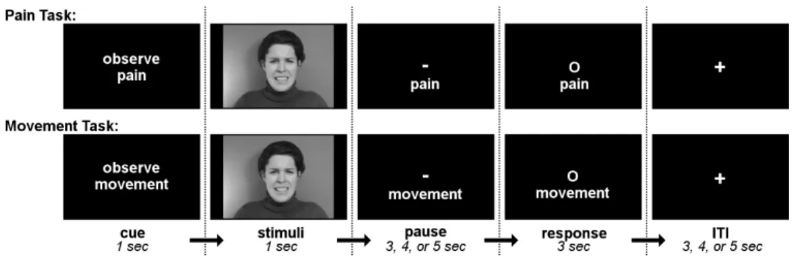
Figure 1. Trial structure for pain evaluation and movement discrimination tasks. ... 33 Figure 2. Effects of pain expression (Contrast A; blue) and amount of pain perceived (Contrast D; orange). ... 38 Figure 3. Effects of task... 41
Chapter 2 – Article 2:
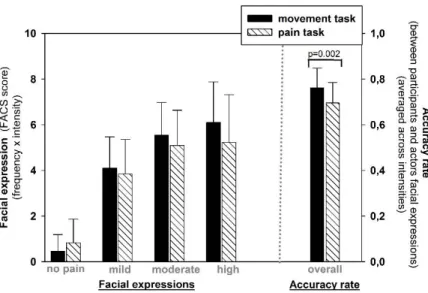
Figure 1. Trial structure for pain expression and movement imitation tasks. ... 65 Figure 2. Results of FACS analysis of facial expressions. ... 72 Figure 3. Areas commonly activated during observation and execution phases of the pain task (Obs∩Exec(PT)). ... 74 Figure 4. Effects of pain during both observation and execution of pain expressions (Obs∩Exec(Pain;PT)). ... 77 Figure 5. Effects of task during both observation and execution of pain expressions. ... 80
Chapter 3 – Discussion:
Figure 3.1. General model for perception of pain via facial expression, as used in this work. ... 105
List of Abbreviations
ACC anterior cingulate cortex aINS anterior insula
aIPS anterior intraparietal sulcus
AMY amygdala
CMA cingulate motor area (monkey) CMZ cingulate motor zone (human) FACS Facial Action Coding System IFG inferior frontal gyrus
INS insula
IPL inferior parietal lobule IPS intraparietal sulcus MNS mirror neuron system mOFC medical orbitofrontal cortex mPFC medial prefrontal cortex
MT movement task (in context of Articles 1 and 2) MTL mentalizing system
NFR nociceptive flexion reflex
pACC posterior anterior cingulate cortex PFC prefrontal cortex
pMCC posterior middle cingulate cortex PoCG postcentral gyrus
Acknowledgements
First and foremost, my deepest gratitude is to my advisor, Pierre Rainville, for giving me the opportunity to join his lab and do this work, and without whose endless patience, generosity, and kindness I never would have completed my studies. I could not have imagined having a better advisor and mentor for my Ph.D studies – Pierre’s enthusiasm for research, his immense knowledge, and his compassion make him not only a wonderful professor, but also a valued friend. It has been a long journey, with many bumps along the way, but Pierre’s unwavering belief in my ability to get to the end is why I have finally done so.
Besides my advisor, I would also like to thank my thesis committee – John Kalaska, Hugo Théoret, Petra Schweinhardt, and Laurent Mottron – for reviewing my work and giving feedback and questions that helped me better understand my own methods and conclusions. In addition, I am indebted to Franco Lepore, who introduced me to the program in the first place, and who has provided invaluable advice about research, writing, and my studies over the years, as well as a warm welcome to Montreal.
I am grateful to Miriam Kunz, a colleague, co-author, and friend, who helped with project design and taught me a lot about statistics, pain in patient populations, the theory behind FACS, and how to streamline my writing. I would also like to acknowledge Phillip Jackson, Jen-I Chen, and the rest of the pain lab, all of whom contributed and helped with different parts of my research including, but not limited to, stimuli creation and validation; protocol design; lab equipment set–up; subject wrangling; fMRI data acquisition, analysis, and interpretation; and writing. Special thanks goes to Mathieu Piché, for studying for our courses together and making the material much more memorable; to Nadine LeBlanc, who helped with
Hurst, who provided expert technical support and advice and ensured my subjects’ safety in the scanner, and whose friendship and humor made the long scanning sessions fly by.
In regards to the completion of my second article and my thesis manuscript, my sincerest thanks goes to Sarah Bruat, who provided assistance leading to the completion of the second article and helped jumpstart what was at the time a very much stalled process; to Sophie Pickford and the other members of the writer’s group for their advice, inspiration, and encouragement; and to thesis angel Sarah M. Byers, who cheered, encouraged, commiserated and proofread my entire thesis manuscript, doing so via choppy Skype connections from all over the world. Her complete understanding of, and lack of judgement regarding, my anxiety over writing is one of the main reasons I finally got over it.
This work would not have been possible without the inspiration and encouragement of several friends. First, I would like to express my deepest gratitude to my good friend Natasha Lepore for her support. It is hard to describe all the ways in which she’s helped me throughout the entire experience of graduate school, and it is pretty amazing how there has never been a question she couldn’t answer or a problem she couldn’t help me solve. Also amazing is how patient she is in the face of endless questions and doubts, and how she never questioned my ability to finish. Her experience and scientific advice have been invaluable. I’d also like to say thank you to friends from Boston who remained in touch all throughout my time in Montreal, giving me a warm Boston home to return to and the constant reminder that life exists outside of grad school. Sioux and Dick, you didn’t teach me neuroscience but your lessons in aikido, and in life, are deep in my heart. You are missed, more than words can say.
I would also like to thank my parents, Josephine and Bill Budell, and my brother, Chase, for their unconditional support, love, and encouragement over the years. They have believed in me and their cheerful faith in me gave me the motivation to keep going and to finish, even when it seemed it was too late to do so. I very much appreciate that they were able to attend the defense and could be with me to celebrate afterwards (and, in the case of my mother, to arrive early and help prepare!). Additionally, my gratitude goes to Francis Sorin and Ghislaine Toutain, for their enthusiastic support and, in particular, their help with the French translation of my abstracts. I am so glad they made the overseas trip to attend my defense, despite the cold Canadian winter.
Last but not least, I’d like to thank my daughter Abigail for her hugs and moral support, and my husband Fabien Sorin, who has been with me throughout this entire experience and who has provided unending love, encouragement, and patience during this long project. Thank you for all the big things – like discussing my work and research ideas and helping with stats – and all the little things, like shopping with me for a defense outfit. There are no words to describe how much your companionship has meant to me, but I know I could never have done this without you.
Chapter 1: Introduction
1.1. A pain story
Imagine the following scenario: You and a friend are on vacation somewhere warm, close to the beach. Being students, you are sharing a room in a cheap motel. Due to a lack of space in the tiny room, your friend has placed her suitcase on the floor in one corner, underneath the television, which is anchored onto a metal frame jutting out from the wall at about head-height. One morning as you are getting dressed, your friend is bent over under the television, rummaging through her suitcase for her bathing suit. Suddenly you hear her cry “Oh!”
You turn quickly to look: she’s now standing with her right hand pressed to the top of her head, her gaze fixed on the sharp corner of the metal TV stand. Her eyes are scrunched in a squint, her nose is drawn up and wrinkled, and her teeth are bared in a grimace.
‘Oh!” you say, feeling alarmed, “I was afraid that would happen! It must really hurt! Are you okay?”
Your unlucky friend has just experienced a noxious stimulus – a painful bump of the head against a hard, sharp object. Although she does not explicitly state “I have bumped my head on the TV stand and am now experiencing pain”, you not only have figured out what physical event took place, but you are also aware of how your friend feels, internally, in response to this event. There are several potential sources of information regarding the event, such as your pre-existing awareness of the threat posed by a sharp object in the room, and perhaps the sound of the bump. However, as a social creature, your main source of information about what has happened to your fellow human, and her reaction, is her behavioral response to it: her vocalization – “oh!”, her hand rubbing her head, and last, but definitely not least, her facial expression, which identifies the emotional component of her response as negative and withdrawing. Once you realize what has occurred, you may also have a memory of bumping your own self on a sharp corner and be able to reference this memory for further knowledge of what your friend may be feeling. All of these things contribute to your understanding of her pain.
Now, rewind back to the moment you hear your friend say “Oh!”, and consider a different scenario. When you turn to look at her, she’s standing with her hand pressed to her head, as before, but this time her face is different: her eyes and mouth are open, but her lips and nose are relaxed rather than drawn up and wrinkled, and her teeth are not showing.
“Oh!” you say, feeling no alarm this time. You look down to see, in her other hand, the sunglasses she was certain she’d lost in the ocean while swimming in the ocean the day before. “You weren’t wearing them after all!”
In this case, the emotional content is clearly different, and it is possible to realize this simply via the expression on your friend’s face. Even without realizing the exact cause of the expression, the emotional content is clearly not negative or withdrawing, and thus in this second version, the first visual information you receive tells you a different story about what your friend has experienced and is feeling, compared to that of the first scenario.
* * *
The above story is an example of pain communication and pain empathy. There are many ways for one person to transmit to another the message that they are suffering. Verbal cues can be the most obvious – e.g. “I hit my head on that damned TV corner!” Non-verbal cues can be equally so, particularly if the observer witnesses the triggering event and therefore has access to information about the noxious stimuli. However, even if this is not the case – for example, when a doctor examines a crying infant but does not yet know if the problem is an earache or a stomachache – the message can be easily transmitted via vocalizations and physical behaviors.
to the self, and that the other has their own mind with thoughts, beliefs, and feelings that may or may not be shared by the observer. Thus, it is in fact a sophisticated and complex feat of social cognition. As such, it involves multiple brain regions, and evidence suggests it utilizes networks involved in the perception of pain in the self, as well as those involved in theory of mind and emotional mirroring.
Pain empathy is important, as it has many implications: the information that someone feels pain can help the observer avoid a threat, or it can provoke the observer to provide aid. In many situations involving acute pain, however, the observer is not explicitly told of what is happening. In this case, how does the observer know the subject feels pain? Moreover, how is pain detected in those who cannot verbalize what they are feeling, such as infants, older patients with dementia, or patients who are otherwise incapacitated and unable to communicate verbally? Obviously, the perception of pain in others via non-verbal routes can be critical.
1.2. Objectives and general outline
The general objective of this manuscript is to contribute to the understanding of pain communication, specifically the decoding component. In Chapter 1, the introduction will begin with a brief description of the perception of pain in the self and how self-pain leads to, and is encoded by, the facial expression of pain. Next, it will continue into a discussion of pain empathy and the perception of pain in others and provide a background for the original research described in the following two chapters.
Chapter 2 is a published manuscript describing the first neuroimaging study, which examines pain decoding. Specifically, this work investigated how the brain response to the facial expression of pain is modulated by the intensity of the observed pain, and how the brain perceives the affective content of a pain expression, versus the physical movements of the face.
Chapter 3 is a published manuscript describing a second neuroimaging study, which investigated the overlap in brain representations for both observed and executed pain expressions. The design of this second study was intended to both reproduce and extend the findings of the first study.
Chapter 4 is a discussion of the results of the two imaging studies. First, it will summarize the findings from the two studies, and address similarities and differences in their findings. Then it will address the results in the context of the literature on pain empathy. In light of results that implicate both neural systems for pain in the self as well as more general systems involved in social cognition, the discussion will then broaden to include the concepts of motor mirroring and mentalizing, and how the work presented above fits into a more general model of social cognition that incorporates these two components. The general idea of overlapping cognitive processes for both self- and other- experiences – pain, specifically – will be the core theme of this discussion. Next, the discussion will address some limitations of the methods and alternative interpretations of the results. Finally, possible future directions for this work will be explored, as well as the contributions it could offer to the understanding of pathological conditions in which perception of emotion in others is abnormal.
1.3. Pain in the self (execution)
Before discussing how the pain experience is conveyed to, and perceived by another person, it is helpful to take a brief look at the pain experience in the self: what is happening, physiologically, during an acute pain experience? This section will briefly and simply introduce the concept of pain, describe the pathways for the perception of acute self-pain from peripheral nociceptors in the skin to the spinal cord and brain, and give an overview of pain responses and their underlying brain mechanisms, particularly in regard to the facial expression of pain.
The International Association for the Study of Pain (IASP) defines pain as “an unpleasant sensory and emotional experience associated with actual or potential tissue
aware its slippery texture, but once she had it in hand and began to get to her feet, it is likely she was no longer consciously attending to this aspect of her sensory experience. Furthermore, the mechanoreceptors in her skin responsible for carrying the sensory input generated by the gentle contact with the suit’s material adapt to this contact and become less responsive. If she were to hold her hand still, she would quickly become unable to feel the material much at all. The forceful contact of her head to the television corner, however, triggers different types of mechanoreceptors which do not adapt so quickly to continuous stimulation. Likewise, the sensation of this contact demands much more of her attention, and her conscious experience of it persists for a much longer time. Unpleasant as it may be, this fact is quite useful; digging through one’s suitcase for prolonged periods of time causes no harm, whereas repeated trauma to the head certainly will.
Pain can be described in terms of two dimensions – sensory and affective. The former refers to the sensory aspects of the pain: when it occurs (right…now), where it occurs (the scalp), its quality (sharp, aching), and its intensity (strong). The latter refers to the hedonic qualities of the pain (very unpleasant), the emotional arousal it causes, and its motivational effects on behavior (i.e. attentional orientation and motor facilitation). Together, these create the subjective feeling that this sensation is negative, salient, and requires attention and action. These two dimensions, while closely linked, are nonetheless distinct; this can be observed in subjective reports about both clinical and experimental pain, as well as in experimental protocols where the two dimensions are modulated independently of each other (Horn et al., 2012; Price et al., 1987; Rainville et al., 1999) As will be discussed later in this introduction, this distinction is also reflected in brain activity underlying the pain experience (Danziger et al., 2006; Hofbauer et al., 2001; Kulkarni et al., 2005; Rainville et al., 1997).
Finally, it is important to note that classically described nociceptive pathways do not necessarily provide a direct link between a given noxious stimuli and experienced pain, for several reasons. Noxious stimuli do not always trigger nociceptive pathways and the subjective feeling of pain. Lesions or other interruptions to these pathways do not always disrupt or halt the pain experience. Factors other than the characteristics of stimuli can affect both nociception and the subjective experience of pain; the intensity and type of the noxious stimulus is only one component in the creation of the pain experience: attention (and distraction), expectations, context, individual differences in pain sensitivity, and physiologic
mechanisms also contribute. While a more in-depth discussion of these points is beyond the scope of this manuscript, the important concept they illustrate is that pain is not always the equivalent of nociception, and vice-versa. Ultimately, the subjective experience of pain is comprised of both physiological and psychological elements, all of which are subject to various internal and external factors: a given noxious stimulus could be perceived as less painful when a subject is distracted by a cognitive task or a competing sensory stimulus, or more painful when the subject is primed with a negative emotional cue. The target of this manuscript is one type of visible evidence of the subjective experience of pain: the facial expression of pain. Non-visible evidence of the subjective experience of pain will not be addressed, nor will be objective measures such as activation of the peripheral nervous system (i.e. as detected via conductive skin response or heart rate) or spinal reflexes such as the nociceptive flexion reflex.
1.3.1. Body to brain: pain perception via afferent pathways from
periphery to brain
1.3.1.1. Periphery to spinal cord: nociceptors to spinal tracts
There are five different afferent pathways that carry information about noxious stimuli from the tissues of the body to the brain; the primary of these is the spinothalamic tract, which carries most of the information about the location and nature of the pain (intensity, burning versus stinging, etc.), while the other pathways contribute more to the response to pain. The following section will summarize the pathways linking peripheral nociceptors to the thalamus
branch of the fibers terminates in free nerve endings in the skin, where they are triggered by physical changes such as pressure and mechanical distortion of the skin by the sharp metal corner, and the other terminates in a synapse in either the chief sensory nucleus in the pons, or the spinal trigeminal nucleus in the medulla.
If the noxious stimulus was to the shoulder instead, the pathway would be slightly different in that the body of the first-order neuron is found in a dorsal root ganglion next to the spinal cord, and its secondary axon fiber terminates in a synapse with a second-order neuron in the dorsal horn of the spinal cord. In both the head and shoulder examples, the axon of the second neuron crosses the anatomical midline and ascends towards the thalamus. In the former case it does so via the ventral trigeminal thalamic pathway, terminating in the ventral posterior medial thalamic nucleus (VPM), and in the latter case, via the lateral spinothalamic tract, ending in the ventral posterior lateral thalamic nucleus (VPL).
1.3.1.2. Pain in the brain: thalamus to cortex
Pain is a multidimensional phenomenon and, as such, its neural representation requires a complex interaction of multiple brain regions rather than a single “pain center.” Noxious sensory input from spinal tracts enters the thalamus and propagates through a pain-responsive, though not necessarily pain-specific, network of cortical brain regions, including SI, SII, PFC, ACC, and the INS. These areas have been identified in several ways: in monkeys, viral neural tracing of STT targets in the thalamus and cortex and single-unit recordings provides direct evidence of activation in, and connections between, neurons responsive to noxious stimuli. The results of these studies can then guide human research, which uses methods such as the assessment of abnormalities in pain perception due to lesions, triggering of pain sensations via direct cortical stimulation (less commonly done), and neuroimaging techniques such as functional magnetic resonance imaging (fMRI) and positron-emission tomography (PET).
Primate work has traced direct connections from the thalamus to primary and secondary sensory cortex, the posterior insula and supracallosal regions of the ACC – particularly medial-wall cingulate motor regions, the human equivalent of which are consistently activated in response to pain (Craig, 2003; Dum et al., 2009; Gingold et al., 1991; Kenshalo et al., 1980) – and studies using evoked potentials provide evidence that these
regions are involved in early responses to pain in humans, as well ((Frot et al., 2008), see review (Craig, 2003)). In humans, the best evidence for a direct link between a particular structure and its role in pain would, in theory, come from lesion studies showing reduced or abnormal pain perception associated with cortical damage. For example, one case study showed a patient with unilateral damage to SI and SII resulting in a deficit in the perception of the sensory aspects of noxious heat stimuli (Ploner et al., 1999). However, other studies have failed to find similar impairments in regard to primary sensory cortex (see reviews (Bushnell et al., 1999; Craig, 2003) and, overall, a clear connection between cortical lesions and abnormal pain perception has only been established for the insula. Furthermore, the insula is one of only two regions where direct cortical stimulation has been found to trigger pain sensations, the other being the secondary somatosensory cortex ((Mazzola et al., 2009, 2006), also see review (Garcia-Larrea, 2012)). Nevertheless, as will be explained below, there is a clear pattern of structures consistently activated during pain, and thus the lesion and stimulation data simply provide further evidence that pain is a multidimensional, distributed process.
Most research into the brain mechanisms of pain perception in humans has employed fMRI, which while unable to directly measure neural activity or trace pathways as is possible with more invasive methods, has nonetheless provided strong evidence for a network of regions commonly activated during pain experiences that includes the thalamus, SI, SII, supracallosal ACC (within BAs 24 and 23), insula (posterior and anterior portions), as well as the SMA, PFC, and subcortical regions of the striatum, cerebellum, and periaqueductal grey (Apkarian et al., 2005; Coghill et al., 1994; Duerden and Albanese, 2013; Garcia-Larrea and Peyron, 2013; Peyron et al., 2000). This collection of areas has been commonly referred to as
a parametric response for pain intensity, not discriminating between innocuous and low-intensity noxious stimuli (Bornhövd et al., 2002; Büchel et al., 1998; Frot et al., 2007). However, when considering the role of each pain matrix region in the pain experience, it is helpful to consider the two dimensions – sensory/discriminative and affective/motivational – described earlier; none of the studies mentioned above distinguished between the pain intensity (sensory aspect) and pain unpleasantness (affective aspect).
Thalamocortical projections are sometimes described in terms of ‘lateral’ and ‘medial’ pain systems, with those from lateral thalamic nuclei to sensory and posterior insular cortices thought to carry primarily sensory information such as location and quality, and those from medial thalamic nuclei to the ACC and the INS to carry more general information, such as arousal, that contributes more to the affective-motivational dimension of pain (Albe-Fessar et al., 1985; Garcia-Larrea and Peyron, 2013; Treede et al., 1999).
Responses of the somatotopically-mapped primary sensory cortex code for stimulus location and intensity (Bornhövd et al., 2002; Bushnell et al., 1999; Kulkarni et al., 2005; Peyron et al., 2000; Porro et al., 1998). Increasing the perceived intensity, versus unpleasantness, of a pain stimulus via hypnotic suggestion increases the magnitude of the response of SI (Hofbauer et al., 2001; Rainville et al., 1997), as does increased attention to the location, versus the unpleasantness, of the stimulus (Kulkarni et al., 2005). Damage to the somatosensory cortex has been observed to selectively disrupt the perception of sensory, but not affective, aspects of noxious heat stimuli (Ploner et al., 1999). Thus, SI and SII are typically described as processing the sensory components of a pain experience.
The involvement of the insula in pain processing has been well established. Years of neuroimaging research in humans have proved that the insula responds to all types of pain, anywhere in the body, in both clinical and experimental conditions (Apkarian et al., 2005; Coghill et al., 1994; Duerden and Albanese, 2013; Price, 2000). The posterior portion receives direct connections from thalamic nuclei (VMpo) that receive STT input from noxious stimuli (Treede et al., 2000, 1999), and the magnitude of its response correlates with self-reports of pain intensity (Bornhövd et al., 2002; Coghill et al., 1999; Frot et al., 2007). It shows somatotopic organization for both painful and non-painful stimuli, and direct electrical stimulation of this area elicits painful sensations (Brooks et al., 2005; Mazzola et al., 2009; Ostrowsky et al., 2002). Post-stroke damage in the opercular-insula region has been associated
with deficits in pain perception coupled with a type of central pain similar to that produced by lesions to the upper spinothalamic tract (Garcia-Larrea et al., 2010). These findings have led to the proposal that the posterior portion of the insula might represent a sort of ‘primary cortex for pain’ (Garcia-Larrea, 2012). However, looking beyond pain, it is clear that the insula is not performing purely first-order sensory processing. In a broader context, the insula is known to be a sensory association area where information about a wide variety of internally-felt sensations related to homeostasis and physiologic status are integrated into a coherent sense of interoceptive awareness (Craig, 2009, 2002; Critchley et al., 2004). Insula response has been associated with the perception of bodily and visceral sensations such as itch, thirst, air hunger, and stomach distension (Craig, 2009), all of which are examples of sensory stimuli associated with the internal physical state and which are highly salient in that they indicate the need for attention and action. Pain is perhaps one of the clearest examples of such stimuli, and the findings gathered for insula response to a wide range of seemingly disparate conditions – e.g. pain and thirst – illustrate how the insula transforms information about what a noxious stimulus is (the sensory aspects) into what it means (the affective aspect). In other words, it is helping to answer the global question “How am I?” using information from basic sensory processes that has already answered the questions “How is my body positioned? What sensation do I feel on my scalp?”
This transformation appears to occur in a posterior to anterior progression, as is suggested by the timing of pain response in each subregion (Frot et al., 2014; Pomares et al., 2013). Passing from posterior to anterior, the information is transformed from an initial somatosensory representation to a subsequent subjective feeling; one study comparing processing of stimulus intensity versus reported pain intensity observed that the response of
It is thus clear that the insula participates in both sensory and affective processing of pain, creating and monitoring a sensory representation of the bodily state, using it to build a subjective feeling state that is then linked to emotional and motivational drives. This last step is postulated to occur in connection with the anterior cingulate cortex; as discussed above, these two regions are nearly always indicated in pain studies as key regions involved in the affective experience of pain, and their responses during pain and other interoceptive sensations are highly coordinated (Craig, 2009).
Along with the anterior insula, the supracallosal portion of the ACC is widely reported in studies on self-pain. Early single-neuron recoding work in cingulotomy patients reported neurons responsive to noxious stimulation (Hutchison et al., 1999), and subsequent neuroimaging work on self-pain further defined pain-related activation of the ACC, in particular the supracallosal, midcingulate area comprising BAs 24 and 32 (Apkarian et al., 2005; Duerden and Albanese, 2013; Shackman et al., 2011; Torta and Cauda, 2011), a region referred to as the posterior ACC (pACC) as well as the anterior middle cingulate cortex (aMCC). Typically, response of this area is associated with the affective/motivational dimension of pain, and more specifically, emotional and motivational drives that affect behavioral and physiological responses such as arousal, attentional orientation, and motor responses.
In a broader context, the ACC is believed to play a key role in the modulation of autonomic arousal, especially in regard to emotion processing, monitoring of action outcomes, and adaptive behavioral control (Paus, 2001; Rushworth et al., 2007; Vogt, 2005), and there is a well-established link between ACC response and affect – particularly negative emotional states (Kober et al., 2008; Shackman et al., 2011; Vogt, 2005). Its response reflects cognitive and motor task difficulty and is correlated with associated markers of stress and arousal (e.g. heart rate variability) (Critchley et al., 2003, 2000; Paus et al., 1998). Likewise during painful stimulation, cingulate response has been correlated with specific markers of autonomic response such as changes in skin conductivity (Dubé et al., 2009; Piché et al., 2010), as well as with affective pain ratings (i.e. unpleasantness) (Rainville et al., 1997). It has also been associated with attentional orientation towards aversive stimuli, including pain (Frot et al., 2008; Peyron et al., 1999; Tölle et al., 1999). Taken together, these findings support the idea
that the ACC contribution to the affective and motivational aspects of pain includes the modulation of autonomic arousal and the orientation of attention.
In addition to emotional and attentional modulation, the ACC participates in motor responses to pain. In monkeys, the medial supracallosal region receives direct nociceptive input from the spinothalamic tract (Dum et al., 2009; Vogt et al., 1987; Vogt and Pandya, 1987), but also has clearly defined motor properties: individual neurons here have been demonstrated as active during motor tasks, and tracing studies have identified projections from this area to premotor and primary motor cortex, as well as to motor neurons in the spinal cord (Dum and Strick, 2005; Picard and Strick, 1996). The human homologue of this ‘cingulate motor area’ has similarly been found to project to premotor, primary motor, and supplementary motor cortical areas, as well as the spinal cord; to display response profiles implicating involvement in the planning and control of voluntary movement; and to respond to pain (Dum et al., 2009; Picard and Strick, 1996; Shackman et al., 2011).
The combination of pain- and motor-responsiveness in this subregion of the ACC has led to the hypothesis that this region has a motor function in the pain response, such as motor readiness or priming, and/or response inhibition. Indeed, ACC response has been associated with motor withdrawal in response to aversive stimuli (Isomura and Takada, 2004), and studies using go/no-go paradigms have associated increased button-press reaction times as well as inhibition of motor responses with modulations in ACC activity (Morrison et al., 2007a; Rubia et al., 2001). In addition to voluntary motor control, it may also be involved in reflexive motor responses to pain (Frot et al., 2008; Piché et al., 2010).
This motor-related and pain-responsive portion of the ACC/MCC receives direct input from the amygdala (Morecraft et al., 2007; Shackman et al., 2011; Vogt and Pandya, 1987;
perception (Legrain et al., 2011; Mouraux et al., 2011). Nevertheless, this network is reliably associated with pain experience across different types of noxious stimuli (e.g. heat, cold, mechanical pain), body location, and experimental contexts (Duerden and Albanese, 2013). The pain experience comprises a collection of physiological, emotional, and cognitive factors and, while it is unlikely that the above-described network of brain regions is pain-specific, pain is supported by a common and identifiable brain network.
1.3.2. Brain to body: the response to pain, and pain behaviors
There are several responses to a painful experience: physiological, cognitive, emotional, and behavioral. Returning to the example in the story presented earlier, a hard bump of the head against a sharp metal corner would likely provoke increased physiological arousal, which manifests as changes in heart rate and skin conductivity. The cognitive response includes orientation of attention towards the potential threat, priming the motor system for action, and the production of conscious thoughts about the situation (“Is my head bleeding? Do I need help? How can I avoid bumping my head again in this tiny room?”). The emotional response in this case is the immediate feeling that something negative and unpleasant has occurred.
The behavioral response is the most visible, e.g. a quick jerk of the head away from the corner, vigorous rubbing of the scalp with the hands, movement of the entire body a few steps away while keeping the head ducked low in order to avoid additional contact. This illustrates one of two general functions that can be ascribed to pain behaviors: withdrawal from noxious stimuli and protection of the body from further damage. Another behavioral response in this example is the display of a facial expression of pain to the second person, illustrating the second general function of pain behaviors – communication to others, either to warn of a potential threat or to solicit help (Williams, 2002).
Part of the behavioral response is automatic and reflexive and does not necessarily require cortical involvement. Spinal motor responses allow ‘flinching before feeling’ – the reflexive withdrawal of a body part away from the noxious stimulus, via neural circuits connecting peripheral nerves, the spinal cord, and skeletal muscles. However, more complex motor responses such as head rubbing or facial expression require cortical-level processing
(Morecraft et al., 2004; Müri, 2016). The subsequent sections will focus on the facial expression of pain as a behavioral response, as it is intended for the purpose of pain communication.
1.3.3. Encoding pain in the face
There are many behavioral responses that can convey the sufferer’s experience of pain: verbalizations (e.g. crying out), body movements (e.g. flinching away from a sharp corner, rubbing a sore head), and postures (e.g. hunching or guarding). The facial expression of pain can occur with or without these other behaviors. It is nonspecific in that it does not in itself offer information about the cause of the pain nor the body part affected, but it is universal in that the same core expression has been consistently observed across a range of different types of acute pain in experimental or clinical settings, as well as in chronic pain patients (LeResche et al., 1992; Prkachin, 1992). There is not, for example, one expression for head bumps and another, completely different, expression for low back pain. Likewise, observers can easily differentiate between this fundamental expression and those of other negative emotions such as anger, sadness, disgust, and fear (Boucher, 1969; Simon et al., 2008).
Any emotional expression can be described by its component facial movements and the muscles involved in effecting those movements. The most commonly used tool for the qualification of expressions, including pain, is the Facial Action Coding System (FACS) created by Ekman (Ekman and Friesen, 1978). Using this tool, a trained evaluator determines which facial muscle groups, called ‘Action Units’ (AUs), are engaged throughout an expression, and the amplitude of the movement of those groups. The prototypical pain face involves four core elements, the primary of which is a lowered, crinkled brow (AU 4, the
experimental settings as well as in clinical applications, particularly those in which the subjects are unable to give clear self-reports about their pain.
The ‘core’ pain expression is an innate behavioral response. It is observed in newborns (Craig et al., 1994; Grunau and Craig, 1987) and blind subjects (Kunz et al., 2012a), and, as with other facial expressions of emotion, does not change with age (Kunz et al., 2008b; Williams, 2002). It also does not differ significantly between males and females (Kunz et al., 2006). However, encoding different intensities of pain is most likely learned through observation and can be modified by various intrinsic and extrinsic factors (Craig et al., 2011; Kunz et al., 2011). Intrinsic factors that can effect pain displays include the severity and nature of the pain (for example, a head bump versus childbirth) and concurrent emotional states (Craig et al., 2011). Individual differences in pain expressiveness exist as well, with some subjects showing greater facial reactivity to painful stimuli over a range of stimulus intensities. These differences may be due in part to variations in pain sensitivity, with higher pain expressiveness correlated with greater sensitivity to pain (Kunz et al., 2008a). Expressiveness during a particular pain experience can also be influenced by extrinsic factors such as the sociocultural context, in which display rules create a broad sense of what people should or should not show, and situation-specific cues, where aspects of the immediate context guide displays of pain (Hadjistavropoulos et al., 2011; Williams, 2002).
Nevertheless, at the individual level, pain expression correlates with self-reports of pain ratings (Kunz et al., 2004), encodes both the intensity of pain and its unpleasantness (Kunz et al., 2012b), and can be distinguished from other concurrent emotions (Williams, 2002). One study even found that observers were able to determine stimulus intensity based on the facial responses (Patrick et al., 1986). Thus, while there is variation in how individuals encode pain, pain expression is nonetheless a good reflection of the subjective pain experience; for this reason it is a valid way to assess pain and a relevant target for research on how we perceive pain in others.
As discussed above in regard to pain behaviors in general, the facial expression of pain may be considered as having two functions: first, a physical response for self-protective withdrawal, with the goal of reducing exposure to the noxious stimuli; and second, a socio-behavioral response, with the goal of communicating the pain experience to others.
1.4. Perception of pain in others (observation)
Pain communication is important for two reasons: it can help the sufferer solicit help from others, and it can provide others with information about an aversive stimulus and warn them of potential harm. Prkachin and Craig present a model that structures nonverbal pain communication as a three-step process, described as A -> B -> C., wherein given an episode of pain, A represents the internal state of the pain sufferer, B represents the encoding of this internal state via a specific set of expressive features, and C represents the decoding performed by the observer ((Prkachin and Craig, 1995)). Once the pain message is encoded and broadcast by the sufferer, it must then be observed and decoded by the observer. The previous sections have discussed the first steps – A, the internal state (self-pain, sensory and affective dimensions), and B, encoding (the facial expression of pain); the material presented in the following sections, as well as in the work described in the following two chapters, will be concerned with the third step, C – decoding the pain experience.
1.4.1. Pain empathy in the brain
An ever-growing body of imaging research has provided evidence that the decoding aspect of pain communication – pain empathy – shares a neural basis with the experience of pain in the self (see review (Lamm et al., 2011)). This work has used a variety of stimuli to investigate the perception of pain in others, such as cues that a loved one is in pain (Singer et al., 2004), still images or videos of body parts in painful situations (X Gu and Han, 2007; Jackson et al., 2005; Morrison and Downing, 2007), or even descriptive narratives (Bruneau et al., 2012b; Xiaosi Gu and Han, 2007)).
subjectively and in terms of its saliency, where an overlap with observed pain occurs (Singer et al., 2004). Similarly, the anterior insula has also been found to show overlapping responses for observation and experience of other affective states, such as disgust and gustatory pleasure (Jabbi et al., 2008, 2007; Wicker et al., 2003), suggesting a role for the insula in empathy for affective states in general, not just for pain.
Later work employed images and videos of body parts in painful situations, with similar results showing overlap for observed versus experienced pain in the anterior insula and the ACC (Jackson et al., 2005; Morrison et al., 2004). It is in this body of work where the role of the ACC in observed pain was more closely examined. For example, Morrison and colleagues compared ACC response to that of SI, for both noxious and innocuous felt and seen pain conditions, finding that the former responded only to the noxious stimuli in both conditions, while the latter responded to felt stimuli only, whether or not they were painful. They concluded that these results suggest that the same idea of a functional dissociation of sensory and affective/motivational aspects of self-pain applies to observed pain, and that the ACC plays a role in organizing behavioral responses, most likely via modulation of motor regions (Morrison et al., 2004). Following this reasoning, the authors presented additional work showing that viewing pain in others facilitates motor responses to self-pain, and that this behavioral effect is reflected in the ACC response (Morrison et al., 2007a, 2007b). Later work has further shown that viewing images of body parts in painful situations lowers the threshold for reflexive spinal responses to self-pain (Vachon-Presseau et al., 2011).
Taken together, these results support the hypotheses that, as in the case of pain in self, the role of the ACC in the perception of pain in others is to create motivational drives influencing behavior (specifically, motor responses), whereas the anterior INS is involved in representing physical saliency.
In all of the above studies, however, the source of the pain is known to the observer; either it is witnessed outright, as in the videos and pictures of limbs receiving painful stimuli, or it is known but not seen, as in the cue-based paradigms. More importantly, neither type of protocol provides information about the subjective response to the pain stimuli, such as withdrawal responses or facial expressions of pain. As in the opening pain story, there are many real-world examples of pain communication in which the observer may only see the pain response, not the actual event causing the pain. In fact, it can even be said that these
protocols are not examining pain communication at all, as there is no pain message to be decoded. Thus, an important subsequent question concerns what happens when an observer does witness pain-related behavior – specifically, the facial expression of pain. While there has been extensive work looking at the brain response to facial expressions of emotion (e.g. (Fusar-Poli et al., 2009; Sabatinelli et al., 2011)), prior to the preparation of the first study presented in this manuscript only three published works had specifically looked at the response to pain expressions (Botvinick et al., 2005; Saarela et al., 2007; Simon et al., 2006).
1.4.2. Faces as the channel: decoding pain from the face
In most protocols, the type of noxious stimuli and the affected body part is known – a needlestick to the finger, an arm dunked in ice water, an electrical shock to the foot, or the manipulation of an injured shoulder are some examples. However, pain communication does not require the observer to witness the injury or to be given a narrative describing it; as in the introductory story, an observer can receive the message that an injury has occurred simply by viewing the facial expression of the other.
Perceiving the emotional content of a facial expression is a complex feat of social cognition that involves several steps: basic visual perception, recognition of a configuration of visual features as a meaningful whole object classified as a ‘face’ (face detection), and lastly, recognition of the emotional message being conveyed by this face.
1.4.2.1. Perception of faces
Livingstone, 2008). This distinction can also be seen in observations of patients with the disorder known as prosopagnosia, who show deficits in recognizing familiar faces without similar deficits for non-face objects (Kress and Daum, 2003; McNeil and Warrington, 1993).
Furthermore, this difference is also reflected in the brain response, with certain regions showing clear preference for faces and face-like stimuli. Following early processing in the primary visual cortex, face processing begins with recognition of the face-like configuration of features in the fusiform gyrus of the occipitotemporal cortex, an extrastriate region dubbed the fusiform face area (FFA) (Allison et al., 1994; Kanwisher et al., 1997; Sergent et al., 1992), as well as in the occipital face area (OFA), a portion of the inferior occipital gyrus (Grill-Spector et al., 2004; Johnson et al., 2015; Tsao and Livingstone, 2008). Whereas these two regions are associated with initial face detection and recognition, which involve processing invariant features and their configurations, a third region – the posterior superior temporal sulcus (pSTS) – is recruited for the processing of dynamic features such as gaze direction and movement of facial features (Allison et al., 2000; Haxby et al., 2000, 2002; Phillips et al., 1997). Together, these three regions comprise the core system for cortical face detection and recognition.
1.4.2.2. Perception of emotional expression
While face identification depends on occipitotemporal pathways, and the perception of facial emotion recruits a wider network of regions, evidence suggests these are not distinct, separate processes, but rather interactive mechanisms, with even early face processing showing sensitivity to emotional content, most likely via fast subcortical pathways and top-down modulation of the fusiform by the amygdala (Vuilleumier et al., 2004).
Facial expression recognition, however, is a multi-level, multi-process task preceded by face detection and dissociated from face identification (recognition of gender, age, or identity); this distinction is reflected in the timing and location of brain responses, with recognition of emotional expression occurring subsequent to the initial face detection associated with the earliest brain responses, although evidence suggests that emotional content can then modulate the fusiform response via feedback from subcortical pathways involving the amygdala (Geday et al., 2003; Vuilleumier, 2007; Vuilleumier and Driver, 2007). Looking
again at the case of prosopagnosia, it has been observed that patients often retain the ability to recognize facial expression despite deficits in facial recognition (Humphreys et al., 2007; Palermo et al., 2011).
There does not seem to be one dedicated neural circuit for the perception of all emotional expressions; rather, much differentiation has been observed for different emotions (Fusar-Poli et al., 2009; Hennenlotter and Schroeder, 2006). Processing facial affect has been linked with increased activation in numerous brain regions, including prefrontal, temporal, parietal, visual, limbic, and subcortical areas (Fusar-Poli et al., 2009; Vuilleumier, 2007). Despite a large body of research on the subject, there is still not a consistent picture of any of the basic emotions most frequently studied – anger, fear, disgust, happiness, sadness and surprise – although some patterns have emerged. For example, fear expressions are strongly linked with amygdala response, in line with the amygdala’s known role in fear conditioning and aversive learning, whereas disgust faces activate the anterior insula but not the amygdala (Hennenlotter and Schroeder, 2006; Phillips et al., 1998). This insula response overlaps with that for other aversive stimuli such as unpleasant tastes and smells (Jabbi et al., 2008; Wicker et al., 2003), providing an example of an interoceptive emotion showing recruitment of the same regions for both self- and other- experience.
1.4.2.3. Perception of pain expressions
Although there is generally a wide body of work looking at the brain mechanisms for the perception of facial expressions, less attention has been given specifically to pain expressions. As of the time the study reported in Chapter 2 of this manuscript was designed and carried out, only three earlier studies had looked specifically at brain response during the
in the ACC and the anterior insula, consistent with previous studies on the perception of pain in others. Consistent with the broader body of work on the perception of emotional expression in general, activation was also observed in the amygdala and the OFC.
The second study, also using video clips, compared expressions of pain to those of anger, as well as neutral expressions (Simon et al., 2006). In this protocol, subjects performed a gender discrimination task while viewing the stimuli; this task was intended to result in implicit processing of the pain content of the expressions, as subjects’ attention was at least partially directed away from the pain content of the faces. Here, the most significant results were found when comparing brain response to pain as shown by male models, versus female models. Face-processing regions of the FFA and STS, as well as the amygdala, responded more strongly to pain expressions, versus angry or neutral expressions. This is consistent with a subcortical pathway for emotional modulation of face processing via the amygdala, discussed above. Compared to anger, pain expressions also provoked greater response in the vmPFC, the anterior insula, and SII/posterior insula. However, all of these effects were seen only in response to male models and, additionally, there were no results reported in the supracallosal portion of the ACC identified in other studies on pain empathy. The authors hypothesized that these results reflected gender differences in the perceived social role of pain expression, with male pain faces representing greater potential threat, and that this perception of threat may prevent or reduce processes of pain empathy. However, it could also be that engagement of self-pain regions during pain empathy requires attention to the pain content of the face. Testing this possibility would require a protocol using two task conditions to manipulate subject attention.
Another important question is how the brain may code for the intensity of an observed pain expression. As described earlier, the intensity of self-pain modulates the response of the INS and ACC, and pain expressions are a reliable indicator of the subjective experience of pain. It is possible that information about the intensity of observed pain is decoded via the same mechanisms that encode pain in the self. Indeed, the response of the ACC has been shown to code for perceived intensity of other pain when viewing images of body parts in painful situations (Jackson et al., 2005). Thus, the third study looking at brain response to pain faces attempted to answer the question of pain intensity coding (Saarela et al., 2007). Here, subjects viewed photos of ‘provoked’ versus ‘chronic’ pain expressions displayed by chronic
pain patients, in addition to neutral expressions displayed by healthy volunteers. Following the scanning session, subjects rated the intensity of pain displayed in each image, and a correlation was found between these ratings and the response of the ACC and the aINS during the viewing of the stimuli. However, as with the first study, task instructions during scanning were simply to attend to the images; subjects were not specifically instructed to judge how much pain was displayed.
Taken together, the evidence from these studies suggests that the perception of pain faces, like that of other cues relating to pain in others, does recruit brain regions involved in self-pain. However, some questions remain. In particular, the influence of attentional and task demands on the recruitment of these areas is an important factor; in the studies described above, subjects were either instructed simply to attend to the visual stimuli (Botvinick et al., 2005; Saarela et al., 2007), or they performed a non-pain-related task (i.e. gender discrimination) (Simon et al., 2006). In the latter case, this can be considered implicit processing of the affective content of the expressions, as subjects were instructed to attend to another aspect of the stimuli. Observing an emotional expression, as has been discussed above, involves both the perception of a complex, dynamic object, as well as perception of the meaning of that object. In using only one task, or only passive viewing, these earlier works do not address the potential difference in coding for the dynamic visual attributes of an expression versus its affective content. In other words, is recruitment of brain regions involved in the processing of self-pain linked to explicit attention to the pain content of an expression? Further, how is the response of these areas during explicit processing of pain modulated by the perceived pain intensity? These questions were therefore the motivation for the first study, described in Chapter 2.
In Study 1, we hypothesized that the explicit evaluation of pain expressions would recruit brain regions involved in the processing self-pain and that a subset of these regions would also show a parametric effect in response to the perceived pain intensity. Further, we hypothesized that evaluation of the meaning of the expressions would reveal greater responses in midline cortical regions involved in thinking about the mental states of others, whereas evaluation of the movement shown in the expressions would result in greater responses in motor regions identified in action observation and motor mirroring.
In Study 2, our hypotheses were similar to, and expanded on, those of Study 1. Once again, we expected that evaluation of the emotional content of a pain expression would recruit brain regions involved in the processing self-pain, and that attending to the meaning of the expressions would more strongly engage the frontal portions of the action observation/mirroring network, whereas attending to the movement in the expressions would more strongly engage parietal regions of this network. While the first study included only observational conditions, the experimental protocol of Study 1 added an execution condition to allow greater confidence in the identification of ‘mirroring’ processes.
Article 1: Brain responses to facial expressions of pain: emotional
or motor mirroring?
Title: Brain responses to facial expressions of pain: Emotional or motor mirroring? Authors: Lesley Budell1, 2 Phillip Jackson3 Pierre Rainville2, 4 Affiliations:
1 Département de physiologie, Université de Montréal, Montréal, Québec, Canada
2 Groupe de recherche sur le système nerveux central (GRSNC) and Centre de recherche de
l’Institut universitaire de gériatrie de Montréal (CRIUGM)
3 École de psychologie and CIRRIS and CRULRG, Université Laval, Québec, Canada 4 Département de stomatologie, Université de Montréal, Montréal, Québec, Canada
Corresponding author: Pierre Rainville, PhD
Département de stomatologie Faculté de médecine dentaire Université de Montréal C.P. 6128, Succursale Centre-ville (Tel) 514-343-6111 #3935 (Fax) 514-343-2111 Montréal, Québec, H3C 3J7 Pierre.Rainville@Umontreal.ca Publication status:
Manuscript published in NeuroImage, Vol 53, Issue 1, May 2010.
Authors' contributions: LB, PLJ and PR contributed to the conception and design of the study; LB acquired and analyzed the data; LB, PLJ and PR contributed to the analysis design and interpretation; LB drafted the manuscript. All authors contributed important intellectual content and approved the final version.
ABSTRACT
The communication of pain requires the perception of pain-related signals and the extraction of their meaning and magnitude to infer the state of the expresser. Here, BOLD responses were measured in healthy volunteers while they evaluated the amount of pain expressed (pain task) or discriminated movements (movement task) in 1-second video clips displaying facial expressions of various levels of pain. Regression analysis using subjects’ ratings of pain confirmed the parametric response of several regions previously involved in the coding of self-pain, including the anterior cingulate cortex (ACC) and anterior insula (aINS), as well as areas implicated in action observation, and motor mirroring, such as the inferior frontal gyrus (IFG) and inferior parietal lobule (IPL). Furthermore, the pain task produced stronger activation in the ventral IFG, as well as in areas of the medial prefrontal cortex (mPFC) associated with social cognition and emotional mirroring, whereas stronger activation during the movement task predominated in the IPL. These results suggest that perception of the pain of another via facial expression recruits limbic regions involved in the coding of self-pain, prefrontal areas underlying social and emotional cognition (i.e. ‘mentalizing’), and premotor and parietal areas involved in motor mirroring.
Keywords: functional magnetic resonance imaging, anterior cingulate cortex, anterior insula, human mirror neuron system, empathy
Abbreviations:
ACC = anterior cingulate cortex aINS = anterior insula
IFG = inferior frontal gyrus INS = insula
IPL = inferior parietal lobule MNS = mirror neuron system mPFC = medial prefrontal cortex PCG = post central gyrus