Silver-containing diamond-like carbon deposited by
plasma as versatile antibacterial coatings
Thèse en cotutelle
Doctorat en génie des matériaux et de la métallurgie
Maxime Cloutier
Université Laval
Québec, Canada
Philosophiae doctor (Ph.D.)
et
Université Pierre et Marie Curie
Paris, France
Docteur
Silver-containing diamond-like carbon deposited by
plasma as versatile antibacterial coatings
Thèse en cotutelle
Doctorat en génie des matériaux et de la métallurgie
Maxime Cloutier
Sous la direction de :
Diego Mantovani, directeur de recherche
Michael Tatoulian, directeur de cotutelle
iii
Résumé
Les infections associées au milieu hospitalier demeurent une cause majeure de mortalité et de morbidité dans le monde, malgré plusieurs décennies dédiées à promouvoir une meilleure surveillance et des méthodes de désinfection plus complètes. La capacité des bactéries pathogènes à survivre sur des substrats solides a été identifiée comme un facteur clé de la pathogenèse de ces infections, en multipliant les sources de transmission et de contamination. Au niveau de la recherche, cette situation s’est récemment traduite par un intérêt marqué pour le développement de revêtements antibactériens novateurs pouvant constituer une ligne de défense complémentaire contre la colonisation bactérienne de surfaces, pourvu qu’ils puissent résister à l’environnement rigoureux des établissements de santé.
Dans cette thèse, nous avons émis l'hypothèse qu'un revêtement antibactérien avec une stabilité supérieure pouvait être déposé en utilisant un procédé plasma modulable, de sorte que les propriétés du revêtement résultant pourraient être adaptées aux exigences de différentes situations ou applications. Par conséquent, des revêtements nanocomposites de carbone amorphe adamantin contenant de l'argent (Ag-DLC) ont été développés et étudiés comme plate-forme polyvalente pour des surfaces antibactériennes. L’intérêt de ce matériau réside dans la combinaison des excellentes propriétés mécaniques, de la résistance à l'usure et de l'inertie chimique du carbone amorphe adamantin avec les propriétés antibactériennes à large spectre des nanomatériaux d'argent au sein d’un même revêtement déposé par plasma.
Ce travail a d'abord identifié les défis de conception spécifiquement associés au développement de revêtements antibactériens pour le milieu hospitalier. Des analyses approfondies des revêtements Ag-DLC ont ensuite démontré une bonne efficacité antibactérienne in vitro ainsi qu’une stabilité des propriétés, de la structure et de l’état chimique des revêtements dans le temps. L'étendue de la polyvalence des revêtements Ag-DLC a été évaluée au travers de l’identification des mécanismes de croissance principaux, permettant d’obtenir des informations essentielles sur la façon dont les propriétés des films, telles que la dureté, la teneur et la distribution d’argent, pouvaient être contrôlées en ajustant des paramètres spécifiques du dépôt plasma. De plus, un traitement de surface in situ a été développé pour surmonter les problèmes de délamination et a montré la capacité de favoriser l'adhérence de
iv revêtements DLC sur des substrats métalliques. Dans l'ensemble, cette étude a mis en évidence l'importance de la stabilité dans l'application des revêtements antibactériens et a démontré le vaste potentiel des procédés plasma pour le dépôt de revêtements antibactériens stables avec des propriétés adaptables.
v
Abstract
Healthcare-associated infections remain a major cause of mortality and morbidity worldwide, with a substantial financial burden on society, despite decades of monitoring and disinfection efforts. The ability of pathogenic bacteria to survive on solid substrates has emerged as a key contributing factor in the pathogenesis of these infections by multiplying the sources of transmission and contamination. This has prompted investigations into the development of innovative antibacterial coatings, which could provide a complementary barrier against bacterial colonization of surfaces provided that they can withstand the harsh operating environment of healthcare facilities.
In this thesis, we hypothesized that an antibacterial coating with superior stability could be deposited using a tailorable plasma process, so that the resulting coatings’ properties could be adapted to match the requirements of different situations or applications. Therefore, silver-containing diamond-like carbon (Ag-DLC) nanocomposite coatings were developed and investigated as a versatile platform material for antibacterial surfaces. The interest of this material lies in the combination of the excellent mechanical properties, wear-resistance and chemical inertness of diamond-like carbon with the broad-spectrum antibacterial properties of silver nanomaterials in a single, plasma-deposited coating.
This work first identified the specific design challenges associated with the development of antibacterial coatings for healthcare environments. Thorough investigations of Ag-DLC coatings then revealed good antibacterial efficacy in vitro as well as stability of the coatings’ properties, structure, and chemistry over time. The extent of the tailorability of Ag-DLC coatings was also assessed through the identification of the main growth mechanisms, providing insights on how the film’s properties, such as the hardness, silver content, and silver distribution, could be controlled by adjusting specific plasma deposition parameters. Furthermore, an in situ interface plasma treatment was developed to overcome delamination issues and showed the ability to promote the adhesion of high stress DLC coatings on metallic substrates. Overall, this study highlighted the importance of stability in the application of antibacterial coatings and demonstrated the vast potential of plasma processes for the deposition of stable antibacterial coatings with tunable properties.
vi
Table of contents
Résumé ... iii Abstract ... v List of tables ... x List of figures ... xiAbbreviations & Symbols ... xvi
Acknowledgements ... xviii
Foreword ... xxi
1 Introduction ... 1
1.1 Context and motivation ... 1
1.2 Challenge ... 3 1.2.1 Healthcare-associated infections ... 3 1.2.2 Role of surfaces ... 6 1.2.3 Prevention strategies... 9 1.2.4 Bacteria ... 10 1.2.5 Antimicrobial agents ... 14 1.3 Proposed platform ... 18
1.3.1 Surface engineering by plasma ... 19
1.3.2 Deposition techniques ... 25
1.3.3 Diamond-Like Carbon ... 28
1.4 Summary of approach and research objectives ... 33
2 Antibacterial coatings: challenges, perspectives and opportunities ... 35
2.1 Résumé ... 37
2.2 Abstract ... 38
2.3 Antibacterial surfaces in health applications ... 39
2.3.1 Advances in biomedical engineering prompted by the development of new materials ... 39
2.3.2 Nosocomial infections and the role of surfaces ... 39
2.3.3 Importance of antibacterial coatings ... 40
2.4 Relevance of release-based antibacterial coatings ... 45
2.4.1 Recent developments in antibacterial strategies ... 45
2.4.2 Key challenges ... 46
2.5 Release-based coatings ... 46
2.6 Control of release kinetics ... 48
2.6.1 Passive approaches ... 48
2.6.2 Active approaches – stimuli-responsive materials... 51
2.6.3 Bacterial triggers approaches ... 52
vii
2.7.1 Multi-release coatings ... 55
2.7.2 Multi-approach coatings ... 55
2.7.3 Multi-property (smart) coatings ... 56
2.8 Long-term stability ... 57
2.9 Conclusions and perspectives ... 61
3 Adhesion enhancement of DLC coatings on 316L stainless steel surfaces by in situ plasma carburation ... 63
3.1 Résumé ... 64
3.2 Abstract ... 65
3.3 Introduction ... 66
3.4 Materials & Methods ... 67
3.4.1 Sample preparation ... 67
3.4.2 Characterization ... 68
3.4.3 Adhesion tests ... 68
3.5 Results and discussions ... 69
3.5.1 Effect of interfacial treatment on composition ... 69
3.5.2 Depth profiling ... 74
3.5.3 Adhesion measurements ... 75
3.6 Conclusions ... 77
4 Controlled distribution and clustering of silver in Ag-DLC nanocomposite coatings using a hybrid plasma approach ... 79
4.1 Résumé ... 80
4.2 Abstract ... 81
4.3 Introduction ... 83
4.4 Experimental... 84
4.5 Results ... 86
4.5.1 General composition and morphology of Ag-DLC coatings ... 86
4.5.2 Effect of silver ion flux ... 89
4.5.3 Effect of ion energy ... 91
4.6 Discussion ... 93
4.7 Conclusions ... 96
4.8 Acknowledgements ... 97
5 On the long term antibacterial features of silver-doped diamond-like carbon coatings deposited via a hybrid plasma process ... 98
5.1 Résumé ... 99
5.2 Abstract ... 100
5.3 Background ... 101
5.4 Materials &Methods ... 104
viii
5.4.2 Plasma deposition of thin film ... 104
5.4.3 X-Ray Photoelectron Spectroscopy ... 104
5.4.4 Silver release analysis ... 106
5.4.5 Bacterial strain and culture preparation ... 106
5.4.6 Antibacterial activity test ... 106
5.4.7 Live/dead bacterial viability assay ... 106
5.4.8 Modified Kirby-Bauer diffusion test ... 107
5.5 Results and discussion ... 108
5.5.1 Chemical composition ... 108
5.5.2 Antibacterial activity evaluation ... 110
5.5.3 Release of silver ions from the coating... 112
5.6 Conclusions ... 114
5.7 Competing interests ... 115
5.8 Authors’ contributions ... 115
5.9 Acknowledgements ... 115
6 Long-term stability of hydrogenated DLC coatings: Effects of ageing on the structural, chemical and mechanical properties ... 116
6.1 Résumé ... 117
6.2 Abstract ... 118
6.3 Introduction ... 119
6.4 Materials & Methods ... 120
6.4.1 Sample preparation and storage ... 120
6.4.2 Characterisation ... 121
6.5 Results and discussion ... 122
6.5.1 Structural properties ... 122 6.5.2 Surface chemistry ... 125 6.5.3 Surface morphology ... 127 6.5.4 Mechanical properties ... 128 6.5.5 Tribological properties ... 131 6.6 Conclusion ... 132 6.7 Acknowledgment ... 133 7 General discussion ... 134
7.1 Summary and significance of contribution ... 134
7.2 Ag-DLC as a tunable platform ... 137
7.2.1 Choice of substrates ... 137
7.2.2 Structure and mechanical properties ... 138
ix
7.2.4 Antibacterial properties... 140
7.3 Investigated areas of applications of Ag-DLC coatings ... 141
7.3.1 Healthcare environmental surfaces ... 141
7.3.2 Catalytic coating for depollution ... 142
7.4 Limitations ... 143
7.4.1 Limitations of this study... 143
7.4.2 Limitations of Ag-DLC coatings ... 144
8 Conclusions and perspectives ... 146
9 References ... 147
Annexes ... 174
A.Stability and robustness assessment of Ag-DLC coatings ... 174
Abstract ... 175
Introduction ... 175
Experimental... 176
Ag-DLC coatings deposition... 176
Stability and robustness assessment ... 177
Conclusions ... 178
B. Report on the performances of Ag-DLC as catalytic coatings in an ozonation process ... 180
Context ... 180
Materials & Methods ... 181
Catalysis of ozone degradation ... 182
Catalyst in the degradation of a model pollutant ... 184
Stability of silver as a catalyst ... 185
Stability of Ag-DLC coatings under water flow ... 187
Conclusions ... 189
C.List of publications ... 190
x
List of tables
Table 1.1: Modes of transmission of pathogens. Adapted from [23, 31] ... 6
Table 1.2: Key factors facilitating surface-mediated transmission of infections. ... 7
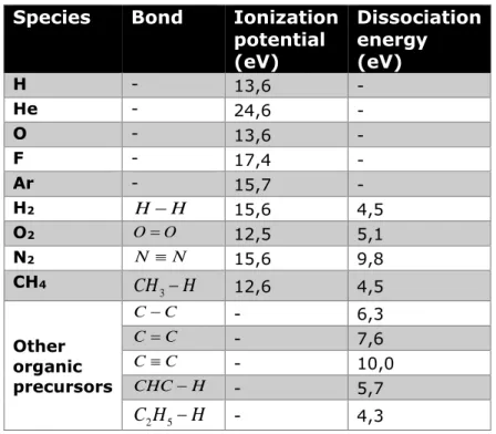
Table 1.3: Ionization potential and dissociation energy of relevant atoms and molecules. Data from [99-101] ... 21
Table 1.4: Important parameters of plasma discharges and their respective impact on the deposition process. The selected examples are taken from DLC literature... 27
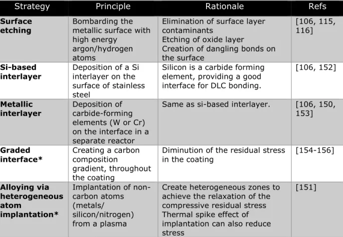
Table 1.5: Interface modification strategies for the optimisation of adhesion strength. ... 32
Table 2.1: Main antibacterial compounds in release-based coatings ... 42
Table 2.2: Uses of plasma processes for antibacterial coatings and surfaces ... 59
Table 3.1 : Plasma treatment parameters ... 68
Table 3.2 : Chemical surface composition of untreated and treated samples measured by XPS. ... 72
Table 5.1: Critical properties to consider in the design of antibacterial coatings. Comparison between implantable devices and environmental surfaces. ... 103
Table 6.1: Hardness of DLC films measured by nanoindendation ... 131
Table B.1: Contact angle (water) of stainless steel and Ag-DLC coatings before and after 24h exposure to water flow ... 189
xi
List of figures
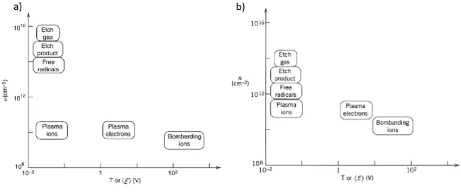
Figure 1.1 : Pathogens associated with four of the most common HAIs in intensive care unit surveillance, 2003 (adapted from [20]). Gram-negative bacterial species are shown in color, gram-positive species are shown in black and white... 4 Figure 1.2: Persistence of relevant pathogens on environmental surfaces (adapted from [26]). The estimated survival time, in months, is based on the average life of infective strains on dry, inanimate surfaces at room temperature. The reported persistence for Klebsiella pneumoniae (30 months) and Salmonella typhimurium (50 months) are not shown to their full extent in the graph. ... 8 Figure 1.3: The multiplication of pathogen reservoirs and infection transmission routes due to contaminated surfaces (adapted from [27]). ... 8 Figure 1.4: Cell wall structure of Gram-negative and Gram-positive bacteria (from [53]) ... 11 Figure 1.5 : Model of the development of biofilms from planktonic cells and dispersal of bacteria from a mature biofilm [55]. ... 12 Figure 1.6: Genetic and biochemical mechanisms of the development of bacteria resistance to biocidal metals (from [65]). ... 13 Figure 1.7: Mechanisms of action of silver against bacteria (from [78]) ... 15 Figure 1.8: High-angle angular dark-field scanning TEM (HAADF STEM) images showing the interactions of silver nanoparticles with bacteria: (left) E. coli, (middle) S. typhus, (right) P. aeruginosa (adapted from [71])... 16 Figure 1.9: Maxwellian energy distribution of electrons in a weakly ionized plasma (from [98]). Another function that can be used to describe electron energy distribution is the Druyvesteyn function. ... 21 Figure 1.10: Ion energy distributions for an Ar plasma at different rf-bias power showing the characteristic double energy peak of ions [103]. ... 22 Figure 1.11: Range of energies and densities for various species found in CCP (left) and ICP (right) processing plasmas (low pressure, RF discharges). From [96] ... 26 Figure 1.12 : (left) Parallel plate reactor (for CCP) and (right) inductively driven planar source (for ICP) [96, 124]. ... 27 Figure 1.13: Ternary diagram of Diamond-Like Carbon. [140] ... 29 Figure 1.14: a) Subplantation and relaxation processes leading to the formation of sp3
bonding in a:C-H film [125]; b) Sp3 fraction as a function of the bias voltage/ion
energy [144]; c) The various plasma-surface processes playing in a role in the deposition of DLC [125]... 31 Figure 1.15: Graphical representation of the different topics addressed in this doctoral thesis. ... 34 Figure 2.1: Schematics and images illustrating various passive strategies to control the release kinetics and antibacterial properties of coatings. a) AFM images showing the change of topography and nanotexture of Ti surfaces after an acid etching treatment [227]. b) SEM micrographs of cyclodextrin-based hydrogels with tunable porosity [228]. Porous coatings with a lower crosslinking density exhibit faster release kinetics than more crosslinked hydrogels. c) The deposition of a thin plasma polymer film can be used as a diffusion barrier for release-based antibacterial coating, enabling the
xii
control of the antibacterial agents rate of release by adjusting its thickness [229]. d) SEM images of TiO2 nanotubes (NTs) used as tunable reservoirs for Ag NPs [230]. e) Other special architectures, such as dendrimers, can be used for loading and delivery of antibacterial compounds from a coating [221]. Figures reproduced with permission from Elsevier (a-b, d-e). ©2009 ACS (c) ... 50 Figure 2.2: Designing antibacterial coatings within a 4D perspective. The design strategies to control the release of antibacterial agents over space and time can be grouped under three main categories. a) Passive approaches. By tuning the coating’s properties, it is possible to impose specific preloaded release kinetics, giving the possibility to produce a variety a release profiles, including rapid bursts (left) or linear release (right) from AB coatings. b) Active approaches. External stimuli can be used to trigger the local release of embedded compounds. c) Bacterial triggers approaches. Bacteria-responsive coatings release antibacterial agents locally when challenged by bacteria. Inset: examples of representative release profiles for each approach, showing the release rate as a function of time. ... 54 Figure 2.3: Control of release kinetics - Bacteria-triggered release. Coatings have been engineered to release antibacterial agents when subjected to two different bacterial triggers, the acidification of the local environment from bacterial metabolism and bacteria secreted enzymes. When challenged, the antibacterial compounds can be released by different mechanisms from simple bond cleavage to charge balance within the coating. ... 54 Figure 3.1 : a) XPS core-level spectra of C1s after plasma carburation treatment b) Example of decomposed spectrum. The c) Comparison of the C-Me/C-C ratio for the different treatment biases... 71 Figure 3.2 : XPS core level spectra of C1s at the interface of samples treated with (a)-100V and (b)-250V plasma carburation treatment recorded at take-off angles from 15° to 80°. ... 72 Figure 3.3 : XPS core-level spectra of (a) Cr2p and (b) Fe2p after plasma carburation treatment. ... 73 Figure 3.4 : a) XPS depth profile of a DLC coated, interface treated sample (Vb
=-650V). b) Evolution of the atomic concentration of the main metals (Fe and Cr) with the C-Me contribution measured using the C1s peak. ... 75 Figure 3.5 : Maximum tensile strength recorded during the pull-off experiment for specimens treated with etching (black, left) and plasma carburation at -650V (red, right). Each bar represents an individual DLC deposition and is the average of at least two pull-off tests. ... 76 Figure 3.6 : Optical micrograph showing a combination of adhesive and cohesive failure of an interface-treated (-650V) DLC coating after a pull-off test. The different zones observed are 1) 316L substrate (adhesive failure), 2) damaged DLC film (cohesive failure), and 3) intact DLC film. The stud initially covered zone 1 and 2. Scale bar, 250µm... 76 Figure 3.7 : Representative optical micrographs showing the self-delamination and peeling of DLC coatings occurring for non-treated interfaces. Scale bar, 100µm. ... 77 Figure 4.1 : Graphical abstract ... 82 Figure 4.2: Example of XPS depth profile of Ag-DLC films (Vb =-100V, VAg =-750V) showing (a) the atomic percentages (at.%) of carbon, silver, oxygen and silicon with a zoom-in (inset) on the first 200s of sputtering, (b) the Ag3d5 peak binding energy, and
xiii
(c) the evolution of the silver modified Auger parameter as a function of the profiling time. ... 87 Figure 4.3: (a, b) Representative SEM images (n=4) of Ag-DLC coatings. (VAg =-750V, Vb =-100V). c) Size distribution histogram of silver clusters (N=1197, d= 1 nm). Dotted curves show the best fit simulated LND functions for each mode (M1 and M2) of the bimodal distribution. The solid curve is the sum of the two components. Counting was limited to particle diameter above 4.5 nm due to image resolution limits. ... 88 Figure 4.4 : AFM topography (left panels) and phase contrast (right panels) images of DLC (a, b) and Ag-DLC coatings (c, d). Representative images taken from n =5. Please note the scale height/phase differences. ... 89 Figure 4.5 : Effect of the variation of the silver target bias /silver flux on silver distribution in Ag-DLC films. a) Representative silver concentration profiles (n=2) measured by XPS profiling. b) Surface and bulk concentrations for each condition. Bulk Ag concentration corresponds to the average value between 200 and 300 s sputtering time. ... 90 Figure 4.6 : Effect of Ag flux on silver clusters size and density in Ag-DLC. Representative AFM height images (1 x 1 m, n=3) of coatings deposited with (a) low (-400V) and (b) high (-750V) silver target bias (VAg). ... 91 Figure 4.7: Representative XPS depth profile (n=3) analysis showing the effect of the ion energy (Vb) on silver dispersion and clustering in Ag-DLC samples. (a) Silver concentration (at.%) profiles, (b) Binding energy and (c) FWHM of the Ag3d5/2 peak for different substrate bias. Higher FWHM /BE shifts indicate the presence of smaller clusters. ... 92 Figure 4.8: Effect of ion energy on silver surface distribution and cluster size in a-C:H:Ag films. Representative AFM height images (1 x 1 m, n=3) of coatings deposited at a substrate bias (Vb) of (a) 0V (b) -50V (c) -100V and (d) -150V substrate bias (Vb). Please note the scale height differences. ... 93 Figure 4.9: Schematics of silver distribution for two different growth modes of a-C:H:Ag. Hydrocarbon ions subplantation leads to subsurface film densification and growth. The process also induces a thermally activated diffusion of Ag which, combined with the inability of Ag atoms to implant in the film due to their size, ultimately causes Ag segregation at the surface of Ag-DLC coatings where it diffuses to form nanoclusters. Surface growth occurs at all deposition biases (Vb) but is the only mode
of growth in polymer-like coatings. This leads to a more uniform distribution of Ag through the coatings depth, with less fluctuation in cluster size. ... 96 Figure 5.1 : a) Deposition procedure of the Ag-DLC films b) Schematic of the plasma reactor 1. Quartz window, 2. Spiral antenna, 3. Matching and tuning networks, 4. RF generator (13,56 MHz, Max: 1000 W) of the ICP plasma source, 5a. VLF generator (3 kHz) for biasing of the silver target, 5b. Heating current supply 6. Vacuum system (throttle valve + turbo mechanical pump + primary mechanical pump), 7. LF generator (90 kHz) for biasing of the sample holder, 8. Sample holder (4 inches diameter), 9. Reactor door and observation window, 10. Silver target (silver wire 99,99% purity, biased and heated), 11. Mass flow controllers, 12. Gas cylinders. ... 105 Figure 5.2: Results of XPS analyzes: a) typical XPS survey of Ag-DLC (Ag 2.4 at.%), b) Silver concentration (at.%) of Ag-DLC films for different negative bias on the silver cathode and c) Depth profile showing the position of Ag3d5/2 peak (black squares) and
oxygen concentration (red triangles) . In inset, high resolution Ag3d peak showing the influence of the oxidation state on the peak position. ... 109
xiv
Figure 5.3 : Antibacterial activity of Ag-DLC coatings against E. coli. Fluorescent optical micrographs showing the distribution of Live (green)/Dead (red)-stained cells on a) uncoated silicon and b) 2.4at.% Ag-DLC coated silicon. c) Quantitative antibacterial activity test of Ag-DLC with different silver concentration. ... 111 Figure 5.4: a) Silver release (in µg/L or ppb) from Ag-DLC (1.7 at% silver) coatings in deionized water, for up to two weeks. b) Modified Kirby-Bauer diffusion test with a 2.4 at% Ag-DLC coating at the middle. ... 112 Figure 6.1: Single-wavelength Raman spectroscopy (λ=488 nm) analysis of DLC samples. a) Typical Raman spectra showing both deconvoluted peaks and the fitted linear background. b) Pos(G) c) I(D)/I(G) ratio d) FWHM(G) and e) hydrogen content of as-deposited (squares) and aged (triangles) DLC films as a function of deposition power. ... 125 Figure 6.2: Representative XPS survey spectra of DLC films (deposition power 200 W) before and after ageing. Only carbon (C1s) and oxygen (O1s) were detected on both samples. ... 125 Figure 6.3: High resolution (HR) XPS analysis of the investigated films. a) HR XPS C(1s) spectrum of the DLC films with corresponding peak deconvolution (deposition power 150 W, post-ageing). b) Proportion of oxygen components of the C(1s) peak for the as-deposited and aged DLC films. ... 127 Figure 6.4: Representative water contact angle measurements of a) as-deposited (81±3°) and b) aged DLC coatings (65±3°) ... 127 Figure 6.5: Characteristic tapping mode AFM height images (1 µm x 1 µm) of DLC films a) as-deposited and b) aged (deposition power 250 W). Please note that the vertical scale bars represents only 1 nm, for both images. ... 128 Figure 6.6: Compressive stress measured experimentally in as-deposited (square, black) and aged (triangle, red) DLC coatings, as a function of deposition power. .... 130 Figure 6.7 : Representative nanoscale friction measurements (deposition power 150 W) of as-deposited (square, black) and aged (triangle, red) ... 131 Figure 7.1: The different investigated strategies for the controlled release of Ag+ ions
from silver-based plasma coatings. ... 140 Figure A.1: Silver concentration (measured by XPS) as a function of sputtering depth for coatings deposited at 0V (red circles) and -100V (black square) bias voltage. The hardness and silver ion release values for each conditions are also shown. Both coatings were deposited at the same silver flux, [H] and [CH] concentrations. ... 177 Figure A.2 Representative SEM images of a) Vb=0V coating after 20 wear cycles and b) Vb=-100V coating after 1000 wear cycles. c) Silver content at the surface of 0V, 50 and 100V Ag-DLC coatings for an increasing number of cleaning cycles. ... 178 Figure B.1 Schematic of the setup used in the ozone degradation experiment. ... 182 Figure B.2 : Typical reaction kinetics of ozone in solution, measured by UV-Spectroscopy. Because of the upper sensitivity detection limit of the setup, the initial stage of the decomposition kinetics could not be observed and were labeled over-saturation stage. ... 183 Figure B.3 : Ozone decomposition curves when in contact the different silver-based catalyst investigated. The time constants presented on the right figure are associated with the first-order decomposition kinetics measured in the second stage (decompostion stage) of the experiment. ... 184
xv
Figure B.4 : Degradation of pyruvic acid after 1 hour. ... 185 Figure B.5 : Representative images of sputtered silver coatings (on COC substrates) after 1 cycle of ozonation experiment. ... 186 Figure B.6 : Representative images of Ag-DLC coatings on stainless steel before (as-deposited) and after ozonation. Top pictures are optical microscopy (zoomed X20). 186 Figure B.7 : Optical microscopy images (50X) of Ag-DLC coated microchannels after 24h under a dynamic water flow. (Top) Inside of microchannels (Bottom) Ridge between pairs of microchannels. ... 187
xvi
Abbreviations & Symbols
Below is a list of abbreviations and terms used throughout this project. The large majority of the listed abbreviations are already defined in the body of the thesis but are grouped here for ease of reference.
AB: Antibacterial a-C: Amorphous carbon
a-C:H: Hydrogenated amorphous carbon AFM: Atomic force microscopy
Ag-DLC: Silver-containing diamond-like carbon AMP: Antimicrobial peptides
BE: Binding energy
CFU: Colony-forming unit DC: Direct current
DLC: Diamond-like carbon
FWHM: Full width at half maximum
GF-AAS: Graphite furnace atomic absorption spectroscopy HAI: Healthcare-associated infection
ICP: Inductively-coupled plasma IED: Ion energy distribution LbL: Layer-by-layer
LND: Lognormal distribution
LTE: Local thermodynamic equilibrium LOD: Limit of detection
xvii LOL: Limit of linearity
LOQ: Limit of quantification
MRSA : Methicillin-resistant staphylococcus aureus NP: Nanoparticle
PECVD: Plasma-enhancement chemical vapor deposition PEG: Polyethylene glycol
PEM: Polyelectrolyte multilayer PVD: Physical vapor deposition
QAC: Quaternary ammonium compound QS: Quorum-sensing
RF: Radio frequency
ROS: Reactive oxygen species SEM: Scanning electron microscopy SS316L: Stainless steel 316L
VLF: Very-low frequency
XPS: X-ray photoelectron spectroscopy
α': Modified Auger parameter RRMS:: Root mean squared roughness
σM: Geometric standard deviation or shape parameter Te: Electron temperature
VAg: Bias voltage applied to the silver target Vb: Bias voltage applied to the substrate holder
xviii
Acknowledgements
A ma femme Héléna et à mes parents, Ann et Richard, Cette thèse est autant votre réussite que la mienne, merci.
To my wife Héléna and to my parents, Ann and Richard, This thesis is as much your success as it is mine, thank you.
xix Autant le travail d’écriture d’une thèse peut être un exercice solitaire et qui rejoint, au final, qu’un nombre très restreint de lecteurs, autant la thèse elle-même n’est possible que par l’apport et le soutien d’une foule de personnes. Ces quelques lignes sont dédiées à tous ceux qui ont contribué, de près ou de loin, au succès de mon parcours doctoral.
Je tiens à remercier tout d’abord mes superviseurs, Pr Diego Mantovani et Pr Michael Tatoulian, pour la confiance qu’ils m’ont accordée en acceptant de mettre en place et d’encadrer ce travail doctoral. Votre accessibilité, votre ouverture, et votre soutien continuel (même pour régler mes trop nombreux soucis administratifs) ont contribué autant que votre créativité et votre encadrement scientifique à la réalisation de cette thèse. Mais surtout, vous avez réussi à créer, chacun de votre côté, des laboratoires où la science autant que les relations humaines sont mis de l’avant et dans lesquels j’ai eu un plaisir réel à évoluer durant les dernières années.
Un grand merci aux nombreuses personnes ayant eu la tâche (que j’espère n’avoir pas trop rendue ingrate) de me superviser à une certaine étape de mon parcours scientifique : Dr Pascale Chevallier, Pr Paul De Koninck, Dr Cédric Guyon, Dr François Lewis, Dr Stéphanie Ognier, Dr Andranik Sarkissian, et Dr Stéphane Turgeon. Le chemin est assez long et complexe entre le baccalauréat et la fin du doctorat. Vous m’avez aidé à le parcourir à ma façon, tout en m’évitant pas mal d’embûches.
Bien qu’on réussisse à s’éloigner de notre laboratoire quelques journées par année, le quotidien d’un doctorant y reste malgré tout lié. Je veux donc remercier du fond du cœur tous les collègues avec lesquels j’ai pu échanger, apprendre, et grandir durant mon passage. A mes collègues de Paris – Alex, Bradley, Cédric, Dia, Diane, Eric, Fred, Olivier, Mengxue, Rafik, Simon, Xi et j’en passe - merci pour l’accueil toujours chaleureux, les discussions, les vendredis gras, les soirées, et les énigmes. Les moments passés chez vous ont toujours parus trop courts. A mes collègues de Québec (il y en a beaucoup trop pour tous les nommer) – Andrée-Anne, Audrey, Bernard, Carlo, Caroline, Daniele, Éléonore, Essowe, Farid, Fred, Gad, Jean, Julien, Laurence, Linda, Livia, Lucie, Mahrokh, Marie, Mathieu, Meryem, Morgane, Myriam, Nina, Olivier, Pascale, Ranna, Sébastien, Sergio, Stéphane, Vanessa et les autres. Vous avez toujours réussi à faire de notre petit centre de recherche un endroit où l’on prenait plaisir à y retourner jour après jour, malgré l’absence de soleil!
xx Une pensée bien spéciale aussi à ceux et celles qui ont été présents pendant une bonne partie de mon parcours. J’espère bien que nos routes continueront à se croiser régulièrement dans l’avenir. Pascale – Merci pour m’avoir pris sous ton aile dès mon premier stage, pour tes conseils, ton soutien et pour toutes les fois où tu as passé mes échantillons XPS en priorité! Stéphane – Le seul probablement toujours partant pour discuter science, système à vide ou DLC! Merci pour ta rigueur et ton sens du savoir-faire, des qualités qui j’espère auront déteint un peu sur moi. Olivier – Mon « analogue » DLC côté français, mais surtout quelqu’un avec qui j’ai un eu plaisir fou sur deux continents. Merci pour toutes les discussions, les voyages, les soirées et pour l’accueil. Tu reviens au Québec quand tu veux! Éléonore et Vanessa – Vous veniez presque toujours en paire donc aussi bien vous garder comme cela. Je vous ai peut-être fait vieillir prématurément avec toutes les mini crises cardiaques créées en me croisant dans les corridors. Merci de m’avoir enduré depuis le début et pour tous les souvenirs.
Un merci du fond du cœur à mes parents, Ann et Richard. Outre votre contribution génétique non négligeable, vous avez aussi su instiller en moi une curiosité, un désir d’apprendre et une certaine forme d’entêtement dès mon plus jeune âge qui ont fait de moi la personne que je suis aujourd’hui. Ça a sans doute contribué plus que tous autres facteurs à la réussite de ce doctorat. Je vous confie maintenant (en partie) la tâche de faire de même avec la prochaine génération. Une pensée spéciale à mon fils, Félix, et à son petit frère qui s’en vient, qui ont fait des derniers mois de ma thèse une période beaucoup moins stressante que je ne l’aurais imaginé initialement.
Enfin, mes derniers remerciements vont à mon extraordinaire femme, Héléna, qui a parcouru tout ce chemin (et bien plus) avec moi. Je ne t’ai pas fait la vie facile avec mes habitudes nocturnes, mon horaire de travail bizarre et mes allers-retours entre la France et le Québec. Merci pour tes sacrifices, tes conseils, ton aide, pour m’avoir épaulé et supporté pendant les hauts et les bas de ma thèse, et pour m’avoir constamment démontré que la vie est toujours plus belle lorsqu’elle est partagée avec toi. C’est la seule raison pour laquelle tu n’auras pas besoin de m’appeler Dr Cloutier!
xxi
Foreword
From the reader’s perspective, the study presented herein could be looked at strictly as a work on antibacterial surfaces and plasma deposition. However, from the author’s standpoint, it should also be seen through a broader lens, particularly in regard to the objective and scope of the research effort. Instead of focusing strictly on an optimisation process, this thesis was aimed at addressing the challenge of creating a flexible and tunable material platform, capable of exhibiting multiple properties or functions. As such, its potential resides not only in finding application in different fields, from water decontamination to antibacterial hospital surfaces, but also in our ability to adapt the produced materials’ properties to match specific aspects of a chosen environment. Therefore, this thesis explores the ways in which we can understand, improve and control the properties of the Ag-DLC nanocoatings rather than evaluating their performances in a particular setting.
The intrinsic multidisciplinarity and breadth of work that characterize this project called for its realisation in a strongly collaborative environment. For this reason, this thesis was pursued as a “cotutelle” between the Laboratory for Biomaterials and Bioengineering (LBB) at Université Laval (Québec, Canada) and the Laboratoire Procédés, Plasmas, Microsystèmes (2PM) at Université Pierre et Marie Curie (UMPC-Sorbonne, Paris, France). The rich collaborative history, complementarity of expertise and quality of research teams in both laboratories were paramount in the realisation of this thesis.
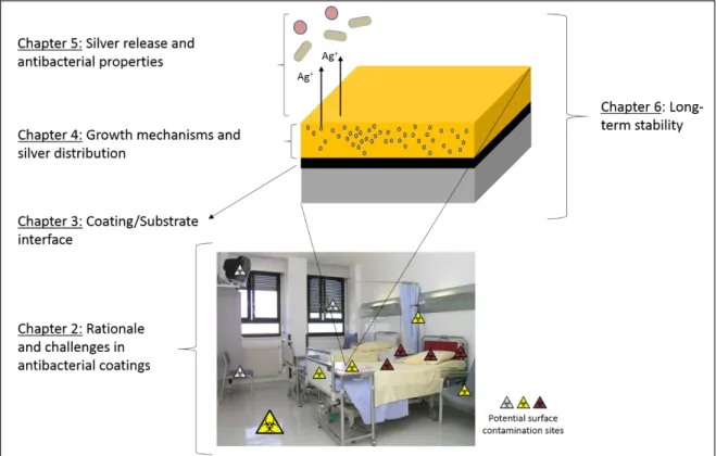
This work is not an extension of previous research from either groups but rather a new investigation. Therefore, the general objectives of the thesis were to first synthetize Ag-DLC coatings using a plasma-based deposition process and then investigate their potential as a material platform through the exploration of four different aspects: i) the interface between metallic substrates and DLC coatings; ii) the analysis of growth mechanisms and distribution
xxii of silver; iii) the measurement of silver-release and antibacterial properties; iv) the evaluation of long-term stability. These results are presented as four published articles making up chapters 3, 4, 5 and 6, respectively, of the current thesis. In addition, the field of antibacterial coatings and the role that silver and plasma-based coatings like Ag-DLC can potentially play within that field have been reviewed and published in a comprehensive review article, presented in chapter 2. Additional unpublished materials that bear relevance to this thesis are presented at the end of this manuscript in annexes.
Further details on the published articles integrated to this thesis are given below.
Chapter 2: Antibacterial Coatings: Challenges, Perspectives, and Opportunities
Authors: Cloutier, M., Mantovani, D., Rosei, F.
Published October 2015 in Trends in Biotechnology, 33 (11), 637-652.
All authors jointly identified the scope of the review article. MC reviewed the literature and wrote the manuscript, under the supervision of DM and FR.
Chapter 3: Adhesion enhancement of DLC coatings on 316L stainless steel surfaces by in situ
plasma carburation
Authors: Cloutier, M. Turgeon, S., Chevallier, P., Tatoulian, M., Mantovani, D.
Journal: Applied Surface Science (to be submitted)
MC designed and performed all the experiments, analyzed the data and wrote the manuscript. MC and ST came up with the original idea for plasma carburation. ST and PC helped with the
xxiii acquisition and analysis of XPS. MT and DM edited the manuscript. All authors discussed the results and implications and commented on the manuscript at all stages.
Chapter 4: Controlled Distribution and Clustering of Silver in Ag-DLC Nanocomposite
Coatings Using a Hybrid Plasma Approach
Authors: Cloutier, M. Turgeon, S., Busby, Y., Tatoulian, M., Pireaux, J.J., Mantovani, D.
Published July 2016 in: ACS Applied Materials & Interfaces, 8 (32), 21020-21027.
MC designed and performed all the experiments, analyzed data and wrote the manuscript.
MC and ST designed the study. MC, ST and YB planned and performed the XPS experiments and analyzed the results. MC wrote the manuscript. JJP provided conceptual advice on the XPS investigation. All authors discussed the results and implications and commented on the manuscript at all stages.
Chapter 5: On the long term antibacterial features of silver-doped diamondlike carbon
coatings deposited via a hybrid plasma process
Authors: Cloutier, M. Tolouei, R., Lesage, O., Lévesque, L., Turgeon, S., Tatoulian, M., Mantovani, D.
Published June 2014 in Biointerphases, 9 (2), 029013.
MC, OL and ST designed the study. MT and DM supervised the project. MC, RT, OL and ST performed non-biological experiments. LL designed and performed the antibacterial tests and helped in the results analysis. MC and OL wrote the manuscript. All authors discussed the results and edited the manuscript.
xxiv
Chapter 6: Long-term stability of hydrogenated DLC coatings: Effects of aging on the
structural, chemical and mechanical properties
Authors: Cloutier, M., Harnagea, C., Hale, P., Seddiki, O., Rosei, F., Mantovani, D.
Published September 2014 in Diamond and Related Materials, 48, 65-72.
MC designed the experiments. PH and OS carried out the first set (year 0) of experiments. MC performed the second set (year 3) of experiment and analyzed the data. CH performed the friction experiments. MC wrote the manuscript. CH, FR and DM oversaw the redaction and edited the manuscript.
1
1 Introduction
1.1 Context and motivation
Healthcare associated infections (HAIs) continue to be a major public health concern in healthcare units worldwide and has been attracting the attention of public health stakeholders for several decades. Although it is difficult to evaluate, their cost, both human and financial, remains staggering, even in this day and age. The number of HAIs in the US, makes it one of the most common adverse event in healthcare. A 2011 survey in acute care hospitals revealed an estimated 725 000 HAIs cases, amongst which 75 000 resulted in deaths [1]. This number goes up to 1.7 million cases of HAIs and 99 000 death annually, when considering the entire healthcare system [2]. Similarly high prevalence rates of HAIs are reported throughout developed countries, 5.4% in France and 8.0 % in Canada, with some units (e.g. pediatric intensive care, transplant units) experiencing rates close to 30% [3-6]. In low- and middle-income countries, the consequences of HAIs are even more dire, with an average prevalence rate of 15.5% [5]. While negligible compared to morbidity and mortality, the economic impacts of bacteria related diseases also put an additional burden on the healthcare system and global productivity of communities. Estimates of annual direct costs of HAIs vary due to differences in methodologies but remain significant in all cases; between 7 and 23 billion USD [2, 7] and up to 7 billion € in Europe [5, 8]. Furthermore, the topic of HAIs has become highly politicised during the last decade, negatively impacting the perception of healthcare systems and tarnishing the image of medical practitioners around the world [9].
Lately, clinical and epidemiological evidences have further underlined the importance of a new area of concern in the fight against HAIs [10-12]. Pathogens are capable of surviving for weeks on environmental surfaces (inanimate objects in the immediate vicinity of a patient such as bed rails, lavatory sinks, over-bed tables, floors, etc.), which become reservoirs of pathogens. Healthcare workers, visitors or the patients themselves may contaminate their hands or gloves by touching contaminated surfaces, thereby multiplying the potential sources of pathogen transmission to patients [13-15].
The use of antibacterial surfaces has been advocated as a viable solution to reduce the risks posed by contaminated environmental surfaces. In this case, however, their design entails a number of stringent requirements, different from those of surfaces intended for medical
2
devices applications. For them to be effective in near-patient clinical areas, such antibacterial surfaces must be used in conjunction with, and therefore resist, frequent cleaning and disinfection procedures. Thus, they must exhibit chemical stability, good wear-resistance, and superior mechanical properties, in conjunction with long-term antibacterial properties.
3
1.2 Challenge
1.2.1 Healthcare-associated infections
1.2.1.1 Historical perspective
While many bacteria are either beneficial or harmless to the human body, history has shown the dire consequences that some pathogens can have. Famous examples of deadly bacteria abound: the Black Death (Yersinia Pestis), one of the most devastating pandemics in human history that peaked during the middle of the 14th century, typhus (Rickettsia), with known outbreaks ranging from the Classical Greece to the second world war and tuberculosis (Mycobacterium tuberculosis), which was a major killer in Europe in the past and still causes many deaths in Africa. Despite an increasing knowledge of microorganisms, marked by Antonie van Leeuwenhoek’s first observation of unicellular organisms in 1674, Louis Pasteur’s experiments regarding germ theory in the early 1860s and Robert Koch’s postulates formulated in 1884 establishing a causal relationship between an infection and a causative microbe [16], and the consequent establishment of modern hygiene practices, pathogens are still a major cause of disease and death nowadays.
1.2.1.2 Definition and classification of HAIs
Healthcare-associated infections1 (HAIs) are defined as infections acquired while receiving medical treatment in a healthcare facility [17]. As presented previously, HAIs are a major cause of morbidity and mortality, as well as a significant financial burden. Their existence obviously predates any modern literature, but the role of pathogens in HAIs has been closely monitored by public health agencies since the inception of global surveillance programs in the 1970s. While the term pathogen used previously actually englobes all types of infecting organisms such as bacteria, viruses, fungi or even prions, HAIs are for the vast majority caused by bacteria. A notable exception is the norovirus, the most common cause of viral gastroenteritis in humans [18]. Clinically, HAIs are classified based on the location of the infection. Common examples include urinary tract infections (UTI), pneumonia, bloodstream infections (BI), surgical site infections (SSI) and C. difficile infections [17].
4
Any pathogenic organism may theoretically cause an HAI under the right conditions, but 8 pathogens account for about 80% of all HAIs [19]. Furthermore, each bacteria is not restricted to a single type of infection, each of them has been reported as a causative pathogen for all of the most noteworthy HAIs (Figure 1.1). In general, pathogen distribution and prevalence amongst infected patients have been showed to vary with time, location/department type and patient type, amongst other factors [6, 20-22].
Figure 1.1 : Pathogens associated with four of the most common HAIs in intensive care unit surveillance, 2003 (adapted from [20]). Gram-negative bacterial species are shown in color, gram-positive species are shown in black and white.
1.2.1.3 Etiology of HAIs
The development of an infection entails the presence of three elements: a source of pathogens, a susceptible host, and a mode of transmission for the infectious agent [23]. Public health researchers and agencies have looked within this epidemiology triad to develop infection prevention strategies.
Sources
Major sources of infection agents in healthcare settings are either humans, (patients, healthcare workers or visitors), inanimate environmental surfaces or the endogenous flora of patients [23]. Although one of the primary source in healthcare settings, carriage of pathogens by human is
5
typically short-lived. However, some human sources, such as incubating, asymptomatic or transiently colonized carriers, are considerably more difficult to detect than carriers with active infections [23, 24]. Epidemiological and clinical studies have confirmed the role of environmental surfaces as reservoirs of pathogens [25-27]. Since surfaces also play an important role in infection transmission, this topic will be discussed in more details in section 1.2.2.
Transmission
There are six possible modes of transmission for infection-causing pathogens (Table 1.1). The role of direct, droplet and airborne transmission in the spread of HAIs have been well-recognized for several decades. Bacterial infections are more commonly passed on by direct contact, usually by the hand of healthcare workers [28]. This has resulted in the implementation of several strategies targeting these modes of transmission, with programs to improve compliance with hand hygiene best practices as their cornerstone [29, 30]. However, the role of surfaces, and consequently of indirect contact transmission, have emerged much more recently. Researchers have shown that several pathogens can contaminate hospital surfaces and play an important role in transmission events of HAIs. This will be described in more details below. Lastly, although they are important from an epidemiological standpoint, neither vehicle nor vector-borne transmission play a significant role in the transmission of HAIs.
6
Table 1.1: Modes of transmission of pathogens. Adapted from [23, 31]
Host
The susceptibility of the host, the final link in the infection chain, is linked with genetic and intrinsic factors such as age, severity of underlying illness, loss of skin, and degree of immunosuppression [32]. Furthermore, a patient’s susceptibility of contracting an HAI has been also strongly correlated with the use of indwelling medical devices. Catheter-associated UTIs remain one of the most common healthcare infections [33]. Similarly, the use of ventilators and central venous catheters have been respectively associated with an increased incidence and severity of pneumonia and BIs [5, 34, 35].
1.2.2 Role of surfaces
The intricate array of events leading to a patient being infected has complicated the estimation of the full extent of the role played by environmental surfaces in the spread of HAIs. Most of the epidemiological proofs linking pathogens found on environmental surfaces with specific infections has been indirect. Weber et al. have showed patient-to-patient transmission to be directly proportional to the local level of environmental contamination [36]. Likewise, many
Mode of transmission
Description Important
dissemination factors Contact Direct Person to person transmission
without intermediate object/person.
Ungloved contact, skin cuts or abrasions
Indirect Contact with a contaminated intermediate object, usually inanimate or contaminated gloves.
Near-patient surfaces (tables, beds, floors), patient-care devices
Droplet Transmission through large
(>5µm) particles. Coughing, sneezing or talking.
Non-contact
Airborne Transmission via droplet nuclei (<5µm) or dust particles.
Ventilation systems.
Vector-borne Insect or animal vectors Mosquitoes, flies, rats, etc. Vehicle Single contaminated source
spreading the infection (e.g. infected batch of food)
Food, water, medication, etc.
7
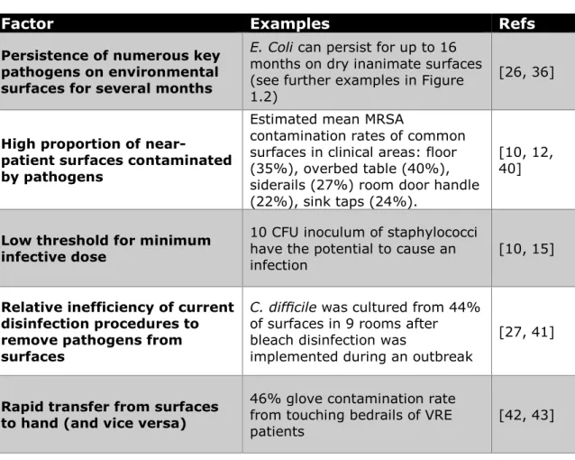
studies have linked improved room disinfection procedures to lower incidence of HAIs [12, 37-39]. Still, the substantial body of evidence accumulated over the last decade have led the scientific and medical communities to recognize the key role of environmental surface in the transmission of HAIs. Key factors influencing HAIs transmission via surfaces are presented in Table 1.2.
Table 1.2: Key factors facilitating surface-mediated transmission of infections.
Factor Examples Refs
Persistence of numerous key pathogens on environmental surfaces for several months
E. Coli can persist for up to 16 months on dry inanimate surfaces (see further examples in Figure 1.2)
[26, 36]
High proportion of
near-patient surfaces contaminated by pathogens
Estimated mean MRSA
contamination rates of common surfaces in clinical areas: floor (35%), overbed table (40%), siderails (27%) room door handle (22%), sink taps (24%).
[10, 12, 40]
Low threshold for minimum infective dose
10 CFU inoculum of staphylococci have the potential to cause an
infection [10, 15]
Relative inefficiency of current disinfection procedures to remove pathogens from surfaces
C. difficile was cultured from 44% of surfaces in 9 rooms after bleach disinfection was
implemented during an outbreak
[27, 41]
Rapid transfer from surfaces to hand (and vice versa)
46% glove contamination rate from touching bedrails of VRE patients
[42, 43]
Environmental surfaces contribute to the multiplication of infection pathways to patients. They may act as reservoirs or as transient sources of pathogens, from which patients or healthcare workers may get contaminated (Figure 1.3). Studies have shown contamination by pathogens of numerous near-patient surfaces including, but not limited to, door handles, clothes and linens (bed sheets, patient gowns, healthcare workers clothes), hospital beds (including overbed table, siderails), bathroom surfaces (sink, bathroom door handle) and portable equipment [10, 28]. The majority of these surfaces are likely to be touched by hands
8
on a daily basis. Operating rooms have also encountered environmental surface contamination issues, with floors and anesthesia equipment showing high contamination rates [44].
Figure 1.2: Persistence of relevant pathogens on environmental surfaces (adapted from [26]). The estimated survival time, in months, is based on the average life of infective strains on dry, inanimate surfaces at room temperature. The reported persistence for Klebsiella pneumoniae (30 months) and
Salmonella typhimurium (50 months) are not shown to their full extent in the graph.
Figure 1.3: The multiplication of pathogen reservoirs and infection transmission routes due to contaminated surfaces (adapted from [27]).
9
1.2.3 Prevention strategies
Increased recognition of the importance of environmental surfaces in the transmission of HAIs have spurred the search for new ways of preventing bacterial colonization of surfaces in healthcare settings. The inanimate environment is probably one of the transmission factors that can be managed most efficiently with proper preparation [44]. It is estimated that as many as 55% to 70% of HAIs may be preventable if necessary measures are undertaken to reduce the level of contamination currently found on environmental surfaces [7]. The inhomogeneity and variations in pathogens distribution have led scientists to call for the use of broad, horizontal approaches, which can target multiple bacterial species at the same time for infection prevention, rather than vertical, species-specific interventions [45].
Several potential approaches of decreasing the level of contamination of environmental surfaces in healthcare settings have been proposed. Integrating more thorough and frequent cleaning protocols, using disinfectants rather than detergent and water, is seen as the most affordable approach [46]. Studies have shown decreases in surface contamination and reductions of HAIs after substituting detergent-based cleanings with active oxygen-based compounds or hypochlorite solutions [47, 48]. However, there are several issues with disinfectant approaches. First, high levels of compliance (>80%) are needed to ensure reduction of HAIs [48]. Based on similar experiences and due to the complexity of the hospital environment, these compliance levels are not sustainable on the long term [49]. Second, the use of a cleaning agent that is not completely effective against a target microorganism can spread pathogens to other surfaces [27]. Third, metals and equipment may get damaged by using liquid disinfectants, especially if they contain chlorine [50]. Finally, the liberal use of disinfectants, which are discharged into the environment, may foster the development of resistance in bacteria and have other, more general environmentally damaging effects in addition to being potentially harmful to users [27].
Novel environmental decontamination technologies such as ultraviolet germicidal irradiation have been studied as potential dry disinfection technologies, circumventing a lot of the issues plaguing traditional disinfectants. However, their ineffectiveness at eliminating pathogens not in a direct line of sight of the generator make them an incomplete solution [51].
A principle that have progressively taken hold among the different interested parties and experts is that infection prevention must be viewed as a holistic process. Therefore, it is better
10
serve by a comprehensive multibarrier approach, where disinfection control programs are combined with antibacterial surfaces. The development of antibacterial coatings for near patient areas can play an important role in the global mitigation strategy of HAIs, by reducing microbial loads on a surface without outside intervention. Therefore, such coating would provide an additional, complementary barrier to pathogen transmission, while acting in conjunction with normal cleaning and disinfection procedures. The suitability of engineered materials for such task typically depends on their stability and general ability to withstand harsh operating conditions. Chapter 2 presents a complete literature review of antibacterial coatings.
1.2.4 Bacteria
1.2.4.1 Generalities
As one of the first life forms to emerge on our planet, bacteria constitute a large and diverse domain of prokaryotic (unicellular) microorganisms, englobing millions of species. Typically between 0.5 and 5 µm in length, bacteria display a wide variety of morphologies: spherical (cocci), rod-shaped (bacilli), curved-rods (vibrio), spiral-shaped (spirilla), etc. Bacterial populations can reach extremely rapid growth, achieved through binary fission of individual unicellular organism. Under optimal conditions, populations of HAI-relevant bacteria like E.
Coli and S. Aureus can double in 17 and 27 minutes, respectively [52].
The bacterial cell is surrounded by a lipid membrane, encasing the cytoplasm of the cell and acting as a barrier to hold all of its components (nutrients, proteins, etc.) within it. Unlike eukaryotes, bacteria do not have nucleus. Their genetic material is loosely located in the cytoplasm and consists of a single circular DNA chromosome. In addition to the cell membrane, most bacteria also possess a cell wall. Differences in cell wall’s structure and composition is the basis for one of the most common classification dichotomy within the bacteria domain: gram-negative (thin cell wall) and gram-positive (thick cell wall) bacteria (Figure 1.4). Differences in structure usually induce differences in antibiotic susceptibility and should be taken into account when developing antibacterial approaches.
11
Figure 1.4: Cell wall structure of Gram-negative and Gram-positive bacteria (from [53])
1.2.4.2 Biofilms
Bacteria undergo major changes during their transition from the planktonic state, where cells are isolated and free in the environment, to a sessile state (attached to a surface) [54]. In the latter case, the microorganisms can form a complex community, with new phenotypic characteristics, called a biofilm. Biofilms constitute a protected mode of growth that allows survival of bacteria in a hostile environment [55, 56]. Planktonic bacteria are exposed to harmful agents in their environment, such as biocides or antimicrobial agents in clinical/industrial settings or even phages in nature. Inversely, once on a surface, the cells are embedded within a self-produced matrix of Extracellular Polymeric Substances (EPS), isolating them from the outside and preventing penetration of antibacterial agents in the full depth of the biofilm [55]. Hence, cells in the biofilm are in a slow-growing or starved state. Slow growth, together with stress response, biofilm heterogeneity and slower compound penetration, increases biofilms resistance to antibiotics and biocides by 10-1000 fold compared with wild type, planktonic bacteria [57, 58]. As a result, biofilms cannot be easily destroyed with conventional antibiotics or without the use of intense mechanical cleaning processes [55]. Mature sessile biofilms can release active planktonic individuals that can rapidly multiply and disperse (see Figure 1.5). Studies have shown that nearly 80% of chronic bacterial infections in developed countries are attributable to microbes in biofilms [55, 59].
12
The planktonic-sessile transition and the initial steps of biofilm formation are complex mechanisms, with several cell-cell and cell-surface interactions involved. Environmental conditions usually trigger such transitions, but they are also deeply dependent of the bacteria itself; P. Aeruginosa will form biofilms under almost any growth conditions while E. Coli O517:H7 form biofilms only in low-nutrient media [54]. Among the multiple mechanisms, mechanical (via flagella and type-IV pili) interactions were shown to play a crucial role in the early events of biofilm development by P. Aeruginosa and P. Fluorescens, as was protein synthesis and subsequent adsorption on the surface [54, 60]. Similarly, Palmer observed that if the cell-surface composition is changed, by the modification of the cell-surface chemistry for example, the bacteria biofilm forming ability was altered [61].
Figure 1.5 : Model of the development of biofilms from planktonic cells and dispersal of bacteria from a mature biofilm [55].
1.2.4.3 Resistance mechanisms
The increased resistance of bacteria to antibacterial agents constitutes arguably one of the greatest risk to human health [62]. Nowhere is the problem of resistant bacteria more apparent than in the medical field. Epidemic antibiotic resistance has been described for numerous pathogens, including several multidrug resistant species (e.g. Clostridium Difficile, Acinetobacter,
13
Pseudomonas Aeruginosa) and the well-known methicillin-resistant Staphylococcus aureus (MRSA),
vancomycin resistant enterococci (VRE) and carbapenem-resistant Enterobacteriaceae (CRE) [63]. The basic mechanisms governing the development of bacterial resistance are now rather well understood. They essentially involve either reduction in the target access (impermeability and efflux), modification in specific antibiotic target sites by mutation, protection of target sites, or antibiotic degradation/inactivation (enzyme-catalysed modification or hydrolysis) [64-66]. For metallic elements that also fulfil certain cellular functions, other resistance mechanisms have been observed, such as sequestration and metabolic bypass (see Figure 1.6). However, thanks to the diversity in microbial targets of antibacterial metals, no single mechanism provides universal resistance to all toxic metals [65]. In the case of antibacterial metals with multiple mechanisms of actions, like silver, studies have shown that the development of resistant bacterial phenotype comes with high fitness costs, limiting the likelihood of resistance in the absence of constant selective pressure [67]. For high degrees of impairment, resistant bacterial strains present a severely reduced competitive fitness compared with parent strains and, therefore, a decreased likelihood to survive and proliferate in the environment. This explains why metallic nanomaterials (especially Ag and Cu) continue to have strong, broad spectrum antibacterial properties despite being used since antiquity.
Figure 1.6: Genetic and biochemical mechanisms of the development of bacteria resistance to biocidal metals (from [65]).
14
1.2.5 Antimicrobial agents
The main mechanisms of action of antimicrobial agents are generally well documented. Antibiotics inhibit essential cellular functions by interacting with specific targets within the bacteria, inducing cell death (in most cases) or preventing cell growth. Known mechanisms associated with drug-target interactions include interference with protein synthesis (the mode of action for antibiotics such as aminoglycosides and tetracyclines), DNA synthesis (quinolones), RNA synthesis (rifamycins) or cell wall synthesis (glycopeptides, β-lactams) [68]. Antibiotics have been ubiquitous in hospitals since the discovery of penicillin in 1928, completely revolutionizing clinical practices and paving the way for the development of modern medicine. However, their high effectiveness and easy access prompted overuse in several areas, leading to the widespread problem of antibiotic resistance (see Section 1.2.4.3). The declining pipeline of potent antibiotics have spurred the investigation of a broad range of antibacterial compounds, including antimicrobial peptides, metals, enzymes, organic cationic compounds, etc. Their role and mechanisms of action will be reviewed in more details in Chapter 2. Amongst them, metals and especially silver, have been regarded as an interesting broad-spectrum alternative to antibiotics.
1.2.5.1 Silver
The understanding of the mechanisms by which silver inhibits bacterial growth is quite recent, despite the fact that the bactericidal properties of silver have been known for centuries. Although most studies on the antibacterial silver were made on colloidal nanoparticles solutions [67, 69-72] or ionic solutions (either from salts or silver electrodes) [73-77], there is valuable information to be taken from these experiences to guide the design of an effective silver-doped coating. In fact, silver interacts in a number of ways with the bacteria to disrupt vital functions, either under ionic form (Ag+), metallic nanoparticles (Ag NPs) or as a catalyst for reactive oxygen species (ROS), so it might be convenient to distinguish the effects of each of them (Figure 1.7).
15
Figure 1.7: Mechanisms of action of silver against bacteria (from [78])
On one hand, silver ions can infiltrate the cytoplasmic membrane and either kill or prevent growth of bacteria by various mechanisms, such as creating agglomerate of condensed DNA [73], interacting with ribosomal subunits to suppress the production of proteins essential to ATP production [74, 79] or uncouple the respiratory control (electron transport) from ATP synthesis [77]. Although getting across the cellular membrane is necessary for these mechanisms to occur, silver-specific transporters are not needed since silver ions, as +1 charged particles, can access the interior of the cell through transmembrane proteins that normally function to transport other ions (K+, Na+).
On the other hand, Ag ions can also interact with the membrane itself, disrupting the permeability to protons and phosphate and stopping further ionic exchanges [75, 79] and even cause the detachment of the cytoplasm membrane from the cell wall [73]. These specific interaction can be explained by the high affinity of ionic silver with thiol (-SH) groups. Silver ions can bind to thiol groups of essential enzymes [73, 75, 80] and catalyze the formation of disulfide bonds, changing the conformation of proteins [81]. Both interactions lead ultimately to the deactivation of enzymes crucial in the metabolism of the bacteria. Thereby, the major consequences of the interaction of Ag ions with the bacteria are the denaturation of the DNA, which prevent replication, and the inhibition of the respiratory chain, leading to the cell’s death.
16
Figure 1.8: High-angle angular dark-field scanning TEM (HAADF STEM) images showing the interactions of silver nanoparticles with bacteria: (left) E. coli, (middle) S. typhus, (right) P. aeruginosa (adapted from [71])
Reported antibacterial mechanisms of silver NPs are similar to those of silver ions and include, among others, the inhibition of nucleic acids and essential proteins synthesis [67] and the disturbance of the membrane permeability and respiration [70, 71]. Even if silver NPs do produce ions, the nanoparticles themselves exhibit antibacterial properties when isolated from their ionic counterpart [71]. However, not all silver NPs are equally effective; smaller nanoparticles (between 1 and 10 nm) showed an increased antimicrobial activity [71, 82], probably due to their greater surface area to volume ratio. The shape of the NPs was also considered, with particles with more active, electron-dense facets having a more pronounced effect [83].
Moreover, silver also exhibits catalytic effects, forming reactive oxygen species, such as H2O2, HO• and O2- that can attack multiple sites on the bacteria. Conversely, non-aerated conditions drastically supressed the bactericidal activity of silver [84, 85]. In all cases, the biocidal effect of silver, with its broad spectrum of activity including bacterial, fungal, and viral agents, could be achieved at submicromolar concentrations and the mechanisms were observed on both Gram-positive and Gram-negative bacteria [67, 70]. Interestingly, studies have demonstrated the ability of Ag+ to potentiate antibiotics efficiency against a wide range of bacteria and biofilms [86]. Finally, no minimum exposition time was required for the observation of an inhibitory effect on bacteria, although the maximum effectiveness of silver was not instantaneous [67]. The number of products entering the market that incorporates silver nanoparticles have exploded in recent years, raising questions over the potential health, environmental and ecotoxicological issues [87-89]. The high stability of Ag nanoparticles (compared with ionic
![Figure 1.3: The multiplication of pathogen reservoirs and infection transmission routes due to contaminated surfaces (adapted from [27])](https://thumb-eu.123doks.com/thumbv2/123doknet/5539324.132472/32.918.128.780.683.928/figure-multiplication-pathogen-reservoirs-infection-transmission-contaminated-surfaces.webp)
![Figure 1.4: Cell wall structure of Gram-negative and Gram-positive bacteria (from [53])](https://thumb-eu.123doks.com/thumbv2/123doknet/5539324.132472/35.918.131.782.109.371/figure-cell-structure-gram-negative-gram-positive-bacteria.webp)
![Figure 1.5 : Model of the development of biofilms from planktonic cells and dispersal of bacteria from a mature biofilm [55]](https://thumb-eu.123doks.com/thumbv2/123doknet/5539324.132472/36.918.128.797.420.764/figure-model-development-biofilms-planktonic-dispersal-bacteria-biofilm.webp)
![Figure 1.6: Genetic and biochemical mechanisms of the development of bacteria resistance to biocidal metals (from [65])](https://thumb-eu.123doks.com/thumbv2/123doknet/5539324.132472/37.918.134.786.689.943/figure-genetic-biochemical-mechanisms-development-bacteria-resistance-biocidal.webp)
![Figure 1.14: a) Subplantation and relaxation processes leading to the formation of sp 3 bonding in a:C-H film [125]; b) Sp 3 fraction as a function of the bias voltage/ion energy [144]; c) The various plasma-surface processes playing i](https://thumb-eu.123doks.com/thumbv2/123doknet/5539324.132472/55.918.186.785.107.564/subplantation-relaxation-processes-formation-fraction-function-processes-playing.webp)