HAL Id: tel-01071399
https://pastel.archives-ouvertes.fr/tel-01071399
Submitted on 14 Oct 2014
HAL is a multi-disciplinary open access
archive for the deposit and dissemination of
sci-entific research documents, whether they are
pub-lished or not. The documents may come from
teaching and research institutions in France or
abroad, or from public or private research centers.
L’archive ouverte pluridisciplinaire HAL, est
destinée au dépôt et à la diffusion de documents
scientifiques de niveau recherche, publiés ou non,
émanant des établissements d’enseignement et de
recherche français ou étrangers, des laboratoires
publics ou privés.
MODIFICATION OF CELLULOSE MEMBRANES
FOR THE ECO
2
-FRIENDLY PREPARATION OF
IMMUNOASSAY DEVICES
Julie Credou
To cite this version:
Julie Credou. SIMPLE, BIOCOMPATIBLE AND ROBUST MODIFICATION OF CELLULOSE
MEMBRANES FOR THE ECO
2-FRIENDLY PREPARATION OF IMMUNOASSAY DEVICES.
Thèse de doctorat de l’École Polytechnique
Spécialité : Science des matériaux
Présentée par
Julie CREDOU
S
IMPLE
,
BIOCOMPATIBLE AND ROBUST MODIFICATION OF CELLULOSE
MEMBRANES FOR THE ECO
²-
FRIENDLY PREPARATION OF
IMMUNOASSAY DEVICES
M
ODIFICATION SIMPLE
,
BIOCOMPATIBLE ET ROBUSTE DE MEMBRANES
DE CELLULOSE POUR LA PREPARATION ECOLOGIQUE ET ECONOMIQUE
DE DISPOSITIFS D
'
IMMUNOANALYSE
Soutenance prévue le 18 Septembre 2014 devant le jury composé de :
Dr. Vincent HUC
Pr. Nicole JAFFREZIC
Pr. Vincent SOL
Pr. Laura FIONI
Dr. Thomas BERTHELOT
Université Paris-Sud
Université Claude Bernard Lyon 1
Université de Limoges
Ecole Polytechnique
CEA Saclay
Rapporteur
Rapporteur
Examinateur
Examinateur
Directeur de Thèse
Ces travaux de thèse ont été réalisés au CEA de Saclay, dans le Laboratoire d'Innovation en Chimie des Surfaces et
Nanosciences (CEA / DSM / IRAMIS / NIMBE / LICSEN) et le Laboratoire d'Études et de Recherches en Immunoanalyse
(CEA / DSV / iBiTec-S / SPI / LERI).
Je tie s tout d’a o d à e e ie Se ge Pala i et He v Volla d de ’avoi a ueillie da s leu s la o atoi es espe tifs
LCSI, a
t e du LICSEN, et LERI . Me i d’avoi u e a apa it à e e à ie e p ojet.
Je
souhaite e suite e e ie tout pa ti uli e e t Tho as Be thelot, d’a o d e ad a t puis di e teu de cette thèse,
pou ’avoi guid e tout au long de ces trois années. S’il est fa ile pou u supe viseu de poi te du doigt les hoses ui
ne conviennent pas, il paraît beaucoup moins naturel de féliciter un travail satisfaisant. Pourtant, Thomas a toujours su
ho o e et aspe t du t avail d’e ad a t et ’a ja ais a h sa satisfa tio deva t les t avau a o plis, u’il s’agisse de
la da tio d’a ti les ou de sultats d’e p i e tatio s. Ce fut u aspe t t s app ia le de so e ad e e t. Il est
très motivant de savoir que son travail est apprécié et estimé
, ue l’o vous fait o fia e. Me i do Tho as pou la
o fia e ue tu ’as a o d e au long de ces trois années.
J
’aimerais e suite e e ie tous eu ui ’o t consacré un peu (voire beaucoup) de leur temps et m’ont aidée au
laboratoire
. Tout d’a o d, e i à Julie Da o pou ’avoi i iti e au a ipulatio s de Biologie et transmis son
savoir-fai e da s l’ la o atio de a delettes de diag osti . U g a d e i gale e t à El se Ya g pou ’avoi i iti e à la
bactériologie, avoir pris le temps de me coacher dans les manipulations et avoir répondu à toutes mes questions du
ieu u’elle pouvait. Merci à Xavier Lefevre pour avoir accompagné mes premiers pas dans le laboratoire de Chimie.
Merci à Cédric Zobrist et son sbire Thibault Percerou, les magiciens du laboratoire, qui faisaient sortir les produits finis de
leur chapeau
…ou plutôt du frigo. Merci à Laure Fillaud pour m’avoir, le temps d’une journée, fais rencontrer les petits
canards de l
’électrochimie. Merci à Pascale Jegou et Jocelyne Leroy, les reines de l’XPS.
Je tiens également à remercier mes compagnons de galère, les autres doctorants - Jérôme Laporte, Ingrid Kikkas, Tiphaine
Bourgeteau, Romain Brisse, Claire Soum - pour leur écoute et leur soutien tout au long de ces trois ans. Romain, je te
laisse les clés du bureau. Tu es le nouveau concierge. J
’espère que tu ne te sentiras pas trop seul pour les nocturnes.
Claire, bon courage pour la fin. Tu es la prochaine sur la liste.
Je remercie également mes compagnons de nocturne au bâtiment de Biologie :
Hervé Bernard et Alexandre Charcosset.
Merci également aux compagnons de jour, notamment Karine Moreau pour sa bonne humeur communicative. Côté
Chimie, merci aux sportifs : Bruno Jousselme, le roi du catch en mousse, et Guy Deniau, le spécialiste Rugby. Merci à la
« Team Berthel
’ », en particulier à sa doyenne, Cécile Baudin, et à l’Anglais de l’équipe, Alexandre Causier. Merci aux
LEMuriens avec qui nous avons fusionné : Vincent Derycke, notre nouveau chef, et Stéphane Campidelli, mon
compatriote Ciel & Blanc (Vive le Racing !). Merci à tous les membres du LICSEN, aux stagiaires de passage que j
’ai plus ou
moins connus. Merci à « la maman » du labo, ou plutôt la marraine la bonne fée, Catherine Julien, pour son écoute, son
soutien et son incroyable capacité à rendre les méandres de l
’administratif moins tortueux.
Ensuite, je voudrais remercier chaleureusement ceux sans qui cette thèse n
’aurait jamais commencé, Pascal Viel et
Vincent Huc, et celle sans qui elle n
’aurait pas fini aussi bien, Rita Faddoul. Docteur Faddoul ! Je ne saurais dire à quel
point j
’ai apprécié faire équipe avec toi pour cette dernière ligne droite. Merci pour tous ces moments passés ensemble
au laboratoire, les discussions devant le MEB ou l
’imprimante, les pauses (café ou non), et tes nombreux conseils. Et
Vincent, merci d
’avoir toujours été là quand j’en ai eu besoin, d’avoir été à l’écoute. Et un grand merci d’avoir accepté
d’être rapporteur de cette thèse. Et merci de m’avoir présenté Pascal.
Je voudrais particulièrement adresser un énorme merci à Pascal Viel et Ekaterina Shilova pour leur soutien inconditionnel,
au labo comme en dehors. Et pour ce qui est d’en dehors, je voudrais également remercier mes consultants en
mécanique des matériaux et amis : Aurélien Souto Lebel, Benoît Beaubier, Pierre Gaborit et tous les membres du
LMT-Cachan. Merci pour votre soutien moral tout au long de ces trois ans.
Pour finir, je voudrais remercier mes amis et ma famille de m
’avoir épaulée et d’avoir supporté sans broncher mon mode
de vie « aléatoire » pendant les phases de rédaction. Merci Maman pour les croq
’ de 6h du matin, et Stall pour les
montagnes de vaisselle.
Point-of-care diagnostics; Bioactive Paper; Immunoassay; Biosensor; Cellulose; Surface Chemistry
Abstract
Since the papyri, cellulose has played a significant role in human culture, especially as paper. Nowadays, this ancient product has found new applications in the expanding sector of bioactive paper. Simple paper-based detection devices such as lateral flow immunoassays (LFIAs) are inexpensive, rapid, user-friendly and therefore highly promising for providing resource-limited settings with point-of-care diagnostics. Recently, paper-based biosensing technology has trended towards three-dimensional microfluidic devices and multiplexed assay platforms. Yet, many multiplexed paper-based biosensors implement methods incompatible with the conventional LFIA carrier material: nitrocellulose. It thus tends to be replaced by pure cellulose. This major material change implies to undertake a covalent immobilization of biomolecules on cellulose which preserves their biological activity.
Furthermore, the current global issues have stimulated the search for both ecologically and economically friendly (eco²-friendly) materials and processes. As a sustainable and affordable biopolymer, cellulose is an ideal material for developing diagnostic devices. However, the frame material is not the only aspect to consider. The whole device design and production, as well as the biosensing material immobilization or the non-sensing membranes treatment, should be as eco²-friendly as possible. Hence, the spatially controlled modification of cellulose surface seems crucial in the development of such devices since it enables to save expensive matter and to pattern surface properties. In any case, modification procedures should abide by the economic and ecological objectives aforementioned.
In this perspective, three processes allowing easy, robust and sustainable modification of cellulose sheets were developed. All are environmentally friendly, simple, time and cost-saving, and versatile.
The first procedure is a functionalization of cellulose membranes for covalent antibody immobilization. While cellulose chemical modification is usually operated under harsh conditions in organic solvents, the diazonium-based procedure developed was performed in water, at room temperature, in a single step. Paper sheets have thus been modified and bear different chemical functions which enable to graft biomolecules by common bioconjugate techniques and to perform LFIAs.
The second is a chemical-free photoimmobilization procedure which allowed antibodies to be immobilized on cellulose without any photocoupling intermediate nor any biomolecule or substrate pretreatment. This immobilization technique was further combined to inkjet printing to localize the antibodies according to any desired pattern. Native antibodies have thus been printed and immobilized on paper sheets which therefore enable to perform LFIAs. Membranes’ performances were evaluated in terms of visual detection limit and challenged nitrocellulose performances.
The third is a modification of cellulose membranes by polymer grafting. Unlike the two previous processes, this technique was developed in order to increase the functionality of the non-sensing cellulose parts of paper-based devices. Yet, it may be employed as another functionalization method for covalent antibody immobilization on cellulose. While cellulose graft copolymerization is usually performed through complex and expensive procedures, the diazonium-based approach employed was performed in water, at room temperature, in a short single step. Cellulose sheets have thus been grafted with several acrylic polymers, first globally through a dipping procedure and then locally by inkjet printing.
All the strategies developed herein would be helpful to immobilize sensitive proteins on selected specific areas of cellulose sheets. More generally, these are powerful tools for easy and rapid modulation of cellulose surface properties according to complex designs, under soft and biocompatible conditions.
I
NTRODUCTION
... 11
C
HAPTER
1
CELLULOSE
:
FROM BIOCOMPATIBLE TO BIOACTIVE MATERIAL
... 15
1. INTRODUCTION ... 17
2. CELLULOSE: A BIOCOMPATIBLE MATERIAL ... 18
2.1. FEATURES ... 18
2.1.1. Structure ... 18
2.1.1.1. Molecular structure ... 18
2.1.1.2. Supramolecular structure ... 19
2.1.1.3. Morphological structure ... 19
2.1.2. Bioavailability and fiber components ... 20
2.1.3. Biodegradability ... 22
2.2. PHYSICOCHEMICALPROPERTIES ... 22
2.2.1. Mechanical properties: “the branch bends but does not break” ... 22
2.2.2. Chemical reactivity: functional cellulose derivatives ... 23
2.2.2.1. Oxidation ... 23
2.2.2.2. Amination ... 24
2.2.2.3. Esterification and etherification ... 24
2.2.2.4. Radical Copolymerization ... 24
2.3. FROMPAPYRUSTONANOMATERIAL... 25
2.3.1. Paper ... 26
2.3.2. Bioactive paper ... 26
3. BIOMOLECULE-BEARING CELLULOSE: A BIOACTIVE MATERIAL ... 27
3.1. PHYSICALMETHODS ... 27
3.1.1. Direct adsorption ... 28
3.1.2. Adsorption of carrier particles: bioactive inks ... 28
3.1.3. Confinement ... 29
3.2. BIOLOGICALMETHODS:BIOAFFINITYATTACHMENT ... 29
3.2.1. Cellulose-binding domain (CBD) / Cellulose ... 29
3.2.2. Protein / Ligand ... 30
3.2.3. Protein A, G or L / Antibody ... 30
3.2.4. Metal ion / Chelator ... 30
3.3. CHEMICALMETHODS ... 31
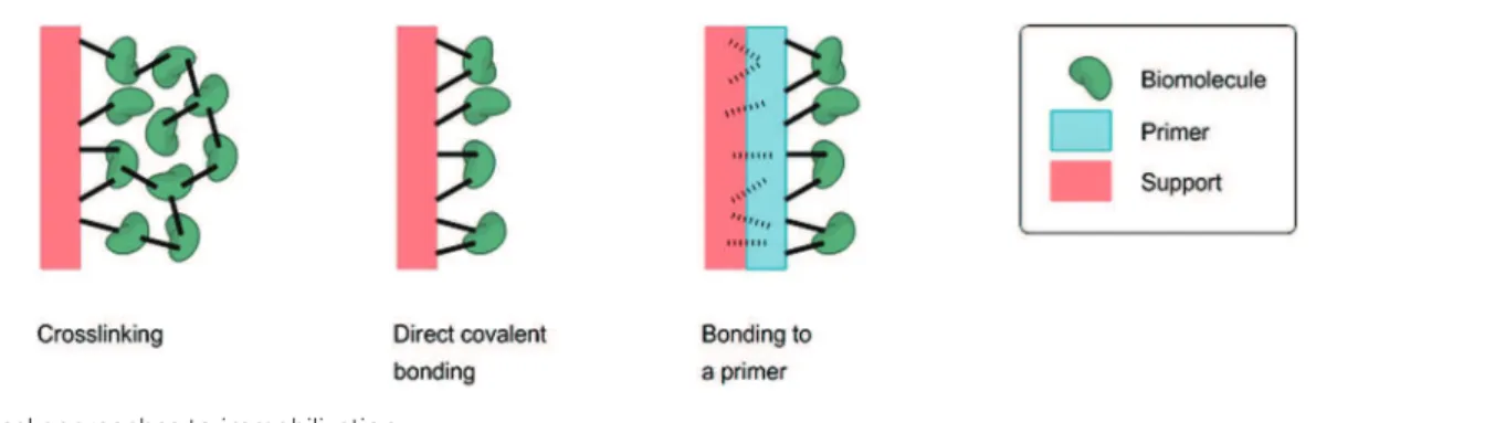
3.3.1. Crosslinking... 31
3.3.2. Direct covalent bonding ... 31
3.3.3. Bonding to a polymeric primer ... 33
4. SUMMARY AND OUTLOOK ... 33
REFERENCES ... 35
C
HAPTER
2
A ONE
-
STEP AND BIOCOMPATIBLE CELLULOSE FUNCTIONALIZATION FOR
COVALENT ANTIBODY IMMOBILIZATION ON IMMUNOASSAY MEMBRANES
... 41
1 INTRODUCTION ... 43
2 EXPERIMENTAL ... 44
2.4.1 Paper activation: EDC/NHS activation of carboxyl groups ... 45
2.4.2 Paper activation: SATA derivatization of primary amine groups... 45
2.4.3 Antibody activation: Preparation of maleimido-antibodies ... 45
2.5 IMMUNOASSAYS ... 45
2.5.1 Preparation of colloidal-gold-labeled antibodies ... 45
2.5.2 Detection of the grafted antibodies (incubation assay) ... 46
2.5.3 Detection of antigen by sandwich immunoassay (incubation assay) ... 46
2.5.4 Immunochromatographic strips (LFIA) ... 46
3 RESULTS AND DISCUSSION ... 46
3.1 ONE-STEP CELLULOSE SHEET FUNCTIONALIZATION UNDER SOFT CONDITIONS ... 46
3.2 COVALENT BINDING OF ANTIBODIES TO CELLULOSE PAPER SHEETS ... 48
3.3 CONSERVATION OF THE BIOLOGICAL ACTIVITY ... 49
3.4 USE IN LATERAL FLOW IMMUNOASSAY ... 50
4 CONCLUSIONS ... 51
REFERENCES ... 51
SUPPLEMENTARY MATERIAL ... 53
C
HAPTER
3
CHEMICAL
-
FREE PHOTOIMMOBILIZATION OF ANTIBODIES ONTO CELLULOSE FOR
THE PREPARATION
OF IMMUNOASSAY MEMBRANES
... 55
1. INTRODUCTION ... 57
2. EXPERIMENTAL ... 58
2.1. REAGENTSANDMATERIALS ... 58
2.2. PHOTOIMMOBILIZATIONOFANTIBODIES ... 58
2.2.1. General procedure ... 58
2.2.2. Variable parameters ... 59
2.3. IMMUNOCHROMATOGRAPHICASSAYS(LFIA) ... 59
2.3.1. Preparation of colloidal-gold-labeled antibodies ... 59
2.3.2. Preparation of immunochromatographic strips ... 59
2.3.3. Evaluation of the immobilization rate ... 60
2.3.4. Evaluation of the activity rate ... 60
2.4. PHOTOIMMOBILIZATIONOFPROBEANTIBODIES ... 60
3. RESULTS AND DISCUSSION ... 60
3.1. OPTIMIZATIONOFIMMOBILIZATIONPARAMETERS ... 60
3.1.1. Photoenergy ... 60
3.1.2. Pre-irradiation drying step ... 61
3.1.3. Post-irradiation washing step ... 62
3.1.4. Wavelength ... 62
3.1.5. Optimal procedure ... 62
3.2. USEOFVARIOUSPAPERSUBSTRATES... 63
3.3. AGEINGOFTHEMEMBRANES ... 63
3.4. STRENGTHOFTHEIMMOBILIZATION ... 64
3.5. PROPOSEDMECHANISM ... 64
4. CONCLUSION ... 64
PREPARATION OF IMMUNOASSAY MEMBRANES
... 69
1. INTRODUCTION ... 71
2. EXPERIMENTAL ... 72
2.1. REAGENTSANDREACTIONMATERIALS ... 72
2.2. CHARACTERIZATIONMATERIALS ... 73
2.3. SUBSTRATESPRETREATMENT ... 73
2.4. IMMOBILIZATIONPROCEDURE ... 73
2.4.1. Printing ... 73
2.4.2. Immobilization ... 74
2.5. IMMUNOCHROMATOGRAPHICASSAYS(LFIA) ... 74
2.5.1. Preparation of colloidal-gold-labeled antibodies ... 74
2.5.2. Preparation of immunochromatographic strips ... 74
2.5.3. Assessment of the immobilization ... 74
2.5.4. Assessment of the biological activity and determination of the visual detection limit ... 74
2.6. PATTERNEDPHOTOIMMOBILIZATIONOFPROBEANTIBODIES ... 75
3. RESULTS AND DISCUSSION ... 75
3.1. LOCALIZEDIMMOBILIZATIONOFPROBEANTIBODIES ... 75
3.2. FROMCLASSICALAUTOMATICDISPENSINGTOINKJETPRINTINGOFANTIBODIES ... 75
3.3. INKJETPRINTINGOFANTIBODIESONTOVARIOUSSUBSTRATES ... 76
3.3.1. Inks ... 77
3.3.1.1. Composition ... 77
3.3.1.2. Rheology ... 77
3.3.2. Initial substrates ... 77
3.3.2.1. Molecular structure ... 77
3.3.2.2. Surface chemical analysis ... 78
3.3.2.3. Surface morphological structure ... 78
3.3.3. Printed substrates ... 79
3.3.3.1. Surface chemical analysis ... 79
3.3.3.2. Surface morphological structure ... 80
3.3.4. Lateral Flow Immunoassays (LFIAs) ... 80
3.4. INKJETPRINTINGOFCOMPLEXDESIGNS ... 81
4. CONCLUSION ... 82
REFERENCES ... 82
C
HAPTER
5
ONE
-
STEP AND ECO
-
FRIENDLY MODIFICATION OF CELLULOSE MEMBRANES BY
POLYMER GRAFTING
... 87
1. INTRODUCTION ... 89
2. EXPERIMENTAL ... 90
2.1. REAGENTSANDREACTIONMATERIALS ... 90
2.2. CHARACTERIZATIONMATERIALS ... 90
2.3. CELLULOSEGRAFTCOPOLYMERIZATION ... 90
2.3.1. Dipping procedure ... 90
2.3.2. Printing procedure ... 91
2.3.2.1. Ink formulation ... 91
3.1.2. Surface chemical analysis ... 92
3.1.3. Surface morphological structure ... 92
3.2. SPATIALLYCONTROLLEDCELLULOSEGRAFTCOPOLYMERIZATION ... 94
3.2.1. Ink behavior ... 95
3.2.2. Surface chemical analysis ... 95
3.2.3. Surface morphological structure ... 95
3.3. INKJETPRINTINGOFCOMPLEXPATTERNS ... 95
4. CONCLUSION ... 97
REFERENCES ... 97
C
ONCLUSION
... 101
Socio-economic context
In various domains such as clinical diagnosis [1–5], drug screening [6–9], food quality control [10–12], and environmental monitoring [13–16], there is a need for easy and rapid detection of target molecules. Several methods have been developed for manufacturing biosensors, biochips, microarray and other immunoassay devices [7,17–22]. According to the World Health Organization (WHO), diagnostic devices for developing countries should be ASSURED: Affordable, Sensitive, Specific, User-friendly, Rapid and robust, Equipment free and Deliverable to end-users [1,23,24]. Furthermore, the current ecological and economic global issues have resulted in an increasing will for sustainable technologic development. Hence, the search for renewable-resources-based procedures and environmentally friendly materials and processes, as well as cost-saving approaches, has been stimulated widely [25].
Scientific context
As the main component of plant skeleton, cellulose is an almost inexhaustible raw material [26,27] and the most abundant form of worldwide biomass (about 1.5 x 10¹² tons per year) [28]. It is therefore an affordable biopolymer with lots of appealing properties such as large bioavailability, good biodegradability and biocompatibility [26,27,29,30]. Moreover, cellulose is insoluble in most usual organic solvents. It swells but does not dissolve in water, hence enabling aqueous fluids and their contained components to penetrate within the fibers matrix and to wick by capillarity with no need for any external power source. With special regard to cellulose paper, porosity combined to biocompatibility allows biological compounds to be stored in the paper device [31]. Besides, cellulose sheets are available in a broad range of thicknesses and well-defined pore sizes, easy to store and handle, and lastly safely disposable [1,32,33]. All of its features make cellulose an ideal material for creating novel diagnostic devices and improving point-of-care (POC) testing [29]. Paper-based assays such as dipstick tests or lateral flow assays have already been marketed and extensively employed for point-of-care (POC) diagnostics and pathogen detection since the 80s (diabetes and pregnancy tests being the most famous) [24,34–40]. Yet, the recent impetus given to paper-based microfluidics by American, Canadian and Finnish research teams [41–43] has resulted in the development of new paper-based bioanalytical devices with complex designs allowing multiplex diagnosis [2,14,21,44–48].
The preparation of efficient immunoassay devices requires the robust immobilization of a large number of biosensing molecules on a support [49]. Because of its ability to immobilize all kind of proteins by a combination of electrostatic, hydrogen, and hydrophobic interactions involving the nitro functions displayed on its surface [37], nitrocellulose constitutes the most commonly used support material for preparing immunochromatographic devices [35–37,50]. However, nitrocellulose is an expensive, fragile and inflammable material [51,52], which was shown to be incompatible with most procedures implemented in the development of new multiplex biosensors such as lab-on-paper devices, microfluidic paper analytical devices (µPADs), or other paper-based analytical devices [7,21,33]. In addition, some agents such as spores and some bacteria may have difficulty in migrating along nitrocellulose. For these reasons, nitrocellulose tends to be replaced by cellulose [25,33].
Objectives
Several methods for immobilizing biomolecules onto cellulose are known. They may be classified into three major families which are presented in Chapter 1. Each of these methods displays specific advantages and drawbacks which are also discussed in this chapter. However, cellulose does not immobilize proteins by adsorption as well as nitrocellulose. Recent findings revealed that about 40% of antibody molecules adsorbed onto cellulose paper can actually desorb from the fibers [53]. Direct adsorption of antibodies onto cellulose is therefore too weak to allow the permanent immobilization required in the development of effective immunoassay [49]. Biomolecules should therefore be covalently bound to the paper [32,49] and thus cellulose needs to be functionalized or activated [32,54,55]. Ideally, chemical covalent bonding should be conducted in mild conditions, with few side reactions, in few steps, with a minimum denaturation of the immobilized biomolecule which needs to keep its original functionality [32,49].There is therefore an ongoing need for time and cost-saving methods allowing immunoassay devices to be prepared by robust and sustainable binding of biomolecules to cellulose.
amounts of reagents, solvents and adjuvants. Hence, the spatially controlled modification of cellulose surface seems crucial in the development of such devices since it enables to save expensive matter and to pattern surface properties [56]. In any case, modification procedures should abide by the economic and ecological objectives aforementioned.
Achievements
In this perspective, three processes allowing easy, robust and sustainable modification of cellulose sheets were developed. All are environmentally friendly, simple, time and cost-saving, and versatile. The first procedure (Chapter 2) is a functionalization of cellulose membranes for covalent antibody immobilization. While cellulose chemical modification is usually operated under harsh conditions in organic solvents, the diazonium-based procedure developed was performed in water, at room temperature, in a single step. Paper sheets have thus been modified and bear different chemical functions which enable to graft biomolecules by common bioconjugate techniques and to perform LFIAs. The second (Chapter 3) is a chemical-free photoimmobilization procedure which allowed antibodies to be immobilized onto cellulose without any photocoupling intermediate nor any biomolecule or substrate pretreatment. This immobilization technique was further combined to inkjet printing to localize the antibodies according to any pattern desired (Chapter 4). Native antibodies have thus been printed and immobilized onto paper sheets which therefore enable to perform LFIAs. Membranes’ performances were evaluated in terms of visual detection limit and challenged nitrocellulose performances. The third is a modification of cellulose membranes by polymer grafting (Chapter 5). Unlike the two previous processes, this technique was developed in order to increase the functionality of the non-sensing cellulose parts of paper-based devices. Yet, it may be employed as another functionalization method for covalent antibody immobilization onto cellulose. While cellulose graft copolymerization is usually performed through complex and expensive procedures, the employed diazonium-based approach was performed in water, at room temperature, in a short single step. Cellulose sheets have thus been grafted with several acrylic polymers: first globally through a dipping procedure and then locally by inkjet printing. All the strategies developed herein would be helpful to immobilize sensitive proteins on selected specific areas of cellulose sheets. More generally, these are powerful tools for easy and rapid modulation of cellulose surface properties according to complex designs, under soft and biocompatible conditions.
Outline
To sum up, this work focuses on the eco²-friendly preparation of paper-based immunoassay devices by means of simple and sustainable modification of cellulose membranes. To this end, Chapter 1 first provides an overview of cellulose structural features and physicochemical properties and then reviews current techniques for the immobilization of biomolecules onto cellulose membranes. Then, two aspects of paper-based immunoassay devices were considered: on one hand, the biosensing material immobilization and resulting membranes’ performances (Chapter 2 to Chapter 4); and on the other hand, the modification of the non-sensing membranes’ properties (Chapter 5) (see Figure 1). Hence, the two methods developed for immobilizing proteins are described in Chapter 2 and Chapter 3. Chapter 4 emphasizes the advantages of using inkjet printing as biomolecule dispensing technique and displays the results of its combination to the immobilization method described in Chapter 3. Finally, Chapter 5 presents the polymer grafting method elaborated to increase cellulose functionality.
material
Contents
1. INTRODUCTION ... 17
2. CELLULOSE: A BIOCOMPATIBLE MATERIAL ... 18
2.1. FEATURES ... 18
2.1.1. Structure ... 18
2.1.1.1. Molecular structure ... 18
2.1.1.2. Supramolecular structure ... 19
2.1.1.3. Morphological structure ... 19
2.1.2. Bioavailability and fiber components ... 20
2.1.3. Biodegradability ... 22
2.2. PHYSICOCHEMICALPROPERTIES ... 22
2.2.1. Mechanical properties: “the branch bends but does not break” ... 22
2.2.2. Chemical reactivity: functional cellulose derivatives ... 23
2.2.2.1. Oxidation ... 23
2.2.2.2. Amination ... 24
2.2.2.3. Esterification and etherification ... 24
2.2.2.4. Radical Copolymerization ... 24
2.3. FROMPAPYRUSTONANOMATERIAL... 25
2.3.1. Paper ... 26
2.3.2. Bioactive paper ... 26
3. BIOMOLECULE-BEARING CELLULOSE: A BIOACTIVE MATERIAL ... 27
3.1. PHYSICALMETHODS ... 27
3.1.1. Direct adsorption ... 28
3.1.2. Adsorption of carrier particles: bioactive inks ... 28
3.1.3. Confinement ... 29
3.2. BIOLOGICALMETHODS:BIOAFFINITYATTACHMENT ... 29
3.2.1. Cellulose-binding domain (CBD) / Cellulose ... 29
3.2.2. Protein / Ligand ... 30
3.2.3. Protein A, G or L / Antibody ... 30
3.2.4. Metal ion / Chelator ... 30
3.3. CHEMICALMETHODS ... 31
3.3.1. Crosslinking... 31
3.3.2. Direct covalent bonding ... 31
3.3.3. Bonding to a polymeric primer ... 33
4. SUMMARY AND OUTLOOK ... 33
Credou, J.; Berthelot, T.
Cellulose: from biocompatible to bioactive material.
J. Mater. Chem. B 2014. DOI: 10.1039/C4TB00431K.
The immobilization of biomolecules onto cellulose paper turns this environmentally friendly material into a
platform for diagnostic devices.
Cellulose: from biocompatible to bioactive material
Julie Credou and Thomas Berthelot*
Since the papyri, cellulose has played a significant role in human culture, especially as paper. Nowadays, this ancient product has found new scientific applications in the expanding sector of paper-based technology. Among paper-based devices, paper-based biosensors raise a special interest. The high selectivity of biomolecules for target analytes makes these sensors efficient. Moreover, simple paper-based detection devices do not require hardware or specific technical skill. They are inexpensive, rapid, user-friendly and therefore highly promising for providing resource-limited settings with point-of-care diagnostics. The immobilization of biomolecules onto cellulose is a key step in the development of these sensing devices. Following an overview of cellulose structural features and physicochemical properties, this article reviews current techniques for the immobilization of biomolecules on paper membranes. These procedures are categorized into physical, biological and chemical approaches. There is no universal method for biomolecule immobilization. Thus, for a given paper-based biochip, each strategy can be considered.
1.
Introduction
Cellulose is the most abundant organic chemical on earth. This natural polymer was rst mentioned by the French chemist Anselme Payen in 1838.1 He suggested that the cell walls of
almost all plants are constructed of the same substance. He described that a resistant brous solid remains behind aer
treatment of various plant tissues with ammonia and acids, and aer subsequent extraction with water, alcohol and ether. By elemental analysis, he deduced its molecular formula to be C6H10O5. The term “cellulose” was rst used one year later in a
report of the French Academy of Sciences on Payen's work.2,3
The current economic and ecological situations have led to an increasing ecological awareness and a growing will for sustainable technologic and economic development. Thus, scientists are urged to search for environmentally friendly materials and renewable resources. As the main component of
Julie Credou entered the Ecole normale sup´erieure de Cachan (ENS Cachan) in 2007. She received her MSc degree in Chemistry from ENS Cachan and Paris-Sud University in 2011. She is currently a PhD student in the Laboratory of Innovation in Surface Chemistry and Nanosciences (LICSEN) at the French Alternative Energies and Atomic Energy Commission (CEA) (Saclay, France). Under the supervision of Dr Thomas Berthelot and Dr Herv´e Volland, her work focuses on the development of paper-based immunosensors for point-of-care diagnostic applications.
Dr Thomas Berthelot obtained his PhD in Organic Chemistry in 2005 in the Bordeaux I Univer-sity aer interdisciplinary studies at the frontier of Biology and Chemistry. In 2006, he joined the French Alternative Energies and Atomic Energy Commission (CEA) for a post-doctoral fellowship to develop biomimetic polymer membranes for fuel cells. In 2007, he was appointed permanent CEA researcher and he participated in the creation of a surface chem-istry start-up (Pegastech SA, France). He currently works at the Laboratory of Innovation in Surface and Nanosciences (LICSEN). His research focuses in NanoMedicine. He is the (co)author of more of than 30 peer review articles in international journals and 24 patents.
CEA Saclay, IRAMIS, NIMBE, LICSEN (Laboratory of Innovation in Surface Chemistry and Nanosciences), F-91191 Gif sur Yvette, France. E-mail: [email protected]; [email protected]; Fax: +33 169084044; Tel: +33 169086588
Cite this: DOI: 10.1039/c4tb00431k
Received 18th March 2014 Accepted 8th May 2014 DOI: 10.1039/c4tb00431k www.rsc.org/MaterialsB
Materials Chemistry B
FEATURE ARTICLE
P u b li she d on 12 M ay 2014. D ow nl oa de d b y CE A S ac la y on 24/ 06/ 2014 14: 47: 09.View Article Online
plant skeleton, cellulose is an almost inexhaustible raw mate-rial.4,2 It is therefore a key source of sustainable materials.5
Moreover, thanks to its biocompatibility and biodegradability, cellulose is gaining more and more importance and appears as a grade one material.6 Apart from its large bioavailability and
good biodegradability, cellulose has lots of appealing features. It is rigid, highly crystalline, insoluble in common organic solvents, and therefore an ideal structural engineering mate-rial.6 With special regard to cellulose paper, its wicking
prop-erties enable components to travel by capillarity with no need for any external power source. In addition, its biocompatibility and porosity allow biological compounds to be stored in the paper device.7 Besides, cellulose sheets are inexpensive,
avail-able in a broad range of thicknesses and well-dened pore sizes, easy to store and handle, and nally safely disposable.8
Because of all these features, a new technological sector has developed and has kept growing within the last ten years: paper-based technology.9Paper has attracted scientists' interest since
the 19thcentury. The rst urine test strips were developed by the French chemist Jules Maumen´e in 185010and marketed by the
English physiologist George Oliver in 1883.11,12A century later,
in 1943, Martin and Synge invented paper chromatography13,14
in order to analyze the amino-acid content of proteins. Contemporaneously, in 1949 M¨uller and Clegg carried out a study on the preferential elution of a mixture of pigments in a restricted channel designed on paper,15hence laying the
tech-nical basis of paper-based microuidics. Few years later, in 1957, the rst paper-based bioassay used an enzyme immobi-lized onto paper in order to detect glucose in urine.16In 1982,
paper-based immunoassays such as dipstick tests or lateral ow immunoassays (LFIAs) were further developed and mar-keted.17–20They were then extensively employed for point-of-care
(POC) diagnostics and pathogen detection,21,22 with diabetes
and pregnancy tests being the most famous.23,24 Recently,
further impetus was given to paper-based microuidics by Whitesides' research group with the development of three-dimensional microuidic paper analytical devices (mPADs).25
This opened the way to many other multiplex paper-based analytical devices.26–33Meanwhile, the Sentinel Bioactive Paper
Network was formed in Canada in 2005,34thereby setting the
paper-based bioassay as a whole new section of biosensing research. Thus, cellulose is not anymore the “brous solid that remains behind”, it is a material platform used to create novel devices for diagnostics, microuidics, and electronics.
According to the World Health Organization (WHO), diag-nostic devices for developing countries should be ASSURED: Affordable, Sensitive, Specic, User-friendly, Rapid and robust, Equipment free and Deliverable to end-users.21,35,36The
afore-mentioned appealing characteristics of cellulose therefore give paper-based devices a great potential to comply with these requirements and to improve point-of-care (POC) testing. Besides, it would be only logical for this natural biopolymer which is available anywhere to be readily available for use everywhere it is needed.
Among paper-based devices, bioactive papers raise a special interest because they can be useful in many elds including clinical diagnosis28,35,37,38and environmental monitoring.29,39–41
They are the main materials for developing paper-based point-of-care (POC) diagnostic devices and therefore will be the main subject of this paper. Thus, this review focuses on the way to develop a bioactive material from the biocompatible cellulose material. We will therefore concentrate on cellulose as a support for biomolecule immobilization. Aer describing the related cellulose features such as ber physicochemical properties, we will then present the existing strategies for biomolecule immobilization onto pure cellulose.
2.
Cellulose: a biocompatible material
According to IUPAC Recommendations 2012, biocompatibility is dened as the ability to be in contact with a living system without producing an adverse effect.42As a ubiquitous naturalbiopolymer, cellulose is by denition a biocompatible material.
2.1. Features
2.1.1. Structure. As a polymer, cellulose is a macromolecule and therefore needs to be dened on three structural levels: molecular, supramolecular and morphological levels. On the molecular level, cellulose is described as a single macromole-cule. Its chemical constitution, its reactive sites and its potential intramolecular interactions are considered. On the supramo-lecular level, cellulose is described as a pack of several macro-molecules interacting and ordering each other. Importance is attached to aggregation phenomena, crystalline organization and brils formation. On the morphological level, structural entities formed by cellulose are described. Layouts made of different supramolecular arrangements are studied.
2.1.1.1. Molecular structure. Cellulose possesses the simplest structure among polysaccharides since it is composed of a unique monomer: glucose under its b-D-glucopyranose
form (Fig. 1). Cellulose is a polydisperse, linear, syndiotactic polymer. Glucose molecules are covalently linked through acetal functions between the equatorial hydroxyl groups of C4 and the C1 carbon atoms. This succession of glycosidically linked anhydroglucose units (AGUs) results in a long chain b-1,4-glucan.2,3,6
The chain length, also called the degree of polymerization (DP), is expressed as the number of AGUs constituting the chain. The average DP value not only depends on the origin of the raw material, but also on the potential extraction treat-ments. For example, cellulose from wood pulp has average DP values around 300 and 1700. In the case of cotton and other
Fig. 1 Cellulose molecular structure (n ¼ DP, degree of polymerization).
P u b li she d on 12 M ay 2014. D ow nl oa de d b y CE A S ac la y on 24/ 06/ 2014 14: 47: 09.
plant bers, DP values range from 800 to 10 000. Similar values are reported in bacterial cellulose.2
Each AGU ring adopts the4C1chair conformation (Fig. 2).
Since the ring substituents and the glycosidic bonds are all in the ring plane (equatorial), this conformation ensures the less van der Waals and steric repulsion between them. It is the most stable conformation and thus the thermodynamically preferred conformation. To comply with this conformation and to accommodate the preferred bond angles of the acetal bridges, adjacent AGUs have their mean planes at an angle of 180" to
each other. Hence, two adjacent AGUs dene the disaccharide cellobiose (Fig. 1).2,6
Furthermore, both ends of the cellulose chain are different (Fig. 1). At one end, the glucose unit is still a closed ring and displays an original C4–OH group. This is the non-reducing end. At the other end, both pyranose ring structures (cyclic hemiacetal) displaying an original C1–OH group and an alde-hyde structure are in equilibrium (Fig. 3), thereby conferring reducing properties. This is the reducing end.
As a result of the glucose structure, cellulose contains a large amount of free hydroxyl groups located at the C2, C3, and C6 atoms. These hydroxyl groups, together with the oxygen atoms of both the pyranose ring and the glycosidic bond, form an extensive hydrogen bond network. This network is composed of both intra- and intermolecular hydrogen bonds. While the intramolecular hydrogen bonds are partly responsible for the linear integrity and rigidity of the polymer chain, intermolec-ular hydrogen bonds result in crystalline structures and other supramolecular arrangements. The main intramolecular hydrogen bond is the O3H–O50 bond; it is shared by most
allomorphs. O2H–O60 hydrogen bonds also occur in some
allomorphs. Both are shown in Fig. 46,43
2.1.1.2. Supramolecular structure. Pure cellulose exists in several allomorphic forms. Native cellulose I crystallized simultaneously in two forms in which chains are packed in parallel: Iaand Ib. On the other hand, chains in regenerated or
mercerized cellulose II are arranged antiparallel. Treatment of cellulose I and II with liquid ammonia leads to cellulose III1and
III2, respectively, and each allomorph may be converted back to
the starting cellulose material. Heat treatment of cellulose III1
and III2leads to cellulose IV1and IV2, respectively, which can
also be converted back to the original cellulose.44
With respect to cellulose I, the Ia/Ib ratio depends on the
origin of the cellulose. The Ibform prevails in woody plants and
cotton whereas the Iaform dominates in primitive organisms
such as bacteria or algae.3,45Cellulose I
ahas a triclinic unit cell
including one chain whereas Ib has a monoclinic unit cell
including two parallel chains. The Ibform is thermodynamically
more stable than the Iaform.
Cellulose II is the most stable among cellulose crystal structures. This allomorph can be produced from cellulose I by mercerization (treatment with aqueous sodium hydroxide) or by dissolution and following precipitation (regeneration of a crystalline form of cellulose). This transformation is considered to be irreversible.43Cellulose II has a monoclinic unit cell which
includes two antiparallel chains.2
As stated above, intermolecular hydrogen bonds are greatly responsible for the supramolecular structure of cellulose. They make the chains group together in a highly ordered structure. Cellulose I and II differ by their inter- and intramolecular hydrogen bonds, resulting in different packings: parallel and antiparallel, respectively (Fig. 5). The main intramolecular O3H–O50 hydrogen bond is shared by both polymorphs. The
intramolecular O2H–O60 hydrogen bond only occurs in
cellu-lose I (both Iaand Ib). Cellulose I has O6H–O300intermolecular
hydrogen bonds whereas cellulose II has O6H–O200
intermo-lecular hydrogen bonds.2,3
The chains are usually longer than the crystalline regions. As a consequence, one chain can run from one crystalline region to another, passing through amorphous areas, and thereby holding the ordered regions together.46,47The intermolecular
hydrogen bonds in the crystalline regions are strong, hence ensuring the resultant ber is strong as well and insoluble in most solvents. They also prevent cellulose from melting. In the amorphous regions, the intermolecular hydrogen bonds are fewer and looser, enabling the chains to form hydrogen bonds with other molecules such as water. This imparts macromo-lecular cellulose its hygroscopic and hydrophilic features. Thus, cellulose swells but does not dissolve in water.46
Cellulose bers have amorphous and crystalline regions. Their ratio, or crystallinity rate, depends on the origin of cellulose. Cotton, ax, ramie and sisal have high degrees of crystallinity which range from 65% to 70% whereas crystallinity of regenerated cellulose only ranges from 35% to 40%.6
2.1.1.3. Morphological structure. Gathering different supra-molecular arrangements of cellulose (crystalline and amor-phous areas) results in brillar elements of nanometer-scale diameters and micrometer-scale lengths.43,48 These are called
brils or microbrils. Assembling these microbrils together
Fig. 2 b-D-glucopyranose conformations.
Fig. 3 Reducing end equilibrium.
Fig. 4 Intramolecular hydrogen bonding in cellulose.
P u b li she d on 12 M ay 2014. D ow nl oa de d b y CE A S ac la y on 24/ 06/ 2014 14: 47: 09.
results in macrobrils of micrometer-scale diameters and millimeter-scale lengths. Micro- and macrobrils represent the building block of the cellulose ber cell wall.
Plant bers consist of different cell-wall layers (primary and secondary walls, middle lamellae) surrounding the central lumen. The lumen takes part in the water uptake behavior of plant bers. The primary cell wall must be capable of growth and therefore be exible. The secondary cell wall has to be rigid in order to avoid buckling.49The secondary cell wall accounts
for approximately 80% of the entire cell wall thickness. It therefore determines the mechanical properties of the ber.46,50
The secondary cell wall is made up of three layers. The thickest is the middle layer which consists of a series of helically wound cellular microbrils. The angle between the ber axis and the microbrils is called the microbrillar angle. Its average value varies from one ber to another. Features of each cell-wall layer are provided by the particular brillar layout and the amount of other components such as lignin (see next Section 2.1.2).6,43
Thus, cellulose forms the basic material of all plant bers. Fig. 6 presents how cellulose molecules and resultant brils take part in the cell walls of plant bers.
2.1.2. Bioavailability and ber components. Cellulose is the most abundant form of worldwide biomass.51It is the main
material of plant cell walls, and therefore the most important skeletal component in plants. Apart from plants which are the dominant cellulose suppliers, cellulose is also produced by algae, bacteria and fungi. Thus, about 1.5 $ 1012 tons are
biosynthesized annually, thereby leading cellulose to be considered an almost inexhaustible polymeric raw material.2
The conventional sources of cellulose are wood pulp and cotton linters.6 The seed hairs of the cotton plant provide
cellulose in almost the pure form. In contrast, the cell wall of woody plants provides a composite material mainly made of cellulose, hemicelluloses and lignin. It may also contain pectin, extractives such as waxes, or even proteins.2,4,6
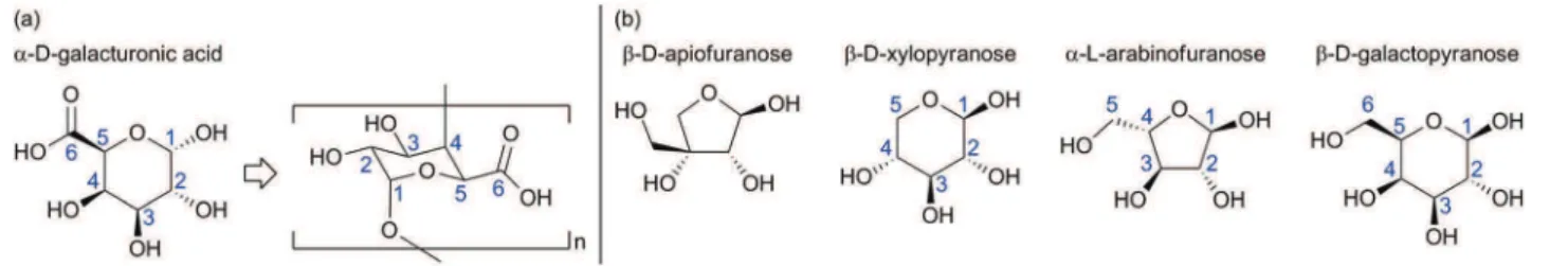
Hemicelluloses are water soluble polysaccharides of low degree of polymerization (100–200). While cellulose is a linear homopolymer of glucose, hemicelluloses are branched hetero-polymers made of many different sugars such as glucose, mannose, galactose, xylose and arabinose (see the most abun-dant sugar monomers in Fig. 7). Sugar ratio changes from plant to plant.3,6
As for lignin, this is a non-linear polymer made of phenyl-propanoid units. Its whole structure has not been fully resolved yet (see monomers and a representative fragment structure in Fig. 8) and its monomer ratio changes from plant to plant as well. While cellulose is the main building block of wood, lignin is the cement which binds the wood cells together. It is cova-lently linked to hemicellulose and thus crosslinks poly-saccharides, thereby giving rigidity to the plant.6,52In addition,
lignin plays a key role in controlling the water content within the cell wall and conducting water in plant stems. Whereas polysaccharides of plant cell walls are highly hydrophilic and thus permeable to water, lignin contains both hydrophilic and
Fig. 5 Supramolecular distinction between cellulose I and cellulose II lies in inter- and intramolecular hydrogen bonds.
Fig. 6 Contribution of cellulose to the cell wall of plant fiber.
P u b li she d on 12 M ay 2014. D ow nl oa de d b y CE A S ac la y on 24/ 06/ 2014 14: 47: 09.
hydrophobic groups which make it much less hydrophilic. Since lignin is crosslinked between polysaccharides, it stands in the way and prevents water absorption into the cell walls, thereby enabling water driving. Finally, because of its aromatic nature, lignin is mainly responsible for the color in wood. This feature appears as a drawback regarding papermaking industry. That is why processes such as pulping and bleaching have been developed in order to remove lignin from the wood matrix (see Section 2.3.1).3
Pectins are complex heteropolysaccharides mainly composed of (1 / 4)-a-D-galacturonic acid residues. The most
abundant pectic polysaccharide is a linear homopolymer of 1,4-linked-a-galacturonic acid called homogalacturonan. The other pectic polysaccharides are made of a backbone of 1,4-linked-a-galacturonic acid residues decorated with side branches consisting of different sugars and linkers.53 These
backbone and sugars are presented in Fig. 9. The amount, structure and composition of pectins vary from plant to plant, but also within a plant depending on the location and the age. Pectins are soluble in alkaline water. They provide exibility to plants. They also play a role in plant growth, development, morphogenesis, defense, cell–cell adhesion, wall structure, signaling, cell expansion, wall porosity, binding of ions, growth
factors and enzymes, pollen tube growth, seed hydration, leaf abscission, and fruit development.6,53
The protein content of wood cells is usually low (less than 1%), but can be higher in some grasses. The encountered proteins are structural proteins such as hydroxyproline-rich glycoproteins, glycine-rich proteins and proline-rich proteins.4
The extractives are all substances resulting from wood extraction processes that are not an integral part of the cellular structure. They are made soluble by extraction processes and can be removed by dissolution in solvents that do not dissolve cellulose such as water, ether, alcohol or benzene. The extrac-tive content of the wood material is about 2 to 5%.3Extractives
can be chemicals such as fats, fatty acids, fatty alcohols, phenols, terpenes, steroids, resin acids, rosin, waxes, etc. These chemicals may be encountered as monomers, dimers or poly-mers.4
Waxy layers contribute to render the ber impermeable to water.
All these alien substances associated with the cellulose matrix are important and should be kept in mind when further dealing with cellulose chemical modications. Indeed, they occur naturally in cellulose-containing materials and their ratio depends on the source of the cellulose (see distribution of these additives within some typical cellulose-containing materials in
Fig. 7 The most abundant monomers of wood hemicelluloses.
Fig. 8 (a) The three monomers of lignin. (b) A representative fragment of the lignin structure.
P u b li she d on 12 M ay 2014. D ow nl oa de d b y CE A S ac la y on 24/ 06/ 2014 14: 47: 09.
Table 1).43 Thus, depending on the source of the cellulose
material and the effectiveness of the purication process, these compounds may occur in the nal cellulose product and even-tually interfere with cellulose chemical modication.
2.1.3. Biodegradability. The increasing ecological aware-ness and the growing will for sustainable technologic and economic development have stimulated the search for envi-ronmentally friendly materials. In particular, the waste disposal problem has to be addressed quickly. These trends have tempted a large part of scientists to search for materials that can be easily biodegraded or bioassimilated.6 To these scientists,
cellulose therefore appears as a grade one material.
First of all, it is important to notice that cellulose is digestible by all grass-, leave- and wood-eating species, such as cows, pandas, beetle larvae and termites. This ability results from a lignocellulose-degrading symbiotic ecosystem located in their digestive tract. This ecosystem consists of bacteria or protozoa depending on the species which produce enzymes dedicated to break down cellulose.54–57The main glycolytic enzymes involved in
the biological conversion of cellulose to glucose are nases, cellobiohydrolases and b-glucosidases. While endogluca-nases randomly hydrolyze 1,4-b bonds along the cellulose chains, cellobiohydrolases split off cellobiosyl units from non-reducing end groups and b-glucosidases cleave glucosyl units from non-reducing end groups.54There are also other enzymes which are
dedicated to hydrolyze the other compounds from plant cell walls such as hemicellulase and xylan 1,4-b-xylosidase.55,57
Some fungi are also able to break down cellulose. Actually, fungi are among the most degradative organisms inducing biodeterioration of paper-based items.58 Many fungal species
(over 200) are involved in paper biodeterioration. The effec-tiveness and the rate of the deterioration process are affected by environmental conditions (e.g. temperature, humidity, light).59,60Their main strength is that a single cell is enough to
induce proliferation over most solid surfaces. Moreover, they can be “sleeping” for years as spores and then be reactivated under a certain set of conditions.61
Because of its sustainability, biocompatibility and biode-gradability, cellulose is a material of growing interest to the current economic and ecological climate.
2.2. Physicochemical properties
2.2.1. Mechanical properties: “the branch bends but does not break”. As stated above, plant cell walls are responsible for the proper growth and structural integrity of plants. As their main component, cellulose plays a key role in the shape and mechanical strength of living plants.49,62
Yet, the term strength may not make much sense by itself. In the informal language strength is synonymous with solidity, rmness or rigidity. But actually, the mechanical denition of the strength of a material mainly takes two properties into consideration: (i) the stiffness of the material, which is measured by its Young's modulus and (ii) the tensile strength (or ultimate tensile strength) of the material, which is the maximum stress that a material can withstand while being stretched before breaking. Considering that “the branch bends but does not break” means that plant bers have low Young's modulus but high tensile strength. The main asset of cellulose ber is therefore its resilience.
The tensile strength and Young's modulus of commercially important bers are detailed in Table 2.50,63,64Cellulose bers
have relatively high strength (tensile strength), medium stiff-ness (Young's modulus), and low density. Considering their lower density, the natural bers compare quite well with glass ber, but are not as strong as carbon bers or Kevlar.
Mechanical tests of whole plant or solid wood (macroscopic scale) provide information about their elementary mechanical properties which are partly inuenced by tissue interactions. Additionally, the tensile testing of single cellulose ber provides more information about the effects of the cell-wall structure on the mechanical properties of plant ber.50The tensile strength
of elementary bers is about 1500 MPa. Their Young's modulus depends on their diameter. It ranges from 39 GPa to 78 GPa for bers having diameters from 35 mm to 5 mm, respectively. From bulk natural bers to cellulose molecules, the elastic modulus
Fig. 9 (a) Galacturonic backbone of pectins. (b) The most abundant sugars of pectins.
Table 1 Chemical composition of some typical cellulose-containing materials
Source
Composition (%)
Cellulose Hemicellulose Lignin Extract
Cotton 95 2 1 0.4 Flax (retted) 71 21 2 6 Jute 71 14 13 2 Hemp 70 22 6 2 Corn cobs 45 35 15 5 Hardwood 43–47 25–35 16–24 2–8 Sowood 40–44 25–29 25–31 1–5 Bagasse 40 30 20 10 Coir 32–43 10–20 43–49 4 P u b li she d on 12 M ay 2014. D ow nl oa de d b y CE A S ac la y on 24/ 06/ 2014 14: 47: 09.
values range as follows: 10 GPa for wood bulk ber, 40 GPa for cellulose ber (aer pulping process), 70 GPa for microbril, and 250 GPa for the cellulose chain (from theoretical calcula-tions).46
In other words: “the smaller, the stronger”.
2.2.2. Chemical reactivity: functional cellulose derivatives. According to the molecular structure of cellulose (Fig. 1), hydroxyl groups in glucose units are responsible for its chemical activity. Under heterogeneous conditions their reactivity may be affected by their inherent chemical reactivity and by steric hindrance stemming either from the reagent or from the supramolecular structure of cellulose itself.47 Therefore, the
accessibility and reactivity of the hydroxyl groups depend on their degree of involvement in the supramolecular structure. In other words, it depends on their involvement in the hydrogen bond network. Intramolecular hydrogen bonding between adjacent AGUs particularly affects the reactivity of the C3 hydroxyl group, which hydrogen binds strongly to the ring oxygen on adjacent AGUs (O3H–O50hydrogen bond) whatever
the allomorph and is therefore not available to react.6 In
contrast, C2 and C6 hydroxyl groups have multiple and variable options to hydrogen bind, what may result in a lower statistical involvement in the hydrogen bond network, and thus a higher reactivity.3 Among the three hydroxyl groups in each glucose
residue, the one at 6-position (primary alcohol) is described as the most reactive site, far more than hydroxyl groups at 2- and 3-positions (secondary alcohols). However, the relative reactivity of the hydroxyl groups can be generally expressed in the following order: OH–C6 [ OH–C2 > OH–C3.47
The accessibility to these reactive hydroxyl groups also depends on the crystalline structure of the ber. Chemical reagents cannot penetrate the crystalline regions but only the amorphous area (see Section 2.1.1.2).47Activation treatments
can enhance the accessibility and the reactivity of cellulose for subsequent reactions. These treatments implement methods such as (i) widening surface cannulae, internal pores and interbrillar interstices, (ii) disrupting brillar aggregation, in order to make available additional areas, (iii) troubling the crystalline order, and (iv) modifying the crystal form and
therefore changing the hydrogen bonding scheme and the relative availability of the reactive hydroxyls. Among all activa-tion treatments, swelling is the most frequently used procedure and aqueous sodium hydroxide solution is the most common swelling agent. Swelling agents usually penetrate the ordered regions, and split some hydrogen intermolecular bonds. Aer alkali treatment (such as mercerization), the structure of native cellulose bers stays brillar but the degree of disorder increases, and so does the accessibility.47
When cellulose chemically reacts through its hydroxyl groups, the average number of hydroxyl groups per glucose unit that have been substituted denes the degree of substitution (DS) of the cellulose derivatives. Thus, its value ranges from 0 to 3. Because of the relative reactivity and accessibility of the hydroxyl groups, this value is oen lower than two, though. Besides, it is not desirable to have all of these hydroxyl groups react in order to keep the structure cohesion and integrity.65
Considering that the DS value is oen between 0 and 1.5,66it is
laborious to determine if we are only graing small molecules onto cellulose.65
The ways used to modify the chemical composition of synthetic polymers cannot be applied to natural cellulose because regarding cellulose these features are determined by biosynthesis. Chemical modications have to be conducted on the whole cellulose polymer. Though, introducing functional groups in the nal polymer is a way around the problem. These functional groups may impart new properties to the cellulose without destroying its many appealing intrinsic properties.47
Many approaches to cellulose functionalization already exist,67 and many others are under development.8,68,69 This
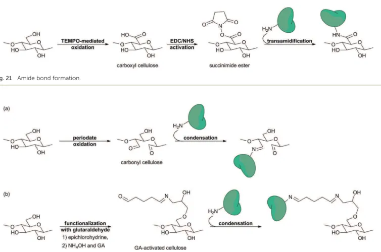
review focuses on cellulose as a support for biomolecule immobilization and its use for diagnostic devices. Therefore, not all the chemical modications of cellulose will be presented here. Instead we will concentrate on the chemical modications which play a role in biomolecule immobilization (see Section 3). 2.2.2.1. Oxidation. Carbonyl and carboxyl groups are very useful for biomolecule immobilization since they can react with primary amines from biomolecules to form imine and amide bonds, respectively (see Section 3.3.2). Carbonyl groups are already present at the reducing end of cellulose chains. Addi-tional carbonyl and carboxyl groups may stem from extraction and purication processes.2 Yet, those are not sufficient for
functionalization and biomolecule immobilization purposes. Therefore, more carbonyl or carboxyl groups would be obtained by oxidation of the hydroxyl groups from the cellulose. Depending on the experimental conditions, the oxidation may be accompanied by the opening of the pyranose ring (Fig. 10).70
The most used method of forming carbonyl groups onto the cellulose skeleton is periodate oxidation. Secondary alcohol groups of the glucose units (OH–C2 and OH–C3) are oxidized into the corresponding aldehydes by means of sodium periodate (NaIO4).40,71,72 This method results in the
opening of the pyranose ring by cleavage of the C2–C3 bond (Fig. 10b). Hence, the cellulose structure is locally affected. Depending on the oxidation rate, this may disrupt the linearity of the chain and the supramolecular arrangement to a certain extent.
Table 2 Mechanical properties of natural fibers compared to so-called “strong materials”
Fiber Density (g cm%3) Tensile strength (MPa) Young's modulus (GPa) Elongation at break (%) Cotton 1.5–1.6 287–597 5.5–12.6 7.0–8.0 Wood bers (spruce latewood) — 530–675 20.8–60.1 — Rayon 1.6 500 40 1.25 Flax 1.5 351 28.5 2.5 Hemp 1.48 820 29.6 3.5 Jute 1.5 579 26.2 1.5 Viscose (cord) — 593 11.0 11.4 Aramid (Kevlar 49) 1.45 2900 130 2.5 Carbon (NM) 1.86 2700 380 0.7 E-glass 2.54 2200 70 3.1
Portland cement concrete 2.2–2.4 2–5 14–41 —
P u b li she d on 12 M ay 2014. D ow nl oa de d b y CE A S ac la y on 24/ 06/ 2014 14: 47: 09.
The usual method of forming carboxyl groups onto the cellulose chain is TEMPO-mediated oxidation. Primary alcohol groups from cellulose (OH–C6) are oxidized into the corre-sponding carboxylic acids by means of sodium bromide (NaBr), sodium hypochlorite (NaClO) and (2,2,6,6-tetramethyl-piper-idin-1-yl)oxyl free radical (TEMPO).73–75 In this manner, the
pyranose ring is not affected by the process and cellulose keeps its structural integrity (Fig. 10a).
2.2.2.2. Amination. Amination of cellulose was used to covalently bind nitrilotriacetic acid (NTA) onto a cellulose lm.76
Aer loading these lms with nickel cations (Ni2+), it is there-fore possible to immobilize His-tagged proteins by bioaffinity attachment and develop biosensors or purication systems (see Section 3.2.4).
The amination process implements a complex procedure since usually both cellulose and amino compound added need to be activated before they can react with each other. However, the synthesis of the NTA-modied cellulose was achieved in two main steps: (i) the activation of the primary hydroxyl group from cellulose (OH–C6), and (ii) the SN2 nucleophilic substitution of
this activated hydroxyl by an activated NH2-terminal NTA
derivative (amination process). Fig. 11 illustrates the amination process resulting in nitrilotriacetic acid (NTA)-modied amino-cellulose.
First, hydroxyl groups were activated by tosylation. Cellulose was dissolved in a solution of lithium chloride in N,N-dime-thylacetamide (DMA/LiCl) which is the most important solvent system for cellulose in organic synthesis.2Tosyl chloride (Ts-Cl)
was added, together with triethylamine (Et3N). The average DS
value for the tosylation step was 1.45.76On the other hand, the
NH2-terminal NTA derivative was activated by persilylation with
trimethylsilyl chloride (TMS-Cl) in toluene in the presence of triethylamine. This activated NTA derivative nally reacted with the cellulose tosylate in a DMSO/toluene mixture (SN2). This
amination procedure resulted in NTA-cellulose. The average DS value for the amination reaction was 0.45.76
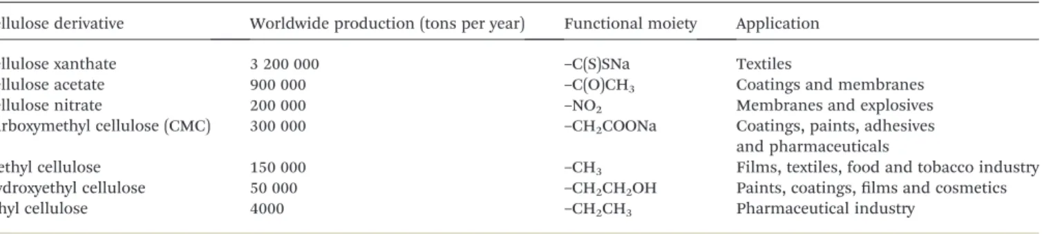
2.2.2.3. Esterication and etherication. Cellulose esters and cellulose ethers are the most important technical derivatives of cellulose.2
They nd their applications in many industrial
sectors including coatings, pharmaceuticals, foodstuffs and cosmetics (Table 3).47,69,77
With regard to biomolecule immobilization, cellulose nitrate (also named nitrocellulose) is the most important cellulose derivative. Biomolecules strongly adsorb to nitrocellulose through a combination of electrostatic, hydrogen, and hydro-phobic forces.20It is therefore the reference material for
per-forming lateral ow immunoassay (LFIA)18–20,78 (see Section
2.3.2). Cellulose nitrate is formed by esterication of hydroxyl groups from cellulose (primary or secondary) with nitric acid (HNO3) in the presence of sulfuric acid (H2SO4), phosphoric
acid (H3PO4) or acetic acid (CH3COOH) (see Fig. 12).47,67
Carboxymethyl cellulose (CMC) is another important cellu-lose derivative used in biomolecule immobilization. It is oen coated and strongly (some might say irreversibly79) adsorbed
onto cellulose (see Section 3.3.3). Thus, it provides carboxyl groups without oxidizing cellulose, thereby avoiding disruption of the hydrogen bond network and breach of the structural integrity. CMC is produced by etherication of hydroxyl groups from cellulose (primary or secondary) with monochloroacetic acid in the presence of sodium hydroxide (NaOH). Cellulose is rst activated with sodium hydroxide in order to enhance the reactivity of the hydroxyl groups as electron donors.43Then the
activated hydroxyl groups will substitute the chloride groups from monochloroacetic acid to yield CMC (see Fig. 13).80,81
2.2.2.4. Radical Copolymerization. Cellulose copolymers can be used for enhancing the rate of functional moieties on the cellulose surface. Therefore, they provide lots of anchoring points for biomolecule immobilization.82,83
Copolymer graing onto cellulose is usually performed by free radical polymerization of vinylic compounds. For initiating a gra side chain, a radical site has to be formed on the cellulose backbone. This radical can stem from the homolytic bond cleavage within the glucose unit caused by high-energy irradia-tion for example, from the decomposiirradia-tion of a funcirradia-tional group such as peroxide, or from a radical transfer reaction initiated by a radical formed outside the cellulose backbone during a redox reaction. The graing is usually conducted on a solid cellulose substrate with the monomer being in solution.47,67
Fig. 10 Main oxidation reactions of cellulose (a) without ring opening and (b) with ring opening.
P u b li she d on 12 M ay 2014. D ow nl oa de d b y CE A S ac la y on 24/ 06/ 2014 14: 47: 09.