Université de Montréal
Pseudopodial MSV-MDCK-INV glycolysis modulates the c-Met phospliory]atïon-dependent celi motility
By
Carlos El Hader
Physiology department f aculty of medicine
This thesis is presented to la faculté des Études Supérieures In the scope of obtaining a
Masters in Science (M.Sc.) li Physiology
December 2003 © Carlos El Hader, 2003 Université de
LI
o
V,’
cq
dl1
de Montréal
Direction des bibliothèques
AVIS
L’auteur a autorisé l’Université de Montréal à reproduire et diffuser, en totalité ou en partie, par quelque moyen que ce soit et sur quelque support que ce soit, et exclusivement à des fins non lucratives d’enseignement et de recherche, des copies de ce mémoire ou de cette thèse.
L’auteur et les coauteurs le cas échéant conservent la propriété du droit d’auteur et des droits moraux qui protègent ce document. Ni la thèse ou le mémoire, ni des extraits substantiels de ce document, ne doivent être imprimés ou autrement reproduits sans l’autorisation de l’auteur.
Afin de se conformer à la Loi canadienne sur la protection des renseignements personnels, quelques formulaires secondaires, coordonnées ou signatures intégrées au texte ont pu être enlevés de ce document. Bien que cela ait pu affecter la pagination, il n’y a aucun contenu manquant.
NOTICE
The author of this thesis or dissertation has granted a nonexclusive license allowing Université de Montréal to reproduce and publish the document, in part or in whole, and in any format, solely for noncommercial educational and research purposes.
The author and co-authors if applicable retain copyright ownership and moral rights in this document. Neither the whole thesis or dissertation, nor substantial extracts from it, may be printed or otherwise reproduced without the author’s permission.
In compliance with the Canadian Privacy Act some supporting forms, contact information or signatures may have been removed from the document. While this may affect the document page count, it does not represent any loss of content from the document.
Université de Montréal
Faculté des Études Supérieures
This thesis entitled:
Pseudopodial MSV-MDCK-INV glycolysis modulates the c-Met phosphorylation-dependent celi motility
Presented by Carlos El Hader
Has been evaluated by a jury consisting of the following individuals:
Jury president: Dr Alfred Berteloot Member ofjury: Dr André Gougoux
Study director: Dr Josette Noél
“7Zinef/&ç 11eveyone sayi Itftii/itern»g sometr»res to /boif r» th’ mr’ror anésee the errd’ncegftiai So gten we a’n ta.fe a moment to say w/iat r» our/ieai arnft/ien) w&n r’ too ht we wr we ,Çad Lin
taiffi a moment now, .9Wom
anfVaaf to te[fyou tiÇr... In t,Çe ,Çr’!î seas gF%Y, you have shîown me thie 4ihiQlianifyouftr
gur my way
q7îan)çftra1r)ays 6e»zg
my supporz
q a pdj andCari
U’/io atways encourageime toftf/bw my %eanri Id?’&ate tlii thes4 }lffrs contenti t/îefrst woréto ille h’st succesi
A
CKNO
WLED GEMENT$
I
would like to extend my appreciation to Dr. Josette Noél for allowing me the privilege of doing my research under her supervision. I offer my sincere thanks and gratitude for lier perpetual guidance, patience, understanding, but especially for lier expert sense of positive criticism. This work liad been realized witli lier personal commitment, support, advice and encouragement. For lier invaluable professional assistance, I am particularly grateful.I
am greatly indebted to the entire secretarial and technical staff of tlie department, botli past and the present. Gratitude is extended to Dr. Ivan Robert Nabi, in the Pathology and Cellular BioÏogy department of the Université de Montréal for lis unselfisli collaboration, especially with pseudopod purification and confocal microscope work.Finally, I am grateful to the GRTM (GEPROM) for the financial support, and to all my colleagues specially David Germain, for tlie teclmical, intellectual and moral support offered throughout tlie duration of this study.
SUMMARY
A metabolic imbalance exists in progressively malignant tumor ceils that have a high aerobic glycolysis. The MSV-MDCK-INV ceils are a Moloney sarcoma virus (MSV) transformed epithelial MDCK ceils, which exhibit multiple f3-actin rich domains where are localized the hepatocyte growth factor receptor c-Met, the glyceraldehyde-3-phosphate dehydrogenase and the N&’/H-exchanger. NHE1. A previous study from the laboratory of Dr No1 had demonstrated that the autocrine activation of the hepatocyte growth factor receptor/c-Met tyrosine kinase induces tumor ceil motility by regulating pseudopodial protrusion. We hypothesized that pseudopodial ATP produced from glycolysis and proton secreted by NHE1 drives pseudopodial formation, and modulates the motile character of MSV MDCK-INV cells.
We present here strong indication of a pseudopodial ATP production by showing the expression and enrichment of the glycolytic enzymes hexokinase, phosphoglucose isomerase and aldolase, and the absence of mitochondrial proteins from
f3
actin-rich pseudopodial domains. Glycolytic enzymes colocalize with [3-actin and phosphorylated c-Met. Incubating celis in glucose-free medium with or without 2-deoxyglucose, or inhibiting glycolysis with iodoacetate in high glucose medium decreases the phosphorylation level of c-Met and leads to the loss of pseudopodia. In contrast, mitochondrial inhibitors such as oligornycin and antimycin-A do flot affect this phosphorylation level in high glucose medium but decrease it in glucose-free medium. Pseudopodial glycolysis was found, in the present study, to be the main source of ATP required to phosphorylate the kinase domain of c-Met and lead to the actin remodelling, which were shown to modulate celi motility.Inhibition of the Na7Htexchanger with ethylisopropylamiloride in the absence of bicarbonate and C02 atmosphere leads to a rapid reduction of p c-Met signal. $ecretion of protons produced by glycolysis may be a
necessary step to drive continuous ATP production required for autocrine c Met phosphorylation. Our resuits suggest a functional link between pseudopodia-associated glycolysis, NHE1 proton secretion, and phosphorylation of c-Met that regulates motility and invasion of MSV MDCK4NV celis.
Key words: c-Met phosphorylation, actin remodeling, proton secretion, invasion.
SOMMAIRE
Un déséquilibre métabolique existe dans les cellules tumorales ayant une forte glycolyse aérobie. Les cellules MSV-MDCK-1NV sont des cellules MDCK transformées par le virus du sarcome de Moloney (MSV) sélectionnées pour leur propriété invasive. Elles présentent des domaines riches en actine
f3
où sont exprimés le récepteur du facteur de croissance hépatocytaire c-Met, la glyceraldehyde-3-phosphate déshydrogénase et l’échangeur Na7H NHE1. Des travaux antérieurs à la présente étude ont démontré que l’activation autocrine du récepteur c-Met module l’activité motile des cellules tumorales MSV-MDCK-NV en régulant la formation des pseudopodes. Nous avons fait l’hypothèse que l’ATP produit de l’activité glycolytique des pseudopodes ainsi que la sécrétion, par NHEI, des protons produit de l’activité glycolytique pseudopodiale sont impliqués dans la formation des pseudopodes et l’acquisition du caractère motile des cellules MSV-MDCK-INV.Nous présentons ici des indices d’une production pseudopodiale d’ATP en montrant l’expression ou l’enrichissement des enzymes de la glycolyse (hexokinase, phosphoglucose isornérase et aldolase) et l’absence des protéines mitochondriales des domaines riches en actine
f3
dans les fractions pseudopodiales purifiées. Les enzymes de la glycolyse colocalisent avec e-Met phosphorylé et l’actinef3
des pseudopodes. L’incubation des cellules dans un milieu sans glucose avec ou sans 2-deoxyglucose et l’inhibition de la glycolyse par l’iodoacétate dans un milieu riche en glucose diminuent le degré de phosphorylation de c-Met et entraînent la perte des pseudopodes. En revanche, les inhibiteurs mitochondriaux tels ciue l’oligomycine et lantimycine A n’affectent pas ce degré de phosphorylation dans le milieu riche en glucose mais le diminuent dans le milieu sans glucose. Ces résultats suggèrent que la glycolyse pseudopodiale est la source principale d’ATP requise pour phosphoryler localement le domaine kinase de c-Met en réponse au facteur de croissance hépatocytaire et pour fournir l’énergienécessaire au remodelage dtactine impliqué dans le phénomène de motilité des cellules MSV-MDCK-ll’JV.
L’inhibition de l’échangeur Na/H par I’éthylisopropylamiloride en l’absence d’ions bicarbonate et de C02 entraîne une réduction rapide du degré de phosphorylation de c-Met. La sécrétion des protons issus de la glycolyse peut donc être une étape nécessaire à une production continue d’ATP, exigée pour l’activation autocrine de c-Met. Nos résultats suggèrent donc l’existence d’un lien fonctionnel entre la glycolyse associée aux pseudopodes, la sécrétion localisée des protons par NHE1 et la phosphorylation de c-Met pour la modulation des activités motile et invasive des cellules tumorales MSV-MDCK-NV.
Mots clés: phosphorylation de c-Met, remodelage de l’actine; sécrétion de protons, invasion.
TABLE 0f CONTENTS Dedication iii Acknowledgements iv Summary V Sommaire vii Table of contents ix
List of figures xiii
List of abbreviations xv
Aimex xvii
Chapter 1. INTRODUCTION
1.1 CelI proliferation and differentiation
1.1.1 Introduction 2
1.1.2 Proliferation 2
1.1.2.1 Tumor growth and ceil proliferation in vivo 2
1.1.2.2 Celis inculture 4
1.1.2.3 Molecular events in ceil proliferation 5
1.1.2.4 Properties oftumor celis in culture 6
1.1.3 Differentiation 8
1.2 Growth factors
1.2.1 Introduction $
1.2.2 Classification ofgrowth factors 9
1.2.2.1 Platelet-derived growth factor family 9
1.2.2.2 Epidermal growth factor family 10
1.2.2.3 Fibroblast growth factor family 11
1.2.2.4 The Insulin family 10
1.2.3 General aspects ofHGF .11
1.2.3.1 Description of function 11
1.2.3.2 Location and control 12
1.2.3.3 Structural classification 13
1.2.4 Growth factor receptors with tyrosine kinase activity 14 1.2.4.1 Hepatocyte growth factor receptor (HGF-R) 15 1.2.4.2 Hepatocyte growth factor/receptor signaling pathways 18
1.3 Energy metabolism in normal and tumor tissues
1.3.1 Aerobic glycolysis ofneoplastic celis: The Warburg effect 19 1.3.1.1 Relationship between respiration and glycolysis 20
1.3.2 General aspects of enzymes in cancer 22
1.3.2.1 The Individual Reactions of Glycolysis 22 1.3.2.2 Glycolytic enzyme pattem in human cancer tissue 26
1.3.3 Anaerobic glycolysis ofneoplastic celis 26
1.3.3.1 Control ofanaerobic glycolysis by proton secretion 28
1.4 Intracellular p11 regulation in normal and tumor tissues
1.4.1 Secretion of protons by normal ceils (pH homeostasis) 28 1.4.1.1 Intracellular aikalinization : normal vs tumor celis 29 1.4.1.2 Extracellular acidification on tumor ceil 30 1.4.1.3 Importance ofNa’7H exchange and intracellular
ph in tumor celis 30
1.4.2 Molecular structure ofVertebrate Na7H exchangers 31
1.4.2.1 Introduction 31
1.4.2.2 Molecular identification of the Na/H exchangers 32
1.4.2.3 Structural features 33
1.4.2.4 The NHE-1 isoforrn 33
1.5 Invasion and metastasis
1 .5.1 Introduction .35
1.5.2 Turnor-host interactions during the metastatic cascade 35
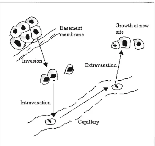
1.5.3 The metastatic cascade 36
1.6 Objectives of flic present study 40
CHAPTER 2. Pseudopodial MSV-MDCK-INV glycolysis modulates the c-Met phosphorylation-dependent ccli motility.
2.1 Summary 47 2.2 Introduction 4$ 2.3 Experimental procedures 51 2.4 Resuits 55 2.5 Discussion 60 2.6 References 64 CHAPTER 3. DISCUSSION 3.1 Study overview $2
3.2 Biochcmica] characterization of tumor ccli pseudopodia $2
3.3 3-actin rich tumor ccli pseudopodia: Role ofglycolysis 85
3.4
Phosphorylated c-Met induces tumor ccli motility by regulatingpseudopodial protrusion $6
3.5 Glycolysis as primary energy source in tumor ccli chemotaxis $7 3.6 Ccli migration requires botli ion transiocation and cytoskeletal
anchoring by the Na-H exchanger .89
3.7 Glycolysis and tumor ccli motility 91
3.8 Conclusion and perspectives 92
LIST 0F FIGURES
CHAPTER 1 Introduction
Figure 1.1 Celi cycle: A schematic overview
Figure 1.2 Model ofcell showing the sequence ofevents following
ligand binding to growth factor receptors.
Figure 1.3 HGF binding to the HGf-R’Met tyrosine kinase receptor
leads to activation of different signaling pathways
Figure 1.4 Pathway of glycolysis from glucose to pyruvate Figure 1.5 Schematic representation of the TCA cycle showing
enzymes, substrates and products
Figure 1.6 The general topology and regulatory sites ofthe Na7H
exchanger.
Figure 1.7 Tumbling down the metastatic cascade
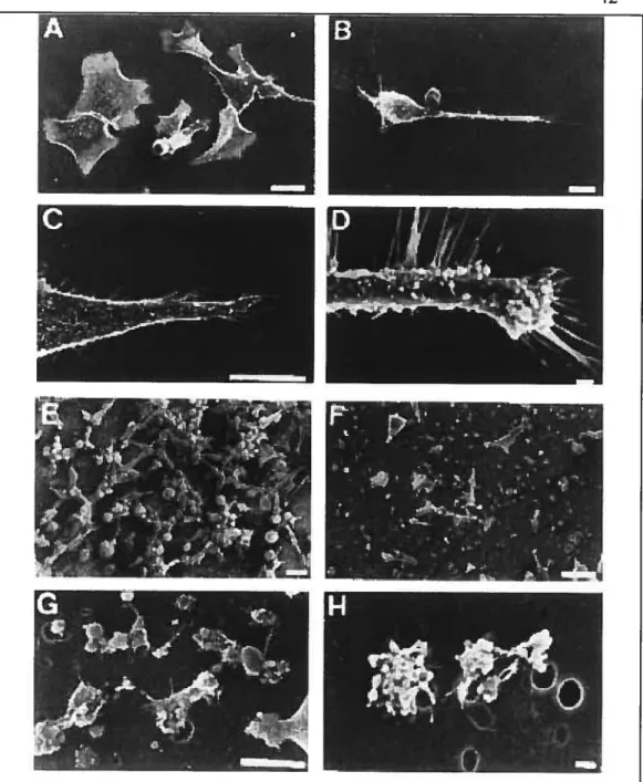
Figure 1.8 13-Actin-rich pseudopodia of MSV-MDCK-LNV celis are highly blebbed as visualized by scanning electron rnicroscopy
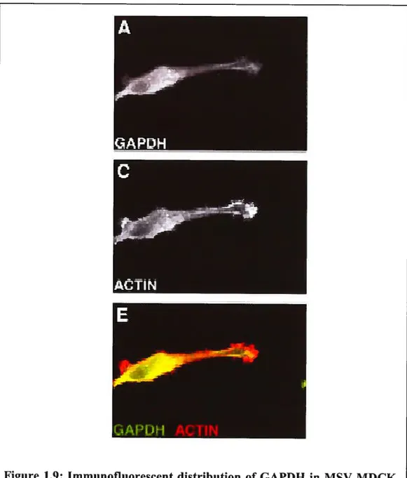
Figure 1.9 Immunofluorescent distribution of GAPDH in MSV
MDCK-INV ceils.
Figure 1.10 NHE1 is localized to b-actin-rich pseudopodia in MSV
MDCK-INV ceils.
CHAPTER 2 Article
Figure 2.1 Colocalization ofglycolytic enzymes with p-c-Met in
f3
-actin rich pseudopodia ofMSV-MDCK-fl’W celis
Figure 2.2 Pseudopodia ofM$V-MDCK-INV celis are divoided
Figure 2.3 Glycolytic enzymes are present or concentrated in pseudopodial fraction, and mitochondrial proteins are absent
Figure 2.4 Glycolytic activity regulates the actin dynamics and the phosphorylation level of c-Met
Figure 2.5 Phosphorylation degree of c-Met is higher in high glucose medium
compared to a glucose-free one
figure 2.6 Treatment with iodoacetate in high glucose medium resuits in a clear shortening and broadening of pseudopodia similar to that observed in low glucose medium
figure 2.7 Treatrnents with oligomycin or antimycin in glucose free compared to high glucose medium induced the lost ofpseudopodia, and appearance of multiple membrane blebs
Figure 2.8 Inhibition of Na+, H+exchange activity decreases c Met phosphorylation
Figure 2.9 The wound healing linked to the motile character of INV ceils is significantly decreased in the absence of glucose
CHAPTER 3 Discussion
Figure 3.1 The Iink between 13-actin, glycolytic enzymes, c-Met and NHE 1 in regulating ceil motility
LIST 0f ABBREVIATIONS
ALDO Aldolase
ATP Adenosine triphosphate
CB Celi body
c-Met Tyrosine kinase receptor for hepatocyte growth factor
2-DG 2-Deoxyglucose
EIPA Ethylisopropylamiloride
ERK Extra-cellular signal regulated kinase
FGF Fibroblast growth factor
G3PDH Glyceraldehyde-3-Phosphate dehydrogenase
HGF Hepatocyte growth factor
HGF-R Hepatocyte growth factor receptor
HK Hexokinase
LAA Iodoacetate
IGF Insulin-like growth factor
INV Invasive
kDa Kilodalton
MDCK Madin Darby canine kidney
MSV Moloney sarcoma virus
NHE1 Isoform 1 ofthe Na, H exchanger
Oligo Oligomycin
PDK1 Mitochondrial pyruvate dehydrogenase kinase
PGI Phosphoglucose isomerase
pHe Extracellular pH
pHi Intracellular pH
Pi liorganic phosphate
PK Pyruvate kinase
PPF Pseudopodia fraction
RTK Receptor tyrosine kinase
SF Scatter factor
Annex
Joseffe No1 My research supervisor. She directed ail the experiments. With her advice and suggestions, she participated in the design and the correction of the manuscript.
1. Robert Nabi He allows us the use of the confocal microscopy. He made suggestions and correction on the manuscript.
Zong Jiang Jïa He gave me advice conceming the pseudopod purification technique. He instructed me on how to use the confocal microscopy.
Yolaine Dodier and Zeinab Daher They made the experimentation concerning the inhibition of the Na/H exchanger with EIPA (Figure 2.9)
Carlos El Hader I performed ail the experirnents including: celi culture, drug treatrnents, immunofluorescence, confocal rnicroscopy, pseudopod and ceil body purification, western blotting, ATP dosage by luminometry, c-Met quantification, and wound healing assays. I perfonned the writing of the manuscript designed in collaboration withrny supervisor.
1.1 Ceil proliferation and differentiation
1.1.1 Introduction
The biology of ceil division and differentiation is exceedingly similar in normal and cancer ceils. The cancer ceil differs from its normal counterpart in that it is aberrantly regulated. Cancer cells generally contain the full complement of biomolecules necessary for survival, proliferation, differentiation, and expression of many celi-type-specific fiinctions. However, failure to regulate these functions properly results in an altered phenotype, which often leads to cancer (1).
Three cellular functions tend to be inappropriately regulated in a neoplasm: First, the normal constraints on cellular proliferation are relaxed (2). This is a necessary but often insufficient requirement for tumor formation. Second, differentiation can be distorted. The tumor cells may be blocked at a particular stage of differentiation, or they may differentiate into an inappropriate or abnormal cell type (3). Third, chromosomal and genetic organization may be destabilized such that variant cells arise with high frequency. Some variants may have an increased growth advantage, others may be resistant to killing by chemotherapeutic drugs or radiation and others may have increased motility or production of enzymes that permit invasion and metastases (4).
1.1.2 Proliferation
1.1.2.1 Tumor growth and ceil proliferation in vivo
In terms of population kinetics, the growth of any tissue depends on three parameters: 1) rate of individual cell division, 2) growth fraction of the ceil population and 3) cdl loss from the growing population through differentiation or cell death.
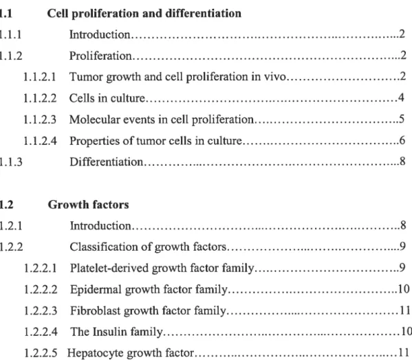
In an organism, the rate of ceil division is a tightly regulated process that is intimately associated with growth, differentiation and tissue turnover. Generally, ceils do not undergo division unless they receive signais that instnict them to enter the active segments of the ceil cycle. Resting cells are said to be in the G0 phase (quiescence) ofthe ceil cycle(Figure 1.1).
Figure 1. Schematic representation of the mammalian ce!! cycle. Competence factors such as PDGF and FGF promote entry into the early G1 phase. Sequential treatment with progression factors, IGF or EGF, promote progression through the G1 phase restriction point. Competence of the cyclin Dl/Cdk4 complex is induced by mitogens. The cyclin D1/Cdk4 complex phosphorylates the pRB protein leading to sequentiai phosphorylation by cyclin E/Cdk2 and release of free E2f. The phosphorylation ofpRB and relief of transcriptional repression by pRB induces genes involved in the induction Figure adaptedftom (S)
The signais that induce celis to divide are diverse and trigger a large number of signal transduction cascades. Generally, signals that direct ceils to enter the cell cycle are called growth factors, cytokines, or mitogens.
Normal cells reach a steady state of growth that provides a balanced economy for the body as a whole. Each organ maintains tight control over the growth rate, growth fraction, and cell loss. Some normal tissues grow faster than cancers under physiologie conditions, so it is flot simply rapid growth at a single time and place that distinguishes neoplasia.
In the early phases of turnor cell growth, it is generally believed that neoplastic cells multiply exponentially, then as the tumor mass increases, the rate of growth declines (6). Several mechanisms have been invoked to explain this change in growth rate with larger tumors: 1) decrease in the growth fraction, 2) increase in cdl loss or death, 3) nutritional depletion of tumor cells due to outgrowth of available blood supply, or 4) lengthening of cdl cycle time (7). Experimental tumor models suggest that cell cycle time changes only slightly when tumor growth decreases (1). Under adverse conditions, tumor cells oflen leave the growth fraction and enter a non growing state (Go or prolonged G1) although these same celis can reenter the division cycle when conditions improve or when stimulated by growth factors.
1.1.2.2 Celis in culture
The importance of the individual cdl in cancer is clear since a single cancer ccli injected into an appropriate animal is sufficient to give rise to a tumor. Many studies have therefore been performed with isolated normal and tumor celis in culture. Both normal and tumor-derived mammalian cells can be grown and compared in culture, although many are not readily established in culture initially (8-li). Most studies have been done on fibroblasts since this cdl type is easily cultured and is most likely to grow out of a tissue explants.
Cells are grown in a medium containing salts, amino acids, glucose, vitamins, and senim or growth factors. Normal celis, plated on a plastic surface to which they may attach, can grow until they have forrned a confluent monolayer, whereupon growth ceases. Growth also ceases when ceils have exhausted an essential nutrient or factor provided by serum, or when such substances are removed by changing to a deficient mcdium. Thus, growth can be manipulated in culture.
1.1.2.3 Molecular events in ce!! proliferation
When a quiescent cell in culture is stimulated, for example by the addition of serum, the activated chain of events leads eventually to formation oftwo cells. This requires duplication of a multitude ofmolecules in the original cdl (12). Multiple growth factors provided in serum have been found to act sequentially following resumption of proliferation of fibroblasts: platelet-derived growth factor (PDGF), epidermal growth factor (EGF), insulin-like growth factor (IGF-1), and hepatocyte growth factor (HGf) (13). These small polypeptides activate cells by binding to specific receptors on the cdl surface (13, 14).
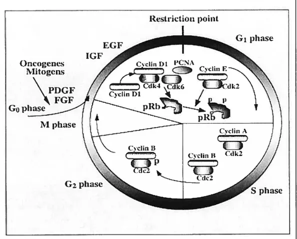
Growth factor receptors are complex large proteins that span the plasma membrane. On the outside of the cell they have a specific domain that recognizes the growth factor, and their cytoplasmic portion may have an enzymatic function, such as protein tyrosine kinase. Binding of a growth factor or ligand to its receptor can induce transmission of a signal to the cytoplasm through activation ofthe kinase (15) (Figure 1.2). The next step is the transduction of the cytoplasmic signal to cell nucleus (16). This is accomplished by a heterogeneous group of molecules known as second messengers. They include various proteins that are phosphorylated by kinases such as transcription factors, small molecules such as inositol phosphates and cyclic AMP, and also ions, including Ca2, H, and Zn2. Within the nucleus, genes are then activated in response to these second messengers.
1.1.2.4 Properties of tumor cells in culture
Tumor and normal ceils can be distinguished in culture by several tests. Tumorigenic cells are less sensitive to the presence of other ceils in their immediate vicinity than are normal celis. Normal cells typically cease proliferation as the culture density increases, but turnor ceils can reach several-fold higher densities in culture. If they are plated on a dense layer of non proliferating, homogenous normal cells, tumor cells can continue to grow and form foci of clustered cell colonies. Such colonies show altered interactions by the tumor cells, which grow randomly, criss-crossing one another and forming clusters of viable and necrotic celis. When these cultures are fixed and stained, number of tumor colonies is easily quantitied against the background population of normal cells. This is the basis of the commonly used “focus forming assay” also called “anchorage independent growth” for detecting transformation, e.g., the capacity of mutagens or oncogens to produce neoplastic transformation within a population of non tumor ceils (3).
Cells of normal solid tissue lie on a secreted extracellular matrix (ECM), cornposed of various proteins that stimulate ceil growth (17). Transformed celis are ofien partly or cornpletely independent of ECM for optimal growth, and they may secrete littie matrix material (18). In addition the cytoskeleton within tumor cells tends to be less well organized, and its actin filaments are less highly polymerized. Tumor cells often can be grown in the absence of a substratum, as within a semisolid medium containing agar. This formation of colonies in suspension is also used as a test ofneoplastic transformation (3).
Fi%ure 1.2: Model of ceil showing the sequence of events following ligand binding to growth factor receptors.
Figure adaptedfrom (19)
Extracellular (ligand binding)
(_)
C)
Ç
I—-’‘«se
1÷.
•.•
transmembrane( ‘r’
) (
É’
‘ktrac ellular (signalrng)
n
Ccli membrane
(
Addligand
Ligandbinding induces dimerisaif on
C)
‘aw.
• —c
__w Receptor dimr •C)
= activatedPh o sp h orylation if tran sph o sph 071 afi on May not happen until AVE ER clustering
=204 (phosphate)
Recruiunent of second messenger molecules & clustering
1.1.3 Differentiation
Ail tumor celis show abnorrnalities in the regulation of ceil proilferation (neoplasia). In addition, most, if flot ail, tumor celis show abnormalities in differentiation (anapiasia). The anaplasia of tumors can provide insights into their etiology, degree of malignancy, prognosis, and sensitivity to therapeutic intervention by differentiation or maturation-inducing agents (3).
It is obvious that although somatic ceiis are geneticaily equal they are flot phenotypically equal. Thus, skin fibroblasts are different from T lymphocytes, muscle cells differ from gastric mucosal cells, and so forth. However, within an organism, all ceils have an identical complement of DNA. Differences in phenotype arise from differences in gene expression, not in gene content (3).
1.2 Growth factors
1.2.1 Introduction
The evolution of multiceiiular organisms has involved the development of intercellular communication required for such processes as embryonic developrnent, tissue differentiation, as well as systemic responses to wounds and infections. These complex signaiing networks are in large part mediated by growth factors, cytokines and hormones. Such factors can influence cell proiiferation in positive or negative ways as well as inducing a series of differentiated responses in appropriate target cells. The interaction of a growth factor with its receptor by specific binding in tum activates a cascade of intracellular biochemical events that is ultimately responsible for the biofogical responses observed. Cytoplasmic molecules that mediate these responses have been tenued second messengers. The eventual transmission of biochemical signals to the nucleus leads to effects on the expression of cassettes of genes involved in mitogen and differentiation responses (20).
Over the past few years it has become increasingly evident that the pathogenic expression of criticai genes in growth factor signaling pathways can contribute to aitered ceil growth associated with malignancy. The v-sis oncogene of simian sarcoma virus, which encodes a growth factor homologous to the B chain of human platelet derived growth factor (PDGF B), is the paradigm for such genes (21, 22). The normal homologs of other oncogenes have been shown to encode membrane spanning growth factor receptors (23, 24). Other genes that act eariy in intracellular pathways of growth factor signal transduction, for example ras, have been implicated as oncogenes as weli. Present knowledge indicates that the constitutive activation of growth factor signaling pathways through genetic alterations affecting these genes contributes to the deveiopment and progression of most if not ail human cancers.
1.2.2 Classification of growth factors
Growth factors are proteins that bind to receptors on the cell surface, with the primary resuit of activating ceiiuiar proiiferation andlor differentiation. Many growth factors are quite versatile, stimulating cellular division in numerous different cell types, while others are specific to a particular ceii type (20).
1.2.2.1 Platelet-derived growth factor family
Platelet-derived growth factor is a cationic protein that consists of two reiated but non-identicai polypeptide chains designated A and B (also caiied PDGF-1 and PDGF-2), which is the major growth factor in human serum (21, 22). PDGF molecules exist as AA and BB homodimers as weii as an AB heterodimers (25, 26). PDGF-AB is the major PDGF form found in platelets and is released into serum upon blood clotting. Efforts to identify factors that control angiogenesis recently led to the identification of a new growth factor, the vascular endothelial growth factor (VEGF), that is a potent mitogen for vascular endothelial cells of small and large vessels, but has no effect on
fibroblasts, lens epithelial ceils, comeai endothelial celis, keratinocytes, or adrenal cortex cells (21). VEGF was shown to be a critical factor which stimulates neovascularisation of tumor ceils, in that way, favoring the deveiopment ofmetastasis.
1.2.2.2 Epidermal growth factor family
EGF purified from mouse sub-maxillary glands was found to promote precocious eyeiid separation by enhancing epidermal growth and keratinization while it induced eariy incisor eruption by eiihancing the differentiation of the lips of treated animais (27). Other members of the EGF family including tumor growth factor-a (TGF-a), amphiregulin (AR) and poxvirus growth factors share sequence similarities, high binding affinity to the EGF receptor and mitogenic effects on EGF-responsive celis. EGF is normally expressed in kidney and sub-maxiiiary glands and is produced in response to GI tract injury (27, 28) as well TGF-Πappears to be normally expressed by a variety of epithelial ceils (29).
1.2.2.3 Fibroblast growth factor family
There are seven known members of the fibroblast growth factor (FGF) family, whose targets include celis derived from mesoderm and neuroectoderm. Because heparin can bind to and modulate the biological activity of these proteins, they have aiso been termed heparin-binding growth factors (HBGFs) (30, 31).
1.2.2.4 The insulin family
The diversity of metabolic effects of insulin has been studied intensiveiy for decades (32). Its primary in vivo functions involve the regulation of rapid anaboiic responses such as glucose uptake, lipogenesis and amino acid and ion transport. Besides its effects on metabolism, insulin stimulates DNA synthesis and ceii growth. The activities of insuiin-like growth factors (IGF-I
and IGF-II) were first recognized as serum factors, antigenically distinct from insulin, that interacted with growth hormone in stimulating growth of skeletal tissues and were, as a resuit, termed somatomedins (33). Subsequently it was determined that somatomedin C is identical to IGF-I, while a polypeptide known as multiplication stimulating factor (MSA) is homologous to IGF-IT.
1.2.2.5 Hepatocyte growtli factor
A growth factor apparently specific for hepatocytes (HGf) was isolated from plasma (34) or platelets (35). Unexpectedly, the predicted amino acid sequence of HGF was found to be related to plasminogen (36). In addition to the 38% sequence identity to plasminogen, including its serine protease domain within the alpha chain, HGF was shown to possess disulfide bond linked intrachain structures known as “kringles” which are typical of prothrombin. Neither plasminogen nor plasmin have HGf-like activity, and HGF is flot likely to be a protease since the histidine and serine residues in the region corresponding to the catalytic site are replaced by other amino acids.
1.2.3 General aspects ofHGF
Hepatocyte growth factor (HGf) is a heparin binding, secreted basic protein (37) initially identified as a potent hepatotrophic factor responsible for vigorous regeneration of the liver. It has become a well-characterized multipotent growth factor (more properly called a cytokine) with biological functions that reach far beyond the original identifications, operating in virtually every tissue of the body (3$). Cellular targets include hepatocytes and other epithelial celis (e.g. lens epithelial celis) melanocytes, endothelial and haematopoietic celis.
1.2.3.1 Description offunction
HGf, the natural ligand for the c-Met proto-oncogene product, is a mesenchymal- or strornal-derived multipotent polypeptide that mediates
epithelial-mesenchymal interactions (39). During embryogenesis, HGF stimulates celi proliferation (e.g. placental cytotrophoblasts), differentiation, motility and invasiveness via its membrane-spaiming tyrosine kinase receptor, c-Met. HGf induces angiogenesis, is involved in haematopoiesis, chondrogenesis, and supports organogenesis and morphogenesis (40) of various tissues and organs, including the liver, kidney, lung, mammary gland, tooth, skeletal system, etc. In aduit tissues, HGF elicits a potent organotrophic function, which supports regeneration of organs including the liver, kidney, and lung.
HGF has been shown to play a pivotal role in integrin-mediated adhesion and transmigration of neutrophuls to sites of acute inflammation through cytoskeletal rearrangement (41). In neoplastic tissue, HGF is involved in tumor invasion and metastasis, through tumor-stromal interactions. Studies have shown anti-tumor activity for certain species of carcinoma celis, and in particular, growth of most hepatoma cells is inhibited by HGF.
1.2.3.2 Location and control
HGF is the ligand for the transmembrane tyrosine kinase (c-Met), and is initially secreted as a single-chain, biologically inert glycoprotein precursor (pro-HGF) (38). Under appropriate conditions, pro-HGF is converted into its bioactive form by proteolytic digestion; four proteases are reported to activate HGF within the so-called dibasic site (uPA) , a serine protease. This
processing takes place in the extracellular environment; cleaving of the zymogens induces a conformational change in the ligand, and interactionlactivation of the receptor follows. The HGF precursor can also be processed by a senim-derived serine protease. This soluble glycoprotein, known as HGF activator, may bring about quantitative activation of pro-HGF in response to the triggering of the blood coagulation cascade, as in the case of tissue injury, whereas uPA may effect a more restricted activation of the
precursor in the tissues and on the membrane of target ceils under conditions other than trauma or injury (42).
Expression of HGF at the transcription level is regulated by various factors. Interleukin- 1, platelet-derived growth factor, acidic and basic fibroblast growth factor, epidermal growth factor, and prostaglandin’s are potent inducers of HGF expression (39). Polysaccharides, such as heparin and heparan-suiphate, stimulate HGF synthesis, but only affect post transcriptional processes (39, 40). In fact heparin is crucial for inducing HGF dimerization (3-6 fold higher levels). Moreover, the fact that cellular responses to HGF depend on glycosaminoglycan composition of the ceil membrane further supports the hypothesis that receptor activation requires a co-operative participation of multiple surface and soluble components (38). In contrast, transforming growth factor-131 and glucocorticoids suppress the gene expression of HGf, acting transcriptionally (39).
1.2.3.3 Structural classification
Mature HGF is a heterodimer with a 69-kDa a-chain and a 34-kDa 13-chain, linked by a single disuiphide bridge (43). The Œ-chain contains the N-terminal hairpin structure and 4 homologous “kringle dornains”, the first of which carnes receptor-binding determinants (38). Although the kringle domain (80 amino acids) is thought to play a role in protein-protein interaction, its physiological function is unknown (44). However, a hydrophobic core and a distinct folding pattem determined by 3 intramolecular disulphide bonds, characterize the dominant structural feature. The 13-chain lias serine protease like motif (37) and thus lias structural homology (3 8%) with plasminogen but 2 arnino acid residues are different at the “protease active site11 conferring it no biological activities.
1.2.4 Growth factor receptors witli tyrosine kinase activity
Membrane spanning tyrosine kinase receptors contain severaÏ discrete dornains including their extra cellular ligand binding, transmembrane, juxtamembrane, protein tyrosine kinase and carboxy-terminal tau domains (45, 46). Interaction of a growth factor with its receptor at the cell surface leads to a tight association, so that growth factors are capable of mediating their activities at low nanomolar concentrations. following ligand binding, the growth factor-receptor complex is internalized leading to increased turnover of the receptor. It has been proposed that the growth factor activation signal might be mediated by its internalization (46). However, there is substantial evidence that activation of the receptor tyrosine kinase is the trigger for the biochernical cascade of events that follows. It is possible that conformational changes induced by ligand binding to the receptor’s external domain are somehow transrnitted through the transmembrane domain to induce the conformational alterations of the receptor kinase resulting in its activation (45, 46). li an alternative model more generally accepted, ligand binding induces receptor dimerization or oligomerization (46). By this latter mechanism, molecular interactions between adjacent cytoplasmic domains lead to activation of kinase function.
The tyrosine kinase domain is the most conserved a;nong tyrosine kinase receptors and an intact protein tyrosine kinase domain is absolutely required for receptor signaling. For example, mutation of a single lysine in the ATP binding site (45), which blocks the ability of the receptor to phosphorylate tyrosine residues, completely inactivates receptor biological ftinction. Yet, such kinase mutants retain the ability to bind ligand with high affinity and exhibit normal internalization and down regulation as well (45).
The carboxy terminal domain of the receptor is thought to play an important role in regulation of kinase activity. This region typically contains several tyrosine residues, which are phosphorylated by the activated kinase. In fact,
the receptor, itself, is ofien the major tyrosine phosphorylated species observed following ligand stimulation. Tyrosine phosphorylation of the carboxy terminal domain has been postulated to modulate kinase catalytic activity, an&or the ability of the kinase to interact with substrates. Thus, mutations, which alter individual tyrosine sites or deletions of the carboxy terminal domain, have the effect of attenuating kinase ftinction in those receptors so far analysed (45, 47).
1.2.4.1 Hepatocyte growth factor receptor (HGF-R)
The receptor for HGF, the c-met protooncogene (4$), is a transmembrane protein that is derived from a 1 70-kDa precursor. Afler processing by furin (49), the mature c-met is composed of a 50-kDa
c’
subunit that is linked by 2 disulfide bonds to a 145-kDa B subunit (50). The Œsubunit is extracellular andheavily glycosylated, whereas the B subunit consists of an extracellular portion to which HGF binds, a membrane spanning dornain, a distinctive juxtarnembrane domain, a cytoplasmic tyrosine kinase domain and carboxy
terminal sequences acting as a docking site for adaptors (Figure 1.3).
Autophosphorylation of the tyrosine residues in positions 1230, 1234, 1235 has a positive regulatory effect on the enzyme activity whereas autophosphorylation of a serine residue in position 1003 in the juxtarnembrane domain negatively regulates the kinase (3$). The carboxy terminal portion includes the tyrosine residues 1359 and 1365 that, when phosphorylated, together form a specific docking site for multiple signal transducers and adapters. Growth factor receptor-bound protein 2 (GRB2) binds preferentially to the second tyrosine residue and triggers the ras signal transduction pathway (32).
Cytcoi
IAG
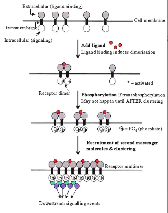
Figure 1.3: HGF binding to the HGF-RJMet tyrosine kinase receptor leads to activation of different signalïng pathways. HGF, upon binding to its receptor c-met, induces its dimerization as well as autophosphorylation of tyrosine residues. Phosphorylation of tyrosine residues within multifunctional site triggers a pleiotropic response involving multiple signal transducers. The synchronous activation of several signaling pathways is essential to conferring the distinct invasive growth ability of the HGF receptor. In epithelial ceils, HGF functions as a scattering (dissociation!motility) factor.
figttre adaptedftom (51) r.Jiiii:r.iKhc:i u Pt.: IIG[ I_IEc, I
C.
tdlaL i:coI txt ric.II u latThe polypeptide growth factor hepatocyte growth factor/scatter factor (HGF/$F) (44, 52-54) and its receptor MET, the product of the c-MET proto oncogene (55), play essential roles in the developrnent of epithelial organs such as the placenta and liver (56, 57) and in the migration of myogenic precursor ceils (58) and motor neurons (59, 60).
HGF/SF and MET are also involved in the spreading of a variety of epithelial tumors as a resuit of MET chromosomal rearrangements (61), somatic and/or germ une mutations in the MET kinase (62) or, more oflen, over expression in tumor ceils of an unreananged and unmutated MET gene (63).
Met signaling clearly has a role in normal cellular processes. When this signaling pathway is deregulated, it is implicated in tumor development and progression. Met signaling can increase tumorigenicity, induce ceil motility, and enhance invasiveness in vitro and metastasis in vivo (56, 64, 65). In addition, Met signaling can increase the production of protease and urokinase, which are associated with extracellular matrix/basal membrane degradation and are important for metastasis (66).
Following c-Met activation by HGF in tumor celis, phosphorylation of beta catenin occurs, together with loss of intercellular adhesion and a gain in the motile and invasive nature of the celi. It was shown that c-met is co-localized with beta-catenin and E-cadherin at regions of cell-cell contact in MCF7 and MDA MB 231 BCC unes (transformed mammary celi unes). Immunoprecipitation studies demonstrated an association between c-met and members of the cadherin adhesion complex in these epithe1ia tumor ceils, along with the membrane tyrosine protein phosphatase, PTPmu (67).
Activating Met mutations leading to increased levels of tyrosine phosphorylation and enhanced kinase activity toward an exogenous substrate when compared with wild type Met could contribute to papillary renal carcinoma and other human malignancies (56, 68). Mutant Met induced
motility of Madin-Darby canine kidney celis and metastasis of NIH 3T3 celis while transgenic mice expressing the oncogenic form of Met developed metastatic manmiary carcinoma.
1.2.4.2 Hepatocyte growth factorireceptor signaling pathways
Foïlowing c-Met dimerization and autophosphorylation of tyrosine residues, various adaptors and signal transducers bind to the docking site’s phosphorylated residues. Interaction of growth factor receptor bound protein 2 (Grb2), gabl (Grb-2 associated binder 1), p$5-Pl3kinase, phospholipase C y (PLC y), the isoform 3 of signal transducer and activator of transcription factor (STAT-3) and the $rc homology/collagen (SHC) protein via Src homology domain 2 (SH2) triggers various signal transduction pathways (Figure 1.3). The synchronous activation of several signaling pathways is essential to conferring the distinct invasive growth ability of the HGF receptor.
HGF functions as a scattering (dissociationlmotility) factor for epithelial celis, and this ability seems to be mediated through the activation of STAT-3 (Miller and Padanilam 2001). Phosphorylation of adhesion complex regulatory proteins such as ZO- 1, 3 catenin, and focal adhesion kinase (FAK) may occur via activation of c-src. Another Bc12 interacting protein termed BAG-1 mediates the antiapoptotic signal of HGF receptor by a mechanism of receptor association independent from tyrosine residues. Akt phosphorylation through the Pl3kinase pathway also participates in the anti-apoptotic action of c-Met (51)
1.3 Energy metabolïsm in norma] and tumor tissues
1.3.1 Aerobic glycolysis of neoplastic cetts: 11w Warburg effect
A metabolic imbalance exists in progressively malignant tumor ceils. The observation that rapidly growing tumor celis have a high aerobic glycolysis was first reported by Warburg (69), who considered that elevated glycolysis was a consequence of impairment in the respiratory mechanism of celis caused by carcinogens. Warburg noted that in ail of the tumors he examined respiration was iow and glycolysis was high. In Warburg’s proposai, the carcinogenic agent is presumed to interfere with cell respiration and the celi either dies or adopts a fermentative mechanism to derive energy necessary for survival. The carcinogenic agent, whether it is a virus, anaerobiosis, X-ray, or chemical carcinogen, is presurned to damage respiration either by interfering with the extent of respiration or by impairing the effect of respiration, rendering it incapable of suppressing glycolysis. Warburg’s theory implied that damage to respiration should be ineversibie, but as yet there has flot been an unequivocai demonstration that the respiration of tumors is distributed. following Warburg’s initial observation ofa low rate of respiration in tumors, it was demonstrated that in some turnors the rate of respiration is normal and that severai none-cancer tissues have appreciable rates of aerobic glycolysis, such as the retina, leucocytes, kidney, medulia and intestinai mucosa (70).
Mitochondria from neoplastic tissues have a full complement of the enzymes of the citric acid cycle and also functional respiratory pigments, which serve as the components ofthe electron transport chain (71). Further, as emphasized by Weinhouse (72), neoplasms exhibit a normal Pasteur effect when measured in terms of the Meyerhof oxidation quotient, that is, the oxygen, which is consumed by malignant cells, is just as effective in quantitatively inhibiting the formation of glycolysis end products as that consumed by normai tissues. One of the most potent arguments against the Warburg’s hypothesis is the demonstration that the malignant celis respire at rates, which do not appear to
differ from that of their normai counterpart. Thus aerobic glycolysis is flot necessariiy an essential feature ofmaiignancy.
Despite the failure to observe a unique biochemical pattern of energy metabolism in ail tumor celis, a correlation of growth rate with increasing glycoiysis is observed in the hepatoma series (73). However, the question as to whether this is related to the primary event in oncogenesis is unanswered.
Racker (74) has proposed a new hypothesis, which focuses on “the findings of Warburg as a significant feature of cancer pathogenesis”. He proposes, “Tumors can be caused by a number of different primary lesions” ail of which “have in common the ability to cause a persistent alteration in the intracellular pH thereby upsetting the normal reguiatory mechanism that prevents uncontrolled growth”. Thus the so-cailed Warburg effect remains, and, in this authors opinion, is deserving of further study in view of the reported correlation between growth rate and glycoiysis (73). The increased aerobic glycolysis may reflect cellular alterations, which favor growing ceils.
1.3.1.1 Relationship between respiration and glycolysis
Before discussing possible explanations for an elevated aerobic glycolysis, it is perhaps useful to discuss the regulation of carbohydrate metabolism, particuÏarÏy the interrelationships of respiration and glycolysis. The fttndamentai controls, which operate on these processes at the celiuiar levei, are part of the basic system on which the action of extemal controls, such as hormones and growth factors, is superimposed.
Inhibition of glycolysis by oxidative phosphorylation is known as the Pasteur effect. The uncouplers of oxidative phosphorylation reverse the inhibition of glycolysis, indicating that phosphorylation of ADP by inorganic phosphate (75) and not respiration is important to the control mechanisrn. As pointed out by Racker, the Pasteur effect is expiained as follows: “the enzymes of glycolysis can metabolize glucose oniy when (a) inorganic phosphate and
ADP are available for the oxidation of glyceraldehyde-3-phosphate, and (b) the ATP: ADP ratio and the concentration of Pi and other ailosteric effects of glycolytic enzymes are suitable for catalytic action” (74).
Some key enzymes of glycolysis are reguiated allosterically by the ATP: ADP ratios as weii as by Pi levels. As first demonstrated by Bucher and Russman (76), the key enzymes subject to regulation are those whose substrates and products are displaced from equilibrium in the ccli. As an example, the phosphofructokinase reaction is inhibited by high concentrations of ATP but stimulated by Pi. further, inorganic phosphate reverses product inhibition of hexokinase-l by glucose-6-phosphate; aiso, inorganic phosphate as well as the NAD: NADH ratio regulates the oxidation of giyceraldehyde-3-phosphate. The NAD: NADH ratio is dependent on the availability of an electron acceptor such as pyruvate, and pyruvate formation from phosphoenolpyruvate is dependent on ADP availability. k is thus apparent that the levels of ATP, ADP, and Pi determine the rate of giycoiysis by their interaction at several key steps in the fermentation scherne (70).
The rate of respiration is also markedly dependent on the availabiiity of ADP and Pi and this dependence is known as respiratory controi. Thus the cofactors of the phosphoryiation system are common to both giycolysis and respiration. Further, there is competition between these two systems for ADP and Pi, which are in Iimited supply. As a chemical work (e.g., biosynthesis) or an osmotic work such as ion transport (e.g. Na and K by the Na pump) is performed, energy is utilized and ADP and Pi become availabie for further generation of energy. The inhibition of glucose utiiization by oxygen then is attributed in part to the iesser availabiiity of cofactors of the phosphorylation system under aerobic conditions (70).
1.3.2 Generat aspects ofglycolytic enzymes in cancer
One of the most notable developments in biochemistry during the past 50 years has been the recognition and description of the individual enzymes that mediate the various steps ofintermediary metabolism in tissues.
1.3.2.1 The individual reactions of glycolysis
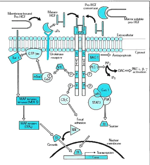
The pathway of glycolysis adapted from King et al., (Figure 1.4) can be seen as consisting of 2 separate phases
The first is the chemical priming phase requiring energy in the form of ATP, and the second is considered the energy-yielding phase. In the first phase, 2 equivalents of ATP are used to convert glucose to fructose 1 ,6-bisphosphate (Fi, 6BP). In the second phase Fi, 6BP is degraded to pyruvate, with the production of 4 equivalents ofATP and 2 equivalents ofNADH.
Some of the enzymes catalyze the reaction in one direction as, for example, glucokinase (or hexokinase IV) and hexokinase I, II, and III mediating the interaction of glucose with ATP to form glucose 6-phosphate, whereas the reverse reaction is mediated by glucose 6-phosphatase. Many other enzymes catalyze reversible reactions, and equilibrium is attained no matter from which direction the reaction is started.
The hexokinase reaction: The ATP-dependent phosphorylation of glucose to form glucose 6-phosphate (G6P) is the ftrst reaction of glycolysis, and is catalyzed by tissue-specific isoenzyrnes known as hexokinase. The phosphorylation accomplishes two goals: First, the hexokinase reaction converts nonionic glucose into an anion that is trapped in the cytoplasm, since cells lack transport systems for phosphorylated sugars. Second, the otherwise biologically inert glucose becomes activated into a labile form capable of being further metabolized.
Glucose
ATP—J hexokinase ADP ..‘1glucokinase Glucose-6-phosphate phosphohexose isom erase Fructose-6-phosphate ATP phospho ADP fwctokinase-1 Fructose-1 ,6-bisphosphateGlyceraldehyde-3-phosphate 1.Di hydroxyacetone
triosephosphate phosphate isomerase glyceraldehyde-3-pho sp hate dehydrogenase NADH+ R I ,3-bisphosphoglycerate ADP phosphoglycerate kinase 3-phosp[1oglycerate
f
phosphoglycerate+
mutase 2-phosphoglycerate enolase Phosphoenolpyruvate ADP pyruvate kinasePyruvate + NADH 4 Lactate + NAD
Fiure1.4: Pathway of glycolysis from glucose to pyruvate. Substrates and products are in blue; enzymes are in green. The two high-energy intermediates whose oxidations are coupled to ATP synthesis are shown in red. Under aerobic conditions, pyruvate in most celis is further metabolized via the ICA cycle. Under anaerobic conditions pyruvate is converted to lactate by the enzyme tactate dehydrogenase (LDH), and the lactate is transported out of the ce!! into the circulation. figitre adaptedftom (77)
Phosphohexose isomerase: The second reaction of glycolysis is an isomerization, in which G6P is converted to fructose 6-phosphate (F6P). The enzyme catalyzing this reaction is phosphohexose isomerase (also known as phosphoglucose isomerase). The reaction is freeÏy reversible at normal cellular concentrations of the two-hexose phosphates and thus catalyzes this interconversion during glycolytic carbon flow and during gluconeogenesis.
6-Phosphofructo-1-kinase (phosphofructokinase-1, PfK-1): The next reaction of glycolysis involves the utilization of a second ATP to convert F6P to fructose 1,bisphosphate (fi, 6BP). This reaction is catalyzed by 6-phosphofructo-1-kinase, better known as phosphofructokinase-1 or PFK-1. This reaction is not readily reversible because of its large positive free energy tAG0 = +5.4 kcal/mol) in the reverse direction. Nevertheless, fructose units
readily flow in the reverse (gluconeogenic) direction because of the ubiquitous prescnce of the hydrolytic enzyme, fructose-1, 6-bisphosphatase (F-l, 6-BPase).
Aldolase: Aldolase catalyses the hydrolysis of Fi, 6BP into two 3-carbon products: dihydroxyacetone phosphate (DHAP) and glyceraldehyde 3-phosphate (G3P). The aldolase reaction proceeds readily in the reverse direction, being utilized for both glycolysis and gluconeogenesis
Triose phosphate isomerase: The two products of the aldolase reaction equilibrate readily in a reaction catalyzed by triose phosphate isomerase. Succeeding reactions of glycolysis utilize G3P as a substrate; thus, the aldolase reaction is pulled in the glycolytic direction by mass action.
Glyceraldehyde-3-phosphate dehydrogenase: The second phase of glucose catabolism features the energy-yielding glycolytic reactions that produce ATP and NADH. In the first of these reactions, glyceraldehyde-3-phosphate dehydrogenase (G3PDH) catalyzes the NADtdependent oxidation of G3P to 1,3-bisphosphoglycerate (l,3BPG) and NADH. The G3PDH reaction is
reversible, and the same enzyme catalyzes the reverse reaction during gluconeogenesis.
Phosphoglycerate kinase: The high-energy phosphate of 1,3-BPG is used to form ATP and 3-phosphoglycerate (3PG) by the enzyme phosphoglycerate kinase. Note that this is the only reaction of glycolysis or gluconeogenesis that involves ATP and yet is reversible under normal celi conditions. Associated with the phosphoglycerate kinase pathway is an important reaction of erythrocytes, the formation of 2,3BPG by the enzyme bisphosphoglycerate mutase. 2,3BPG is an important regulator of hemoglobin’s affinity for oxygen. Note that 2,3-bisphosphoglycerate phosphatase degrades 2,3BPG to 3-phosphoglycerate, a normal intermediate of glycolysis. The 2,3BPG shunt thus operates with the expenditure of 1 equivalent of ATP per triose passed through the shunt. The process is flot reversible under physiological conditions.
Phosphoglycerate mutase and enolase: The remaining reactions of glycolysis are aimed at converting the relatively low energy phosphoacyl-ester of PG to a high-energy form and harvesting the phosphate as ATP. The 3-PG is first converted to 2-3-PG by phosphoglycerate mutase and the 2-3-PG conversion to phosphoenolpyruvate (PEP) is catalyzed by enolase
Pyruvate kinase: The final reaction of aerobic glycol ysis is catalyzed by the highly regulated enzyme pyruvate kinase (PK). In this strongly exergonic reaction, the high-energy phosphate of PEP is conserved as ATP. The loss of phosphate by PEP leads to the production of pyruvate in an unstable enol form, which spontaneously tautomerizes to the more stable, keto form of pyruvate. This reaction contributes a large proportion of the free energy of hydrolysis ofPEP.
1.3.2.2 Glycolytic enzyme pattern in human cancer tissue
Because of the high glycolytic activity of tumors and the assumption that the activities of glycolytic enzymes might be elevated in turnors, Warburg and Christian (7$) conceived the possibility that the blood leaving the tumor and entering the general circulation might show elevation of serum glycolytic enzymes. Indeed, they found that serum aldolase activity was elevated in rats bearing the Jensen sarcoma. These observations were confirmed by $ibley and Lehuinger (79) who, in addition, extended the studies to human cancer.
Since these early studies, other enzymes in the glycolytic sequence and, indeed, in other metabolic sequences have been studied with regard to their appearance in human serum and their elevation in patients with cancer. $everal factors may be involved in the elevation of a serum enzyme activity in cancer: 1- the damage of membranes of tumor cells or normal cells, so that one or more enzymes pass into the extracellular fluid and then into the circulation, 2- the size of the tumor or organ which is damaged and the concentration of enzymes in the tumor or normal organ and 3- the rate of disappearance of the enzyme from the circulation, whether by rnetabolisrn or by excretion. Elevations in serum enzyme activities may be characterized by varying degrees of specifity.
1.3.3 Anaerobic glycolysis of neoplastic celis
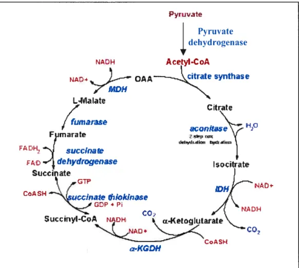
Under aerobic conditions, acetyl-CoA in most celis is ftirther metabolized via the TCA cycle (Figure 1.5). Under anaerobic conditions and in erythrocytes under aerobic conditions, pyruvate is converted to lactate by the enzyme lactate dehydrogenase (LDH), and the lactate is transported out ofthe celi into the circulation. The conversion of pyruvate to lactate, under anaerobic conditions, provides the cell with a mechanism for the oxidation of NADH Qroduced during the G3PDH reaction) to NAD, which occurs during the LDH catalyzed reaction. This reduction is required since NAD is a necessary
Pyruirate
Pyruvate dehydrogenase
NAEH Acetyl.CoA
NAD4 oAA—_,Scresynihase
L41aIate
f
Citrate/
furnaia.ç (y Fimarate ‘ t iIflriI,.III’n 141i FA Dit k f.D‘1,
ciehydrogenase Sticcinate I-
\
tw
CO» i Iccfnate
iok!nase jç GDP4ri/7
$cccFnyI-CoA NACH ci4(etci kiLarte
C
KGDH
Figure 1.5: Schematic representation of the TCA cycle showing enzymes, substrates and products. The abbreviated enzymes are: IDH = isocitrate
dehydrogenase and cx-KGDH = a-ketoglutarate dehydrogenase. The GTP
generated during the succinate thiokinase (succinyl-CoA synthetase) reaction is equivalent to a mole of ATP by virtue of the presence of nïtcÏeoside dthosphokinase. The 3 moles of NADH and 1 mole of f ADH2 generated during each round of the cycle feed into the oxidative phosphorylation pathway. Each mole of NADH leads to 3 moles of ATP and each mole of FADH2 leads to 2 moles of ATP. Therefore, for each mole of acetyl-CoA, which enters the TCA cycle, 12 moles ofATP can be generated.
substrate for G3PDH, without which glycolysis will cease. Normally, during aerobic glycolysis the electrons of cytoplasmic NADH are transferred to mitochondrial carriers of the oxidative phosphorylation pathway generating a continuous pool of cytoplasmic NAD.
Aerobic glycolysis generates substantially more ATP per mole of glucose oxidized than does anaerobic glycolysis. The utility of anaerobic glycolysis to a celi when it needs large amounts of energy stems from the fact that the rate of ATP production from glycolysis is approximately 1 OOX faster than from oxidative phosphorylation. Cells do flot need to energize anabolic reaction pathways. The requirement is to generate the maximum amount of ATP in the shortest time frame.
1.3.3.1 Control of glycolysis by proton secretion
There are several factors, which can contribute to the availability of the phosphorylating system and to the control of energy states in the cdl. Aerobic glycolysis of glucose to pyruvate requires two equivalents of ATP to activate the process, with the subsequent production of four equivalents of ATP and two equivalents of NADH. Thus, conversion, of one mole of glucose to two moles of pyruvate is accompanied by the net production of two moles each of ATP and NADH and 2 protons ($0).
1.4 Intracellular pH regulation in normal and tumor tissues
1.4.1 Secretion of protons by normal ceils (pH homeostasis)
The maintenance of an appropriate pH within mernbrane-enclosed compartments is a constant challenge for all living beings, from the simplest prokaryotes to complex multicellular organisms. Accordingly, cells have evolved a variety of specialized proton-translocating devices ($0).
ht mitochondria, cytochromes exploit the flux of electrons to extrude protons, thus producing a proton gradient that is used to generate useftil chemical energy in the form of ATP. Other organelles consume ATP to pump protons into the lumen to generate the acidic pH required for the maturation and processing of secretory proteins and for the dissociation and recycling of endocytosed materials. Proton-exchanger, ionophores of interest, allows an efficient extraction andlor release of cations by a simple control of the pH ($0).
1.4.1.1 Intracellular aikalinization: normal vs tumor celis
Measurement of pH in vivo has shown that the microenvironment of tumors is generally more acidic than in normal tissue, with median pH values of about 7.0 for tumors and 7.5 for normal tissue (69, 81, $2). The studies ofWarburg in the early part of the century (69) showed that tumor ceils preferentialïy convert glucose and other substrates to lactic acid, even under aerobic conditions (83). Since then, positron emission tomography (PET) and magnetic resonance spectroscopy (MRS) have consistently demonstrated the increased use of glycolysis in tumors (84-87). The increased use of the glycolytic pathway matched with the compromised vasculature of a tumor results in the poor removal of lactic acid and is believed to be the main cause of acidity within solid tumors (88).
Traditionally, most estimates of pH in tissue were obtained by insertion of pH electrodes (81). These were usually quite large in comparison to a tumor celi (83), and primarily measured interstitial or extracellular tissue pH. for rnost purposes, the parameter of interest is intracellular pH (pHi), and since lactic acid production can lead to intracellular acidosis, tumors were thought for many years to have a more acidic pHi (89). However, with the advent of MRS imaging for non-invasive measurements of pHi (83), both human and animal tumor cells have been shown to have intracellular pH similar to normal celis, near neutrality (pH 7.0) (88, 90), or even slightly alkaline (pH
7.1-7.2) (89, 91). This gives rise to a reversed pH gradient across the celi membrane between normal tissues and tumors.
1.4.1.2 Extracellular acidification on tumor celi
Despite the more acidic tumor microenvironment, most in vitro experiments are stili performed at the relatively alkaline extracellular pH (pHe) of 7.4. This is significant because slight changes in pHe can have profound effects on ceil phenotype (92). It has been shown that the metastatic potential of tumor ceils depends directly on the degree of acidification (93). More specifically studies by Martinez-Zaguilan and co-workers (92) have shown that the incubation of human melanoma cells under conditions of low pHe causes them to become more invasive and migratory. Furthermore, tumor angiogenesis may be regulated by pH. The switch to the angiogenic phenotype depends on a net balance of positive and negative angiogenic factors released by the tumor. Recent evidence from several laboratories (94-96) support a role for pH in this process, and indicate that tumor ceils at low pH, increase the expression of positive angiogdnic factors, such as vascular endothelial growth factor (VEGF) and interleukin-8 (IL-8), probably in order to increase the vasculature ofthe tumor.
1.4.1.3 Importance of Nat’H exchange and intracellular pH in tumor celis
Proper regulation of intracellular pH (pHi) is critical for the optimal function of almost every biologic process. In many cell types, pHi is regulated primarily by a family ofNa/ff’ exchangers (NHEs), transmembrane proteins that mediate the electroneutral exchange of an intracellular proton for an extracellular sodium ion. NHEY was the first Na/H exchanger isoform to be cloned and sequenced in 1989 (97). Since then, rnuch effort has been directed to identifying novel members of the Na/H exchanger family and
understanding their roles in a range of physiologie and pathophysiologic cellular processes, from cellular proliferation to apoptosis (98, 99).
The Na/H exchanger has been recently examined in several models of human turnors. They suggest that, in at least some celi types, the Na7H exchanger and intracellular pH regulation associated with this protein play an important role in tumor ceil growth (100). Early studies showed that the Na7H exchanger deficient ceils (101) lost or severely reduced their capacity to grow tumors in vivo in immune deficient mice. In addition, a variety of evidence suggested that activation of the Na/H exchanger and the resultant increases in intracellular pH may be required for mitogenesis in some celi types (98). In HC03 free media, Na7H exchanger deficient cells caimot proliferate in media of low extemal pH (101). In addition, extracellular Na is limiting in growth factor-dependent proliferation (102), and amiloride and its analogs can block changes caused by growth factor stimulation, especially in HC03 free or low Na medium (103).
Because of the important role of the Na7H exchanger in tumor and cell growth, arniloride and its analogs have been tested for use in tumor selective therapy.
1.4.2 Molecular structure of vertebrate Na/H exchangers
Na/H exchangers (NHE), also called antiporters, are vital transmembrane transporters involved in multiple cellular functions including the regulation of intracellular pH, the control of ceil volume and transepithelial ion transport (104, 105).
1.4.2.1 Introduction
Na7H antiport (exchange) is one of the primary mechanisms involved in the extrusion of H from vertebrate celis. Originally described by Murer et al., (106) in vesicles from brush-border membranes of kidney tubules, the
transporter has since been identified in the plasma membrane of virtually all eukaryotic cells. Biochemical studies and, more recently, molecular cloning have provided increasing evidence about the structure, functional features and regulation of the Na7H exchangers, referred to as NHE (107). Na7H exchangers are integral plasma membrane proteins that catalyze the electroneutral exchange of extracellular Na for intracellular H with a stoichiometry of one for one. An essential feature of these exchangers is their altosteric activation by intracellular protons, which are presumed to interact at a ‘modifier’ site that is separate from the sites involved in Na and H transport (107).
The activity and expression level of the exchangers can be modulated by a remarkably wide variety of stimuli, including growth factors, tumor promoters, hormones (107) and chronic extracellular acidification (108), as well as by physical factors, such as changes in ceil volume (109) or cell spreading (110). The most widely studied NHE isoform, NHE-1, is ubiquitously expressed and is involved in a variety of cellular functions by virtue of its ability to govern intracellular pH. It is inhibited by the diuretic compound arniloride and by the 5-amino-substituted derivatives of amiloride. Besides NHE- 1, many other isofonns, initially characterized by their lower sensitivity to amiloride, have been identified and cloned, principally in epithelia where they perform more specialized ion transport (NHE-2, NHE-3, NHE-4, NHE-8). NHE-6 and NHE-7 are specialized isoforms involved in organelle pHi regulation and Na transport (99).
1.4.2.2 Molecular identification ofthe Na17H exchangers
Using a genetic strategy, Sardet et al., (1989) were the first to identify a Na7H exchanger isoform fully by cloning a hurnan cDNA encoding the amiloride-sensitive growth-factor-activatable Na’7H antiporter. This cDNA, cloned by complementation of a Na7H antiport-deficient cell line, was shown to restore fully the biochemical and physiological features of the
transporter, namely pHi regulation, when transfected into antiporter-deficient ceils. This human exchanger cDNA, now referred to as NHE-1, is ubiquitously expressed in tissues and celis and, in polarized epithelial ceils, generally resides in the basolateral membrane, although exceptions have been reported (11 1). Pharmacological, kinetic and regulatory properties of Na7H exchangers in various ccli types, tissues and species had predicted the existence of multiple isoforms ofNa17H exchangers (112).
1.4.2.3 Structural features
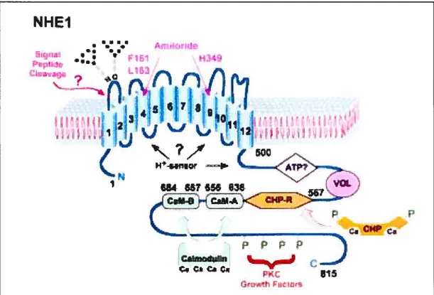
On the basis of their hydropathy profiles, all Na/H exchangers exhibit similar topologies. The molecule has two separate functionai domains: an N-terminal hydrophobic domain (made up of 10—12 membrane-spanning domains) with approximately 500 amino acids, and a C-terminal hydrophilic domain with around 300 amino acids (Figure 1.6). The N-terminal transmembrane part of NHE- 1 is necessary and sufficient to catalyze the ion exchange, whereas the cytoplasmic C-terminal domain determines the pH set point value of the exchanger and is crucial for mediating the activation of the exchanger by growth factors, hormones and hyperosmotic stress (113).
1.4.2.4 The NHE-1 isoform
Amiloride and its 5-amino-substituted derivatives are reported to inhibit the transporter by competing with Na for its extemai binding site (114). The amiloride-binding site appears to be iocated in the N-terminal domain of the exchanger since NHE- 1, afier rernoval of the entire cytoplasmic domain, remains sensitive to amiloride (113). NHE-1 is the isoform that is most sensitive to amiloride and its analogues (103, 115).
Most mitogens activate the NHE-1 isoform, leading to an intracellular alkalization (107), most easily detectable in the absence of bicarbonate (116, 117). At least three groups have reported that mitogenic agents activate the antiporter by shiffing the pH-dependence of the modifier site, adjusting the set