Published in: Trends in Neuroscience (2014), vol. 37, n°6, pp. 334-342 Doi: http://dx.doi.org/10.1016/j.tins.2014.03.005
Status: Postprint (author’s version)
*These authors contributed equally to this article.
Thinking out of the dish: what to learn about cortical
development using pluripotent stem cells
Jelle van den Ameele
1,2*
, Luca Tiberi
1*, Pierre Vanderhaeghen
1,3,4,5, and Ira
Espuny-Camacho1,4,5*1Université Libre de Bruxelles (ULB), Institute for Interdisciplinary Research (IRIBHM), and ULB Institute of Neuroscience (UNI), B-1070
Brussels, Belgium
2Department of Neurology, Ghent University Hospital, 9000 Ghent, Belgium 3 WELBIO, B-1070 Brussels, Belgium
4 VIB Center for the Biology of Disease, 3000 Leuven, Belgium 5 Center of Human Genetics, KU Leuven, 3000 Leuven, Belgium
ABSTRACT
The development of the cerebral cortex requires the tightly coordinated generation of dozens of neuronal subtypes that will populate specific layers and areas. Recent studies have revealed how pluripotent stem cells (PSC), whether of mouse or human origin, can differentiate into a wide range of cortical neurons in vitro, which can integrate appropriately into the brain following in vivo transplantation. These models are largely artificial but recapitulate a substantial fraction of the complex temporal and regional patterning events that occur during in vivo corticogenesis. Here, we review these findings with emphasis on the new perspectives that they have brought for understanding of cortical development, evolution, and diseases.
Glossary
Embryonic stem cell (ESC): pluripotent cell lines, typically derived from the inner cell mass of the early embryo (blastocyst),
capable of indefinite selfrenewal and differentiation into the derivatives of all three primary germ layers: ectoderm, endoderm, and mesoderm.
Induced pluripotent stem cell (iPSC): a pluripotent (ESC-like) cell that is obtained through reprogramming of differentiated cells,
typically through ectopic re-expression of a defined set of transcription factors.
Intermediate progenitor cells (IPC): neurogenic cortical progenitors that mostly undergo symmetric divisions to generate two
neurons, or sometimes two IPC. IPC are located in the subventricular zone (SVZ) and lack a defined polarity. Neuroepithelial (NE)
cells: non-neurogenic cortical progenitors that undergo symmetric divisions leading to the amplification of the initial progenitor pool.
NE cells are located in the ventricular zone (VZ) at the earliest stages of corticogenesis (Figure 2, main text).
Outer radial glial (oRG) cells: neurogenic cortical progenitors that can undergo symmetric or asymmetric divisions to generate
neurons and self-renew. oRGC reside in the outer SVZ (OSVZ). They display a distinctive polarised morphology characterised by a basal process towards the pial surface, whereas they typically lack an apical process. They are particularly prominent in higher mammals, such as primates (Figure 2, main text).
Outer/inner subventricular zone (OSVZ/ISVZ): proliferative compartment that is located at the basal side of the SVZ, where oRG
cells reside. It is particularly prominent in primate species.
Pial surface: the outer surface of the cortex closest to the meninges.
Radial glial cells (RG): neurogenic cortical progenitors that can undergo symmetric or asymmetric divisions to generate neurons
and to undergo selfrenewal. RG cells are located in the cortical VZ. They display a distinctive polarised morphology characterised by an apical process towards the ventricule and a long basal process towards the pial surface (Figure 2, main text).
Subventricular zone (SVZ): proliferative compartment that is located at the basal (i.e., away from the ventricle) side of the VZ,
where the IPC reside.
Telencephalon: most anterior part of the central nervous system. It comprises the dorsal telencephalon (pallium) and the ventral
telencephalon (subpallium). Ventricular zone (VZ): the most apical (i.e., closest to the ventricle) proliferative zone in the developing cerebral cortex, where NE and RG cells reside.
One for all? Cortical diversity and stem cell pluripotency
The cerebral cortex is among the most complex of all biological structures, and the major site of higher cognitive functions specific to our species. The mechanisms underlying its development and evolution are at the core of what makes us humans, and could have major implications for a variety of human-specific diseases [1]. In correlation with its elaborate functions, the cerebral cortex displays multiple levels of complexity. It contains dozens of different types of neurons populating specific cortical areas and layers, and cortical neuron number and diversity are thought to be at the core of its powerful computational capacities. A first subdivision among cortical neurons distinguishes two main cell classes. Pyramidal neurons constitute >85% of cortical neurons, they are glutamatergic, and send long-range projections to other cortical or subcortical targets. The remaining 15% of cortical neurons are GABA-ergic interneurons that display only local connectivity. Pyramidal neurons and interneurons can be further subdivided into dozens of subtypes, characterised by specific molecular and functional properties [2,3].
Pluripotent embryonic stem cells (ESC; see Glossary) [4] have emerged as a promising tool for neurobiology, allowing the in vitro recapitulation of many events that occur during brain organogenesis as well as the directed differentiation of specific cell types [5]. In parallel, the advent of induced PSC (iPSC) [6,7] has provided the opportunity to use ESC or iPSC-based neural differentiation to model human brain diseases [8,9].
Here, we review recent progress on the generation of cortical neurons from PSC, illustrating that much, but not all of the complexity of cortical development can be recapitulated with surprisingly simple in vitro conditions, providing novel insights into corticogenesis.
Forebrain identity: less is more
The cerebral cortex is formed within the telencephalon, the anterior-most part of the forebrain. Forebrain or telen-cephalon identity is thought to constitute a primitive pattern of neural identity, which is acquired and retained through local inhibition of caudalising morphogen signals [10]. In vitro studies using ESC have confirmed and extended this model in both mice and humans (Figure 1A). When ESC are cultured as single cells in a minimal medium devoid of any added extrinsic cues, they start to express neural markers within hours and, after few days, most of them adopt a forebrain identity [11-16]. Telencephalic and/or cortical identity is best achieved when the medium is supplemented with inhibitors of bone morphogenetic protein (BMP)⁄Nodal and Wnt pathways [17-20], demonstrating how little extrinsic information is needed for telencephalic induction in vitro. The telencephalon then undergoes patterning along the dorsoventral axis, primarily through induction of ventral identities by the morphogen Sonic Hedgehog (SHH) [21]. This regionalization process is intimately linked to the specification of the two main populations of cortical neurons: pyramidal neurons and interneurons are generated from distinct populations of progenitors located in the dorsal and the ventral part of the telencephalon, respectively [21-23]. The same binary logic is observed during ESC-derived telencephalic induction (Figure 1A). During mouse ESC differentiation, SHH inhibition leads to the generation of dorsal telence-phalic progenitors, which subsequently generate mostly pyramidal neurons [11,17,24,25]. Intriguingly, SHH inhibition is not strictly required during human ESC corticogenesis, which is likely to be due to lower endogenous SHH signalling levels [18,26,27]. Conversely, specification of ventral telencephalic cells from both human and mouse ESC does require SHH stimulation, alone or together with Wnt inhibition [17,25,26,28-32], suggesting a conserved pathway to generate ventral-like identity from forebrain progenitors. The concentration and timing (onset and duration) of exposure to SHH will lead to different types of ventral progenitors and, hence, to distinct subtypes of neurons, from hypothalamic and striatal projection neurons to cortical and striatal interneurons [25,32,33], reminiscent of the time dependence of SHH signalling in vivo [34]. Moreover, the identity of ESC-derived ventral telencephalon progenitors can be further refined through the manipulation of anterior and posterior regional patterning cues, such as fibroblast growth factor 8 and 15 (FGF8 and FGF15), and activin, to generate specific subtypes of interneurons [25,35].
Collectively, these data demonstrate that ESC and iPSC, whether of human or mouse origin, can generate either ventral or dorsal telencephalic progenitors, depending on the levels of SHH signalling. Eventually, these progenitors produce specific types of neurons, whereby dorsal cells generate essentially glutamatergic pyramidal neurons, whereas ventral cells generate mostly GABA- ergic neurons. Interestingly, human and mouse PSC do not differ in this respect, in line with results obtained in human ex vivo cultures where, as in the rodent, the ventral telencephalon appears to be the site of origin of most, if not all, cortical interneurons [36,37].
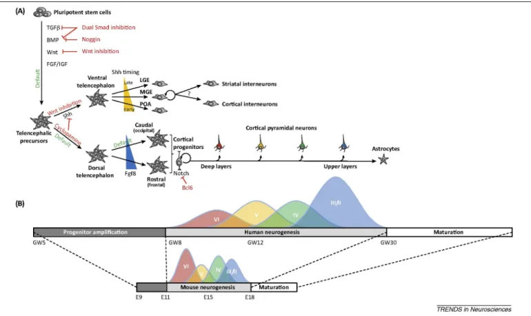
Figure 1. In vitro pluripotent stem cell (PSC)-derived Corticogenesis recapitulates in vivo developmental milestones. (A) PSC cultured under minimal conditions or in the presence of Wnt/transforming growth factor (TGF)-β/one
morphogenetic protein (BMP) morphogen inhibitors, undergo differentiation towards forebrain or telencephalic identity. As in vivo, further specification into ventral telencephalic progenitors and neurons requires activation of the Sonic Hedgehog (Shh) pathway. Different types of ventral progenitor [preoptic area (POA), medial ganglionic eminence (MGE), and lateral ganglionic eminence (LGE)] are generated, including cortical interneurons, depending on the timing and concentration of Shh. Conversely, in absence or low levels of Shh signalling, PSC will mostly differentiate into a collection of progenitors of dorsal telencephalon or cortical identity. Subsequent generation of cortical pyramidal neurons, under the balanced influence of Notch and pro-neurogenic factors, such as B cell CLL/lymphoma 6 (BCL6), follows a temporal patterning, with deep layer neurons being generated earlier than upper layer neurons, eventually followed by a switch to astrocyte production, similar to in vivo. (B) Human corticogenesis and neuronal maturation follow a more protracted time-course than their mouse counterparts, which is also observed in vitro. Abbreviations: E, embryonic day; FGF, fibroblast growth factor; GW, gestational weeks; IGF, insulin-like growth factor.
Cortical neurogenesis: a tale of transitions
The mammalian neocortex is organised into six different layers, each of which comprises a collection of neurons displaying specific patterns of gene expression and connectivity [2,38-40] (Figure 1B). Which layer a neuron settles in, is tightly linked to its birthdate, with deeper layer neurons being generated earlier than upper layer neurons. This process of temporal patterning is central to the generation of layer-specific types of cortical neurons. In vivo and in vitro studies have shown that it involves the progressive restriction of progenitor competence, which is controlled by both intrinsic, cell-autonomous programmes and by environmental influences [2,38-40].
Surprisingly, both mouse and human PSC-derived corticogenesis recapitulate robustly temporal patterning in vitro: neurons that express molecular markers and project to targets specific of upper layer neurons are generated consistently later than neurons with a deep layer identity [11,18,24,41,42]. From clonal cell analyses, it was furthermore shown that mouse ESC-derived neural progenitors are initially multipotent and can change and restrict their competence over time [11], similarly to what was previously demonstrated using ex vivo cultures of early cortical progenitors [43]. However, whereas in vitro systems of corticogenesis display remarkable similarities with in vivo developmental processes, it still differs in significant ways from in vivo corticogenesis, depending on culture conditions. Indeed, whereas in vivo deep and upper layer neurons each represent approximately half of the cortex, ESC- derived pyramidal neurons are strongly skewed towards a deep layer identity following monoadherent culture in minimal differentiation conditions in the absence of added morphogens [11,18]. Importantly, this was also the case when native
mouse cortical progenitors were grown ex vivo at clonal densities [43]. Conversely, a higher proportion of upper layer neurons appears to be generated when ESC are first differentiated at high density and/or supplemented with extrinsic cues, such as retinoic acid [27] or as cell aggregates [24,44]. Although direct comparison between various studies is not always straightforward because of different markers being analysed, these findings suggest that extrinsic cues that may be missing in minimal culture systems, are required for the proper generation of the upper layer neurons [45]. Consistent with this hypothesis, whereas human PSC-derived cortical progenitors cultured in minimal conditions generate only a few upper layer neurons even after prolonged periods in vitro, they generate many upper layer neurons following transplantation into the mouse newborn cortex [18]. These data suggest that cortical progenitors competent to generate neurons of all six layers can be generated following minimal in vitro conditions from ESC, but that cues present in the mouse newborn brain are necessary to enhance upper layer neuron production from these cells. Such cues could be either produced by cortical progenitors and neurons themselves, or derived from extrinsic sources, such as meninges, cerebrospinal fluid, or the vascular niche [45]. PSC-derived corticogenesis may provide a useful platform to identify these cues and their mechanisms of action. Specifically, it will be interesting to determine whether and how these cues act to change the competence of cortical progenitors to generate upper layer neurons, and if they act on the specification or amplification of specific types of progenitors, such as intermediate progenitors or outer radial glial cells, as explained further below.
In parallel with temporal patterning, a key transition for the generation of cortical neurons is neurogenesis itself, whereby cortical progenitors differentiate into cortical neurons, either directly or indirectly following brief amplification through intermediate progenitors [46,47]. The rate and timing of neuronal differentiation from cortical progenitors is dynamically regulated by various intrinsic and extrinsic cues, including Notch and proneural factors, such as neurogenins, which collectively control the number and fate of cortical neurons [45,48-50]. ESC-derived cortical neurogenesis closely recapitulates these processes [11,24], and has been used to gain mechanistic insights into the factors involved in this process (Figure 1A). The mouse ESC cell-based model of corticogenesis was used to screen for novel transcription factors involved in neurogenesis, leading to the identification of a novel potent pro-neurogenic gene, B cell CLL/lymphoma 6 (BCL6) [41]. Whereas BCL6 was uncovered in an ESC-based screen, it was subsequently found to be expressed in cortical progenitors in vivo, during the transition from cortical progenitors to pyramidal neurons. Furthermore, the analysis of BCL6 knockout mice led to the conclusion that BCL6 is required for proper cortical neurogenesis in vivo [41]. Combined studies on the ESC system and in vivo cortex converged to demonstrate that BCL6 acts through direct and stable epigenetic repression of the Notch target Hes5 promoter, thereby enabling the neurogenic transition to proceed irreversibly. This study illustrates how the use of ESC- based models of corticogenesis can lead to novel insights into the in vivo mechanisms of cortical neuron generation.
Cortical areal identity: intrinsic insights from transplantation experiments
In addition to layer-specific identity, neurons from different cortical areas also develop selective patterns of gene expression and connectivity. The patterning of cortical areas is a complex process resulting from the interplay between factors intrinsic to the cortex, as well as extrinsic factors from outside the brain [22,51]. Surprisingly, in vivo transplantation experiments revealed that mouse ESC- derived cortical neurons seem to acquire mainly limbic and visual (occipital) identities [11]. Following transplantation, ESC-derived cortical neurons send axons to specific visual and limbic targets, with a pattern of projection that is strikingly similar to grafted embryonic visual cortical tissue [11,52]. Importantly, these results were all obtained with grafts within the frontal cortex, suggesting that the highly selective pattern of projections was not due to in vivo respecification of the grafted neurons. Confirming this hypothesis, examination of the molecular identity of ESC-derived cortical progenitors and neurons before grafting revealed that most of them expressed typical markers of the occipital cortex, in particular chicken ovalbumin upstream promoter transcription factors (Coup-TF) I and II [11]. By contrast, the areal fate of ESC-derived cortical progenitors in vitro could be modified by the addition of extrinsic cues known to induce frontal cortical fates in vivo, such as FGF8 [24,53].
Corticogenesis from human ESC in similar minimal conditions also results in many Coup-TFI/II-expressing progenitors that display visual and limbic-like patterns of axonal projections shortly after transplantation into the frontal cortex of neonatal mice [18]. However, unlike in the mouse, when the grafts were left for longer periods, the transplanted cells tended to lose markers of occipital identity and their axonal projections corresponded to a wider range of areal identities [18]. These observations are consistent with a model whereby specific patterns of visual or limbic areal identity may be acquired during in vitro minimal differentiation from human ESC, as in the mouse, but that, following grafting, a substantial fraction of the cells can be specified to other areal identities over time, perhaps in relation to their relatively earlier stage of maturation at the time of grafting and, therefore, higher susceptibility to extrinsic cues from the frontal cortex. This is reminiscent of data obtained with rodent native cortical cells, which show different patterns of areal fate specification depending on their degree of commitment and maturation at the time of grafting [54]. These issues need to be dissected further, but the data available so far suggest the existence of a conserved programme of specification into visual and limbic areal identity from PSC in minimal conditions, which might perhaps correspond to an ancient ‘primitive’ pattern of areal specification.
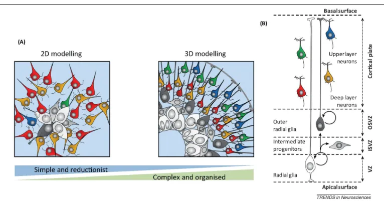
Figure 2. Different models of in vitro corticogenesis from 2D to 3D. (A) 2D models are more reductionist but
nevertheless recapitulate key aspects of corticogenesis and can be used to study various aspects of cortical neuron generation and function, including following in vitro screens or in vivo transplantation. 3D models can recapitulate in a strikingly faithful way the in vivo organisation of cortical progenitors and neurons, thereby providing unique tools to study spatial patterning and cytoarchitecture formation. (B) Schematics of the relations between the various cellular players of corticogenesis found in vivo. Abbreviations: OSVZ/ISVZ, outer/inner subventricular zone.
Cytoarchitecture in a dish: how far can we go 3D?
The cortex is much more than merely a sum of its parts and, if one wishes to model this in vitro, its 3D cytoarchitecture should also be recapitulated. We now know that at least part of the intricate organisation of many organs can also emerge in vitro: a striking example was provided through the autonomous formation of a fully formed optic cup and derived retinal structure from ESC aggregates [55]. For corticogen- esis, this goal is far from being completely achieved and, in principle, would be more challenging; however, some key aspects of the patterned organisation of the developing cortex can be recreated in vitro (Figure 2). Even in 2D systems, neural progenitors exhibit spatial patterns, with cells often organised as rosettes displaying apicobasal polarity and including features typically found in the neuroe-pithelium and ventricular zone, such as interkinetic nuclear migration [11,19,27]. When mouse or human ESC are cul-tured as bowls of cells and differentiated into cortical-like progenitors, this leads to a more robust polarised cellular organisation [24], with progenitors occupying deeper layers of the bowls, and neurons accumulating at their periphery, following an organisation highly reminiscent of a nascent cortical primordium, including the ventricular zone and the cortical plate. A crucial challenge of 3D models is to keep them for long periods of culture while preserving access to gas and nutrients. This important aspect was recently improved to allow neural and cortical differentiation to go on for several months and to examine later aspects of development, including the emergence of specific domains of progenitors and neurons [53,56]. Most strikingly, using a long-term cortical 3D model, the final position of the neurons within a cortical plate-like structure was found to depend on their neuronal birthdate, where neurons born earlier were found in deeper positions than later-born ones, thus recapitulating the fundamental inside-out pattern of cortical neurogenesis [53]. It will be fascinating to determine whether this spatial layer-like arrangement of neurons is estab-lished in a similar way as in vivo (i.e., by relying on active neuronal migration along the radial scaffold and on complex guidance by extrinsic cues, such as Reelin).
Altogether, these data constitute a first proof of principle that a cortical-like cytoarchitecture can also emerge in vitro. Similar to the observation that the temporal patterning leading to sequential generation of cortical neurons is encoded in neural progenitors themselves [11,43], it is remarkable that morphogenesis and compartmentalisation also emerge as self-organising properties [57]. This constitutes a promising system to decipher some of the underlying mechanisms of cortical patterning, and opens new roads to optimise further in vitro corticogenesis and to model more closely in vivo cortical neuronal networks (Box 1).
PSC-derived Corticogenesis and human evolution: time is the essence
The brain and, most strikingly, the neocortex, have undergone a rapid and considerable increase in relative size and complexity during the past few millions of years of hominid (human and great apes) evolution [1]. This has led to enlargement of the surface and thickness of the cortex, associated with an increased number and diversity of cortical neurons. Thus, many of the species-specific features of the human cortex are thought to be linked to differences in the generation, specification, and differentiation of cortical neurons [1,58,59], which may directly impact on the total number and diversity of cortical neurons [1,60-62].
Although cortical neurogenesis appears to be well conserved among mammals, several divergent features have also been identified, which are thought to be mainly linked to the properties of cortical progenitors [1,59,63-65] (Figure 1B). Neuroepithelial (NE) cells are the first type to emerge during early development, and are characterised by symmetric proliferative divisions that amplify the initial pool of cortical progenitors. This amplification process may have a direct impact on the final number of neurons [61] and is expanded in species displaying brains of larger size, such as primates (taking weeks instead of 2-3 days in mice) [61,66]. NE progenitors then convert into radial glial (RG) progenitors, which constitute the major subtype of neurogenic cortical progenitors [46,48,67-69]. RG cells undergo multiple rounds of asymmetric cell divisions, thereby enabling the generation of diverse types of neurons while maintaining a pool of progenitors, thus, following a stem cell-like behaviour [70,71]. In primates, the process of neuronal production is considerably protracted in time, taking several months instead of several days in the mouse [66].
Remarkably, human ESC-derived corticogenesis presents temporal specificities that are reminiscent of human-specific features of cortical development [18,24,26, 27,44]. Direct comparison between mouse and human cor- ticogenesis from PSC, using exactly the same culture conditions, revealed that the time line for corticogenesis is considerably extended in the human versus the mouse [18]. Human ESC-cortical derived progenitors start to generate postmitotic neurons after a much longer period (i.e., approximately 4 weeks instead of 6-8 days in the mouse) and this was correlated with the appearance of molecular markers of RG cells. Similar to in vivo, the generation of distinct types of cortical neurons is also much protracted in time: whereas mouse ESC corticogenesis takes 2-3 weeks to be completed, it takes 10-15 weeks for human ESC [11]. The underlying mechanisms that enable the primate embryonic brain to generate neurons for a prolonged period of time remain largely unknown [72], but they might be linked to species-specific properties intrinsic to RG cells, such as differential cell cycle control or tuning of self-renewal versus terminal differentiation [73]. In addition, the emergence and/or amplification of different species-specific types of progenitors might be another mechanism that contributes to cortical neuron diversity and to evolutionary changes in cortical neurogenesis. These progenitors include the recently described outer RG (oRG) cells [64,65,74-76]. oRG cells share many features with RG cells, including the potential for self-renewal, but they lack any apical projection reaching the ventricular surface. Most strikingly, whereas human oRG cells can generate neurons directly, their progeny tend to undergo multiple rounds of divisions, thus providing an additional mechanism for increased neuronal output and cortical expansion. The detection of oRG-like cells in vitro from human PSC was reported in 2D [27] and 3D systems [53,56]. Importantly these cells were not found in similar differentiation paradigms from mouse PSC [56], providing evidence of species specificity, although perhaps the most important feature of oRG cells (i.e., their capacity to generate many neurons through transient amplification) still has to be determined in in vitro conditions.
Another important aspect that appears to be species specific is neuronal maturation: human cortical neurons display more prolonged patterns of morphological and electrophysiological maturation, as well as synaptogenesis, which might underlie some of the relative neoteny that characterises human brain maturation [77,78]. Similarly, the in vitro maturation of the human cortical excitatory neurons follows a slower pace compared with their mouse counterparts. In vitro-derived human neurons exhibit immature profiles of gene expression and excitability for several weeks, and extended periods of culture are needed to observe mature patterns of action potentials and signs of significant synaptic activity [18,27,44]. A similarly protracted pattern is observed for cortical interneurons: GABA-ergic neurons present an immature profile of activity even after 8-9 weeks in culture and only start to display more mature functional properties typically after 15-30 weeks [28]. This slow process can be accelerated by co-culture with murine astroglial or embryonic cortical cultures, but only to a partial extent [28,30]. Besides and most strikingly, in vivo transplantation of human PSC-derived excitatory and inhibitory cells into mouse models has shown that the human cells seem to follow a species-specific programme for delayed neuronal maturation and synaptogenesis [18,20,28,30]. Indeed, whereas differentiated human pyramidal neurons extend axons towards specific cortical targets from 4-6 weeks post-transplantation, the full extent of axonal growth from human cortical transplanted neurons was only reached 6 months after transplantation in the mouse neonatal cortex. Dendritic maturation also progressed at a slow pace even within the mouse brain, where neurons presented a mature complex dendrite arborisation pattern, dendritic spines, and functional synaptic activity only 9 months after transplantation [18]. Similarly, the patterns of morphology and functionality of xenotransplanted human interneurons are not yet fully reached even 6 months post-transplantation [28,30].
Overall, these data indicate that the fundamental principles that control the timing of generation and maturation of cortical neurons appear to be species specific and do not depend much on the in vivo context. Instead, they point to cortex-intrinsic mechanisms that control the clock of corticogenesis, for which PSC-based models may provide
at-tractive experimental set-ups to dissect the underlying mechanisms.
It was previously proposed that such protracted neuronal maturation provides a basis for the neoteny that may characterise human cortical neuron development (i.e., the retention of juvenile traits even at an older age) [77]. Although largely speculative, this possibility is attractive enough to explore further, for instance by examining the consequences of the transplantation and integration of ‘juvenile’ human neurons into the mouse cortex at the circuit and behavioural levels.
Modelling pathological cortical development and degeneration
The advent of iPSC technology [7] offers in principle many novel opportunities to model brain diseases, including those that strike the developing cortex [8,79]. At this point, most can probably be learned from modelling monogenic disorders with high penetrance [80], for which gain- and loss-of-function paradigms could confirm the role of the affected gene. However, there are few examples so far of studies that have relied on iPSC-derived cortical cells of defined identity to model cortical neurodevelopmental diseases. Among these, one striking example is Timothy syndrome, a monogenic disorder caused by a mutation in an L-type voltage-gated calcium channel, which is strongly associated with developmental delay and autism. Examination of cortical cells from iPSC derived from patients with Timothy syndrome revealed several interesting phenotypes. These included, as expected, defects in calcium signalling and neuronal activity, but also somewhat more surprisingly defects in the generation of specific types of neurons (i.e., callosal projection neurons), as well as defects in dendrite remodelling [81,82]. These phenotypes were recapitulated in a mouse model presenting a similar mutation, thus confirming the validity of the in vitro cellular human model. Other examples of autistic syndromes are Phelan-McDermid syndrome caused by 22q13 deletion or Rett syndrome caused by methyl CpG binding protein 2 (Mecp2) mutations. The 22q13 deletion syndrome was modelled in a similar way with patient- derived iPSC, which revealed synaptic deficits that could be largely rescued by re-expression of SH3 and multiple ankyrin repeat domains 3 (Shank3), one of the genes involved in the deletion syndrome [83]. To study Rett syndrome, the authors used isogenic ESC lines generated through homologous recombination, which appears to be most promising to cope with the inherent variability of iPSC lines derived from different individuals [79,84]. This approach revealed several defects in the studied neurons, including a neuron-specific global alteration of gene ex-pression that could underlie many of the cellular and functional defects observed in Rett syndrome-affected neurons. Early defects of cortical development that are at the origin of most severe forms of brain malformations and dysfunction constitute other candidate diseases to be modelled by in vitro corticogenesis. For instance, iPSC from a patient with microcephaly affected by a CDK5 regulatory subunit associated protein 2 (CDK5RAP2) mutation [56], have enabled researchers to recapitulate some of the defects previously described in mouse models [85,86]. Cor- ticogenesis from iPSC was also applied to study Down's syndrome, a major cause of mental retardation and neurodegeneration, revealing early synaptic defects in affected neurons [87], as well as amyloid peptide overproduction[88].
These findings illustrate that, when applied to neurons of defined identity, the iPSC modelling technology can yield valuable insight into pathogenic mechanisms of complex neurodevelopmental diseases. In addition, more complex brain developmental disorders, such as schizophrenia [89]or fragile X syndrome [90], although more challenging, could also benefit from modelling using in vitro corticogen- esis. Finally, it could also be applied to degenerative diseases that primarily affect cortical neurons, including Alzheimer's disease and various other forms of dementia. However, in this case, it will be important to find out which critical aspects of these diseases can be recapitulated faithfully in vitro, given the fact that they primarily affect the adult or aging nervous system. Indeed, so far iPSC- derived or directly reprogrammed neurons from patients with familial [amyloid precursor protein (APP) duplications, presenilin 1 or 2 (PSEN1 or PSEN2) mutations] and sporadic Alzheimer's disease, were found to mimic a limited set of the pathological features encountered in diseased brain tissue, including altered APP processing and Tau phosphorylation [87,88,91-94]. By contrast, major hallmarks of the disease, such as Tau neurofibrillary tangle formation, neuronal dysfunction and death, have not been reported so far. In this respect, it will be interesting to explore further artificial ways to accelerate neuronal maturation, or even aging in vitro [95] or try to model neurodegeneration following xenotransplantation in the aging mouse brain.
Box 1. Outstanding questions: What's next, in and out of the dish?
• How much can models of 2D and 3D corticogenesis recapitulate accurately the full repertoire of cortical neuronal identities?
• Can we design and improve the tools of PSC differentiation to generate highly homogenous populations of neurons of a single, physiologically relevant, identity?
• How much can 3D models recapitulate the processes of cortical neurogenesis, including polarised cell movements and asym-metric divisions?
• How much can 3D models recapitulate the layered organisation of the neocortex?
• To what extent are 3D models capable of reproducing the emergence of distinct cortical areal identities?
• What are the patterns of connectivity of cortical neurons generated in 3D models? Can we generate a cortical column in vitro? Do neurons display genuine layer-specific patterns of input and output?
• How much specificity and accuracy of connectivity can be achieved following transplantation of PSC-derived cortical neurons in the brain? Can short- and long-range patterns lead to physiological circuits? If so, how much can lead to cortical function
restoration?
• Are the corticogenesis models robust enough to not only confirm, but also predict mechanisms of development or disease, which could be further explored in vivo?
• Can PSC-derived models of corticogenesis be used to model the events that occur only in the adult or even aging brain, including most aspects of neurodegeneration?
• Can human PSC models be improved to allow cost-effective and robust high-throughput genetic or chemical screens?
Concluding remarks and perspectives
The merge of PSC technology and developmental neurobiology reveals unexpected opportunities to study the forma-tion and maintenance of the cerebral cortex (Box 1). However, to hold its promises, PSC-based modelling will have to contribute significantly to uncover novel features of normal and pathological mechanisms of corticogenesis. One area of interest in this context is to use ESC-based systems for unbiased screens, in line with the discovery of BCL6 as a novel cortical pro-neurogenic factor. Another exciting focus will be to investigate the mechanisms underlying the species-specific features of corticogenesis, by exploring experimentally the genetic links between development and evolution of the human brain. Finally, PSC models may bring novel insights into the pathophysiology of human diseases that are not recapitulated as severely or clearly in mouse models, such as cortical malformations and alterations of higher cognitive functions. However, whatever PSC may reveal will have to be confronted with in vivo data, no matter how challenging this may be. Indeed, models, whether in art or science, can merely represent, but cannot replace, the original complexity of life.
References
1 Lui, J.H. et al. (2011) Development and evolution of the human neocortex. Cell 146, 18-36
2 Molyneaux, B.J. et al. (2007) Neuronal subtype specification in the cerebral cortex. Nat. Rev. Neurosci. 8, 427-437 3 DeFelipe, J. et al. (2013) New insights into the classification and nomenclature of cortical GABAergic interneurons.
Nat. Rev. Neurosci. 14, 202-216
4 Smith, A.G. (2001) Embryo-derived stem cells: of mice and men. Annu. Rev. Cell Dev. Biol. 17, 435-462
5 Gaspard, N. and Vanderhaeghen, P. (2010) Mechanisms of neural specification from embryonic stem cells. Curr. Opin. Neurobiol. 20, 37-43
6 Yamanaka, S. (2007) Strategies and new developments in the generation of patient-specific pluripotent stem cells. Cell Stem Cell 1, 39-49
7 Takahashi, K. and Yamanaka, S. (2006) Induction of pluripotent stem cells from mouse embryonic and adult fibroblast cultures by defined factors. Cell 126, 663-676
8 Dolmetsch, R. and Geschwind, D.H. (2011) The human brain in a dish: the promise of iPSC-Derived neurons. Cell 145, 831-834
9 Gaspard, N. and Vanderhaeghen, P. (2011) From stem cells to neural networks: recent advances and perspectives for neurodevelopmental disorders. Dev. Med. Child Neurol. 53, 13-17
10 Wilson, S.W. and Houart, C. (2004) Early steps in the development of the forebrain. Dev. Cell 6, 167-181
11 Gaspard, N. et al. (2008) An intrinsic mechanism of corticogenesis from embryonic stem cells. Nature 455, 351-357 12 Bertacchi, M. et al. (2013) The positional identity of mouse ES cellgenerated neurons is affected by BMP signaling.
Cell. Mol. Life Sci. 70, 1095-1111
13 Juliandi, B. et al. (2012) Induction of superficial cortical layer neurons from mouse embryonic stem cells by valproic acid. Neurosci. Res. 72, 23-31
14 Ying, Q.L. et al. (2003) Conversion of embryonic stem cells into neuroectodermal precursors in adherent monoculture. Nat. Biotechnol. 21, 183-186
15 Wataya, T. et al. (2008) Minimization of exogenous signals in ES cell culture induces rostral hypothalamic differentiation. Proc. Natl. Acad. Sci. U.S.A. 105, 11796-11801
16 Gaspard, N. et al. (2009) Generation of cortical neurons from mouse embryonic stem cells. Nat. Protoc. 4, 1454-1463
17 Watanabe, K. et al. (2005) Directed differentiation of telencephalic precursors from embryonic stem cells. Nat. Neurosci. 8, 288-296
18 Espuny-Camacho, I. et al. (2013) Pyramidal neurons derived from human pluripotent stem cells integrate efficiently into mouse brain circuits in vivo. Neuron 77, 440-456
19 Chambers, S.M. et al. (2009) Highly efficient neural conversion of human ES and iPS cells by dual inhibition of SMAD signaling. Nat. Biotechnol. 27, 275-280
20 Kirkeby, A. et al. (2012) Generation of regionally specified neural progenitors and functional neurons from human embryonic stem cells under defined conditions. Cell Rep. 1, 703-714
21 Hebert, J.M. and Fishell, G. (2008) The genetics of early telencephalon patterning: some assembly required. Nat. Rev. Neurosci. 9, 678-685
22 Sur, M. and Rubenstein, J.L. (2005) Patterning and plasticity of the cerebral cortex. Science 310, 805-810
23 Wonders, C.P. and Anderson, S.A. (2006) The origin and specification of cortical interneurons. Nat. Rev. Neurosci. 7, 687-696
24 Eiraku, M. et al. (2008) Self-organized formation of polarized cortical tissues from ESCs and its active manipulation by extrinsic signals. Cell Stem Cell 3, 519-532
25 Danjo, T. et al. (2011) Subregional specification of embryonic stem cell- derived ventral telencephalic tissues by timed and combinatory treatment with extrinsic signals. J. Neurosci. 31, 1919-1933
26 Li, X.J. et al. (2009) Coordination of sonic hedgehog and Wnt signaling determines ventral and dorsal telencephalic neuron types from human embryonic stem cells. Development 136, 4055-4063
27 Shi, Y. et al. (2012) Human cerebral cortex development from pluripotent stem cells to functional excitatory synapses. Nat. Neurosci. 15, 477-486
28 Nicholas, C.R. et al. (2013) Functional maturation of hPSC-derived forebrain interneurons requires an extended timeline and mimics human neural development. Cell Stem Cell 12, 573-586
29 Maroof, A.M. et al. (2010) Prospective isolation of cortical interneuron precursors from mouse embryonic stem cells. J. Neurosci. 30, 46674675
30 Maroof, A.M. et al. (2013) Directed differentiation and functional maturation of cortical interneurons from human embryonic stem cells. Cell Stem Cell 12, 559-572
31 Liu, Y. et al. (2013) Medial ganglionic eminence-like cells derived from human embryonic stem cells correct learning and memory deficits. Nat. Biotechnol. 31, 440-447
32 Germain, N.D. et al. (2013) Derivation and isolation of NKX2.1-positive basal forebrain progenitors from human embryonic stem cells. Stem Cells Dev. 22, 1477-1489
33 Ma, L. et al. (2012) Human embryonic stem cell-derived GABA neurons correct locomotion deficits in quinolinic acid-lesioned mice. Cell Stem Cell 10, 455-464
34 Briscoe, J. and Therond, P.P. (2013) The mechanisms of Hedgehog signalling and its roles in development and disease. Nat. Rev. Mol. Cell Biol. 14, 416-429
35 Cambray, S. et al. (2012) Activin induces cortical interneuron identity and differentiation in embryonic stem cell-derived telencephalic neural precursors. Nat. Commun. 3, 841
36 Hansen, D.V. et al. (2013) Non-epithelial stem cells and cortical interneuron production in the human ganglionic eminences. Nat. Neurosci. 16, 1576-1587
37 Ma, T. et al. (2013) Subcortical origins of human and monkey neocortical interneurons. Nat. Neurosci. 16, 1588-1597
38 Gaspard, N. and Vanderhaeghen, P. (2011) Laminar fate specification in the cerebral cortex. F1000 Biol. Rep. 3, 6 39 Leone, D.P. et al. (2008) The determination of projection neuron identity in the developing cerebral cortex. Curr.
Opin. Neurobiol. 18, 28-35
40 Greig, L.C. et al. (2013) Molecular logic of neocortical projection neuron specification, development and diversity. Nat. Rev. Neurosci. 14, 755-769
41 Tiberi, L. et al. (2012) BCL6 controls neurogenesis through Sirt1- dependent epigenetic repression of selective Notch targets. Nat. Neurosci. 15, 1627-1635
42 Shi, Y. et al. (2012) Human cerebral cortex development from pluripotent stem cells to functional excitatory synapses. Nat. Neurosci. 15, 477-486 S471
43 Shen, Q. et al. (2006) The timing of cortical neurogenesis is encoded within lineages of individual progenitor cells. Nat. Neurosci. 9,743-751
Natl. Acad. Sci. U.S.A. 109, 12770-12775
45 Tiberi, L. et al. (2012) Cortical neurogenesis and morphogens: diversity of cues, sources and functions. Curr. Opin. Cell Biol. 24, 269-276
46 Kriegstein, A. and Alvarez-Buylla, A. (2009) The glial nature of embryonic and adult neural stem cells. Annu. Rev. Neurosci. 32, 149-184
47 Fietz, S.A. and Huttner, W.B. (2011) Cortical progenitor expansion, self-renewal and neurogenesis: a polarized perspective. Curr. Opin. Neurobiol. 21, 23-35
48 Gotz, M. and Huttner, W.B. (2005) The cell biology of neurogenesis. Nat. Rev. Mol. Cell Biol. 6, 777-788
49 Okano, H. and Temple, S. (2009) Cell types to order: temporal specification of CNS stem cells. Curr. Opin. Neurobiol. 19, 112-119
50 Guillemot, F. (2007) Cell fate specification in the mammalian telencephalon. Prog. Neurobiol. 83, 37-52
51 O’Leary, D.D. and Sahara, S. (2008) Genetic regulation of arealization of the neocortex. Curr. Opin. Neurobiol. 18, 90-100
52 Gaillard, A. and Roger, M. (2000) Early commitment of embryonic neocortical cells to develop area-specific thalamic connections. Cereb. Cortex 10, 443-453
53 Kadoshima, T. et al. (2013) Self-organization of axial polarity, inside- out layer pattern, and species-specific progenitor dynamics in human ES cell-derived neocortex. Proc. Natl. Acad. Sci. U.S.A. 110, 202842202849
54 Pinaudeau, C. et al. (2000) Stage of specification of the spinal cord and tectal projections from cortical grafts. Eur. J. Neurosci. 12, 2486-2496
55 Eiraku, M. et al. (2011) Self-organizing optic-cup morphogenesis in three-dimensional culture. Nature 472, 51-56 56 Lancaster, M.A. et al. (2013) Cerebral organoids model human brain development and microcephaly. Nature 501,
373-379
57 Sasai, Y. (2013) Cytosystems dynamics in self-organization of tissue architecture. Nature 493, 318-326
58 Bystron, I. et al. (2008) Development of the human cerebral cortex: Boulder Committee revisited. Nat. Rev. Neurosci. 9, 110-122
59 Fish, J.L. et al. (2008) Making bigger brains-the evolution of neural- progenitor-cell division. J. Cell Sci. 121, 2783-2793
60 Bystron, I. et al. (2006) The first neurons of the human cerebral cortex. Nat. Neurosci. 9, 880-886
61 Rakic, P. (1995) A small step for the cell, a giant leap for mankind: a hypothesis of neocortical expansion during evolution. Trends Neurosci. 18, 383-388
62 Rakic, P. (1988) Specification of cerebral cortical areas. Science 241, 170-176
63 Kriegstein, A. et al. (2006) Patterns of neural stem and progenitor cell division may underlie evolutionary cortical expansion. Nat. Rev. Neurosci. 7, 883-890
64 Hansen, D.V. et al. (2010) Neurogenic radial glia in the outer subventricular zone of human neocortex. Nature 464, 554-561
65 Fietz, S.A. et al. (2010) OSVZ progenitors of human and ferret neocortex are epithelial-like and expand by integrin signaling. Nat. Neurosci. 13, 690-699
66 Rakic, P. (2009) Evolution of the neocortex: a perspective from developmental biology. Nat. Rev. Neurosci. 10, 724-735
67 Pinto, L. and Gotz, M. (2007) Radial glial cell heterogeneity: the source of diverse progeny in the CNS. Prog. Neurobiol. 83, 2-23
68 Fietz, S.A. and Huttner, W.B. (2010) Cortical progenitor expansion, self-renewal and neurogenesis: a polarized perspective. Curr. Opin. Neurobiol. 21, 23-35
69 Betizeau, M. et al. (2013) Precursor diversity and complexity of lineage relationships in the outer subventricular zone of the primate. Neuron 80, 442-457
70 Noctor, S.C. et al. (2004) Cortical neurons arise in symmetric and asymmetric division zones and migrate through specific phases. Nat. Neurosci. 7, 136-144
Development 131, 3133-3145
72 Dehay, C. and Kennedy, H. (2007) Cell-cycle control and cortical development. Nat. Rev. Neurosci. 8, 438-450 73 Lukaszewicz, A. et al. (2005) G1 phase regulation, area-specific cell cycle control, and cytoarchitectonics in the
primate cortex. Neuron 47, 353-364
74 Kelava, I. et al. (2012) Abundant occurrence of basal radial glia in the subventricular zone of embryonic neocortex of a lissencephalic primate, the common marmoset Callithrix jacchus. Cereb. Cortex 22, 469-481
75 Reillo, I. and Borrell, V. (2012) Germinal zones in the developing cerebral cortex of ferret: ontogeny, cell cycle kinetics, and diversity of progenitors. Cereb. Cortex 22, 2039-2054
76 Garcia-Moreno, F. et al. (2012) Compartmentalization of cerebral cortical germinal zones in a lissencephalic primate and gyrencephalic rodent. Cereb. Cortex 22, 482-492
77 Petanjek, Z. et al. (2011) Extraordinary neoteny of synaptic spines in the human prefrontal cortex. Proc. Natl. Acad. Sci. U.S.A. 108, 1328113286
78 Defelipe, J. (2011) The evolution of the brain, the human nature of cortical circuits, and intellectual creativity. Front. Neuroanat. 5, 29
79 Sandoe, J. and Eggan, K. (2013) Opportunities and challenges of pluripotent stem cell neurodegenerative disease models. Nat. Neurosci. 16, 780-789
80 Karayiorgou, M. et al. (2012) The best of times, the worst of times for psychiatric disease. Nat. Neurosci. 15, 811-812
81 Krey, J.F. et al. (2013) Timothy syndrome is associated with activitydependent dendritic retraction in rodent and human neurons. Nat. Neurosci. 16, 201-209
82 Pasca, S.P. et al. (2011) Using iPSC-derived neurons to uncover cellular phenotypes associated with Timothy syndrome. Nat. Med. 17, 16571662
83 Shcheglovitov, A. et al. (2013) SHANK3 and IGF1 restore synaptic deficits in neurons from 22q13 deletion syndrome patients. Nature 503, 267-271
84 Li, Y. et al. (2013) Global transcriptional and translational repression in human-embryonic-stem-cell-derived rett syndrome neurons. Cell Stem Cell 13, 446-458
85 Buchman, J.J. et al. (2010) Cdk5rap2 interacts with pericentrin to maintain the neural progenitor pool in the developing neocortex. Neuron 66, 386-402
86 Lizarraga, S.B. et al. (2010) Cdk5rap2 regulates centrosome function and chromosome segregation in neuronal progenitors. Development 137, 1907-1917
87 Weick, J.P. et al. (2013) Deficits in human trisomy 21 iPSCs and neurons. Proc. Natl. Acad. Sci. U.S.A. 110, 9962-9967
88 Shi, Y. et al. (2012) A human stem cell model of early Alzheimer’s disease pathology in Down syndrome. Sci. Transi. Med. 4, 124ra129
89 Brennand, K.J. et al. (2011) Modelling schizophrenia using human induced pluripotent stem cells. Nature 473, 221-225
90 Liu, J. et al. (2012) Signaling defects in iPSC-derived fragile X premutation neurons. Hum. Mol. Genet. 21, 3795-3805
91 Israel, M.A. et al. (2012) Probing sporadic and familial Alzheimer’s disease using induced pluripotent stem cells. Nature 482, 216220
92 Yagi, T. et al. (2011) Modeling familial Alzheimer’s disease with induced pluripotent stem cells. Hum. Mol. Genet. 20, 4530-4539
93 Qiang, L. et al. (2011) Directed conversion of Alzheimer’s disease patient skin fibroblasts into functional neurons. Cell 146, 359-371
94 Kondo, T. et al. (2013) Modeling Alzheimer’s disease with iPSCs reveals stress phenotypes associated with intracellular Abeta and differential drug responsiveness. Cell Stem Cell 12, 487-496
95 Miller, J.D. et al. (2013) Human iPSC-based